Received 3 April 1995/Accepted 14 August 1995
The immediate-early proteins of herpes simplex virus control the cascade of viral gene expression during lytic infection. It is not known which viral or host proteins control the reactivation of the viral genome in latently infected neurons. To determine whether neuronal proteins can regulate a herpes simplex virus immediate-early promoter in vivo, transgenic mice containing the promoter regulatory region of the herpes
simplex virus type 1 immediate-early gene (ICP4) fused to the bacterialb-galactosidase gene were generated.
Two lines of mice, in the absence of viral proteins, displayed ICP4 promoter activity in neurons in specific locations in the central nervous system. The anatomic locations of these neurons were the hippocampus, cerebellar cortex, superior colliculus, indusium griseum, mammillary nucleus, cerebral cortex, and the dorsal laminae of the dorsal horns of the spinal cord. Additional subsets of neurons expressed the ICP4 promoter at lower levels; these included trigeminal ganglia and retinas. In a third line of mice, lower levels of expression were present in many of the above-described neurons. Many types of neurons, nearly all nonneuronal cells in the central nervous system, and some non-nervous system tissues were negative. Viral proteins including VP16 are not necessary to induce transcription from the ICP4 promoter in many neurons and some other cell types but may be required in most cells in vivo. An approximately 100-fold-greater number of neurons in the trigeminal ganglia expressed ICP4 promoter activity in newborn mice compared with adults. These data provide direct evidence that host proteins are sufficient to activate a herpes simplex virus immediate-early promoter in neurons in vivo and that a differential expression pattern for this promoter exists within different neuronal phenotypes and between the same neurons in different ages of mice.
Herpes simplex virus (HSV) infections lead to a variety of diseases in humans; these may include recurrent genital and orofacial lesions, keratitis, conjunctivitis, encephalitis, and dis-seminated infections of newborns (50). An initial lytic infection at peripheral sites is followed by axonal transport of HSV to sensory ganglion neurons, where a lytic or latent infection occurs (9, 10, 38, 45, 46). Regulation of HSV immediate-early (IE) genes is thought to be a critical feature in determining the outcome of infection (39). Regulation of these genes may play a role in cell tropism, establishment of and reactivation from latency (9, 10), and the extent of viral replication and disease. In vitro, the expression of the viral IE genes has been shown to be controlled by protein complexes composed of both viral and host proteins (6, 8, 11, 26, 34, 35, 37, 44, 48). The viral transactivator VP16 (VMW 65) forms a complex with the host DNA-binding protein Oct 1 and another host protein (52) to upregulate transcription of IE genes. The host protein Oct 2 is thought to function as a repressor of IE transcription (8, 18, 21, 22). In addition, some IE proteins regulate other IE genes and also their own promoters (39). It may be that availability of specific cellular DNA-binding proteins in various cells and tissues of animals can determine the level of IE regulation and thus control the outcome of infection of certain cell types and of the whole organism. In situ hybridization studies have shown that the levels of certain transcription factors (such as Oct 1) which interact with IE promoters vary in different neurons (16). In transgenic mice, it has been shown that a mouse homeodomain protein, Hoxa-5 (Hox 1.3), which binds the
se-quence TAAT within the TAATGARAT regulatory sese-quences of the HSV IE promoters alters the course of viral infection and disease (29). It has also been shown that infection with HSV type 1 (HSV-1) induces expression of the Oct 1 gene in sensory neurons (49). The previous experiments do not allow the separation of the role of the host protein from that of viral proteins or the evaluation of variations in the functional ca-pacity of host transcriptional proteins to activate a viral IE promoter in different cells. Since animals are made up of complex and specialized cells and tissues, different cells within a host would be expected to present a variety of different arrays of transcriptional proteins. To what extent these host transcrip-tional proteins may be involved in determining neuronal tro-pism and reactivation of HSV from latency has not been stud-ied. A direct approach has been taken to determine whether there are differences in the availability of factors required for transcription from an HSV-1 IE promoter (ICP4) in different neurons. Transgenic mice which contain the ICP4 promoter fused to the bacterial b-galactosidase coding sequence were generated. In these mice, it was determined that in the absence of viral proteins, subsets of neurons display high levels of ICP4 promoter activity whereas others are negative and that the level of promoter activity in the same neurons varies with age.
MATERIALS AND METHODS
Generation and identification of transgenic mice.The HSV-1 ICP4 promoter regulatory region (nucleotides2372 through124 with respect to the transcrip-tional start site of ICP4) (23, 25, 48) was amplified by PCR from purified HSV-1 F DNA template as previously described (32) except that glycerol (to a final concentration of 15%) was added to the reaction mixture. The primers were CTGCGCACTTCTAGAGGCTCGTATCTCATTACC and GCCCTAGCCAG CTGGTGTCGGCAGCCGCGCTCC. The ICP4 promoter fragment was ligated (40) into the blunt-ended pNlacF plasmid (restriction digested at the SalI site and made blunt ended by treatment with Klenow enzyme) containing the
Esch-* Mailing address: Laboratory of Molecular Medicine and Neuro-science, NINDS, NIH, Building 36, Room 5W21, Bethesda, MD 20892. Phone: (301) 496-9923. Fax: (301) 402-8863.
7942
on November 9, 2019 by guest
http://jvi.asm.org/
erichia colib-galactosidase coding sequence and including a simian virus 40 nuclear translocation signal (27). The XbaI-HindIII fragment from the final construct was isolated and purified, and approximately 200 copies were injected (17, 29) into each (C57BL/63C3H)F13(C57BL/63C3H)F1one-cell embryo.
Three founder animals (designated Tg0002, Tg6305, and Tg6307) were identi-fied, and transgenic lines were established by brother-sister matings. Two of the lines described in this report have been assigned designations according to the standardized rules for nomenclature in transgenic mice: line Tg6305, TgN(HS VieRp)1wm; line Tg6307, TgN(HSVieRp)2wm. Heterozygous transgenic mice and their nontransgenic control littermates were used in this study; these mice were identified by PCR using tail DNA (29, 30, 32) and primers (GCATCGAG CTGGGTAATAAGCGTTGGCAAT and GACACCAGACCAACTGGTAAT GGTAGCGAC) for theb-galactosidase coding sequence.
Analysis of transgene expression in mice.Adult mice of each line (see Table 1 for numbers) were killed, and tissues were dissected, snap-frozen, and stored at
2708C. Ganglia and eyes were fixed as whole tissues in 4% paraformaldehyde for 30 min immediately upon removal from2708C. All other tissues were sectioned with a cryotome at 40mm (a few 8-mm sections were made), attached to poly-lysine-coated glass slides, and fixed in 4% paraformaldehyde for 30 min. Sections or whole tissues were washed for 5 min in 13phosphate-buffered saline (PBS) and incubated in substrate solution (30, 41) for 14 to 18 h at 378C. The substrate solution contained 20 mM potassium ferrocyanide, 20 mM potassium ferricya-nide, 2 mM MgCl2, 1 mg of 5-bromo-4-chloro-3-indolyl-b-D-galactoside (X-Gal)
per ml, and 120ml of 10% Nonidet P-40 and 100ml of 1% sodium deoxycholate per 20 ml. Whole tissues (ganglia and eyes) were washed for 5 min in 13PBS and either sectioned on a vibrotome at 100mm or thinly sliced with a razor blade. Cryotome sections were washed for 5 min in 13PBS. All tissue sections were mounted in PBS, and the coverslips were sealed with Permount. Some 40-mm brain sections were counterstained with 1% neutral red for 30 s and washed three times for 5 min each in water.
Colabeling of neurons andb-galactosidase activity.Cryotome sections (16
mm) of brains and spinal cords of line Tg6305 mice were prepared as described above. The sections were incubated overnight in X-Gal substrate solution as described above. Sections were washed in PBS, and neurons were labeled by the
avidin-biotin alkaline phosphatase method (Vector), using a 1:200 dilution of antibody to neuron-specific enolase (NSE) (Incstar) (42). As a control, a 1:200 dilution of rabbit antibody to the vesicular stomatitis virus G protein was used on adjacent sections in each experiment. Astrocytes were detected inb -galac-tosidase-labeled sections by the avidin-biotin peroxidase method (Vector), using a 1:250 dilution of rabbit antibody to glial fibrillary acidic protein (GFAP) (13).
Virus infections in mice.Three Tg6305 mice and three age-matched control littermates were inoculated intracorneally with 107PFU of HSV-1 strain F per
eye (31). In addition, three Tg6305 mice and three age-matched control litter-mates were mock inoculated with tissue culture medium. Animals were killed after 4 days (acute stage), and corneas and trigeminal ganglia were analyzed as described above for the presence ofb-galactosidase labeling.
Analysis of transgene expression in trigeminal ganglion neurons of newborn mice.To determine whether neuronal differentiation might influence transcrip-tion from the ICP4 promoter, sensory ganglia from newborn Tg6305 mice were assayed forb-galactosidase production. One-day-old Tg6305 mice were killed, and trigeminal ganglia were dissected by using a microscope, snap-frozen, and assayed as described for adult mice. The numbers of positive neurons per gan-glion were counted and compared with those of adult mice of the same line.
RESULTS
Generation of ICP4–b-galactosidase transgenic mice and
response of the transgene to HSV-1 infection.Three founder
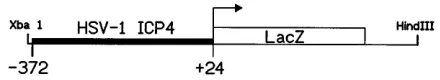
mice containing the ICP4–b-galactosidase transgene (Fig. 1) in a C57BL/63C3H hybrid background were identified by PCR from tail DNA. Transgenic mouse lines (Tg6305, Tg6307, and Tg0002) were established and maintained as heterozygotes by brother-sister matings. To demonstrate that the transgene was functional and responded to viral regulatory signals, Tg6305 transgenic mice were inoculated with HSV-1. Intracorneal in-oculation of Tg6305 mice with strain F resulted in expression of b-galactosidase in moderate to large numbers of corneal cells in six of six corneas from three mice (Fig. 2A), whereas none of six HSV-1-inoculated corneas from nontransgenic con-trol mice were positive forb-galactosidase. Mock-inoculated Tg6305 mice and nontransgenic littermates were negative for
[image:2.612.68.288.71.111.2]b-galactosidase-labeled cells in all of six corneas in each group (Fig. 2B and results not shown). Expression ofb-galactosidase was detected in large numbers of cells in trigeminal ganglia of Tg6305 mice 4 days following intracorneal inoculation with HSV-1 F (Fig. 2C). The labeled cells were large neurons and smaller cells which were probably satellite cells (Fig. 2C). FIG. 1. Diagram of the ICP4–b-galactosidase chimeric transgene. The ICP4
promoter DNA fragment included nucleotides2372 through124 of the ICP4 gene, where 0 is the transcriptional start site. This DNA fragment was fused to the coding sequence for theb-galactosidase gene of E. coli, which also contained a nuclear translocation signal from simian virus 40 (plasmid pNlacF). The re-sulting transgene was used to produce three lines of transgenic mice (Tg6305, Tg6307, and Tg0002).
FIG. 2. The ICP4–b-galactosidase transgene is activated by HSV-1 infection in cells of the cornea and trigeminal ganglia. (A) Cornea of a Tg6305 mouse 4 days after inoculation with HSV-1 F. (B) Cornea of a mock-inoculated Tg6305 mouse. (C) Trigeminal ganglia of a Tg6305 mouse 4 days after inoculation with HSV-1 F. (In the insert, the larger positive cell is a neuron.) (D) Trigeminal ganglia of a mock-inoculated Tg6305 mouse. Bars: (A and B) 35mm; (C and D) 106mm.
on November 9, 2019 by guest
http://jvi.asm.org/
[image:2.612.136.477.494.699.2]Mock-inoculated Tg6305 ganglia contained none (Fig. 2D) or rarely between one and four positive cells. HSV-1 infection activated the ICP4–b-galactosidase transgene in neurons in trigeminal ganglia and in nonneuronal cells in trigeminal gan-glia and in corneas.
Viral proteins are not required for activation of the ICP4
promoter in ICP4–b-galactosidase transgenic mice. In two
transgenic lines (Tg6305 and Tg0002), large numbers ofb -ga-lactosidase-positive cells identified as neurons were present in the brains (Fig. 3, 5, and 6) and spinal cords (Fig. 4) of unma-nipulated adult transgenic mice (Table 1). Smaller numbers of positive neurons were detected in trigeminal ganglia and reti-nas of these mice (Table 1). Four of four mice from line Tg6305 and three of three mice from line Tg0002 displayed similar findings except that in the case of trigeminal ganglia, only two of four Tg6305 mice contained positive neurons (Ta-ble 1). A third line (Tg6307) contained fewer positive cells which were found in fewer mice, but the distribution was sim-ilar to that in the higher-expressing lines (Table 1). Testing of non-nervous system tissues revealed that small numbers of positive cells were present in the kidney (renal tubular epithe-lial cells) (Fig. 7C) and heart (cardiac myocytes) (Fig. 7B) in all three lines (Table 2). Skeletal muscle myocytes were positive in two of three animals in the Tg0002 line. Small numbers of positive cells which resembled neurons were present in the
tunica muscularis (Fig. 7A) and occasionally in the tunica sub-mucosa of the intestine. Positive cells were also present in the adrenal medulla of each of the Tg0002 mice (Table 2). The following tissues were negative in all three lines: liver, lung, thymus, spleen, skin, and cornea (Table 2). Nontransgenic littermates contained no detectable positive cells in any tissues.
The ICP4–b-galactosidase chimeric transgene is expressed
in anatomically distinct subsets of neurons in uninfected adult
mice. Analysis of serial 40-mm brain sections revealed that
[image:3.612.58.560.72.403.2]various numbers of neurons were positive in specific gray mat-ter areas; however, white matmat-ter and most gray matmat-ter areas showed very few to no labeled cells (Table 1; Fig. 3A, 4A and B, and 5A and B). Small to moderate numbers of positive ependymal cells were detected in some sections in the majority of animals of the Tg6305 and Tg0002 lines. The cerebral cortex and hippocampus of each of the Tg0002 and Tg6305 mice contained moderate to large numbers of positive neurons (Fig. 3; Table 1). Sections taken from the same region of nontrans-genic mouse brains showed no labeled cells (Fig. 3B). Neutral red-counterstained sections adjacent to the section shown in Fig. 3A and data not shown demonstrated that theb -galacto-sidase-positive cells in the hippocampus (Fig. 3C) and cerebral cortex (Fig. 3D) were neurons, as judged by morphological criteria. It appeared that more positive neurons were present in the cerebral cortex and hippocampus in Tg0002 mice than in FIG. 3. Neurons of the hippocampus and cerebral cortex express the reporter transgene in uninoculated adult mice. (A) Coronal section through the hippocampus and cerebral cortex of a Tg0002 mouse. Arrows point to labeled neurons in the hippocampus, and arrowheads point to labeled neurons in the cerebral cortex. (B) Coronal section through the hippocampus and cerebral cortex of a nontransgenic control mouse. (C) Higher magnification of the hippocampus showing labeled neurons and surrounding negative cells. (D) Higher magnification of the cerebral cortex showing a labeled neuron surrounded by negative neurons and other cells. Panels C and D were counterstained with 1% neutral red. Bars: (A and B) 950mm; (C and D) 40mm.
on November 9, 2019 by guest
http://jvi.asm.org/
FIG. 4. Neurons of the dorsal laminae of the dorsal horns of the spinal cord express the ICP4–b-galactosidase reporter transgene in all three lines of transgenic mice. (A) Cross section through the spinal cord of a Tg6305 mouse showing bilateral labeling in the dorsal horns. (B) Cross section through the spinal cord of a Tg0002 mouse showing labeled cells in the dorsal horn in the same location as in panel A. (C) Higher magnification of the region marked by an arrow in panel A showing labeled nuclei of cells. (D) Higher magnification of the region marked by an arrow in panel B showing labeled nuclei of cells. (E) High magnification of dorsal laminae of the dorsal horn of the spinal cord of a Tg6307 mouse showing a labeled cell nucleus. (F) Cross section through the spinal cord of a nontransgenic control mouse. Bars: (A, B, and F) 270mm; (C, D, and E) 27mm.
FIG. 5. Granular and Purkinje neurons of the cerebellar cortex express the reporter transgene. (A) Section of cerebellum from a Tg6305 mouse. (B) Section of cerebellum from a Tg0002 mouse. (C) Higher magnification of region marked by arrowheads in panel A. (D) Section of cerebellum from a nontransgenic control mouse. Bars: (A, B, and D) 270mm; (C) 56mm.
on November 9, 2019 by guest
http://jvi.asm.org/
[image:4.612.106.505.441.699.2]Tg6305 mice; however, the distributions were similar. In gen-eral, the positive neurons were in the deeper laminae of the cerebral cortex in both lines; however, not all neurons in any particular location were labeled. In the hippocampus, neurons
of the dentate gyrus, CA1 region, and CA2 region contained labeled nuclei. As in the cerebral cortex, not all neurons in a given region were labeled (Fig. 3C), and fewer neurons were labeled in the CA1 and CA2 regions of the Tg6305 mice. A few scattered positive neurons were present in the hip-pocampus and cerebral cortex of the Tg6307 mice (data not shown).
In the spinal cord, labeled neurons were concentrated in the second and third laminae of the dorsal horns (Fig. 4A to E) in all three lines, and labeling was bilateral (Fig. 4A). Occasion-ally, labeled neurons in other regions of the gray matter could be detected in some sections; however, this was not a consis-tent finding. As in the cerebral cortex and hippocampus, not all neurons in these positive regions were labeled. No labeled cells were detected in nontransgenic control mice (Fig. 4F).
[image:5.612.144.473.70.349.2]Anatomically distinct subsets of neurons were labeled in the cerebellar cortex (Fig. 5; Table 1), and the majority of neurons in the indusium griseum were positive (Table 1). Labeled neu-rons in the cerebellar cortex were located at the junction be-tween the molecular and granular layers and included both granular and Purkinje neurons. All mice which were tested from the Tg6305 and Tg0002 lines showed this localized label-ing in the cerebellum (Fig. 5A to C; Table 1), and one of four of the Tg6307 line showed fewer positive cells in the same region (Table 1). No positive neurons were detected in the cerebellum of nontransgenic control mice (Fig. 5D). In addi-tion to the above-described neurons, the mammillary nucleus, superior colliculus, and central gray area contained moderate numbers ofb-galactosidase-positive neurons (Table 1). A few FIG. 6. ICP4–b-galactosidase-labeled cells which were morphologically identified as neurons are double labeled for a neuronal marker by using an antibody against NSE in an avidin-biotin alkaline phosphatase assay. (A) Section of cerebellum from a Tg6305 mouse showing several NSE-positive neurons in the Purkinje cell layer (P). A double-labeled Purkinje neuron is shown by the arrow. Positive neurons are present in the molecular layer (M), and a few positive neurons are present in the granular layer (G). (B) Section of spinal cord of a Tg6305 mouse showing several neurons (arrows) in the dorsal lamina of the dorsal horn which are labeled forb-galactosidase (nucleus) and NSE (cytoplasm). (C) Section of hippocampus of a Tg6305 mouse showing a neuron labeled for bothb-galactosidase and NSE. (D) Section of cerebellum (white matter) showing several astrocytes labeled by antibody to GFAP in an avidin-biotin immunoperoxidase assay. The GFAP-positive cells were not labeled forb-galactosidase. (E) Section of cerebellum reacted with a control antibody in an avidin-biotin alkaline phosphatase assay. The bar is equal to 19mm and applies to all panels.
TABLE 1. Distribution ofb-galactosidase labeling in neural tissues of mice containing the ICP4–b-galactosidase reporter transgenea
Sample
No. of animals positive
Tg6305 (n54)
Tg0002 (n53)
Tg6307 (n54)
Brain
Cerebral cortex 4 3 2
Hippocampus 4 3 1
Superior colliculus 4 3 1
Central gray 4 3 0
Mammillary nucleus 4 3 0
Indusium griseum 4 3 0
Cerebellar cortex 4 3 1
White matter 0 0 0
Spinal cord
Dorsal horn 4 3 1
White matter 0 0 0
Trigeminal ganglion 2 3 0
Retina (internal nuclear layer) 4 3 0
a
Serial 40-mm brain sections of each animal were labeled forb-galactosidase and examined by light microscopy. n is the number of animals examined. Many negative brain structures and some gray matter regions that contained only a few scattered labeled cells which could not be consistently localized are not listed. No samples from any of the above structures in any of four control animals were positive.
on November 9, 2019 by guest
http://jvi.asm.org/
[image:5.612.58.297.517.685.2]gray matter regions contained few positive cells which could not be consistently localized and therefore were not considered positive.
Outside the central nervous system, moderate numbers of neurons in the internal nuclear layer of the retina were labeled in all of the Tg6305 and Tg0002 transgenic mice tested (Table 1). In the trigeminal ganglia, moderate numbers of positive neurons were present in three of three Tg0002 mice, and small numbers of positive neurons were detected in two of four Tg6305 mice (Table 1). No labeling was detected in nontrans-genic control mice.
Cells which were morphologically identified as neurons were
colabeled for ICP4–b-galactosidase expression and NSE. In
double-labeled sections of cerebellum, moderate numbers of cells were labeled for both b-galactosidase and the neuron-specific marker NSE. Most of the double-positive cells were located at the border between the molecular layer and granular layer, and some of these were Purkinje cells (Fig. 6A and results not shown). Some smaller cells which did not appear to be Purkinje cells were also double labeled in this region. Some cells which were labeled for NSE were not labeled forb -ga-lactosidase, and some cells which were labeled forb -galacto-sidase were not labeled for NSE. This latter result may be due to a lowered sensitivity of the b-galactosidase assay in the double-labeling technique. Sections of cerebellum were re-acted with control serum in the same assays (Fig. 6E). Since astrocytes can occur in the above-described anotomical loca-tion, double-labeling experiments using an astrocyte marker were performed on sections of cerebellum. The immunolabel for GFAP demonstrated many astrocytes (Fig. 6D), especially in the white matter; however, only one cell out of numerous
sections was detected which was faintly positive forb -galacto-sidase, and this cell was located in cerebellar white matter.
Sections of spinal cord and hippocampus were examined for
b-galactosidase and NSE double labeling. A moderate number of cells in the dorsal laminae of the dorsal horns of the spinal cord showed double labeling (Fig. 6B). A small number of cells in the hippocampus were double labeled (Fig. 6C). The smaller number of double-labeled hippocampal cells which were ob-served may be due to a decreased amount of cytoplasm of these cells or decreased sensitivity of the marker for these neurons.
Expression of the transgene in trigeminal ganglion neurons
is modulated by age. To determine whether transcriptional
regulation during development would affect the expression of the HSV-1 ICP4 promoter, ganglia from 1-day-old (Fig. 8A and B) and approximately 1-year-old (Fig. 8C) mice of the Tg6305 line were compared. Greater than 100-fold more neu-rons were positive for b-galactosidase in 1-day-old Tg6305 mice than in adult mice of the same line (Fig. 8; Table 3). In this experiment, each group contained eight ganglia from four Tg6305 mice or six ganglia from three Tg6305 mice. Trigemi-nal ganglia of 1-day-old nontransgenic mice contained no pos-itive cells (Fig. 8D).
DISCUSSION
In these experiments, it has been shown that neurons in vivo in the absence of any viral proteins contain sufficient factors to induce expression from the IE (ICP4) promoter as measured byb-galactosidase reporter activity. Particular subsets of neu-rons were positive for b-galactosidase in adult mice without any manipulations. These included neurons in the hippocam-pus, cerebral cortex, cerebellar cortex, superior colliculus, in-dusium griseum, and dorsal laminae of the dorsal horns of the spinal cord. Also, positive neurons were detected in the tri-geminal ganglia and internal nuclear layer of the retina. Sur-face epithelial tissues such as cornea and skin along with sev-eral other non-nervous system tissues were negative.
[image:6.612.82.274.73.329.2]The above-described subsets of neurons do not require the viral VP16 protein in order to initiate transcription from the ICP4 promoter; therefore, there must exist neuronal transcrip-tional proteins which are activators of ICP4 transcription and which do not require viral cofactors such as VP16. As shown in this study, all neurons are not equivalent in the ability to
TABLE 2. Distribution ofb-galactosidase labeling in nonneural tissues of mice containing the ICP4–b-galactosidase
reporter transgenea
Sample
No. of animals positive
Tg6305 (n54)
Tg0002 (n53)
Tg6307 (n54)
Cornea 0 0 0
Kidney (renal tubular epithelium) 4 3 4
Liver 0 0 0
Cardiac muscle 4 3 1
Skeletal muscle 0 2 0
Lung 0 0 0
Spleen 0 0 0
Intestine 1 3 0
Skin 0 0 0
Adrenal 0 3 0
a
At least three 40-mm sections of each tissue from each animal were tested for
[image:6.612.316.555.567.701.2]b-galactosidase labeling. n is the number of animals examined. No samples from any of the above tissues in any of four control animals were positive. FIG. 7. Some cells in nonneural tissue expressed the ICP4–b-galactosidase
transgene. (A) Section of intestine from a Tg0002 mouse showing a single positive cell in the tunica muscularis (ms). (This cell appears to be located in the myenteric plexus and may be a neuron.) Other layers of the intestine which were negative are tunica submucosa (s), tunica mucosa (mu), and tunica serosa (ar-rowhead). (B) Section of heart from a Tg0002 mouse showing a single positive cell. (C) Section of kidney from a Tg0002 mouse showing several positive renal tubular epithelial cells. The bar is equal to 19mm and applies to all panels.
on November 9, 2019 by guest
http://jvi.asm.org/
induce transcriptional activity from the chromosomally located transgenic ICP4 promoter, and many neurons appear to lack the conditions necessary for this activity in the absence of viral proteins. These negative neurons either may lack the required activator proteins which can function in the absence of viral cofactors or may contain repressor proteins. At least one host protein, Hoxa-5 (Hox 1.3), which binds to the TAATGARAT sequence and upregulates HSV replication (29), has been de-tected immunocytochemically in neurons of the mouse cere-bellum and hippocampus (33). The reported anatomical loca-tion of these Hoxa-5-labeled cells corresponds to that of
b-galactosidase-positive neurons in this study. In another study, a transgene composed of the ICP4 promoter and a mutant ICP4 gene has been observed to be expressed in em-bryos in the absence of viral proteins; however, a detailed analysis of the cells giving rise to this expression was not re-ported (43).
It appears that the problem of control of HSV-1 IE tran-scription in neurons in vivo is complex and may not be com-pletely accounted for by using a simple model. For example, the idea that the VP16-Oct 1 complex binds to the TAATGA RAT sequence and results in transcriptional activation and that Oct 2 binding to the TAATGARAT sequence results in repression of IE transcription does not fully explain the obser-vations presented here. To further elucidate the mechanism by which the ICP4 promoter is regulated in neurons, it would be of interest in future studies to compare transgenic mice con-taining mutated TAATGARAT sequences within the ICP4–b -galactosidase transgene with the mice in this report. The knowledge that neurons can differentially control transcription of the ICP4 promoter in the absence of viral proteins suggests that the answer to the control of latent viral transcription and reactivation may lie with neuronal proteins. This is consistent with the idea that neurons regulate latent HSV DNA as has been previously proposed (9, 10, 47, 51).
[image:7.612.58.560.70.318.2]The presence of ICP4 promoter activity in some trigeminal
FIG. 8. The number of ICP4–b-galactosidase-labeled neurons is markedly higher in newborn than adult trigeminal ganglia of Tg6305 mice. (A) Section of trigeminal ganglion from a 1-day-old Tg6305 mouse. (B) Higher magnification of panel A. (C) Section of trigeminal ganglion from an adult Tg6305 mouse showing the only positive neuron detected in this ganglion. (D) Section of trigeminal ganglion from a 1-day-old nontransgenic mouse. Bars: (A and D) 150mm; (B and C) 27mm.
TABLE 3. Expression of the ICP4–b-galactosidase transgene in trigeminal ganglia is greater in newborn
mice than in adult micea
Tg6305
mouse Age Transgene
Avg. no. of neurons positive/ganglion
Expt 1
T84-E Adult 1 None
T84-F Adult 1 None
T84-G Adult 1 2
T84-H Adult 1 1
T84-P Adult 2 None
T84-Q Adult 2 None
T84-R Adult 2 None
T84-S Adult 2 None
T88-A Newborn 1 33
T88-D Newborn 1 183
T88-G Newborn 1 381
T88-H Newborn 1 520
T88-B Newborn 2 None
T88-C Newborn 2 None
T88-E Newborn 2 None
T88-F Newborn 2 None
Expt 2
T99-A Adult 1 None
T99-B Adult 1 None
T99-D Adult 1 None
T99-C Adult 2 None
T99-E Adult 2 None
T98-A Newborn 1 414
T98-C Newborn 1 402
T98-E Newborn 1 453
T98-B Newborn 2 None
T98-D Newborn 2 None
a
Cell counts were made from two wholeb-galactosidase-labeled trigeminal ganglia per animal.
on November 9, 2019 by guest
http://jvi.asm.org/
[image:7.612.315.555.426.709.2]ganglion neurons in the absence of virus might alternatively suggest that a viral product is required to suppress ICP4 tran-scriptional activity in order for these same neurons to maintain the viral genome in a latent state. It is possible (although there is no evidence) that the trigeminal ganglion neurons which express the ICP4–b-galactosidase transgene are different from the cells which maintain the latent viral genome. It is also possible that the latent viral genome DNA is regulated in a different way from the transgene which is within the host chro-mosomal DNA.
Some variability in the level of expression between different lines of transgenic mice as was seen in these mice is common (2, 5, 12, 15, 19, 27, 29). The fact that all three of the lines share the key expression patterns which are the focus of this paper is taken as evidence that the expression pattern is the result of specific activation of the ICP4 promoter by host cell proteins. It is possible that the chromatin structure or sequence ele-ments within the chromosomal DNA adjacent to the transgene can influence this expression; however, since the transgene is randomly inserted, it is assumed that random flanking ele-ments would not produce a consistent pattern of neuronal expression. The copy number of the inserted transgene is an-other variable which has been suggested as a possible cause of varying levels of expression in transgenic lines; however, in many cases, the expression level is unrelated to the copy num-ber of the transgene (4, 7, 14, 15, 19, 20, 36). A high copy number of the transgene does not necessarily indicate a high number of transcriptionally active copies of the transgene (36). More experiments to evaluate the copy number of the trans-gene and the exact location of the transtrans-gene within the chro-mosome as well as a precise understanding of the structure and function of the adjacent DNA would be required in order to better understand the variability in expression between differ-ent transgenic lines.
It is unlikely that sequences within theb-galactosidase cod-ing sequence play a role in determincod-ing the expression pattern, since this gene has been used previously as a reporter in similar studies (3–5, 12, 19, 27). Even though a basal level ofb -galac-tosidase activity was detected in transiently transfected mouse L cells and in transgenic peritoneal macrophages in culture (28), nob-galactosidase activity was seen in any of the tissues listed as negative. These tissues included liver and lung, which contain many macrophages. This observation suggests that the ICP4–b-galactosidase chimeric transgene is regulated differ-ently in cells grown in tissue culture than it is in transgenic animals.
Many of the neuronal subtypes which were labeled are readily infected by HSV-1 (1, 24); however, many neurons and other cell types which did not expressb-galactosidase in this study can also be readily infected with HSV-1. Therefore, it is difficult to determine whether the upregulation of ICP4 pro-moter activity in certain neurons is associated with increased viral replication in these cells. This question will require de-tailed studies employing methods to precisely deliver virus to specific neuronal subsets in the brain.
No detectable increase in activity of the ICP4 promoter has been observed under conditions that should result in reactiva-tion of HSV-1. Neurons of the trigeminal ganglia did not demonstrate activation of the transgenic ICP4 promoter after explant cocultivation for 1, 2, 3, or 4 days postexplant (28). There are a number of possible explanations. The HSV-1 ICP4 promoter may not be the initial viral target for reactivation. The chromosomally located transgene may not be regulated precisely as the ICP4 promoter is in the context of the latent viral genome. Finally, this observation may indicate that the latent regulation is not the result of positive regulation of the
ICP4 promoter by neuronal factors but may be the result of negative regulation by viral factors.
Although it is not yet clear which viral promoter or promot-ers may control transcription from the latent or reactivated viral genome, the ICP4 promoter is a reasonable candidate and provides a good model for investigating the role of neurons in regulating HSV-1 IE genes. This transgenic mouse model dem-onstrates that neurons alone can activate the ICP4 promoter, and it represents a first step toward trying to understand how neuronal transcriptional proteins interact with and control HSV IE promoters in neurons in vivo. Given the expression pattern which has been observed for the transgene, it may also be reasonable to consider using the ICP4 promoter to express foreign proteins in these particular subsets of neurons in the brain.
ACKNOWLEDGMENTS
I thank Henry Webster for support, Patricia Anderson for technical assistance, Ron DeSanto for injection of embryos, Holly Ressetar, Deborah Henkin, and Dalin Yao for reagents and advice on the use of NSE for labeling neurons, Ward Odenwald for advice on construction of the transgene and for the pNlacF DNA, Heinz Arnheiter for advice and critical reading of the manuscript, Nigel Fraser for critical reading of the manuscript, and John Martin for many useful discussions.
REFERENCES
1. Anderson, J. R., and H. J. Field. 1983. The distribution of herpes simplex type 1 in mouse central nervous system after different routes of inoculation. J. Neurol. Sci. 60:181–195.
2. Arnheiter, H., S. Skuntz, M. Noteborn, S. Chang, and E. Meier. 1990. Transgenic mice with intracellular immunity to influenza virus. Cell 62:51– 61.
3. Bieberich, C. J., C. M. King, B. T. Tinkle, and G. Jay. 1993. A transgenic model of transactivation by the tax protein of HTLV-1. Virology 196:309– 318.
4. Bonnerot, C., G. Grimber, P. Briand, and J. Nicolas. 1990. Patterns of expression of position-dependent integrated transgenes in mouse embryo. Proc. Natl. Acad. Sci. USA 87:6331–6335.
5. Brenner, M., W. C. Kisseberth, Y. Su, F. Besnard, and A. Messing. 1994. GFAP promoter directs astrocyte-specific expression in transgenic mice. J. Neurosci. 14:1030–1037.
6. Cleary, M. A., S. Stern, M. Tanaka, and W. Herr. 1993. Differential positive control by Oct-1 and Oct-2: activation of a transcriptionally silent motif through Oct-1 and VP16 corecruitment. Genes Dev. 7:72–83.
7. Dillon, N., and F. Grosveld. 1991. Humang-globin genes silenced indepen-dently of other genes in theb-globin locus. Nature (London) 350:252–254. 8. Douville, P., M. Hagmann, O. Georgiev, and W. Schaffner. 1995. Positive and negative regulation at the herpes simplex virus ICP4 and ICP0 TAATGA RAT motifs. Virology 207:107–116.
9. Fraser, N. W., and T. Valyi-Nagy. 1993. Viral, neuronal and immune factors which may influence herpes simplex virus latency and reactivation. Microb. Pathog. 15:83–91.
10. Garcia-Blanco, M. A., and B. R. Cullen. 1991. Molecular basis of latency in pathogenic human viruses. Science 254:815–820.
11. Gerster, T., and R. G. Roeder. 1988. A herpesvirus trans-activating protein interacts with transcription factor OTF-1 and other cellular proteins. Proc. Natl. Acad. Sci. USA 85:6347–6351.
12. Gow, A., V. L. Friedrich, and R. A. Lazzarini. 1992. Myelin basic protein gene contains separate enhancers for oligodendrocyte and Schwann cell expression. J. Cell Biol. 119:605–616.
13. Gressens, P., and J. R. Martin. 1994. HSV-2 DNA persistence in astrocytes of the trigeminal root entry zone: double labeling by in situ PCR and immunohistochemistry. J. Neuropathol. Exp. Neurol. 53:127–135. 14. Grosveld, F., G. B. Assendeift, D. R. Greaves, and G. Kollias. 1987.
Position-independent high-level expression of the humanb-globin gene in transgenic mice. Cell 51:975–985.
15. Hammer, R. E., R. Krumlauf, S. A. Camper, R. L. Brinster, and S. L. Tilghman.1987. Diversity of alpha-fetoprotein gene expression in mice is generated by a combination of separate enhancer elements. Science 235:53– 58.
16. He, X., M. N. Treacy, D. M. Simmons, H. A. Ingraham, L. W. Swanson, and M. G. Rosenfeld.1989. Expression of a large family of POU-domain regu-latory genes in mammalian brain development. Nature (London) 340:35–42. 17. Hogan, B., F. Costantini, and E. Lacey. 1986. Manipulating the mouse embryo: a laboratory manual. Cold Spring Harbor Laboratory, Cold Spring Harbor, N.Y.
on November 9, 2019 by guest
http://jvi.asm.org/
presses HSV immediate-early genes in cell lines derived from latently in-fectible sensory neurons. Neuron 7:381–390.
22. Lillycrop, K. A., M. K. Howard, J. K. Estridge, and D. S. Latchman. 1994. Inhibition of herpes simplex virus infection by ectopic expression of neuronal splice variants of the Oct-2 transcription factor. Nucleic Acids Res. 22:815– 820.
23. Mackem, S., and B. Roizman. 1982. Differentiation betweenapromoter and regulator regions of herpes simplex virus 1: the functional domains and sequence of a movablearegulator. Proc. Natl. Acad. Sci. USA 79:4917– 4921.
24. McFarland, D. J., and J. Hotchin. 1987. Contrasting patterns of virus spread and neuropathology following microinjection of herpes simplex virus into the hippocampus or cerebellum of mice. J. Neurol. Sci. 79:255–265.
25. McGeoch, D. J., A. Dolan, S. Donald, and D. H. K. Brauer. 1986. Complete DNA sequence of the short repeat region in the genome of herpes simplex virus type 1. Nucleic Acids Res. 14:1727–1745.
26. McKnight, J. L. C., T. M. Kristie, and B. Roizman. 1987. The binding of the virion protein mediatingagene induction in herpes simplex virus type 1 infected cells to its cis site requires cellular proteins. Proc. Natl. Acad. Sci. USA 84:7061–7065.
27. Mercer, E. H., G. W. Hoyle, R. P. Kapur, R. L. Brinster, and R. D. Palmiter. 1991. The dopamineb-hydroxylase gene promoter directs expression of E. coli lacZ to sympathetic and other neurons in adult transgenic mice. Neuron 7:703–716.
28. Mitchell, W. J. 1995. Unpublished observations.
29. Mitchell, W. J., R. J. De Santo, S.-D. Zhang, W. F. Odenwald, and H. Arnheiter. 1993. Herpes simplex virus pathogenesis in transgenic mice is altered by the homeodomain protein Hox 1.3. J. Virol. 67:4484–4491. 30. Mitchell, W. J., P. Gressens, J. R. Martin, and R. J. DeSanto. 1994. HSV-1
DNA persistence, progressive disease and transgenic immediate early gene promoter activity in chronically infected corneal cells of mice. J. Gen. Virol. 75:1201–1210.
31. Mitchell, W. J., R. P. Lirette, and N. W. Fraser. 1990. Mapping of low abundance latency associated RNA in the trigeminal ganglia of mice latently infected with herpes simplex virus type 1. J. Gen. Virol. 71:125–132. 32. Mitchell, W. J., and J. R. Martin. 1992. Herpes simplex virus type 1
repli-cates in the lens and induces cataracts in mice. Lab. Invest. 66:32–38. 33. Odenwald, W. F., C. F. Taylor, F. J. Palmer-Hill, V. Friedrich, M. Tani, and
R. A. Lazzarini.1987. Expression of a homeodomain protein in
noncontact-herpes simplex virus latency. Annu. Rev. Microbiol. 41:543–571. 39. Roizman, B., and A. E. Sears. 1990. Herpes simplex viruses and their
repli-cation, p. 1795–1841. In B. N. Fields and D. M. Knipe (ed.), Virology, vol. 2. Raven Press, New York.
40. Sambrook, J., E. F. Fritsch, and T. Maniatis. 1989. Molecular cloning: a laboratory manual, 2nd ed. Cold Spring Harbor Laboratory Press, Cold Spring Harbor, N.Y.
41. Sanes, J. R., J. L. Rubenstein, and J. F. Nicolas. 1986. Use of a recombinant retrovirus to study post-implantation cell lineage in mouse embryos. EMBO J. 5:3133–3142.
42. Schmechel, D., P. J. Marangos, A. P. Zis, M. Brightman, and F. K. Goodwin. 1978. Brain enolases as specific markers of neuronal and glial cells. Science 199:313–315.
43. Smith, C. A., and N. A. DeLuca. 1992. Transdominant inhibition of herpes simplex virus growth in transgenic mice. Virology 191:581–588.
44. Stern, S., M. Tanaka, and W. Herr. 1989. The Oct-1 homeodomain directs formation of a multiprotein-DNA complex with the HSV transactivator VP16. Nature (London) 341:624–630.
45. Stevens, J. G. 1975. Latent herpes simplex virus and the nervous system. Curr. Top. Microbiol. Immunol. 70:31–50.
46. Stevens, J. G. 1989. Human herpesviruses: a consideration of the latent state. Microbiol. Rev. 53:318–332.
47. Tenser, R. B., W. A. Edris, and K. A. Hay. 1993. Neuronal control of herpes simplex virus latency. Virology 195:337–347.
48. Triezenberg, S. J., K. L. LaMarco, and S. L. McKnight. 1988. Evidence of DNA:protein interactions that mediate HSV-1 immediate early gene activa-tion by VP16. Genes Dev. 2:730–742.
49. Valyi-Nagy, T., S. L. Deshmane, A. Dillner, and N. W. Fraser. 1991. Induc-tion of cellular transcripInduc-tion factors in trigeminal ganglia of mice by corneal scarification, herpes simplex virus type 1 infection, and explantation of tri-geminal ganglia. J. Virol. 65:4142–4152.
50. Whitley, R. J. 1985. Epidemiology of herpes simplex viruses, p. 1–44. In B. Roizman (ed.), Herpes viruses, vol. 3. Academic Press, New York. 51. Wilcox, C. L., and E. M. Johnson. 1988. Characterization of nerve growth
factor-dependent herpes simplex virus latency in neurons in vitro. J. Virol. 62:393–398.
52. Wilson, A. C., K. LaMarco, M. G. Peterson, and W. Herr. 1993. The VP16 accessory protein HCF is a family of polypeptides processed from a large precursor protein. Cell 74:115–125.