CARE HOSPITAL IN KULASEKHARAM
DISSERTATION SUBMITTED TO
THE TAMIL NADU DR.M.G.R MEDICAL UNIVERSITY
IN PARTIAL FULFILMENT OF THE REGULATIONS FOR THE AWARD OF THE DEGREE OF
M.D. BRANCH - IV
MICROBIOLOGY
SREE MOOKAMBIKA INSTITUTE OF MEDICAL SCIENCES, KULASEKHARAM
THE TAMILNADU DR. M.G.R. MEDICAL UNIVERSITY
CHENNAI - 600 032
This is to certify that the dissertation entitled “BACTERIOLOGICAL
PROFILE OF DIABETIC FOOT AMONG THE PATIENT
ATTENDING TERTIARY CARE HOSPITAL IN KULASEKHARAM”
is a bonafide work done by Dr.K.Greesh, Sree Mookambika Institute of
Medical Sciences, Kulasekharam in partial fulfilment of the University rules
and regulations for the award of M.D in Microbiology (Branch IV) of the
TamilNadu Dr.M.G.R Medical University.
Dr.B.L.Umapathy.BSc,M.D. Dr.U.Arunachalam.M.S,MCH.
(Guide) (Co-guide)
Professor Professor& HOD
Department of Microbiology Department of Surgery
Sree Mookambika Institute of Sree Mookambika Institute of
This is to certify that the dissertation entitled “BACTERIOLOGICAL
PROFILE OF DIABETIC FOOT AMONG THE PATIENT
ATTENDING TERTIARY CARE HOSPITAL IN KULASEKHARAM”
is a bonafide work done by Dr.K.Greesh, Sree Mookambika Institute of
Medical Sciences, Kulasekharam in partial fulfilment of the University rules
and regulations for the award of M.D in Microbiology (Branch IV) of the
TamilNadu Dr.M.G.R Medical University.
DIRECTOR
Dr.Rema.V.Nair. MD(OB&G), DGO The Director
Sree Mookambika Institute of
Medical Sciences,
Kulasekharam [K.K District]
Tamil Nadu- 629 161.
PROFESSOR AND HEAD OF THE DEPARTMENT Prof. Dr.P. Indu.M.D
Professor and Head of the Department,
Department of Microbiology,
Sree Mookambika Institute of
Medical Sciences,
Kulasekharam [K.K District]
I Solemnly declare that the dissertation ‘BACTERIOLOGICAL
PROFILE OF DIABETIC FOOT AMONG THE PATIENT
ATTENDING TERTIARY CARE HOSPITAL IN KULASEKHARAM’
was prepared by me at Sree Mookambika Institute of Medical Sciences,
Kulasekharam under the guidance and supervision of
Dr.B.L.Umapathy.BSc.M.D, Professor, Department of Microbiology, Sree
Mookambika Institute of Medical Sciences, Kulasekharam. This dissertation is
submitted to The Tamilnadu Dr. M.G.R Medical University, Chennai in
partial fulfilment of the University regulations for the award of the degree of
M.D (Microbiology).
Place: Kulasekharam.
Date: DR.K.GREESH
throughout my life.
I would like to express my hearty thanks to Dr.Rema.V.Nair. MD
(OB&G),DGO, Director and Dr.VelayuthanNair.M.S (Gen.Surgery),
Chairman for permitting me to conduct the study in SreeMookambika Institute
of Medical Sciences, Kulasekharam.
I would like to express my sincere thanks to Dr.Padmakumar. MS,
MCH, Principal of Sree Mookambika Institute of Medical Sciences for
indulging me to co-ordinate this dissertation using college facilities.
I consider it a great privilege and honour to express my profound
gratitude to my respected post graduate teacher Dr.P.Indu.M.D, Professor &
Head of the Department of Microbiology, Sree Mookambika Institute of
Medical Sciences for her encouragement and guidance throughout the course.
I sincerely express my deep sense of gratitude to my Professor
Dr.B.L.Umapathy.BSc.M.D, Department of Microbiology, Sree
Mookambika Institute of Medical Sciences for his constant monitoring,
support and valuable guidance at every stage of the study.
I also express my sincere thanks to Dr.U.Arunachalam.M.S, MCH,
Professor and HOD of Department of Surgery, Sree Mookambika Institute of
Medical Sciences for permitting me to conduct this study in the Department of
hearted support and encouragement during this study and my post graduate
program.
I am grateful to Dr.R.Nepoleon.M.D, Professor, Department of
Microbiology for his help in the study
I express my special thanks to Dr.R.Premechandran.M.D,
Mr.N.S.Ravichandran and Dr.V.R.Vidhya.M.D, Assistant Professors,
Department of Microbiology for helping and supporting me throughout the
course.
I am very much thankful to the Associate Professors, Assistant
Professors, Tutors, My Senior and junior colleagues, technicians and non-
teaching staffs of the Department of Microbiology for their co-operation
throughout my study.
I am deeply indebted to my parents Mr.Karunakaran and
Mrs.VasanthaKumari, my brother K.Satheesh, my sisters Mrs.K Lekshmi
and Mrs.K.Anitha and my in-laws for their constant support and
encouragement.
I thank my dear wife Dr.Rathija for her whole hearted support and
affection throughout my study.
SL.NO. INDEX PAGE NO.
1. INTRODUCTION 1-7
2. AIMS AND OBJECTIVES 8
3. REVIEW OF LITERATURE 9-47
4. MATERIALS AND METHODS 48-60
5. RESULTS 61-87
6. DISCUSSION 88-94
7. SUMMARY 95-96
8. CONCLUSION 97
9. BIBLIOGRAPHY
ABSTRACT
Introduction: Diabetic foot is a major medical problem worldwide leading to disability.
Aims ad objectives: To determine the bacterial profiles of infected diabetic foot ulcers and the antibiotic sensitivity pattern of the isolates.
Materials and Methods: Samples were collected from 75 patients with diabetic foot ulcers by using sterile swabs, aspirated pus and debridement tissue and they were
processed.
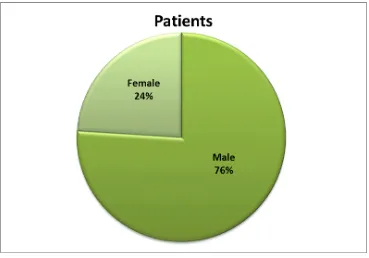
Results: The age group of these patients ranged from 41 to 90 years and the maximum number of patients was in the age group of 51 to 70 years. Gram negative bacilli were
more predominant (73.8%) and the Gram positive cocci (26.2%). Out of 107 isolates,
70 Gram negative bacilli and 20 gram positive cocci were isolated in the age group of
51 to 70. Pseudomonas species was the predominant isolate followed by Klebsiella
species, Proteus species, E.coli, Citrobacter species, Acinetobacter species and
Enterobacter species. Among the Gram positive cocci isolated in this age group (51 to
70years) Staphylococcus aureus was predominant. In most of the infections in the age
group between 51 to 70 years was polymicrobial (31 cases). Gram negative and Gram
positive organisms were highly sensitive to Netilmicin (76% and 81%). Sensitivity to
Amikacin was (59% and 73%). Extended Spectrum Beta Lactamase (ESBL)
approach.
1
INTRODUCTION
Diabetes mellitus is a metabolic disorder characterized by chronic
hyperglycemia and about 150- 170 million people are suffering worldwide
from this diseases, as per WHO reports the prevalence of diabetes will be
double by 2025. Diabetes mellitus is a worldwide phenomenon, type 2
diabetes is the most common form of diabetes in developing countries like
India, hence called diabetic capital of the world. In India prevalence of
diabetes in rural population is about 2.4 %, and in urban population is about
4-11.6 %. Complications of diabetes mellitus are peripheral vascular disease,
cardiovascular disease, nephropathy, retinopathy, neurological and infections.
Uncontrolled hyperglycemia, atherosclerotic vascular disease, sensory
neuropathy are the most important risk factors developing diabetic foot ulcer .1
Twenty five percent diabetic patients have a risk of developing foot
ulcer and limp amputation was 15- 45% higher than non diabetic ulcer.2
PATHOGENESIS:
For development of diabetic foot ulcer, the most important risk factors
are peripheral neuropathy and impaired blood circulation from peripheral
2
Neuropathy:
Development of neuropathy is as a result of hyperglycemia induced
metobolic disorder. The most important one is polyol pathway.
Hyperglycemia state willfavour aldose reductase and sorbitol dehydrogenase
which will convert intracellular glucose to sorbitol and fructose and due to the
accumulation of these sugar products leads to decrease in the synthesis of
myoinositol, which is needed for normal neuron conduction . The
conversion of glucose leads to depletion of nicotinamide adenine dinucleotide
phosphate which is required for detoxification of reactive oxygen species and
for synthesis of vasodilator nitric oxide. This leads to oxidative stress on nerve
cells and increase vasoconstricton leads to is chaemia, which will result in
nerve cell injury and death. This also contributes to abnormal glycation of
nerve cells and leads to inappropriate action of protein kinase C and leads to
further nerve damage.
In diabetic patients neuropathy develops in motor ,sensory ,autonomic
components of nervous system. Imbalance between flexion and extension due
to damage of innervations of intrinsic foot muscles ,leads to foot deformities
that create abnormal bony prominence and pressure points ,which favour for
3
Autonomic neuropathy leads to suppression of the function of sweat
and oil gland. Foot loses natural function of the moisturising the skin and
becomes dry which leads to breakdown and gradually develops infection.
Sensory neuropathy wounds are unnoticed by the patients which
worsenes and exacerbates the development of ulcer.
Vascular disease:
The persistent hyperglycemic state leads to endothelial cell dysfunction
and smooth cell abnormalities in peripheral arteries which result in the
decrease of endothelium derived vasodilator that leads to vasoconstriction .
The diabetes hyperglycemic state leads to increase in thromboxane A2, a
vasoconstrictor, platelet aggregation which promote the risk for hyper
coagulability, and alteration in the vascular extra cellular matrix leads arterial
lumen stenosis. The other factors like smoking, hypertension , hyperlipidemia
contribute to the development of peripheral arterial disease.3
Pathophysiology: In diabetes patients altered protein and lipid
metabolism leads to defective wound healing process. Increased glucose level
in the body end up in uncontrolled covalent bonding aldose sugars to a lipid or
protein without any normal glycosylation enzyme. This product accumulate on
surface of cell membranes, Circulating proteins and structural proteins and this
4
AGE products occurs on extra cellular matrix protein with slow turn over
rate. AGE products alter the properties of matrix protein such as laminin,
collagen and vitronectin through intermolecular cross linking or covalent
bonding of AGE products. AGE products also cross link elastin type on
collagen that leads to increased stiffness and increased synthesis of type III
collagen that forms the granulation tissue. AGE on laminin result in reduced
binding to type IV collagen in the basement membrane, reduced binding of
heparin sulphate proteoglyconand polymer elongation.
Nitric oxide is a stimulator of cell maturation, proliferation and
differentiation and increases fibroblast proliferation and thereby collagen
production in wound healing. Nitric oxide and L-arginine are needed for
proper cross linking of collagen fibres via prolene to maximize the tensile
strength and minimise the scaring of healed tissues. Pulsatile flow of blood
through vessels will activate endothelial cell specific nitric oxide synthase
(EcNOS). Nitric oxide is produced by endothelial cell specific nitric oxide
synthase, maintains the proper blood flow to tissues and diameter of blood
vessels and also regulates angiogenesis, which plays an important role in
wound healing. The diabetic patients reduced ability to produce nitric oxide
from L-arginine, due to high glucose associated kidney dysfuction
ketoacidosis. results in accumulation of nitric oxide synthase inhibitor that
leads to reduced production of nitric oxide synthase and pH dependent nature
5
Diabetic ulcer fibroblasts show various morphological differences, that
usually large and widely spread in the culture flask, compared to the spindle
shaped morphology of the fibroblast in the age matched controls. They often
show numerous vesicular bodies, dialated endoplasmic reticulum and lack of
micro tubular structure. Interpretations of this observations would be that
inspite of high protein turn over and production in diabetic ulcer fibroblasts,
vesicles containing secretory proteins could not travel along the micro tubules
to release the products outside. Diabetic ulcer fibroblasts exhibit proliferative
impairment that contributes to a decreased production of extracellular matrix
proteins and delayed wound contraction and impaired wound healing. For
wound healing, extra cellular matrix not only needs to be lay down and also
able to undergo remodeling and degradation to develop a mature tissue with
appropriate tensile strength.5
Past twenty years major increase in mortality among the diabetic people
is considered to be due to micro and macro vascular complication. Wound
healing process is a step wise repair of last extra cellular matrix (ECM) that
develop the largest component of the dermal skin layer. To avoid over or
under healing, accurate control and rebuilding is essential ortherwise it may
lead to various abnormalities. But in some situation the physiological insult
and certain metabolic disorders that impedes the normal steps of the wound
healing mechanism. One of the examples of metabolic disorder is diabetes
6
inflammatory phase in diabetic wound ulcer which leads to delay in the
formation of mature granulation tissue and reduction in wound tensile
strength. Non healing chronic diabetic ulcers are often treated with
extracellular matrix replacement therapy, advanced moist wound therapy,
negative pressure wound therapy, bio engineered skin or tissue substitute,
growth factors. No therapy is completely perfect as each have their own
disadvantages. Moist wound therapy stimulates keratinocyte and proliferation
and migration, early angiogenesis, collagen synthesis and wound contraction.
Various categories of moist dressings are adhesive, backing film, silicon
coated foam, hydrocolloids and hydro gels etc. Moist wound dressing is not
the best for exudative wounds because it causes fluid retention. In diabetic
ulcer treatment various tissue engineering technologies have come up with a
cellular or cellular skin replacement products. New therapies such as platelet
rich fibrin wound patch which is effective in chronic diabetic ulcer.6
Assessment of diabetic foot ulcers
In 2008 American Diabetes Association of the foot care interested
group recommended components of foot examinations for patients with
diabetes;
1. Visual inspection of bare foot should be performed in a well – lit room .
2. Should include assessment of shoes – inappropriate foot wear leads to
7
3. Should check between the toes for the presence of ulceration or signs of
infection .
4. The presence of callus or nail abnormality should be noted .
5. Temperature difference between feet – suggestive of vascular disease.
6. The foot should also be examined for deformities .
7. To palpate the dorsalispedis and posterior tibial pulses and
characterized as present or absent to asses vascular disease .
Wagner ulcer classification :
Grade Ulcer
0 No open lesion ,skin intact
1 Superficial diabetic ulcer
2 Ulcer involves ligament , tendon ,joint capsule or fascia
3 Grade 2 ulcer with abscess or osteomyelitis
4 Partial fore foot gangrene
5 Extensive foot gangrene.7
In diabetes mellitus patients, one of the major complications is diabetic
ulcer. Fifteen percent diabetes mellitus patients develop diabetic foot ulcer
and leads to 84% of foot amputation.8
The present study was carried out to determine the aerobic bacterial
isolates cultured from diabetic foot infections and their susceptibility to
8
AIMS AND OBJECTIVES
1. To determine the bacterial profiles of infected diabetic foot ulcers and
9
REVIEW OF LITERATURE
In 1973, Burkitt.D.P et al said that diabetes mellitus was a global
burden. Key factors for development of diabetes are environmental socio
economical and metabolic. Complications of diabetes such as neuropathy is to
unbalance glycemia level and is associated with other diseases such as
depression and artheroscelerosis.9
In 1984 Sapico F.L et al said that, diabetic foot infections are treated
empirically according to causative microorganisms may improve the patient
outcome. When optimal sample collection, transport, and culture techniques
are used multiple organisms are isolated from diabetic foot infections.
Interactions of organisms within these polymicrobial mixture leads to
production of virulence factors such as collagenases, hemolysins , proteases
and short chain fatty acids ,that cause inflamation, impede wound healing and
leads to chronicity of infection .10
In 1992 Brike J.A et al said that in india type 2 diabetes is the most
common and 31.7million people are diabetics in India. Complications of
diabetes include peripheral neuropathy, nephropathy, retinopathy,
cardiovascular disease, diabetic foot, hypertension, cerebrovascular disease. In
diabetic patients many factors like trauma ,smoking ,durations of diabetes,
10
disease are the two major factors for developing the foot ulcer. Due to these
risk factors ,untreated minor abrasions can cause foot ulcer, further can get
infection with aerobic and anaerobic bacteria. With the help of broad
spectrum antibiotics and proper foot care may help in healing the ulcer .If at
any point of delay may lead to complications like amputation of foot or limp.11
In 1994 Caputo.GM et al, observed that in India 33 million people are diabetics which was highest in the world, out of which nearly 15% suffer from
the dreaded sequelae of diabetic foot. Certain types of infections are common
in diabetics and others more severe. It is not only the numbers that is
worrisome, situation is different in India due to socio cultural practices as
barefoot walking, religious practices like walking on fire, use of improper foot
wear and lack of knowledge about foot care attributes towards increase in the
prevalence of diabetic foot. Diabetic foot ulcers are not spontaneous ulcers,
but results from the interplay of various factors like peripheral vascular
disease, neuropathy, autonomic neuropathy, alteration in the plantar pressure,
limited joint mobility and defective foot wear. Cell mediated immunity is
mostly affected with abnormalities of polymorphonuclear leukocytes (PMN),
monocytes and lymphocytes. There are abnormalities of adherence,
chemotaxis, phagocytosis, oxidative burst and intracellular killing, also
advanced glycation and products leads to the state of low level persistent
activation in polymorphonuclear cells, which leads to spontaneous activation
11
neutrophil granular components which may lead to burn out or tolerant
polymorphonuclear leucocytes, also may initiate pathologic process leading to
vascular injury. Adapting cellular immunity is also affected with decreased
lymphocyte proliferative response.12
In 1994, Foster.AVM et al, said that in diabetic wound case
management, selection of dressing is also important component, specific
dressing types could prove beneficial depending on the characteristics of the
individual wound. For example saline soaked gauze dressings were
inexpensive, well tolerated, atraumatic, moist wound environment. Foam and
alignate dressings are highly absorbant and decreasing the risk for maceration
in wounds with heavy exudates. However, an ideal dressing should contribute
to a moist wound environment, absorb excessive exudates which increase the
risk for infections.13
In 1995 Steed D, discussed the adjunctive wound care treatment were
under investigation and in practice for diabetic foot ulcer. With the help of
human skin equivalents had been shown to promote wound healing in diabetic
ulcers, through the action of cytokines and dermal matrix components , which
will stimulate tissue growth and wound closure . A recombinant platelet
derived growth factor was also currently in use . Other adjunctive therapies
were hyperbaric oxygen therapy (HBOT) and granulocyte colony stimulating
12
to the patients, which leads to increase oxygen concentration level in the
blood, increase diffusion capacity to the tissues and also increase partial
pressure of oxygen in the tissues, which stimulates neovascularization ,
fibroblast replication, increase phagocytosis and leukocyte mediated killing of
bacterial pathogens in the wound.14
In 1995, Reiber.G E et al, had detected that lower extremity
amputation was the most feared complication of diabetes, in many cases
amputation should be a treatment option, with good rehabilitation patient may
return to normal activities. However, in countries like Poland the supporting
mechanism for amputees are not well developed. Amputation should be
considered in very limited situations. One of the indications for amputation is
serious infection which could be life threatening sepsis. As reconstruction
cannot be performed in ischaemic limb, amputation is considered. And also
the amputation is also considered in significant rest pain which was not
manageable with analgesics.In a major amputation subsequent outcome of the
patient was poor. In 5 years, the mortality rate was as high as 40% to 70%.
The multidisciplinary team approach to diabetic foot has been shown as a
major reduction in amputation incidence.15
In 1995, Gerding.DN, said that anerobes are often participate in a
mixed infections with aerobes, especially in deep tissue infection. But they
13
staphylococcus species, enterococcus species or corynebacterium species are
also represent as true pathogens.16
In 1995, Armstrong.D.G et al, said that the role of anerobes is
particularly unclear because in many studies, samples were not collected
appropreiately for anaerobic culture or due to lack of anaerobic setup in many
institutions. Among those who did with appropriate methods, some reports
that anaerobes play a minimal role, while others have detected 95% prevalence
of anaerobes, in a study with Bacteroidesfragilis being the predominant
anaerobe isolated.17
In 1996, Baird.D et al, identified that diabetic foot ulcer infections are
mostly polymicrobial infections, proper management of these infections
requires an appropriate antibiotic solution based on the culture and
antimicrobial sensitivity testing results.18
In 1997, Stone.J.A et al reported that uncontrolled diabetes is prone to
skin infections, increased blood sugar levels thatleads to inhibit bacteria
fighting cell. Skin infections may be hazadours, even small injury may
progress to ulcer if not properly treated.19
In 1997, Boyko.E.J et al, said that hyperglycemia in diabetes mellitus
further alters cellular function, damage endothelium of vessel valve and
14
the effect of atherosclerosis is as high as 2 to 3 times and calf vessels were
most affected. Peripheral vascular disease may lead to poor healing and
increased risk for amputation. Ischaemia presents as bilateral absence of pedal
pulses and claudication pain. To assess the ischaemia, posterior tibial and
dorsalispedis arteries pulses should be palpated and also to collect the history
of claudication pain. Colour also may be assessed which may be difficult in
dark skin, pale on elevation or rubor on dependency may indicate ischaemia.
Other characteritics were skin temperature (cold on touch), capillary refill
more than six seconds, dry, fissured skin, absence of hair growth, dystrophic
toe nails, presence of oedema, pain or dry gangrene. 20
In 1998, Smith.D et al evaluated that diabetic foot ulceration become
infected approximately 56%. Signs of infections were cellulitis, increase in
local temperature, foul smell, oedema, abscess formation. Due to neuropathy
the pain is absent. Leucocytosis, fever may not be present in about 50%
diabetic patients. Infection may be caused by gram negative, gram positive
bacteria and anaerobes. Short time duration of ulcers were usually infected by
single gram positive organism, but chronic ulcers may yield mixed flora, both
gram positive and gram negative organisms may be with anaerobes.21
In 1998, Armstrong.D.G et al, said that risk factors for ulcer
development were trauma and pressure. It may be necessary to encourage the
15
crutches.Encourage the patient to replace or modify their foot wear. Custom
made shoe inserts (orthoses) may be necessary for pressure reduction or
redistribution. Selection of devices mustbe taken into consideration that the
ability of device to remove pressure, ease of application, cost effectiveness and
ability to gain patient compliance. 22
In 1998 Reiber G.E et al suggested platelet rich fibrin therapy for
chronic or hard to treat diabetic ulcers. Isolation fibrin or plasma from the
patients blood ,which have rich platelets and growth factors to promote natural
healing process. Application of these product to diabetic foot ulcers have been
shown to accelerate healing. Leucopatch is one such product which is a three
layered fibrin patch. It is composed of patients own cells and growth factor
,containing high level of platelets and leukocytes. After six weeks of
treatment with Lekopatch application showed significant reduction of wound
area (65%).23
In 1998, Pathare.N.A et al, had described that dibetes mellitus is one
of the major health problem in world and in India around eighty million
people were diabetic. Asia is contributing more than 60% of worlds population
with diabetes. India and China contritubes the largest. Incident of multidrug
resistant bacteria has been increased in recent years which leads to increased
hospital stay, morbidity, mortality and costs. Diabetic foot infections were
16
factors such as microbial flora of the lower limb, foot hygiene, metabolic
factors and use of antibiotics.24
In 1998, Krasner.D et al proved that neuropathic ulcer, primarily seen
on the plantar aspect of the foot at the base of the metatarsal heads and first
and fifth base of the toe. Ischaemic ulcer mostly occur as distal lesions on the
toe or back of the heal.25
In 1998, Lovery.L.A et al, said that in diabetic patients, once ulcer has
developed, risk of wound progression is increased, that may ultimately leads
to amputation in up to 85% patients of diabetic ulceration may require
amputation. Team approach to wound care, can prevent at least 40% of
amputation in diabetic patients.26
In 1999, Reiber et al, said that factorswhich may affect wound healing
are as follows: wound environment, vascular status, ischaemia, pressure area,
glycemic control and nutrition status. Comorbidities, retinopathy, end stage
renal disease, hypertension, history of amputation and some medications are
also involved in blood glucose control and peripheral vascular disease.27
In 1999, Kelwin.W.S et al, said that in diabetic foot on average of 5 to
6 strains of organisms are often involved with mixture of aerobic and
17
In 1999, Zangaro.G.A et al, discussed about examination of foot
wear, because 55% of traumatic events were result of poorly fitting shoes,
health care providers and clients must be able to assess the appropriateness of
foot wear. The type of shoe, pattrern of wear,fit,linings,seams,insoles or
orthoses and presence of foreign bodies must be assessed and appropriate
intervention implemented. When choosing shoes, clients are advised to shop
late in the day and to be measured both feet. Shoes should be sufficiently
ideal with a deep toe box to accommodate foot changes or deformities. Laces
were preferred to accommodate swelling, natural fibers such as soft leather
more readily conform to the foot and non skid soles and low heals reduce the
risk of falls.29
In 1999, Lipsky. BA identified that Staphylococcus aureus and beta
haemolytic streptococci are the first microorganisms to colonise and infect the
skin. Patients with previously treated or with chronic infections, gram negative
bacilli mainly Enterobacteriaceae was found. Wounds treated with wet
dressing the isolates are specifically Pseudomonas aureginosa.30
In 1999, Tentolouris.N et al, said that diabetic wound infections caused
by Methicillin Resistant Staphylococcus aureus was 30%.31
In 2000, Campbell.LV et al observed that about 60% of diabetic ulcers
were due to neuropathy, about 20% were due to is chaemia and 20% were
18
components which will affect the plan of care. On examination neuropathic
foot generally appear as dry, painless, warm, insensate. Ischaemic foot usually
cold, atrophic skin, dystrophic nails, absent pulses. Sporadic clauducation may
be present. The study stressed to monitor ulcer status and identify change over
time, several important characteristics including size, location and presence or
absence of infections to be assessed and documented.32
In 2000, Fryberg.R.G et al, published that charcotarthropathy is a
structural abnormality in which joint instability due to muscle and ligament
atrophy. Walking a weakened, insensate joint causes structural damage and
results in sprains and stress, fractures to the foot. The acute stage presents as
inflammatory response with bone resorption and then leads to bone destruction
with destruction arch of foot presence as rocket bottom sole and the altered
pressure distribution may increase the risk of ulceration.33
In 2000, Sinacore et al observed that many secondary aging factors
also implicated in delay wound healing. In addition, patient education, healing
potential, physical environment, wound management and quality of life also
are the factors implicated in delayed wound healing. 34
In 2000, Sibbalal et al had detected that intervention to promote wound
healing includes control of infection, tissue debridement, avoid further trauma,
moist wound environment with proper dressing, oxygenation, perfusion,
19
purpose is to remove the dead or devitalized tissue. In the management of
diabetic foot ulcers, common techniques were used such as mechanical
debridement which is irrigation with saline solution, wet to dry dressing.
Autolytic debridement (hydro colloids, hydrogels) and surgical debridement,
which was the method of choice for wounds with large amount of devitalized
tissues or infection, however surgical debridement must be performed only on
tissue with adequate blood supply or blood circulation.35
In 2000, Lipsky.B.A et al, said that several studies have confirmed that,
receiving prior antibiotic treatment for chronic infections or lesions were
usually polymicrobial.36
In 2001, Calhorn et al, observed that in controlling infection,
intervention depends upon the nature of the infection (acute, chronic and
systemic). However general interventions for all wounds promote healing. In
addition to debridement, it is necessary to control bacterial balance, support
host defense and support medical and pharmacological intervention.37
In 2002, Calhoun et al, obsevered that in diabetic wound dressing, no
one dressing was appropriate for all diabetic wounds or the various stages of
healing. Selection of dressing was made on the basis of healing potential and
clinical assessment of ongoing wound status. Wound dressing categories
20
retentive (adherent or nonadherent), absorbant (hydrofibres, foam, hypertonic
saline, algineter) and antimicrobial (cadixomer iodine, silver agents).38
In 2002, Abbott.C.A et al proved that 55% of foot ulcers are due to
pressure from foot wear which can cause trauma to the foot, with reduced
circulation and loss of sensation. Assessment should include checking for
callus foot wear and structural abnormality.39
In 2003, Merza.Z et al, had described that in diabetic foot some of the
risk factors such as biomechanical factors, smoking, level of glycemia were
strongly associated with environmental factors. In certain societies factors
such as average monthly house hold income, racial distribution, education
level may contribute to diabetic foot prevalence.40
In 2003, Jeffcoate et al, discussed about the optimation of wound
environment which involves a number of component. This include assessing
the wound bed for bacterial balance, exudates and need for debridement.
Selection of dressing that can control or manage the wound environment,
maintaining a moist wound bed as required, while keeping the surrounding
wound skin dry, controlling exudates without dessicating the ulcer bed,
eliminate dead space by loosely filling the cavity, ensure that there is adequate
21
In 2004Anadhi C et al said that in diabetic foot patient , risk of leg
amputation was 15-46 times higher than non diabetic patient . Peripheral
neuropathy and poor circulation were the major factors for developing foot
ulcer . In diabetic foot infections were usually polymicrobial ,where the milder
infections are monomicrobial .42
In 2004, Schultz G.S etal suggested to evaluate patient wound status
and compatibility with treatment goals. The wound edge should be examined
to determine the presence of cell migration and wound closure. If wound
healing was not occuring, status of those factors could be corrected.43
In 2004, Farish.P.L et al, said that incase of ischaemic diabetic foot,
Bypass surgery is a common method of treatment and reported long term
result. Ten years limb salvage rate with surgical bypass of lower limb was
upto 90%. In case of multiple occlusion, revascularisation at each point to
restore arterial blood flow and increase the chance for limbs salvage.
Transluminal angioplasty of the iliac artery in conjunction with surgical
bypass in the distal extremity may be implemented and efficiency has been
demonstrated in diabetic patients.44
In 2004, RNAO (Registered nurses association Ontario) conveyed that
nurses have a major role to identify the emerging problem, to promote
22
it have been identified that five primary risk factors that can be quickly
assessed and screened by nurses.
These factors are:
• Circulation.
• Sensation.
• Past history of foot ulcer.
• Structural and biochemical abnormalities.
• Knowledge and selfcarebehaviour.
The presence of one or more of this risk factors is favour for developing
foot ulcer and amputation of lower limb. Nurses contribute a key role by
identifying such risk factors, informing and providing referrals for clients at
risk to prevention strategy.
In 2004, Lipsky.B.A et al, reported that in India 40 million people are
suffering with diabetes mellitus and of equivalent magnitude in other
developing countries. In that upto 20% of patients were struggling with
diabetic foot complications and hence are the most commonly faced surgical
problem. In treatment was not appropriate, it may lead to amputation or
disarticulation of varying levels, atleast once in such patients life time. Most
23
clinical knowledge of the treating doctor and the prevalence of the microbial
pattern in the hospital and locality. It would be prudent if the treatment is
directed based on the hierarchy of the organisms most commonly isolated and
the most common antibiotic sensitivity pattern of these organisms, at the onset
and thus help in a better outcome. Several studies have been conducted world
wide with respect to the bacteriology and antibiotic sensitivity pattern. A
number of studies have found that Staphylococcus aureus and other gram
positive aerobes were the most common causative pathogens, usually isolated
in more than 60% of cases.45
In 2005, Koneman.W.K et al have shown the selection of antibiotic
agent for treating diabetic foot infection require the knowledge of the potential
microbial pathogen and the resistance to the commonly used antibiotics. To
assess the right antibiotic to manage the diabetic foot ulcer infections, the
result of misuse and abuse of specific antibiotic studies were needed.46
In 2005 Lipsky.BA et al ,conducted a multicentre study (‘SIDESTEP’)
showed Ertapenem was ineffective against pseudomonas, compared with
Piperacillin / Tazobactam. Although in some wound cultures involved by
Pseudomonas aeruginosa, revealed similar outcomes. The authors from this
study and several other studies from western countries said that Pseudomonas
aeruginosa was the commensal organism rather than a pathogen, since could
24
aeruginosa, need wound care measures, such as avoiding moisture in the
periwound area, frequent changing of wound dressing and avoiding
hydrotherapy based wound care.47
In 2005, Singh.N et al said that in India diabetes associated problems
are the second most common cause of lower extremity amputation.48
In 2005, Bakker.K et al, said that globally, every 30 seconds one lower
limb is lost due to diabetic foot ulcers.49
In 2006, Kulkarni.J et al reported that 50% of amputed patients will
die within five years of amputation, according to USA data mortality in the
group of diabetic foot complications was comparable to the mortality in some
types of cancer.50
In 2006, Tsae.S.M et al, said that Staphylococcus aureus infection in
the diabetic foot accentruted the inflammatory process, endothelial injury and
blood coagulation, ultimately leading to quicker death.51
In 2007 Citron D .M et al observed that anaerobes was almost always
present in mixed culture . F.magna was predominant organism this was in
contrast to other study ,which failed to isolate anaerobic gram positive cocci
due to not using selective media for anaerobic gram positive cocci ,suboptimal
collecton and transport methods .Most frequently used selective anaerobic
25
vancomycinagar, growsB. Fragilis group and Prevotella species ,but not gram
positive anaerobes .52
In 2007,Stansbury.L.G et al described that in diabetic patients foot
amputation ratio was higher than soldiers taking active part in military, which
incorporate 2.3% of all battle injuries and 7.4% of major limb injuries.53
In 2008, Frykberg.R et al found that in diabetic and neuropathy patient
may develop charcotosteoarthropathy which was characterised by progressive
destruction of bones and joints of the diabetic foot accompanying with
osteopenia. Incidence of this complication, range between 0.1% to 30%. In
about 25% of charcotosteoarthropathy may be missed or diagnoses may be
delayed due to lack of specific markers of charcotosteoarthropathy, which may
lead to significant deformity, ulceration and amputation of foot.54
In 2008 Flynn.N et al, said that Staphylococcus aureus is most
important pathogen among the staphylococci and found in the environment
and anterior nares of 20 to 40% of adults. Other sites of colonisation are axilla,
vagina, skin fold and the perineum. Staphycoccusaureus has a variety of
virulence factors and the ability to develop and expand resistance to broad
spectrum antibiotics. Patients with diabetic foot infection is the major cause of
morbidity. They occur in 15% of diabetic patients and 20% of all hospitalized
26
In 2009 Paul S et al said that foot ulceration and infections are the most
frequent serious complication of diabetes mellitus . The annual incidence of
leg and foot ulcer was 2.6,5.33, times more common than diabetic coronary
disease, stroke, renal failure respectively . About 15% of diabetic patient
develop foot ulcer in their life time . Studies past 25 years on bacteriology of
diabetic foot infections , but the results are varied and contradictory .56
In 2009, Orji.F.A et al, have shown that incidence rate was high in
males. Polymicrobial organisms (56%) were higher than monomicrobials
(53%). Prevalence rate of the bacterial isolates were Clostridium species 51%,
Staphylococcus aureus were 60%, E.coli 20% and Klebsiella aerogenes 12%.
Antibiotic sensitivity pattern of Clotridium species showed sensitive to
Fluoroquinolones and high resistance to beta lactams. E.coli and
Klebsiellaaerogenes showed resistant to beta lactams and aminoglycosides.
All gram negative organisms showed significant sensitivity to
fluroquinolones.57
In 2010 Zubair M observed that prevalence of diabetic foot ulcer in
male was to be 56.6% and female was 30% and ratio of 3.5:1 . bacteriological
evaluation of diabetic foot ulcer infections showed that prevalence of gram
negative organisms were found to be more than gram positive organisms .The
27
patients in india due to misuse of antibiotics , this leads to longer duration of
hospital stay and their treatment could be more coastly .58
In 2010 Shakil.S said that diabetes patients have 10 times higher risk
of being hospitalised than non diabetes for soft tissue and bone infections . By
the year 2025 in India diabetic populations is expected to increase to 57
million.59
In 2011 Chopdekar K.A et al did a study on bacteriological profile of
diabetic foot infection ,out of 113 samples ,a majority of samples that is 96
(85%) showed polymicrobial growth of which, 29 was mixed growth of only
aerobes 67 was mixed growth of aerobes and anaerobes. Out of 290 isolates
,223 were aerobes 67 were anaerobes . Among the aerobes gram negative
bacilli were 133 , gram positive cocci were 90 ,majority were Staphylococcus
aureus (50) , followed by Pseudomonas aeruginosa (47), Acinetobactor
species(2)among the anaerobes ,46 were gram positive cocci, 21 were bacilli
in that 16 were gram negative and 5 were gram positive. The majority of
anaerobes were Peptostreptococcus species 38(57%) , followed by anaerobic
streptococci 8(12%).60
In 2011 Pappu A.K et al did a study on microbiological profile of
diabetic foot ulcer out of 104 samples ,average of 1.08 species per diabetic
ulcer patient . In that more than one organism was isolated in only 7.7%. Gram
28
gram negative aerobes, Pseudomonas was 23% isolated,followed by
Klebsiella(17%) , Proteus mirabilis(15%),E.coli (12%), Acinetobactor(6%),
Proteus vulgaris (2%) and Streptococci (4%) . Second commonest was
Staphylococcus aureus(21%). No anaerobes were isolated .22% of amputees
each were infected with Psedomonasaeruginosa and Proteus mirabilis ,18%
Klebsiella, 14% Staphylococcus aureus in that 6% were methicillin resistant
and Acinetobactor 4% infections in amputees .61
In 2012 Banashankari G.S observed that choice of specimen for
culture and sensitivity was tissue and more specific and sensitive than swab
because it yielded pathogenic organism by eliminating contaminants but the
same time isolation with swab was reliable ,but there is a possibility of
isolating only contaminant, it has to be done with at most care. All ulcers
were thoroughly washed with sterile saline to avoid the colonizer rather than
pathogenic and specimens were collected by scraping from base of ulcer ,
wound curettage or aspiration rather than wound swab . Majority of isolation
was single organisms it was 64% and rest were polymicrobial and about three
or more organisms were 5% .Aerobic facultative organisms were isolated in
98% , anaerobic was 2% . In that 66% gram negative isolates were isolated
with predominant organisms being Proteus which was 18% , E.coli were 16%
and Pseudomonas 13% . Among the gram positive organisms 19% were
Staphylococcusaureus followed by Enterococcus were 9% , coagulase
29
1.5:1 . Minimal contaminants and minimal isolation of Staphylococcus
epidermidis due to tissue technique method (56%), less use of swab . 18.4%
were gram positive organisms,34% were gram negative organisms,remaining
had grown both (gram positive and gram negative organisms) .62
In 2012 Esmat M.M et al observed that diabetes patients have risk of
developing diabetic foot ulcer in their life time as high as 25% . 15 to 45 times
higher risk of limp amputation in diabetic ulcers than ulcers due to other
causes .2
In 2012 Manisha J et al observed that prevalence of diabetes mellitus
depends upon many factors, that is age, sex, socio-economic status, diet,
heredity, physicalactivity, life style and environmental factors. Incidence
ranges in the annual population from 1.0% to 4.1%, prevalence ranges from
4% to 10% and the life time incidence as high as 25%.Males to females ratio
was2.1:1, the mean age of patients was 50.25 + 12.5.63
In 2012, Tiwar.S et al, found that diabetic ulcer with
polymicribialinfection had comparatively higher total leucocyte count
(16,928+ 9,642 versus 14,593+6,687) and haemoglobin level significantly
lower (7.9+2.4 versus 9.2+ 2.2) than monomicrobial infections. HbA1C level
in both groups were similar (9.9% versus 9.5%).Patient infected with gram
negative bacteria were also had significantly lower level of Hb (8.5 + 1.9
30
9771+ 3243). Neutrophils level was higher (77 versus 67), than infected with
gram positive bacteria.While infected with both gram positive and gram
negative bacteria had significantly lower level of Hb (7.6 + 3.2 versus 11.1 +
2.2), than infected only with gram positive microorganism. But this was
insignificant when patient infected with only gram negative micro organism.
In case of culture negative suspicion of infection was high can use molecular
techniques for diagnosis of bacterial infection. To differentiate infected from
noninfected foot ulcers, inflammatory markers were used. However positive
culture and sensitivity result have a priority over the molecular study for the
selection of antibiotics.64
In 2012, Shim.V.R et al, detected that males were more prone for
diabetic foot ulcers than females. This may be due to differences in
biomechanics between male and female, specifically high foot pressure
decrease joint mobility. Male have nearly twice the odd of having insensate
neuropathy as women with diabetes.65
In 2012 McInnes.A.D et al suggested that diabetic foot protection team
working across primary and secondary care could reduce length of hospital
stay for diabetic foot ulcer, also reducing major amputation ratio. Median
length of hospital stay over the period of three years for diabetic foot ulcer ,
31
In 2012 Blumberg.S.N et al suggested that an innovate and promising
treatment for diabetic foot ulcer is stem cell theraphy.67
In 2012, Forbes.J et al, said that, in the treatment of wound amniotic
membrane has been used which is rich in collagen and various growth factors
and it will support the healing process to improve the wound closure and it
will reduce the scar formation. In early they have been used natural amniotic
membrane which obtained from labour and delivery. Recently techniques have
been developed to dehydrate the material while preserving many of these
wound healing attributes to produce temperature stable allograft.68
In 2013 Konar.J et al reviewed that wound healing was a stepwise
repair of lost extra cellular matrix that forms largest component of the dermal
skin layer. Accurate and controlled rebuilding is necessary to avoid over or
under healing that may lead to various abnormality. Sometimes wound healing
was disturbed by certain disorder and physiological insult. One such disorder
was diabetes mellitus, disturbs the normal steps of wound healing process.
Many histopathological studies show in diabetic wound inflammatory phase
will be prolonged, this leads to delay formation of mature granulation tissue
and parallel reduction in wound tensile strength. 67cases out of 150 identified
bacteriology etiology (38%), single organism was isolated in 58 (87%) among
which Pseudomonas aeruginosa was the commonest (21 cases), followed by
32
vulgaris, Enterococcus, Klebsiella pneumoniae were each 2 cases.
Polimicrobial was isolated in 9 cases ,in that Staphylococcus aureus along
with Klebsiella oxytoca was isolated in 4cases, rest of 5 cases isolated
Pseudomonas aeruginosa with Escherichia coli .Among 50 gram negative
bacteria, 23(46%) produced ESBL, 17(33.33%), were Amp C beta lactamase
producers and carbapenamase producers were4(8%); 33 gram negative isolates
were resistant to fluoroquinolones (66%). Extended spectrum beta lactamase
producers were Escherichia coli followed by Pseudomonas aeruginosa.
Carbapenamase producers was exclusively Pseudomonas aeruginosa .Amp C
beta lactamase producers was Pseudomonas aeruginosa followed by Klebsiella
pneumonia andKlebsiellaoxytoca. Out of 19 isolates of Staphylococcus
aureus, 7 were methicillin resistant (36.84%). All Staphylococcus aureus were
sensitive to both linezolid and vancomycin. One Enterococcus faecium was
vancomycin resistant and MIC value was > 64mic.gram/ ml and was sensitive
to teicoplanin, Dalfopristin/Quinupristin and Linezolid. Isolated Enterococcus
faecium were resistant to Penicillin, Gentamycin, Tetracycline,
Fluoroquinolone leaving behind very restricted therapeutic options .69
In 2013 Shanmugam.P said that poor micro vascular circulation in
diabetic ulcer patient, limit the phagocytes and favour the development of
infection .Improper foot wear and local injuries further compromise blood
circulation in the lower extremities .diabetic foot infections are initially treated
33
Many studies in the past 25 years reported on bacteriology of diabetic foot but
the results were varied and often contradictory. These could be due to
differences in the causative organisms occurred over time, geographical
variation, type and severity of infections. Most of the time diabetic foot
infections were polymicrobial , proper management of the infections requires
an appropriate antibiotic selection based on cultures and antimicrobial
susceptibility testing results. In recent years increase in the incidence and
prevalence of extended spectrum beta lactamase and carbapenemase producers
may be due to paucity of data on extended spectrum beta lactamase and
carbapenemaseproducers .70
In 2013, Rani.KL etal observed that out of 150 cases of diabetic foot
107 were males and 43 females, which mean that diabetic foot infections were
common in men than women. This could be because diabetes is more common
in men, and are prone for trauma because of their outdoor work. In this study
they found that diabetic foot infection were common in 40 to 60 year age
group. Out of 150 cases, 98.66% was non insulin dependent diabetes mellitus
(NIDDM) type. In this study 53.6% of the cases was suffering from diabetes
mellitus for more than 6 years. Enterococcus species was isolated only from 4
cases (1.85%) but in most of other studies they isolated in a range of 4 to 30%.
Gram negative bacilli was isolated more commonly in this study which was
188 (87.03%). Among gram negative organism Escherichia coli were the
34
(23.14%) Pseudomonas aeruginosa (13.42%) Klebsiellapneumoniae (12.03%)
and the other gram negative bacilli were Proteus vulgaris (6%)
Citrobacterfreundii (1.85%), Enterobacter species (3.7%),
Morganellamorganii (1.38%).
In the management of diabetic foot infections, extended spectrum
cephalosporins group of drugs shows good results. The patients with
necrotising infections are severely ill, require broader antibiotic coverage.
Sensitivity pattern of Staphycoccusaureus were Vancomycin 100%,
Cefotaxime (65.2%), Ceftazidime (56%), Clindamycin (34.78%), Gentamicin
(34%) and Ampicillin (34%), Ofloxin (34%) and Amikacin (30.4%). This
study showed higher rate of resistance. This due to patients received treatment
early which could have eliminated sensitive organisms and remain only
resistant organism. Sensitivity pattern of Enterococcus species were
Vancomycin (100%), Amikacin (50%), Gentamicin (25%), Netilmicin (25%),
Ofloxacin (25%), Cefotaxime (25%), Ceftazidime (25%), Ceftriaxone (25%).
Gram negative bacilli is more sensitive to Imepenem, Piperacillin/
Tazobactam, Piperacillin, Amikacin, Cefotaxime, Ceftazidime, Ceftriaxone
and Gentamycin, Ofloxacin.
In this study Escherichia coli was the most common isolate (25.46%),
which was sensitive to Imepenem (96%), Piperacillin/ Tazobactam (90%),
35
(54.5%), Ceftazidime (49%), Gentamicin (30.9%) and Ofloxacin (30.9%).
Proteus mirabilis sensitive to Imepenem 98%, Piperacillin/ Tazobactum 90%,
Cefotaxime 60%, Ceftriaxone 58%, 90% sensitivity to Amikacin. Proteus
vulgaris was sensitive to Amikacin 79%, Cefotaxime 38.46% and other
routinely used antibiotics showing maximum resistant.
Enterobacter species sensitivity pattern was Ofloxacin (87.5%),
Cefotaxime (75%), Amikacin (75%), Ceftazidime (62.5%), Gentamicin
(50%), Ceftriaxone (50%). Citrobacterfreundii sensitive to Cefotaxime was
50% and Ceftriaxone 50%. Morganellamorganii was sensitive to Cefotaxime
66%, other antibiotics were 33%. Pseudomonas species was sensitive to
Imepenem 93%, Piperacillin/ Tazobactam 83%, Piperacillin 83%, Amikacin
was 55%, other routinely used antibiotic were less sensitive.
Patient education was the most important aspect, once patient was
diagnosed as diabetes, it is doctors responsibility to advice the foot care in
diabetic and make them understand the complications of diabetic foot
infection.71
In 2013, Turhan.V et al reported that in diabetes foot infections
Staphylococcus aureus was long been recognized as the predominant micro
organism but in this study it was second frequently isolated organisms. In
1990 community acquired MRSA emerged as the important isolates in
36
species. 44.2% methicillin resistance strain isolated in this study. They found
that fusidic acid was effective against all Staphylococcus species, including
MRSA and in mild to moderate diabetic foot infection, Fusidic acid may be
considered as an important therapeutic alternative.72
In 2013, Salihi.S.A et al, did a study on 25 patients with diabetic ulcer.
In that 17 were non insulin dependent diabetes mellitus, 8 were insulin
dependent diabetes mellitus and they belong to age group between 40 to 49
years. This may due to repetitive mechanical force of weight during working.
All the 25 samples isolated showed pure form of isolates. Among that 14
(56%) were E.coli, 7 (28%) were Proteus mirabilis and 4 (16%) were
Staphylococcus aureus. Antibiotiuc pattern of E.coli was more sensitive to
Rifampicin, Ciprofloxacin and Nitrofurantoin. From this study they found that
Gentamicin had highest antibacterial activity to E.coli 85.7%, Proteus
mirabilis 42.8%, Staphylococcus aureus 75%, while antibiotic resistance of
Nalidixic acid to E.coli was 100%, Proteus mirabilis 71.4%. Multidurg
resistant may be due to chronic course of admission and chronicity of the
wound.73
In 2013, Hameedhefni.A.A et al, identified that the diabetic wound
infections begins superficially, but delay in the treatment and impaired body
defence mechanism infection can spread through deeper tissues which leads to
37
In 2013, Jaya.M et al evaluate that maximum number of patients
between the age group of 60 to 65 years. Totally 75 organisms isolated from
50 patients. He isolated monomicrobial and polymicrobial 25% equally.
Among the isolates gram negative bacilli was isolated more than gram positive
cocci. In gram negative bacilli, Pseudomonas species was higher (16%)
followed by E.coli (14.6%) and the other organisms were Methicillin sensitive
Staphylococcus aureus (13.3%), Streptococcus pyogens (10.6%), Klebseilla
species (8%), Acinetobacter species 8%), Methicillin resistant Staphylococcus
aureus (8%), Proteus mirabilis (6.6%), Citrobacter species (5.3%),
Enterococcus species (5.3%), coagulase negative Staphylococcus (2.6%),
Enterobacter species (1.3%).
Among the gram negative bacilli 37.5% of the isolates were extended
spectrum beta lactamase (ESBL) producers, 31% of isolates were
Carbapenamase producers. Diabetic foot ulcers were ischaemic 74.1%
followed by neuropathic type. Most common locations of diabetic ulcer was
toes (big toe followed by second toe) plantar region of the foot and dorsum of
foot, which was >70%.75
In 2013, Madanchi.N et al, did a descriptrive study on 873 diabetic
ulcer patients. He found that mean duration of diabetic foot ulcer prior to
admission was 79.8 days. Mean haemoglobin A1C (HbA1C) level was 9.51%
38
amputation was 16.4% of patient. In that 4% of patients had major amputation
(above ankle), and 12.5% had minor amputation. Majority of patients had
ischaemic type of diabetic foot ulcer was 74%, Neuropathic diabetic foot
ulcer was 17.4% and neuroischaemic (both type) was 8.5%. Diabetic foot
ulcer in the right lower limb is 53.4%, in the left lower limb 38.8% and both
lower limbs are 7.8%. Most common location of diabetic foot ulcer was in big
toe. Followed by second toe. Diabetic foot ulcer at the site of previous
surgeries like debridement, amputation or venous graft removal for coronary
artery bypass graft (CABG).
Out of 873 patients 28.2% of patients underwent lower limb
amputation, in that 38.3% was major amputation, 61.7% was minor
amputation. Mean duration of hospital stay was 16.7+/- 11.3 days, and
mortality rate was 5.2%. Most of the patients developed diabetic foot ulcer in
the mean age of 59.3 years. Other studies also found that diabetic foot ulcer to
be 55- 60 years. In the study population male patients was dominant, which
was 58.1%, Other studies also reported the same, that in the average of 50%-
63.3% in their study population.
Most of the patients 85.6% had uncontrolled diabetes mellitus, their
HbA1C level was more than 7%. It is suggesting that diabetic foot ulcers are
mostly developed in poorly controlled diabetes mellitus. Due to diabetes
39
hospitalization. More than 50% of the patients had renal and cardiovascular,
more than 40% had ophthalmic comorbidities.
Diabetic foot ulcer was more common in patients with past history of
amputation and foot ulceration which was 57 times higher than patients
without this history. 22.4% of patients had previous of hospitalization because
of diabetic ulcer. Previous hospitalization due to other comorbidity of diabetes
mellitus including renal, cardiovascular, cerebrovascular, ophthalmic
complication was 47.2%. Relationship between diabetic foot ulcer and
comorbidity was controversial but many reports found that comorbidity is an
independent risk factor for development of diabetic foot ulcer and amputation.
He found that 74.1% of diabetic foot ulcer was ischaemic type followed by
neuropathic type. To compare right lower limb to left lower limb, diabetic foot
ulcer by ratio, It was 1.37:1, it is because most patients are right dominant,
using more on right which may expose to trauma. Most common locations of
diabetic foot ulcer was toes (big toe followed by second toe), plantar region
and dorsum of foot, which was more than 70% of the patients in the study.
Comparing with proximal part, distal part of limb was more affected. This is
mostly because of is chaemia, diabetic neuropathy and trauma. Diabetic foot
ulcer at the site of previous surgery was identified in 26 patients, which
suggesting to avoid unwanted surgical procedures in left of diabetes mellitus
patients. Average stay of hospitalization was 16.7 days and 28.2% of patients
40
rate will vary from different report in different countries. This study suggested
that multi disciplinary approach (by a team of Orthopaedic surgeon,
Endocrinologist, Vascular surgeon, Infectionist, Internist, Interventional
cardiologist, General practioners, Nurse, Physiotherapist) decreases the
amputation rate, because multidisciplinary approach is for early detection,
prevention, personal education and multiple therapies (dressing, drainage,
frequent debridement, washing along with antimicrobial therapy and daily
assessment of wound healing).76
In 2013, Zycover.S et al, did a randomised, double blind, placebo
control study and he indicated that in the treatment of diabetic foot ulcers,
soluble beta glucans were effective. Soluble beta glucans are available with
the brand name woulganbiogel.77
In 2014 Kaur. N et al did a study on clinical susceptibility profile of
diabetic foot and observed that , out of 106 samples were cultured ,in that 98
was culture positive ,136 organisms was isolated averaging of 1.38 organisms
per positive culture. No growth was obtained in 8 patients (7.6%) . Growth of
one organisms showed in 70 (71.4%) ,growth of 2 organisms was showed in
18 (18.36%) ,and three or four organisms were isolated in 10 patients (10.2%)
. Most commonly isolated organisms were gram negative , which was 67.6%,
gram positive organisms were 28.6% ,while yeast were 3.67% . Among the