DISSERTATION ON
ALTERATION IN APPARENTLY NORMAL BUCCAL
MUCOSAL CELLS OF SMOKERS AND
NONSMOKERS USING SILVER STAINING
NUCLEOLAR ORGANISING REGIONS
A STUDY OF 75 CASES
Dissertation submitted to
TAMILNADU DR.M.G.R. MEDICAL UNIVERSITY
CHENNAI
for
MD (PATHOLOGY)
APRIL(2015)
Under the guidance of
DR.K.VALARMATHY,M.D.,
PROFESSOR,
DEPARTMENT OF PATHOLOGY
GOVT.STANLEY MEDICAL COLLEGE,
CHENNAI
THE TAMILNADU DR.M.G.R.MEDICAL UNIVERSITY,
CERTIFICATE
This is to certify that this dissertation titled “ALTERATION
IN APPARENTLY NORMAL BUCCAL MUCOSAL CELLS
OF SMOKERS AND NONSMOKERS USING SILVER
STAINING NUCLEOLAR ORGANISING REGIONS – A
STUDY OF 75 CASES” is the original and bonafide work done by
Dr.K.Shanmugam under the guidance of Dr.K.Valarmathy,M.D., Professor, Department of Pathology at the Government Stanley medical College & Hospital,Chennai-600 001,during the tenure of his course in M.D.Pathology from May 2012 to April 2015 held under the regulation of the Tamilnadu Dr.M.G.R. Medical University, Guindy ,Chennai-600032
Prof.S.MARY LILLY,M.D
Professor and Head, Department of Pathology,
Government Stanley Medical College, Chennai-600 001.
Prof.AL.MEENAKSHISUNDARAM.M.D.,
DEAN,
Government Stanley Medical Colle ge, Chennai-600 001.
Place : Chennai Place : Chennai
CERTIFICATE BY THE GUIDE
This is to certify that this dissertation titled “ALTERATION
IN APPARENTLY NORMAL BUCCAL MUCOSAL CELLS OF
SMOKERS AND NONSMOKERS USING SILVER STAINING
NUCLEOLAR ORGANISING REGIONS-A STUDY OF 75
CASES” is the original and bonafide work done by Dr.K.Shanmugam
under my guidance and supervision at the Government Stanley Medical College & Hospital ,Chennai-600 001,during the tenure of his course in M.D.Pathology from May 2012-April 2015 held under the regulation of the Tamilnadu Dr.M.G.R. Medical University, Guindy, Chennai-600032
PROF.K.VALARMATHY
Professor,
Department of Pathology,
Government Stanley Medical College, Chennai-600 001.
DECLARATION BY THE CANDIDATE
I solemnly declare that this dissertation titled “ALTERATION IN APPARENTLY NORMAL BUCCAL
MUCOSAL CELLS OF SMOKERS AND NONSMOKERS
USING SILVER STAINING NUCLEOLAR ORGANISING
REGIONS –A STUDY OF 75 CASES” is the original and
bonafide work done by me under the guidance of Dr.K.VALARMATHY M.D., Professor, Department of Pathology at the Government Stanley Medical College & Hospital ,Chennai -600 0001 ,during the tenure of my course in M.D.Pathology from May-2012 to April 2015 held under the regulation of the Tamilnadu Dr.M.G.R. Medical University, Guindy, Chennai-600032
Place : Chennai Signature by the candidate
ACKNOWLEDGEMENT
I take this opportunity to express my heart felt gratitude to DR.S.Mary Lilly,M.D., Professor and Head of the Department of Pathology, Stanley Medical College, Chennai for her keen interest, constant encouragement, guidance and valuable suggestions throughout this study.
I would like to express my sincere gratitude and appreciation for my guide, Dr.K.Valarmathy,M.D., Professor of Pathology, Stanley Medical College for her kind and able guidance, immense help and timely advices towards the completion of my study. Her constant motivation and drive were the key factors for the construction of this study. I am extremely grateful to her.
I am extremely thankful to Dr.P.Arunalatha,M.D., Professor of Pathology, Stanley Medical College who has extended her encouragement and support during the study.
My sincere thanks to Dr.K.Chandramouleeshwari M.D., Professor of Pathology, Stanley Medical College for her immense help and support for the completion of this study.
I am extremely thankful to Dr. A.Jamila M.D.,Professor of Pathology, Stanley Medical College for the constant encouragement and guidance offered by her during the study.
It gives me immense pleasure to thank All Assistant Professors, Department of Pathology, Stanley Medical College who had extended their valuable guidance and support during the study.
Last but not the least, I am grateful to all the faculty members, my colleagues and the technical staff members of the Department of Pathology, Stanley Medical College, my family members and my friends for their constant support and encouragement during the period of study.
CONTENTS
S.NO. TITLE PAGE NO.
1. INTRODUCTION 1
2. AIMS AND OBJECTIVES 4
3. REVIEW OF LITERATURE 5
4. MATERIALS AND METHODS 82
5. OBSERVATION AND RESULTS 85
6. DISCUSSION 95
7. SUMMARY 105
8. CONCLUSION 106
9. BIBLIOGRAPHY
10 ANNEXURE
ABBREVATIONS
Ag+ - SILVER ION
AgNOR - Silver stained nucleolar organizing regions
DNA - Deoxyribonucleic Acid
D.P.X - Dibutyl phathalate xylene
EGFR - Epidermal Growth Factor Receptor 2
GST - Glutathione-S-transferases
HIV - Human Immunodeficiency Virus
ICD - International Classification of Diseases
mAgNOR - Mean AgNOR
NDMA - Nitrosodimethylamine
NNK - Nitrosamine 4-(methylnitrosamino)- 1-(3-pyridyl)-1-butanone
NNN - Nitrosonornicotine
OSCC - Oral Squamous cell carcinoma
PAH - Polycyclic aromatic hydrocarbons
p-value - Probable value
rDNA - Ribosomal DNA
RNA - Ribonucleic acid
SD - Standard deviation
UGTs - Uridine-5′
-Disphosphateglucuronosyltransferases
ABSTRACT
BACKGROUND: Smoking is considered to be initiators of dysplastic changes
in the oral mucosa.
AIM: The aim of this study was to determine and compare the alteration in
apparently normal buccal mucosal cells in smokers and non –smokers due to the effect of tobacco by assessing silver stained nucleolar organiser regions.
(AgNOR)
MATERIALS AND METHODS: The study comprised of 75 subjects divided
into two groups with 25 subjects having smoking habit and 50 subjects who were non smokers. Cytological smears were taken from each subject with the help of a cytological brush. The smear was wet fixed and stained with AgNOR and assessed for nucleolar organising regions.50 cells were counted in each slide.
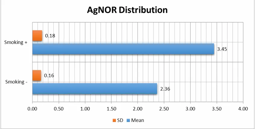
RESULTS: Unpaired T-test and Pearson’s R correlation test was applied.
Cytological changes in smokers revealed an increase in mean AgNOR in comparison with non-smokers.
CONCLUSION: Tobacco smoking produce alterations in apparently normal
INTRODUCTION
Medical science has made considerable progress with respect to infectious diseases. In case of carcinoma, there is a lot to be achieved, although a good deal of innovations and lifesaving therapies have been discovered. In case of oral cancers, it begins with use of tobacco. It is the powdered leaf of a plant which was used in a Y shaped piece of cone called “Tobago”.
Malignancy of the oral cavity is the sixth most common malignancy worldwide.1 It has a dismal 5-year survival, except when it is detected in early stages. The established method for diagnosis is by biopsy, which is carried out only when patient is symptomatic. Hence it is of little value in detecting at an early stage and preventing the progression.
The prognosis is good if detected earlier, in case of mouth cancers. Many methods have been used to identify pre-malignant lesions as a marker of impending malignancy. They are assessment of mitosis, DNA ploidy status, DNA and RNA in situ hybridization, monoclonal antibodies to detect proliferation related antigens. The issue is, they are expensive, time consuming and need sophisticated equipment.
Nucleolar organizing regions are loops of DNA, that transcribe to the ribosomal RNA, which in turn results in synthesis of proteins by the cell. Nucleolar organizing regions are correlated with cellular proliferation.
“Doctors, nurses, midwives, dentists, pharmacists,
chiropractitioners, psychologists and all other professionals
dedicated to health can help people change their behavior. They
are on the frontline of tobacco epidemic and collectively speak
to millions of people.”
Quote by DR.Lee jong-wook,
AIM OF THE STUDY
REVIEW OF LITERATURE
ILL EFFECTS OF SMOKING
People can be broadly classified as non -smokers and smokers .Further,they are subclassified as
Non smokers - a) Never smokers b) Former smokers
Smokers - a) Minimal-who smoke <15cigarettes / day b) Moderate-who smoke between
15-25cigarettes / day
c) Heavy –who smoke >25 cigarettes / day2
They are prone to a wide range of diseases. Smoking can lead to recurrent pulmonary infections, COPD, pulmonary tuberculosis, peripheral vascular diseases, myocardial infarction, stroke, hip fracture. It can have a major role in the etiopathogenesis of various cancers. In 20th century it killed about 100 million People worldwide.
MECHANISM
The cancer causation by cigarette smoking follows these steps
(1) Exposure to the carcinogens.
(2) Establishment of bonds between carcinogens and DNA.
(3) Accumulation of somatic mutations in genes.
Each puff of cigarette has many compounds, of which 60 are well known carcinogens. These carcinogens can be subdivided into various classes, including, aromatic amines, volatile organic compounds, polycyclic aromatic hydrocarbons (PAH). The metabolites of these carcinogens are elevated in blood, breath and mainly urine.3
detoxification and activation of carcinogens shows considerable variation among persons and affects susceptibility to malignancies.
The carcinogens, once activated results in DNA adduct production, which is vital in the carcinogenic process. A few can directly form DNA adducts. Studies found that adduct levels is elevated in smokers .5
The repair system removes DNA adducts. They are direct repair of DNA bases by alkyl transferases, double-strand break repair, and mismatch repair6. If these repair enzymes do not function properly. These adducts may remain and raise the chance of producing somatic mutations.
Persistent DNA adducts miscoding during replication of DNA. Particular DNA adducts leads to specific somatic mutations. KRAS and Tp53 oncogenes are subjected to such changes.
Nicotinic receptors are the sites where nicotine and nitrosamines bind. These activate protein kinase A and protein kinase B7. Smoking stimulates production of EGFR8 and COX-2, which results in changes leading to cell proliferation. These products enhance the carcinogenicity of these constituents of tobacco. The changes which carry an increased risk of cancer, are reverted on cessation of smoking.
An important pathway of enzymatic hyper methylation, which is termed as epigenetic changes9. It produces gene silencing of mainly tumor suppressor genes and can lead to proliferation of cells which are unregulated.
CARCINOGEN EXPOSURE
The exposure of smokers to these carcinogenic compounds is 1.4 to 2.2 milligrams per cigarette. N-nitrosamine and aromatic amines, PAHs are a few carcinogens which are quiet potent and less in quantity. Acetaldehyde and isoprene are higher in quantity, but their potency is quiet less.
Similar to PAH are heterocyclic compounds and they contain nitrogen. one of the compound, which is a liver carcinogen is furan.10
A group of potent carcinogens are N-nitrosoamines. (methylnitrosamino)-1-(3-pyridyl)-1-butanone (NNK) and
N’-nitrosonornicotine (NNN) are two of them.10
Aromatic amines are products that include 4-aminobiphenyl (4-ABP) and 2-naphthylamine which were first recognized as bladder carcinogens due to exposure to various dyes10.
The volatile hydrocarbons are 1,3-butadiene and benzene, a known leukemogen. 1,3-butadiene and benzene are potent carcinogens in cigarette smoke10.
URINE , BREATH AND BLOOD BIOMARKERS
The carcinogens or their metabolites can be quantified by analyzing urine, breath, and blood. The products whose measurement is made in urine are
1. Trans, Anti-Phet and 1-HOP for PAH11
2. Total NNAL (NNAL plus NNAL glucuronides) for NNK
3. MHBMA for 1,3-butadiene;
4. tt-MA , S-PMA for benzene.
Smokers show more amount of the products Benzene, 1,3-butadiene in expired air.
Benzene and styrene, are elevated when measured in the blood of smokers.
Cotinine is a metabolite of nicotine. Cotinine assay is useful to estimate smoking in adults and smoke exposure in non smokers and children.
Metabolism of various carcinogens of cigarette smoke
interact with nucleophilic site in DNA, the ensuing product being DNA adducts. Detoxification reactions compete with metabolic activation. The DNA adducts B[a]P-7,8-diol-9,10-epoxides (BPDEs)12 are potent mutagen which are produced following activation of B[a]P. The BPDE formation involves
1. Metabolism of B[a]P to B[a] P-7,8-epoxide.
2. Hydration of 7,8-epoxide resulting in dihydrodiol B[a]P-7,8-diol.
3. More epoxidation to form BPDE.
One enantiomer reacts with DNA to form adducts at N2 of deoxyguanosine and found to be highly carcinogenic.
Detoxification pathways compete with B[a]P metabolic activation by forming phenols through direct hydroxylation of epoxides and Dihydrodiols by hydration of epoxides formation of glutathione, glucuronide, and sulfate conjugates..
and methylamine. The protein forms cross-links and other products as it interacts with formaldehyde, an aldehyde produced by NNK.13
α-hydroxylation of NNN produces an intermediate which leads to pyridyloxobutyl (POB)-DNA adducts. Denitrosation produces norcotinine which is the way NNN is detoxified13.
7-(2-hydroxyethyl) guanine is a product as ethylene oxide interacts with DNA. Glutathione conjugation and excretion of mercapturic acids is the pathway for detoxification.
Benzene and 1,3-butadiene undergo activation during their metabolism
Benzene is converted to benzene epoxide, on interaction with DNA produces 7-phenylguanine13.
Metabolism of 1, 3-butadiene is through epoxidation and form monoepoxide that further changes to products, which results in DNA adduct formation. Important among these are dihydrodiol epoxide which results in cross-links in DNA13.
Enzy mes in Carcinogen Metabolism
transferases (NATs), epoxide hydrolases, and sulfotransferases GSTs, UGTs. Their involvement in the metabolism of a carcinogen depends on properties of carcinogen properties like lipophilicity, size, polarity and of the property of enzymes which are regulation of expression structure and tissue distribution.
Cytochrome P-450 EnzymesCytochrome P-450 Enzymes
P-450s are microsomal enzymes which involve oxidation of carcinogens. The enzymes P-450s 1A1 and 1B1 are the ones which catalyse the metabolism of PAH. Aromatic amines are processed with the help of the enzyme P-450 1A2.
P-450s 2A6, 2A13 and 2E1 are involved in the sequence of events leading to formation of metabolites from N-nitrosamines 14. P-450 2E1 is the key enzyme in the process of metabolisation of benzene and 1,3-butadiene which involves the process of epoxidation. P-450 1A2 is the catalyst for aromatic amine N-oxidation. Smoking induces and increases the levels of this enzyme in the liver. .
Glutathione-S-Transferases(GST)
the carcinogens. These four GST classes to which the enzyme belong are: alpha (GSTA1-1), mu (GSTM1-1), pi (GSTP1-1), and theta (GSTT1-1)15.
GSTM1-1 and GSTT1-1 are vital for the metabolism of two 1,3-butadiene epoxide metabolites which involves conjugation. The two metabolites mentioned are 3,4-epoxybutene (EB) and diepoxybutane. The glutathione conjugation of benzene oxide produces S –PMA. This is the way by which benzene is detoxified and excreted in urine. GSTM1-1 or GSTT1-1 are involved in the conjugation of benzene oxide .
Ethylene oxide is detoxified by glutathione conjugation. GSTT1 acting as a catalyst of this reaction in smokers expressed increased level of 2-hydroxyethylvaline Hb adducts. The carcinogens involved are ethylene and ethylene oxide in cigarette smoke.
Uridine-5′-Diphosphate-Glucuronosyltransferases
later eliminated. The excretion of the NNN is done after conjugation involving UGT. Detoxification of benzene is by conversion into products formed after conjugation with glucoronic acid.13
Acetyltransferases
Aromatic amines undergo N-acetylation ,which is the process by which they are eliminated. N-hydroxy metabolites of arylamines generated by P-450 (e.g., N-hydroxy-4-ABP), goes through a process of O-acetylation is activates them and forms DNA Adducts13
DNA Adducts and Biomarkers
HPLC, liquid chromatography, electrochemical detection,
32
P-postlabeling and immunoassay are various methods to analyze DNA adducts.
Adducts and sources are
1. 1.7-methyldeoxyguanosine - NDMA, NNK,
2. O6-ethyl-deoxyguanosine and O4-ethylthymidine- ethylating agent (chemically uncharacterized).
3. pyridyloxobutylate DNA-NNK and NNN
5. N6-ethenodeoxy-adenosine, 3, N4-ethenodeoxycytidine - vinyl chloride , ethyl carbamate.
Some studies document clear evidence for elevated levels of adducts resulting from exposure to specific carcinogens such as B[a]P, NNK, or NNN16. .
Carcinogen-albumin and carcinogen-Hb adducts are used to indirectly estimate DNA adducts. Advantages of Hb adducts as surrogates are easy accessibility of Hb in blood and the lifespan of red blood cells allowing adequate time to cumulatively aggregate in sufficient quantities for detection.16
Smokers show higher DNA adducts. DNA adducts are classified into
1. Nonspecific adducts-detected by immunoassay and 32 P-postlabeling
2. Specific adducts, which are detected by structure-specific methods. .
Measuring levels of Hb adducts is very simple way to assess carcinogen exposure of the cell. Accumulation of DNA adducts leads to genetic damage. The propagation of this genetic damage during clonal outgrowth is consistent with the accumulation of multiple genetic changes observed in cancer progression.
Conversion of DNA Adducts to Mutations
Gene promoter hyper methylation can result in loss of gene transcription and gene function is silenced. In initial stages of tumor formation, P16 gene undergoes this process of hyper methylation. AGT promoter hyper methylation, affects TP53 gene and results in tumor proliferation.17
Nicotine or NNK promotes rapid multiplication of cancer cells and new blood vessel formation. Cell-surface receptors and further cytoplasmic kinase activation is carried out by carcinogens present in smoke Among them, the two most important ones are proteins in BCL-2 family 18and NF-κB. These components also play a role in suppressing apoptosis. Genetic and epigenetic events can lead to cancer causation. Critical cellular pathways are involved in proliferation of these transformed cells.
Genetic polymorphisms
Polymorphisms in phase I and II enzymes have been observed. Phase I enzyme result in oxidation of the carcinogen, while phase II enzymes convert the carcinogen into products which are easily excreted. These products are predominantly water soluble. The enzymes which are catalyst for this reaction are seen to play a role in the activation and detoxification of carcinogens. Studies of the autopsy shows polymorphisms of CYP1A1 and
GSTM1 and a raised DNA adduct levels. It can lead to minor and
major variations in the metabolic pathways .This can be assumed as the cause of variation in response to carcinogens in smokers. Many studies have extensively investigated whether variation in metabolizing carcinogens can lead to variation in lung cancer risk.19
Cytochrome-P1A1 Gene (CYP1A1)
Among patients with squamous cell carcinoma (SCC), the homozygous variant genotype was associated with an increased risk of developing lung cancer, especially in those with a lower cumulative dose of cigarette smoke. .
Studies have also associated the ILE462VAL polymorphism of
CYP1A1 with lung cancer risk in Japanese and Chinese
populations.20 Again, the homozygous variant *VAL/*VAL genotype was associated with lung cancer at lower cumulative doses of cigarette smoke. The effects of genetic variability and differential enzymatic activity are more apparent at low doses, when saturation has not been reached.
A study of African Americans and Mexican Americans showed a twofold increase in the risk of lung cancer among light smokers with the MSPI variant genotype. However, a Brazilian study showed an increase in risk with the *ILE/*VAL polymorphism but not with the MSPI polymorphism.
Researchers found an association in Whites between the
CYP1A1 homozygous *MSPI variant and lung cancer risk after
CytochromeP2E1 Gene (CP2E1)
The CYP2E1 gene plays a role in the activation of NDMA, as well as other carcinogens. Le Marchand and colleagues made case-control study with 341 lung cancer cases and 456 case-controls. These researchers found that CYP2E1 polymorphisms were associated with a decrease in risk of lung adenocarcinoma. A Chinese study confirmed this finding. However, the presence of at least one variant CYP1A1 *MSPI allele was associated with an increased risk of SCC. Associations between CYP1A1 and CYP2E1 polymorphisms and subsets of lung cancer indicate PAHs induce SCC and nitrosamines induce adenocarcinomas.21
CytochromeP2A13 Gene (CYP2A13)
specific. The reduction in risk did not reach statistical significance for SCC or other histologies of lung cancer22.
Gluthatione S-TransferaseM1 Gene (GSTM1)
Large variations in enzymatic activity for several GSTs are seen. The GSTM1 enzyme is important in detoxifying carcinogens.
The effect of GSTM1 appears to be increased by gene-environment and gene-diet interactions. The high frequency of
GSTM1 polymorphisms observed across all ethnicities may
contribute to the importance of this variant as a risk factor for developing lung cancer.
CYP1A1 and GSTM1 in Combination
Studies of the effect of combined CYP1A1 and GSTM1 variant genotypes hypothesized that increased PAH activation and decreased PAH detoxification in tobacco smokers might lead to an increase in lung cancer risk. Numerous studies have explored this association.
Combination of the CYP1A1 variant genotype and the GSTM1 null genotype enhanced the risk of smoking-related lung cancers in a Japanese population. Hayashi and colleagues (1992). They found a raise in frequency of the homozygous *VAL/*VAL genotype combined with the GSTM1 null genotype in lung cancer patients . The cigarette dose is low then CYP1A1 and GSTM1 may be an important determinant of susceptibility to cancer 25
Persons with these variant genotypes in both CYP1A1 and
the variant CYP1A1 and wild-type GSTM1 . Studies in Scandinavian populations ,see an increase in the risk of lung cancer with the combination of variant CYP1A1 and GSTM1 genotypes.
Gluthatione S-Transferase P1 Gene (GSTP1)
Polymorphisms in the GSTP1 gene is associated with family of phase II enzyme. One GSTP1 polymorphism includes an base change that leads to an isoleucine→valine substitution, which results in lower enzymatic activity toward 1-chloro-2,4-dinitrobenzene but higher activity toward PAH diol epoxides. In the largest study with 1,042 cases and 1,161 controls, the GSTP1 homozygous variant genotype was associated with a higher lung cancer risk at any level of exposure to smoke than was the wild-type genowild-type.26
In a study of 1,694 cases and 1,694 controls, double variants in GSTM1 and GSTP1, as well as in GSTP1 and TP53, were associated with an increase in lung cancer.
Gluthatione S-Transferase T1 Gene (GSTT1)
Denmark also suggested that the GSTT1 null genotype is associated with a higher risk of lung cancer.27
N-Acetyl Transferase 2 Gene (NAT2)
Polymorphisms of the NAT2 gene are linked with decreased activity or reduced stability of the enzyme. The polymorphisms result in slow or fast acetylation. Most studies report no overall increase in risk with the genotype for either slow or fast acetylation. A study involving 1,115 lung cancer patients and 1,250 control participants, NAT2 genotype and lung cancer risk exhibited no association. It showed significant interaction with smoking. Among nonsmokers, the genotype for rapid acetylation decreased lung cancer risk more than slow acetylation. In smokers persons with the genotype for rapid acetylation had a higher risk. A research from Taiwan said that the NAT2 genotype for fast acetylation is associated with an increased risk of lung cancer among women who were never smokers.
The NAT2 protein is involved in bio-activation and detoxification of the aromatic amines associated with cigarette smoke.
The NAT2 genotype for slow acetylation is associated with an increased risk of bladder cancer, cigarette smoking and occupational exposure to aromatic amines showing cumulative effects28.
Microsomal Epoxide Hydrolase (MEH)
4 polymorphisms, was associated with an increased risk for SCC (Lin et al. 2000).
In nonsmokers, MEH activity is inversely proportional to cancer risk. It is due to inability to counter the effect of environmental pollutants by MEH when in low doses. The adenocarcinomas of the lung has better prognosis if exon3 polymorphism is present. This shows Exon3 polymorphism has a protective role to play.
CONCLUSIONS ON THE MECHANISM OF CARCINOGENESIS IN SMOKERS
1. The doses of cigarette smoke carcinogens are indicated by the level of metabolites in Blood, Urine.
2. DNA adducts are formed by the carcinogens which undergo metabolic activation which is induced by CYP-450
3. In smokers, carcinogens can cause numerous changes at cellular level, which is caused by direct effect of these products.
5. Smoking is associated with methylation of genes like P16,
which are tumor suppressors in lung cancer and other cancers
The smoke constituents such as nicotine and 4-(methylnitrosamino) -1-(3-pyridyl)-1-butanone permit the survival of damaged cells. It is carried out by means of activation of signal transduction pathways.
ANATOMY OF ORAL CAVITY
Oral cavity is defined as the region from vermilion junction of the lips to the line of the circumvallate papillae of the tongue below and junction of the hard and soft palate above (Fig. 1). It can be divided into eight areas:
1. Lip mucosa 5.Retromolar gingival (RMT)
2. Buccal mucosa 6.Floor of mouth (FOM)
3. Lower alveolar ridge 7.Hard palate
[image:39.595.127.451.448.664.2]4. Upper alveolar ridge 8. Anterior two-thirds of tongue(the OT)
EXAMINATION OF THE ORAL CAVITY
A simple clinical examination of the oral cavity is useful in identifying few premalignant and malignant lesion .Studies say that changes in buccal cells offers promise as biomarkers for early detection of oral cancer.
The investigations can be classified as in vivo and ex-vivo
The in vivo tests differentiates normal, precancerous and cancerous epithelial cells.It is done using novel imaging techniques like fluorescence spectroscopy30,photodynamic imaging of tumors that have incorporated photofrin31 .The other in vivo test is to identify high risk premalignant lesion using toluidine blue32. It has been used successfully in clinical studies which correlated with histological progression.
Ex-vivo tests include biopsies of normal and malignant tissues, scraping containing exfoliated buccal cells have been in vogue for quiet sometime33. Tests to identify buccal cell changes will be adjunct to clinical examination of oral cavity of subjects.
HISTORY OF EVOLUTION OF ORAL CYTOLOGY
cytology. This work proved an effective tool for screening malignant disease of the cervix 34 .In the initial stages, comparative studies of oral and cervical cytology was done .They observed cellular changes based on the phase of menstrual cycle.
In late nineteenth century, sputum yielded malignant cells indicative of oropharyngeal carcinoma35. The diagnosis of nasopharyngeal carcinoma was done by using papanicolou staining in oral smears. . Cytology of the oral cavity was considered useful by many researchers. Later, Sandler, by his major work named Veterans Administration studies of oral cytology, opined that this technique helps in early detection of oral malignancies37.
Important events in oral exfoliative cytology
1860 - Beale - sputum in a case of pharyngeal carcinoma showed malignant cells
1940 - Weinmann - Cytological examination of oral cellular keratinisation
1941 - Ziskin et al. - Effects of the menstrual cycle on oral cellular morphology
1943 - Papanicolaou
and Traut - Cytological diagnosis of uterine cancer
1949 - Morrison et al. - Cytological diagnosis of
nasopharyngeal malignancies More recent advances in oral cytology are
Meyer, Rubinstein and Medak (1970)34
Mucosa of the smokers and non-smokers was compared. In both the groups, subjects had clinically healthy mucosa and they found early response to smoking is in the form of change to less keratinized cells. Meyer et al suggested that exfoliative cytology in smokers is a useful investigation to identify alteration in apparently normal buccal mucosa.
Bernstein and Miller (1978)35
They stated oral exfoliative cytology, if properly used could be a tool for the early identification of premalignant and malignant lesions. Advantages of the cytology techniques despite its pitfalls, is simple, painless, inexpensive and rapid
Hillman and Kissin (1980) 36
features and selected indicators of nutritional status and observed that a significant association existed between the cell/nucleus ratios. They observed that poor diet patterns caused an increase in nuclear size and reduction in cell/nucleus ratio.
Cowpe JG and Longmore RB, (1981) 37
Made a morphometric analysis on clinically normal buccal mucosal cells with parameters like nuclear area .There was no significant variation seen in the nuclear size.
Van Molengraft et al (1982)38
Observed that there was no significant difference in cellular and nuclear diameters between solitary and clustered cells.
Scott J et al (1983)39
They said a reduction in nucleus/cytoplasmic ratio with advancing age in the study on normal lingual epithelium.
Cowpe et al (1984)40
Cowpe JG and Semmens HE, (1985) 41
Nuclear and cell size of normal buccal squamous cells are un affected by menstrual cycle. It came to a conclusion that oral smears do not demonstrate the time of ovulation or stage of menstrual cycle
Burkhardt et al (1985) 42
The success in exfoliative cytology in early detection of cervical cancer has motivated numerous studies on oral mucosa. But unlike the uterine cervix, metaplasia does not occur in the oral cavity. It is also observed that only superficial cells are recovered which gives little information about the deeper layers and makes its value very restricted in the oral cavity.
Cowpe, Longmore and Green (1985) 43
Smeulders and Dorst (1985) 44
A sharp definition of the objects to be measured or not to be measured using strict qualitative criteria is therefore essential. The poor quality of slide preparation can cause difficulty in identifying the objects to be measured. Moreover, poor fixation resulting in ballooning and vague outlines and under staining or over staining hinders the segmentation (discrimination between foreground and background). Optimizing staining techniques and the quality of slides is of major importance in quantitative cytopathology.
Abdel Salam et al (1986) 45
Studied the utility of image analysis in distinguishing among oral white lesions. They observed that image cytometry is a upcoming field of research for oral disease. The analysis of morphology of cells and nuclear parameter is useful in understanding behaviour of the disease.
Hill and Gibson (1987) 46
Creath, Shelton and Wright (1988) 48
Studied the prevalence of use of smokeless tobacco by adolescent athletes. The associated abnormal mucosal findings were noted. They observed that oral leukoplakia is more common in those who had dipped smokeless tobacco for more than 2 years would have a high incidence of oral leukoplakia.
Cowpe, Longmore and Green (1988)49
Applied quantitative techniques to the smears collected from the abnormal buccal mucosa and floor of the mouth, which were compared with normal smears. In their study, statistically significant variations were observed in nuclear and cytoplasmic areas .The abnormal smears showed an increase in the nuclear area and a reduction in cytoplasmic area. The result displays a good success rate for identifying premalignant and malignant lesions .The quantitation provides an excellent investigation to detect early oral malignancy.
Chen S.Y. (1989 ) 50
Bramlye and Smith (1990) 51
There are methods other than biopsy such as exfoliative cytology for sampling oral cancer. They stated that it is a very simple procedure. The disadvantage is many false negative results, mainly of the precancerous lesions of oral mucosa.
Ogden, Cowpe and Green (1990) 52
The study made an effort to use semi-automated image analysis techniques on smears obtained from normal oral mucosa .The regions which were suspected of being dysplastic were assessed for the cytomorphologic features and DNA content of buccal mucosal cells .It was found that smears with malignancy had a significant reduction in cell area, in comparison with normal subjects
Gao S et al, (1995) 53
Sugerman P.B. and Savage N. W (1996) 54
They said oral cytology is a non-invasive procedure to assess changes in oral epithelium and its utility is in the screening for dysplasia and carcinoma. They concluded that exfoliative cytology is possible in patients abusing tobacco in the form of betel quid, who can develop oral malignancies. The smears from premalignant and malignant lesions exhibited a raised nuclear cytoplasmic ratio, increased keratinization, and pleomorphism of the nucleus.
Gray T, Bancroft J D, Stevens A (1996) 90
coloured red while the rest are coloured blue. This is superimposed over the digitized image so that the operator can check which objects are to be measured. This can be fixed so that automatic measuring is possible. To display the digital image, each pixel’s memory location is read and redisplayed on the monitor.
Colour selection is performed by defining the unique ratio of red, green and blue for a specific colour, which can be re-expressed as its hue, saturation and intensity (HIS).
T.Ramesh et al (1998) 55
They studied the changes in morphology based on cell diameters and nuclear diameter of squamous cells of oral premalignant and malignant lesions. They suggested that nuclear diameter and Cell Diameter could be utilised in the investigations to identify oral lesions which are premalignant and malignant.
Ogden, Wright and Rice (1999) 56
Heloisa de Castro Sampaio et al (1999) 57
Compared the AgNOR count of cells collected from normal buccal mucosa of cigarette smokers with that obtained from non-smokers. Cytological smears of normal buccal mucosa from 20 smokers and 20 non-smokers were stained for AgNORs. The AgNOR count was established in 100 cells. The mean AgNOR count was higher in cells of smokers than non-smokers.
Amit Chattopadhyay et al (2002) 58
Silver stainable nucleolar organizer regions (AgNORs), as proliferative marker may play a role in identifying dysplasia in tissue specimens. Mean AgNOR is a good parameter for defining objective parameters in dysplasia
Cytology Techniques and Modifications
keratotic cell layers are removed.Few advocated the use of a metal spatula or a sharp spoon for this procedure.
Numerous analytical methods for light microscopy were used. The use of fluorescence microscopy and phase contrast microscopy was explored. Fluorescent DNA-specific dyes like Acridine Orange measure the cellular DNA content . Cytomorphological parameters for malignancy, was done with the help of image cytometry. Besides these applications of the oral cytological studies, Epstein– Barr virus can be demonstrated in hairy leukoplakia. Oral cells can be obtained from a sample of saliva,material from rinsing or by scrapping of the mucosa
The instrument for making a nice cytological smear must show the following features
1. Must be at ease to use even at difficult sites in the mouth
2. Traumatic damage must be very minimal
3. Help in procuring adequate number of cells
combination of smears and image analysis system go a long way in recognizing oral epithelial abnormalities. Pinpoint bleeding is evidence that a full thickness sample has been obtained.
The introduction of the oral brush is important in the history of oral cytology. Use of a brush for cervical cytology demonstrated better cell spreading on objective slides as well as an improve ment in quality and validity of smears compared with smears obtained by using a wooden spatula59. It is a more convenient instrument, than the wooden tongue depressor, in oral lesions. This technique is painless and easy investigation that can be used to assess any suspicious lesion.
Evolution and Modifications in techniqes in oral exfoliative cytology-
1951 - Gladstone - Improved quantities of obtained cells by use of a “sponge biopsy”
1952 - Schneider - Modifications of staining
1960 - Cawson - Modifications of staining
1963 - King - Use of frosted glass slides
1963 - Staats and Goldsby - Comparison of wooden and metal spatula.Recommendation of the metal spatula
1964 - Sandler - Removal of keratotic layers with a sharp Curette
1981 - Dumbach et al. - Smear curettage’. Inclusion of deeper cell layers by use of a curette
Few recent advances in methodology in oral exfoliative cytology are
Ogden G R et al (1989)
and air drying. The observation was no difference found , whatever fixation method used.
Van Diest et al (1989) 61
It investigated mechanical influences of the smear and of preparation techniques on cells and nuclei. Neither method led to an area dependent distribution (area gradient) of the cells or nuclei on the side of induced orientation of the cells or nuclei on the slide or induced orientation of the cells or nuclei.
Herzberg A J, Raso D S, Silver man J F ( 1999 )62
In his color atlas of normal cytology, used number of techniques in the collection of oral epithelial cells for cytologic examination. Instrument like wooden tongue depressors, metal
spatulas, and cotton tipped applicators were commonly used.
rapid. It is accepted by patients and suitable for application in population screening programmes, for early analysis of suspect lesions, and for pre-and post-treatment monitoring of confirmed malignant lesions.
The oral cavity is squamous epithelial lining with difference in surface keratinization at various sites.
SAMPLING METHODS OF THE ORAL CAVITY
The mouth is sampled by smears obtained by scraping. The scrape smears are obtained with a tongue depressor or a small curette. The lesions covered with thick layers of keratin, require a more vigorous scraping with a sharp metallic instrument .
INDICATIONS FOR CYTOLOGIC EXAMINATION
CYTOLOGY OF NORMAL ORAL SQUAMOUS EPITHELIUM
Squamous Epithelial Cells
Normal squamous epithelium of the oral cavity has superficial and intermediate squamous cells. They occur singly or in clusters and are identical with squamous cells that are found in specimens of sputum and of saliva .
Condensation of the nuclear chromatin like a nuclear bar with lateral extensions, has been recorded in superficial squamous cells by Wood et al (1975)63. These cells are seen in the mucosal surface of the adjacent floor of the mouth and the lower lip in healthy people . The change is probably related to “nuclear creases” . Similar cells may be observed in mesothelial cells in the pericardium, surface of the conjunctiva and in other organs.
Other Cells
The nasopharynx and salivary gland duct produces columnar cells rich in mucin. Lymphocytes, singly or in clusters are seen, when base of tongue or tonsillar area is vigorously scraped.
Oral Flora
Entamoeba gingivalis is fairly common. People who have poor oral hygiene harbor many types of bacteria and saprophytic fungi. An unusual organism, Simonsiella species, was described by Greene Baum et al in smears of oropharynx, sputum, and gastric aspirates in 198865. The large bacteria form caterpillar-like chains, made of 10 to 12 individual bacteria. The bacterial chains are readily observed overlying squamous cells (Fig. 21-2B). The organism is nonpathogenic, is observed in mouths of people with rich dietary intake, particularly fat and proteins.
Buccal Squamous Cells in Genetic Counseling and as a Source of DNA
identified in fewer than half of these cells by light microscopy of oral smears stained with Papanicolaou's stain. Peripherally placed chromocenters and focal thickening of the nuclear membrane may mimic Barr bodies. Occasionally, malignant cells may contain two or more Barr bodies, reflecting aneuploidy.66
The presence of Barr bodies in a phenotypic male strongly suggests Klinefelter's syndrome (47 chromosomes, YXX). The absence of Barr bodies in a phenotypic female suggests Turner's syndrome.
Buccal cells collected by mouthwash or by other techniques acts as a source of DNA for various tests, including person identification.
INFLAMMATORY DISORDERS
Multinucleated macrophages may occur in chronic inflammatory process. These conditions express purulent exudate and leukocytes. Plasma cells are also seen, more commonly with smears obtained from posterior pharynx
SPECIFIC INFLAMMATORY DISORDERS
Oral Herpes
This common disorder, characterized by blisters and painful ulcerations, is caused by Herpes virus type 1. Kobayashi et al (1998) observed the pathognomonic cell changes in smears of only 4 of 11 patients in whom the diagnosis could be confirmed by culture.
Moniliasis (Thrush)
CHANGES IN ORAL SQUAMOUS CELLS IN DEFICIENCY DISEASES
In deficiencies of vitamin B12 and folic acid, both the nucleus
and the cytoplasm of the squamous cell show increase in size. Megaloblastic anemia, tropical sprue may show identical changes67. Vitamin B12 and folic acid are vital ingredients for DNA synthesis.
If either one is inadequate, the DNA synthesis is altered, exhibiting cell enlargement.
OTHER BENIGN DISORDERS
Benign Leukoplakia
Heavy keratin formation on the surface of epithelium is a process localized to areas like the palate, parts of gingiva. The benign leukoplakia is milky white appears histologically as a benign squamous epithelium, with layers of keratin. The precancerous leukoplakia may have a similar clinical appearance.
MALIGNANT LESIONS
Invasive Squamous Carcinoma and Its Precursors
RISK FACTORS
Abuse of alcohol and more importantly smoking are the key epidemiologic factors in patients developing malignancies of the mouth. Tobacco contains carcinogens which initiates and promotes cancers in the oral cavity. Tobacco in any form, such as pipe-, cigar-, or cigarette smoking, reverse smokers (people holding the burning end of a cigarette in their mouth), betel-nut chewers (tobacco powder is often wrapped inside the betel leaf) represent high-risk populations. The latter two forms of tobacco use are seen mainly in India and other parts of Southeast Asia. In America, African-Americans have a higher risk of oral squamous cancer than other people (Skarsgard et al, 2000).
Acetaldehyde produced by microflora by the oxidation of ethanol is responsible for alcohol associated carcinogenesis.
Several studies have reported association between poor oral hygiene and oral cancer. Experimental evidence in animals show localization of chemical carcinogens induced tumors to the sites of repeated mucosal traumatization.
Clinical observations in human cases describe carcinomas developing at sites of chronic trauma caused by a broken teeth or ill-fitting dentures. It is certain that the role of inflammation, due to poor oral hygiene, is seen in the pathogenesis of oral carcinoma. It is compounded by known carcinogens such as tobacco, alcohol abuse, nutritional deficiencies.
Dietary deficiencies of Vitamin A, C, and E are associated with oral cancers.Trace elements such as zinc, selenium may contribute to development of oral cancer.A study in china found a strong protective effect of carotenoid, vit.c and fiber intake in oral cancer risk.
Human papillomavirus (HPV) presence in oral cancer has been suspected for some years and has now been documented in lesions of the oral cavity69. Garelick and Taichman (1991) observed HPV types 2, 4, 6, 11, 13, and 32 in the benign lesions, including leukoplakia, and HPV types 16 and 18 in oral carcinomas. Paz et al (1997) observed HPV sequences in only 15% of squamous cancers of the esophagus and the head and neck area. HPV was mainly observed in tumors of the tonsillar area and in some metastases. The presence of HPV had no prognostic significance. Mork et al (2001) considered infection with HPV type 16 as a risk factor in squamous cancer of the head and neck.
Cytology
The biopsy of clinically suspicious lesions yields a diagnosis of invasive squamous cell carcinoma. The ulcerated invasive lesions can be diagnosed cytologically. It is necessary to remove necrotic material before cytologic sampling. The background always shows blood, numerous leukocytes and necrotic material.
squamous cell carcinoma is the highest in comparision with benign and precursor lesions. The cytologic diagnosis of nonulcerated, invasive, keratinizing carcinomas, like verrucous carcinoma , is not clear as abundant “ghost” cells hinder the observation of malignant cells. Reddy and Kameswari (1974) made a study with 165 patients with keratinizing carcinoma of the hard palate in reverse smokers in India and diagnosis was made in only 60% of the patients.70 Similar results were reported by Bànóczy and Rigó (1976). A detailed observation of nuclear abnormalities was made, which occurs in only a few cells. Irregularity of outline, nuclear hyperchromasia, nuclear enlargement, is of diagnostic significance. A biopsy is advised if diagnosis is suspicious.
In poorly differentiated squamous carcinomas, keratinization is not prominent, , but coarse chromatin and nucleoli which is large is seen. In oral cancer, nucleocytoplasmic ratio is usually modified in favor of the nucleus
PRECURSOR LESIONS IN ORAL SQUAMOUS CELL CARCINOMA
Two types of precancerous lesions in the oral cavity:
The common white lesions with irregular, jagged borders, usually referred to as precancerous leukoplakias, similar to the benign leukoplakias, and correspond to precancerous lesions with a heavily keratinized surface and nuclear abnormalities in well-differentiated squamous cells . The white color of the lesion is due to the opaque surface layer of keratinized epithelium. Mild or Moderate dysplasia is often attached to such lesions.71
• The less common red lesions (erythroplakia), corresponding to the nonkeratinizing precursor epithelial lesions, are usually composed of smaller cancer cells with minimal or absent keratinization of surface (carcinomas in situ or severe dysplasia) 71. The red color is because of vascularized stroma underlying the often thin epithelium. The lesion is a precursor of invasive squamous cancer .It is recognized in the studies by Sandler (1962, 1963), Shafer et al (1975), and Mashberg et al (1977). Niebel and Chomet (1964) suggested in vivo staining of the oral mucosa with toluidine blue to demarcate the territories of these lesions.
It is difficult to diagnose precancerous leukoplakia and keratinising carcinoma in situ.The abnormal cells in smears are obscured by anucleated squames or keratinized benign cells. Few visualized cells suggest either a borderline squamous lesion or a well-differentiated squamous cancer with keratinized cytoplasm and nuclear enlargement. In these conditions, it is of prime importance to note the clinical finding .The information from cytology may be minimal, is a indication to proceed for a biopsy
Hong et al opined that a beneficial effect on the size and degree of cellular abnormalities in oral precancerous leukoplakias of some patients is seen with administration of 13-cis retinoic acid.
Nonkeratinizing Lesions
Oral carcinoma in situ or severe dysplasia, are not similar to precancerous leukoplakia. They are seen without significant keratin formation on their surfaces and have malignant epithelial cells. All these lesions present clinically as areas of redness (erythroplakia) .
abnormalityand keratinized cytoplasm. The smear pattern in oral carcinoma in situ is remarkably similar to that of a high-grade squamous precursor lesion of carcinoma of the uterine cervix of well-differentiated type.
Stahl et al (1964), observed the implications of dyskaryosis in oral mucosal lesions, pointed out the necessity of long-term follow-up of patients showing such cells in their smears.
RESULTS OF CYTOLOGIC SCREENING FOR OCCULT CARCINOMA AND PRECURSOR LESIONS
The difficulty in clinical identification of precancerous leukoplakia and carcinoma in situ, both easily curable precursor stages of oral cancer, was appreciated in an extensive cytologic study of mouth lesions which was conducted by the Veterans Administration, guided by Dr. H. Sandler. There were 2,758 patients with visible mouth lesions identified by cytology, and there were 287 histologically documented cases of invasive carcinoma. Many of these lesions were very small, many were not ulcerated, not indurated, and not fixed to the underlying tissue.
Redness of circumscribed areas of oral epithelium, erythroplakia is frequently characteristic of carcinoma in situ.
• Shafer studied the clinical and histologic data on 82 oral
carcinomas in situ diagnosed by biopsy only. The comparison of clinical findings of both the studies showed roughly 50%of Sandler's lesions were red, whereas there were only 16% of such lesions in Shafer's survey, suggesting that even competent observers consider red oral lesions as benign and do not biopsy them. Such lesions should be the prime target for cytologic screening.72
Sandler's, Shafer's, and Mashberg and Meyer's studies pointed out that the floor of the mouth was the most frequently affected site of oral squamous cancer, followed by lateral surface of tongue and soft palate. These areas deserve a careful inspection during routine dental examination.
Within recent years, there has been a revival of interest in cytologic detection of oral cancers based on evaluation of oral smears by a semi automated cell analysis system OralCDx (Sciubba, 1999). A specially designed brush was used to secure cell samples from the visible lesions of the oral cavity. Of the 945 lesions sampled by cytology, 131 were “dysplastic” lesions or carcinomas confirmed by biopsies. In these cases, the smears were judged to be either “positive” or “atypical” .
TABLE 1 CYTOLOGIC DIAGNOSIS OF ORAL
CARCINOMA AMONG BETEL-NUT CHEWERS
Total cases of oral cancer studied 812
Clinically unsuspected
(66 squamous carcinoma, 2 reticulum cell sarcoma, 1 adenocarcinoma)
69
Clinical diagnoses on 69 unsuspected cases
Leukoplakia 26
Ulceration 27
Trismus 9
Dysphagia 4
Tonsillar enlargement 3
Cytologic diagnoses on the same cases
Malignant cells 39
Cells suggestive of cancer 21
Dyskaryotic cells, possibly Malignant
(Prof. P.N. Wahi, Agra, India, personal communication.)
CYTOLOGIC DIAGNOSIS OF RECURRENT ORAL CANCER AFTER TREATMENT
• A close follow-up of all patients is essential when local recurrences after treatment is possible. The have an increased chance of treated patients to have a second malignancy or a recurrence. The addition of cytologic techniques with the follow-up examination may result in the diagnosis of a recurrent or new cancer ,before it is suspected clinically.73
• Hutter and Gerold (1966) used cytologic techniques in the follow-up of patients previously treated by surgery. The application of cytology to the patients without visible lesions, uncovered clinically unsuspected recurrent cancer in 10 of 177 patients investigated. They used material scraped from the general area of prior surgery by an endometrial curette.
AGNOR
Human beings have 23 pairs of chromosomes, 22 pairs of autosomes and 1 pair of sex chromosome. Based on location of centromere they are classified as
1. Metacentric 2. Sub metacentric 3. Acrocentric
The acrocentric chromosomes are 13, 14, 15, 21, and 22.
A satellite chromosome is which shows a secondary constriction with chromatic knobs extending from the arms on stalks which are slender .It may also present with negative hetero pyknosis at the ends of chromosome arms.79
All the acrocentric chromosomes, showed satellites on their short arms which were associated with nucleolus in the prophase cells. They are loops of DNA involved in transcription of ribosomal RNA (rRNA) genes. It functions in the organization of nucleoli. It is therefore called as nucleolar organizer regions.75
of the chromosomes appear at diametrically opposite sides of the nucleolus.
In metaphase nucleolus organizer section become very thin, stain poorly, known as secondary constriction. The second constriction may serve as identifying landmark for specific chromosomes.
[image:73.595.110.488.427.696.2]Acrocentric chromosome approach each other with their satellite more often than randomly expected. Cytogeneticists, started investigating this preferential satellite associations as a cause of chromosomal non disjunctions
PHYSIOLOGY OF AGNOR’S AND CELLULAR KINETICS:
Ultra structure of the human nucleoli shows three substructures76.
These are composed of
a) The dense fibrillar component – packed electron dense fibrils.
b) The fibrillar center – composed of loose network of fibrils.
c) The granular component.
The dense fibrillar component has lightly packed electron dense 3-5nm thick fibrils and is the site for processing of ribosomal RNA precursors.
The fibrillar center is the site for the production of ribosomal RNA and it composes topoisomerase I, RNA polymerase I and ribosomal DNA.
The precursor of ribosomes constitutes the granular component.
‘S’ phase is an indicator of proliferative activity and AgNOR detects the DNA content at this stage.
The size, shape and number of the NORs changes according to nucleolar transcription and are related to the cell cycle. Since nucleolar transcription rate and cell turnover is comparatively high in proliferating cells, assessment of the morphology and quantity of NORs helps in assessing cell proliferation
DEMONSTRATION OF AgNORS:
The NORs can be visualised by various techniques which either demonstrate NOR associated proteins or the ribosomal DNA itself
DEMONSTRATION OF NORs
Reagent Target
Silver colloid (AgNOR) NORAPs
Bismuth ions 100K NORAP Radiolabelled rRNA rDNA
Antibodies NORAP epitopes
silver staining technique identifies acidic NORAPs (Nucleolar Organizer Region Associated Proteins) seen in association with the site of RNA transcription.
AgNORs are aggregated tightly within one or two nucleoli in a normal cell, as seen in cytological smears . The number of AgNORs detected depends upon several of these factors. These include stage of the cell cycle, number of NORs bearing acrocentric chromosomes in their karyotype, level of transcriptional activity of the cell.
There is thus a remarkable difference between AgNOR counts in chromosome spreads and those observed in histology sections of the similar cell preparations. Since the AgNORs congregate within a relatively small nucleolus in histological sections, there is a greater difficulty in visualising the individual AgNORs.
Hence the number of visible AgNORs indicates the number of cells in the current phase of transcription.
HISTORY OF STAINING OF AGNOR
By banding techniques Nucleolar organizing regions appeared as achromatic gaps on short arms of specific chromosomes when metaphase preparations were examined. Thus Howell et al felt the need for a technique to differentially stain these important regions. AS – SAT technique by already existing ammonia silver staining where satellites appeared as dark areas above kinetochore of Acrocentric chromosomes.
Goodpasture and Bloom developed a simple silver staining technique in 1995 and they observed silver stained NORs (AgNORs) as black dots on yellow brown chromosome arms. Only transcriptionally active NOR’s bind silver
Step I: attachment of silver to the protein nucleolin
Step II: Nucleation of further silver on the bound metal,
black appearance is seen
It was postulated by Buys and Osinga that the AgNOR reaction involved protein moiety associated with a non-histone.as the reaction occurred extraction of histone .The reduction in silver solution was brought about by-COOH groups of the non-histone proteins containing
The silver deposits were in the form of large aggregates, which were seen at disulfide and sulfhydryl group site. Protein B23, C23 and RNA polymerase-1 are responsible for silver staining of nucleoli.
An improved version of this method was developed by Howell and Black,77 which required two minutes to perform and used protective colloidal developer (gelatin, de iodized water and formic acid) to control reduction of silver.
Ploton et al further simplified this technique by diminishing
the staining temperature from 600C to 200C by which troublesome background staining was reduced, thereby aiding in better visualization of NOR’s.
showed the pathology, in comparison with normal tissue. A pilot study by them suggested that in prostatic carcinoma several AgNOR nuclear dots were seen.
They opined that the easy applicability of AgNOR staining technique to, tissues with pathology, embedded in paraffin permitted the study of silver stainability and assess the relation to grading of cancer tissues and carcinoma in situ. This was modified technique by Ploton et al, adopted by most workers later.78
Earlier workers counted 50, 100, and 200 nuclei. It was later found that the mean AgNOR with 100 nuclei obtained was the same as when only 50 were counted.80
Crocker et al, described three types of AgNOR configuration
• Solitary rounded argyrophilic structure corresponding to nucleolus. The resting lymphocytes and benign cells are the ones in which these are seen.
• Secondly, as often seen in proliferating cells NOR’s could be seen in the nucleus as partially dissociated foci.
Based on this, they advised two ways of counting AgNORs
Firstly, all silver stained structures should be counted, but when lying in groups each cluster should be treated as one structure.
Secondly, where AgNORs can be seen separately within a nucleolus, each AgNOR could be counted as a unit, in addition to the smaller AgNORs seen outside the nucleoli.
Hall et al81 obtained a linear correlation between Ki-67 immuno reactivity and mean number of NOR in non-Hodgkin’s Lymphoma and suggested that NORs were possibly reflection of cellular kinetics.
Smith and Crocker et al82 showed distinct variation between non neoplastic lesions, benign and malignant lesions Smith et al83 studied the effect of a series of fixatives. Mercurial and dichromatic containing fixatives had detrimental effect while routine 10% normal saline gave good results. Alcohol gave optimal AgNOR staining.
An apparent increase in the mean AgNOR count was noticed in the cells under the following conditions:
• When the cell proliferation was present, the nucleolar dissociation was present, and that the AgNORs were seen throughout the nucleus.
• A defect of the nuclear association results in AgNOR dispersion throughout the nucleus.
• An increase of the AgNOR bearing chromosomes resulting from increased cellular ploidy.
1. The stage of the cell cycle.
2. The number of NORs bearing acrocentric chromosomes in their karyotype
3. The level of transcriptional activity of the cell.
There is thus a remarkable difference between AgNOR counts in chromosome spreads and those observed in histology sections of the similar cell preparations. Since the AgNORs congregate within a relatively small nucleolus in histological sections, there is a greater difficulty in visualising the individual AgNORs.