“
COMPARATIVE STUDY ON OUTCOME OF
VARIOUS SURGICAL PROCEDURES IN
PILONIDAL SINUS
”
M.S. BRANCH - I
GENERAL SURGERY
MADRAS MEDICAL COLLEGE
THE TAMILNADU
Dr. MGR MEDICAL UNIVERSITY
CHENNAI – TAMILNADU
This is to certify that, the dissertation entitled “COMPARATIVE
STUDY ON OUTCOME OF VARIOUS SURGICAL
PROCEDURES IN PILONIDAL SINUS” is the bonafide work done
by Dr. BHARATHIDHASAN. V during his MS (General Surgery)
course 2012-2015, done under my supervision and is submitted in partial
fulfillment for the requirement of the M.S.(BRANCH-I)- General
Surgery, April 2015 examination of The Tamilnadu Dr.MGR Medical
University.
Prof. P. RAGUMANI, M.S., Prof. A. AFFEE ASMA, M.S.,
Professor & Director I/C Professor of Surgery
Institute of General Surgery Institute of General Surgery Madras Medical College Madras Medical College
Chennai-3 Chennai-3
Dr. P. VIMALA, M.D., THE DEAN
I, declare that this dissertation titled “COMPARATIVE STUDY
ON OUTCOME OF VARIOUS SURGICAL PROCEDURES IN
PILONIDAL SINUS” represents a genuine work of mine. The
contributions of any supervisors to the research are consistent with
normal supervisory practice, and are acknowledged.
I also affirm that this bonafide work or part of this work was not
submitted by me or any others for any award, degree or diploma to any
other University board, either in India or abroad. This is submitted to The
TamilNadu Dr. M.G.R Medical University, Chennai in partial fulfillment
of the rules and regulations for the award of Master of Surgery Degree
Branch I (General Surgery).
Date:
As I walk down the memory lane, I realize with a deep sense of humility that what I have done now would not have been possible, but for
certain luminaries, who have enlightened my path to wisdom.
“Surgery is learnt by apprenticeship and not from textbooks, not
even from one profusely illustrated” – Ian Aird.
While I put these words together it is my special privilege and great
pleasure to record my deep sense of gratitude to my revered Professor and
Guide Prof. T. Bavanisankar, M.S., and Prof. A. Affee Asma. M.S.,
but for whose constant guidance, help and encouragement this research
work would not have been made possible. The unflinching academic,
moral and psychological support will remain ever fresh in my memory
for years to come. Words cannot simply express my gratitude to them for
imparting to me the surgical skills that I have acquired.
It is my pleasure to record my profound gratitude and indebtness to
Prof. S. Deivanayagam M.S., and Prof. P. Ragumani M.S., for their
support, keen interest and the constant encouragement they have given
Professors of Surgery, for all of them have given me invaluable advice,
guided me and have been most kind and patient to me.
My heartfelt thanks to the entire Institute of Biochemistry for
granting me permission and helping me to conduct this study.
All along the way, I have been supported and encouraged by all my
associate professors and assistant professors who helped me to reach
where I am.
I also thank my fellow postgraduates, friends and colleagues who
have extended their co-operation in my work.
I thank the Dean, MMC & RGGGH for permitting me to conduct
this study.
I would be failing in my duty if I do not show my deep sense of
gratitude to all the patients who had helped me to become a surgeon and
especially those who consented to be part of this study.
With deep reverence, I salute my parents and I thank the Almighty
for blessing me with a wonderful family to whom I have dedicated this
Background
Pilonidal disease is a condition affecting the young. It is disease that
have high rates of morbidity unless properly identified and treated.
Objectives
This study intends to know the incidence, aetio-pathogenesis, clinical
presentation and management of pilonidal disease with relevance to flap
procedure and prevention of complications and recurrence
Methods
This is a prospective study which includes 50 patients who were
admitted to our hospital, who met with inclusion and exclusion criteria were
subjected to detailed clinical examination and investigations. Depending upon
the site of incompetence and the magnitude of disease, the treatment is carried
out. All the results are evaluated and analyzed by comparing with other standard
results.
Results
In this study, there was a male preponderance noted to be about 5:1.
Mean age at presentation is noted to be in the range of 29.48±5.12years. The
disease was noted to be more common in persons who have work pertaining to
long duration of sitting. Clinical presentations varied from chronic painful
discharging sinuses with acute presentations of pilonidal abscess. Almost
a high body mass index. The organisms cultured from the discharge are mostly
anaerobic in nature. The most common treatments that the patients underwent
were incision and drainage for abscesses and wide excision with healing by
secondary intention, marsupialization and Limberg flap. The complications
noted are wound infection. wound dehiscence and collection. The recurrence
rates for the individual treatments have been studied and noted. The duration of
hospital stay and time taken for complete healing of the would is noted to be
lowest in the Limberg flap procedure.
Conclusion
This study shows that pilonidal disease affects the young with a male
preponderance. Occupation and local anatomical factors play an important role
in the pathogenesis of the disease. The diagnosis of pilonidal disease is
essentially clinical. There are a variety of treatment options available for the
management of pilonidal disease. But the most effective is the Limberg flap
procedure as it has low rates or recurrence and faster healing rates.
Key words
Pilonidal Disease, Natal Cleft, Pain, Sinus, Discharge, Abscess, Incision
and Drainage, Marsupialization, Limberg Flap, Complications, Recurrence,
Submission author: Assignment title: Submission title: File name: File size: Page count: Word count: Character count: Submission date: Submission ID:
This receipt acknowledges that Turnitin received your paper. Below you will find the receipt information regarding your submission.
The first page of your submissions is displayed below.
Bharathi V .Bharathidhasan TNMGRMU EXAMINATIONS
Clinical study on pilonidal sinus pilonidal_post_plagiarism.docx 15.48M 110 13,324 70,471 20-Sep-2014 09:49PM 453184622
Sl. No. Title Page No.
1. Introduction 1
2. Objectives of study 3
3. Review of literature
• Historical aspects
• Relevant anatomy
• Incidence and Aetiology
• Pathophysiology
• Pathoanatomy
• Stages of pilonidal disease
• Clinical features
• Management
4
4. Materials and methods 80
5. Results 84
6. Discussion 106
7. Conclusion 113
8. Summary 114
9. Bibliography
10. Annexure
• Proforma
• Key to master chart
INTRODUCTION
Pilonidal Disease includes Pilonidal Sinus, Pilonidal Cyst and
Pilonidal Abscess.
Though seen in other parts of the body, it is mostly occur in the
sacro- coccygeal area, posing problems that include pain, acute abscess
and chronic discharging sinus . It causes discomfort that may interfere
with education or employment sometimes for prolonged periods.
The origin and the pathogenesis of pilonidal disease has always been
a subject of controversy. It ranges from the age old controversial
congenital theory to the latest more accepted hormonal and acquired
theory.
The diagnosis of pilonidal disease is mostly clinical.
Management of the pilonidal sinus is difficult due to higher
incidence of post operative infection, defective healing and recurrence.
Thus, though not life threatening, it can cause significant morbidity,
considerable time lost from work-which can amount to months and high
rates of recurrence.
methods of treatment, so far no single method can be relied upon to
completely cure the condition and prevent recurrence. Flap
techniques have revolutionized the management of pilonidal disease.
Good technique, less incidence of recurrence, less morbidity, less
duration of hospital stay and good patient compatibility have made these
procedures popular and acceptable with minimal cosmetic disfigurement.
Though many techniques are practiced, the LIMBERG’S
RHOMBOID FLAP is one of the flap technique that was found to be
efficient in the management of this condition.
OBJECTIVES OF STUDY
Comparative study of outcome of various surgical procedures in
management of Pilonidal Disease in relevance to morbidity, hospital stay,
REVIEW OF LITERATURE
HISTORICAL ASPECTS
Pilonidal sinus disease was initially described by Mayo in the year
1833. Later it was also reported by Anderson in 1847. Warren in the year
1854 reported a case of an abscess containing hair in the natal cleft.
Hodges in 1880 was the first to use the name “pilonidal” derived
from Latin “pilus” meaning hair and “nidus” me a n i n g nest. The term
literally means nest of hair, the epithelium lined sinus mostly contains
hair.
It is called as “Jeepers bottom” as it was common in jeep drivers
during World War II.
RELATED ANATOMY
Pilonidal sinus disease can affect different areas of the body, but
most commonly it involves the sacro-coccygeal region of the natal cleft
probably 4 to 5cm above the anal opening . The most common
presentation of pilonidal sinus is a chronic discharging sinus in the
sacro-coccygeal area in the midline natal cleft. Furthermore, the patient may
openings superior to the midline pit. The sinus tract itself is smooth and
lined with squamous epithelium. Eventually, the sinus tract leads to a
subcutaneous cavity lined by granulation tissue and filled with nests of
hair. The sinus tract openings are actually an extension of the deep cavity.
This is why an abscess formation may present either in the midline or
lateral to the midline.
The commonest site is the natal cleft over the sacro-coccygeal
region but the extra natal sites such as finger webs (hair dressers,
barbers, sheep shearers, milkers, dog groomers and people who
work in slaughter houses), axilla, perineum, amputation stump,
chest wall, umbilicus, and ear and supra pubic region may also
[image:17.595.244.373.492.612.2]develop pilonidal disease.
Fig.-3 Pilonidal Sinus in web space of the hand
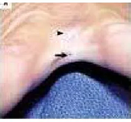
The occurrence of a dimple of the skin in the post anal region is
INCIDENCE
During the Second World War from 1941 to 1943, 78,923
soldiers were affected and managed in Military hospital due to
pilonidal disease. In the year 1973, more than 70,000 patients were
admitted to non government hospital in the US with a primary diagnosis
of pilonidal disease. In 1980, more than 40,000 patients with pilonidal
disease were hospitalized in the United States averaging over 5days in
hospital care. In India, the disease is not uncommon as might be thought
even though incidence statistics are not available.
It primarily affects young adults and teenagers, the maximum
incidence being between 19 and 25years. There is a 4:1 male
predominance. Negroes and American Indians are almost immune
perhaps because of their hair distribution. Caucasians were involved
most commonly by that disease than black African or Asian
peoples. Majority of patients were hirsute with strong wiry dark hair.
AETIOLOGY
The aetiology of pilonidal disease has been a subject of
controversy whether it is congenital or acquired. The congenital theory
postulates secondary infection of a congenital remnant of
epithelium resulting in a pilonidal sinus. The other theory proposes
entrapment of epithelium and hair follicles in the sacro-coccygeal
region. The later acquired origin of pilonidal disease is now more
widely accepted.
The occurrence of disease is related to the appearance of hair for
example thick, curly and profuse growth.
Various factors include:
· Friction in buttock
· Overweight
· Local injury
· Tissue papers for cleaning the perineum
· Increased duration of sitting and sweating
Other correlation factors are
· Overweight
· Same disease in family
· Folliculitis or furuncle in that area
The disease occurs early in females due to earlier attainment of
puberty in females.
PATHOPHYSIOLOGY
Controversy exists in the pathogenesis of sacrococcygeal disease
for many years.
CONGENITAL THEORY
Most of the authors in nineteenth century believe that the
pilonidal sinus disease was congenital in origin and they proposed a
congenital theory based on their studies on the human embryo:
1. Neural canal remnants in the caudal region gets attached to the skin
surface thereby accounting for recurrent cyst formation leading to
rupture to produce a sinus tract.
2. The degenerated epithelial nests were sequestered, leading on to the
formation of dermal inclusions.
3. As the human tail bud involutes because of the lack of development
of the caudal appendix, tractions will be exerted which attracts the
skin into the subcutaneous tissue region thereby resulting in the
ACQUIRED THEORY
Patey and Scarffl challenged the opinion that the lesion is
congenital, and their view has since been supported by Hueston, Currie,
Gibson and Goodall and by Davage. These writers have emphasized the
following observations:
1. In spite of term "pilonidal" (hair bearing or hair growing), no one ever
clearly demonstrated by microscopic sections that the hairs do in fact
grow out follicles the suppurating tract. Such follicles seen in micro
sections belong to hairs growing out of surface of the skin.
2. Hairs found lying within the tract or within zones of suppuration are
loose, unattached dead hairs.
3. Hair found projecting out of the sinus opening is loose hair with
the pointed end (the end furthest from the follicle) pointing into the
lumen of the tract.
4. Lesions pathologically identical to the pilonidal sinus, occurring the
inter digital clefts as an occupational disease of barbers, have been
described by Currie, Gibson and Goodall, Hueston, and Patey and
Scarfft. These inter digital sinuses-which by no stretch of imagination
can be on an embryological basis-contain hair (customers' hair) and
are for varying distances lined by epidermal cells as a down growth
The opponents of the congenital origin of this condition state the
following:
1. The congenital abnormalities are also found in the cervical and dorsal
areas of the vertebral column similar to that in the sacrococcygeal
region except that there is no associated pilonidal sinus.
2. Males and females are equally affected according to congenital
aspects, but they are present more in males than in females
3. Many presents during the adolescent period thereby not getting
concordance with the developmental theory.
4. The pilonidal sinus disease has a occupational prelidection for Jeep
drivers and soldiers and barbers thereby against the congenital theory.
5. Similar description in other sites of the body.
6. Congenital theory postulates the suction of the hair and other skin
appendages during dermal tractions. However there is lack of hair
follicles and other skin appendages in the wall of the sinuses
despite the presence of hair shafts, freely and deeply embedded in
granulation tissue or scar and the lack of lining epithelium in most
Though 50 to 75% of sinuses contain hair shafts during exploration,
Hair has most importantly three roles.
1. De novo in the distended hair follicle can remain unshed and
enhance micro abscess formation.
2. Free hairs from parts of the body can invade the follicles’ open
mouth and create foreign body reaction.
3. Skin hair in close vicinity to pilonidal wound irritates it
mechanically, affecting healing.
Karyadakis postulated the pathogenesis of the pilonidal sinus
disease. He attributed three main factors responsible for the hair
insertion process :
1. The loose hair being the invader
2. And the force causing the insertion
3. The skin in the natal cleft vulnerable to the
insertion of hair
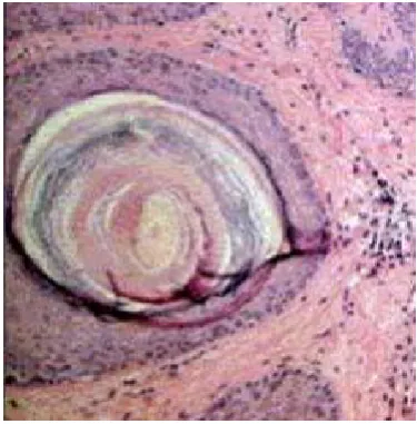
MICROSCOPY
Microscopy revealed Enlarged and distorted hair follicles in the
midline pits of the natal cleft and the basis of this is not known. Most
Pathogenesis being the expansion of the cavity caused by the
Occlusion of the mouth of hair follicles and rupture into the cavity
leading to foreign body reaction and formation of micro abscess. Later
they develops into chronic pilonidal abscess and rupture leading to
foreign body reaction and micro abscesses. These become chronic
pilonidal abscess and epithelialised tracts which are laterally displaced.
The disease becomes a pilonidal sinus when burrowing occurs in the
microabscess.
Microscopic examination of the pits revealed distorted hair follicles.
1) Distended hair follicles have a pit in the midline, which have
the wide ranges of sizes depending on the originally distended
hair follicles. The Normal ones, Moderately distended and
overtly distended hair follicles have pits which were found in
close proximity to each other.
2) Hair in the centre is surrounded by concentric keratin sleeves
were found in the large pits with distorted hair follicles.
3) Germinal hair bud is seen in the pit wall.
4) Pits are seen in the midline in 90% of Pilonidal disease whose
distribution pattern was determined by nearby hairs.
5) There is a considerable evidence that proves distorted hair
Fig.-4 Dilated follicle with central hair. Varying sizes of follicles. Right side has a normal hair follicle with left side showing a pit.
MECHANISM:
Enlargement of the normal follicles occurs by two kinds of forces.
One is the outward force that is due to the accumulated keratin. And the
other one is the inward force by the vacuum. Other forces may also
come into play.
Stages of the pilonidal disease are revealed by sectional microscopic
Fig.-5 Keratin accumulation
STAGES OF THE PILONIDAL DISEASE AND
MICROSCOPIC EVIDENCE :
Infection followed by edema – closes the mouth of the follicle. The
base of the follicle where keratin and hairs come out is the site of the
infection around the enlarged follicle. Contents does not escape and the
follicular mouth is hidden.
Hair shafts left behind in the walls of the abscess will heal. The
hair creates the hole in the epidermis through which it breaks and lie
outside and inside the body.
The outward force driven by the accumulated keratin and the
inward pulling force by the vaccum pushes the pus found in follicular
because of the accumulation of pus and blows out of the follicle’s
bottom.
The above mentioned pathologic process almost results in the
formation of acute pilonidal abscess and such abscess eventually drains
outside. After the drainage of the abscess, again the mouth of the follicle
reopens due to the reduction of the edema.
Remnants of the follicle, whose both ends are opened now, creates a
cavity at the outer end which results in the formation of chronic pilonidal
abscess. This cavity has similar inward and outward forces responsible
for maintaining the cavity. This evidence conclusively proves that hair
follicles which are distorted are midline pits.
Pathogenesis of pilonidal disease is best explained by theories
putforth by Bascom and Karyadakis. Hair and hair Follices are the
important predisposing factors in the pathogenesis of the pilonidal sinus
disease.
WHY MIDLINE IS MORE COMMONLY INVOLVED :
Skin cells and the hairs smash together in the midline areas. The
scales and barbs in hair, and the vertical walls on either side of the
with barbs on hair which imparts motion to hairs, thereby causing hairs
to push against midline cells where the skin in the midline area is
punctured and ultimately results in the accumulation of follicles. So,
midline pits are very common in the pilonidal sinus disease.
MANAGEMENT :
Management plan of pilonidal disease depending upon the
following.
· Nature of presentation and stages of pilonidal sinus.
· During the exploration of the pilonidal sinus through lateral
incisions, the instrument explores the pilonidal abscess cavity to
the underside of the pit which has blown out or to a pit that has
blown in to fat which in turn starts to express a pasty white
material from the abscess cavity.
· These findings implicate midline pits and their contents as a
possible source of the disease.
MECHANISM
· In standing posture, the gluteal tissue is separated away from
the sacrum because of gravity and thus the vaccum is created
which sucks air into the cavity along with the vagrant hairs
· Furthermore, these pass through the follicular remnant to join
the accumulated pus.
· This vaccum was measured by Brearly, who demonstrated
this mechanism by explaining when the patient sits, the
gluteal tissues will be pushed against sacrum and thereby
sealing the exit of the sinus added by the squeeze of the
buttocks together while sitting.
· The pressure required to drain the fluid from inside to outside
of the sinus onto the chair must be more than 125mm Hg and
such pressure was not attained inside during sitting, therefore
the pus was driven through fat, creating tunnels thereby
accounting for the abscess collection from the site distant to
the site of hair follicle of origin.
[image:29.595.141.449.498.705.2]
HORMONAL ORIGIN
· During the period of Pubertal growth, there was a sex hormonal surge
which has major implication in the growth of the pilosebaceous
glands, which results in the formation of hair follicle with keratin.
· This results in the formation of folliculitis, which produces oedema
and follicle occlusion. The infected follicle invades into the
subcutaneous tissue and ruptures to form the abscess thereby
creating a sinus tract which extends into the subcutaneous cavity.
· The hair follicle grows cephalad thereby accounting for 90% cases
having a sinus tract in the cephalad direction and it was
approximately 5 – 8 cm from the anus.
· In some instances, the sinus is located caudally and is found 4 to
5cm from the anus wherein it is formed because of the sinus
overlying the sacrum drains into the skin surface while the original
sinus formed from the natal cleft becomes completely
epithelialized leaving behind the laterally communicating sinus
tract alone which granulates and forms the sinus tract opening.
· The basis of the pilonidal sinus disease is because of the foreign
body reaction of the host tissue to the hairs. Loose hairs are
friction and movement of the buttocks.
· The tip of the hair enters into the cavity and becomes entrapped as
the barbs on the hair prevents it from being expelled.
· Thus, the hair which is trapped inside the cavity elicits a foreign
tissue reaction thereby predisposing to pain, infection and abscess
and eventually chronic discharging sinus from the opening.
· There are rare circumstances, wherein the foreign body other than
human hair causing the pilonidal disease was usually a birds
[image:31.595.176.442.383.679.2]feather, the type used to stuff feather bedding.
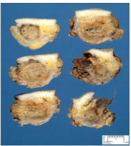
Fig.-8 Gross specimen of a pilonidal sinus
PATHOANATOMY
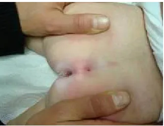
The occurrence of a dimple of the skin the post anal region is
frequently observed. Few authors, while examining recruits during World
War-II, recorded some of the minor anatomical variations in the perianal
region. Out of 3,136 male recruits, a distinct post anal dimple was
observed in 287 individuals, an incidence of approximately 9%. A dimple
in this region has the same significance as a dimple at other sites (e.g.
cheeks, chin, sacral region or knees). Anatomically all these dimples
represent nothing more than a local fixation of skin by dense collagenous
fibers to underlying bone or fascia. However, the occurrence of a dimple
at this site, associated with other factors, may be important in the
Fig.-9 Picture showing a post anal dimple in an infant
In possessing a deep post anal dimple, the use of toilet paper
in an antero- posterior direction may well serve to impact a paste
made up of broken bits of hair plus faecal material deeply into
the dimple with each successive wipe. Davage [13] quotes from a
personal communication from Dr. K. C. Samuel of the Department
of Pathology at Jaipur, India,
"Pilonidal sinus is very uncommon in India. Personal
cleansing after defecation is by ablution, and toilet paper is never
Fig.-10 Pilonidal sinus in post anal dimple
A - Diagram showing a loose hair trapped at the bottom a post
anal dimple and beginning to penetrate epidermis by the sharp pointed
end.
B and C - Successive stages of inflammatory reaction to the
persistent foreign body. D - Epithelization of a portion of the tract so
formed.
Dr. V. L. Parmar, Professor of Surgery, Grant Medical College,
Bombay in a personal communication to Dr. Alan. A. Klass (Associate
Professor, Department of Anatomy, University of Manitoba; Assistant
Sturgeon, Winnipeg General Hospital.) Dated April 4, 1955, states:
"It is an invariable practice for all Indians to use water for
cleansing after defaecation, and not toilet paper. Using of toilet paper is
looked down upon as being very unhygienic, and in this country people
always use water. These pilonidal sinuses and cysts are not common in
this country, and although I have been attached to three general public
hospitals during the last five years, I have not operated on more than
three cases." [9]
In the matter of prophylaxis with individuals subject to pilonidal
suppuration, this triad of post anal dimple, loose bits of hair and fecal
residue are all important. The additional traumatic factor of prolonged
sitting, like in jeep drivers, may provide the final link in the chain of
events leading to suppuration.
The local anatomy of the inter natal cleft in the immediate area of
the post anal dimple has an influence on the spread and persistence of
an inflammatory process. The dense network of collagen firmly attaching
the deep surface of the skin to the subjacent periosteum of the coccyx
and sacrum is noted to prevent spread. The fatty tissue in this central
dense strands of fibrous tissue. In the lateral areas, however, the
proportion of fat to collagen becomes reversed, and large areas of fatty
tissue become interlaced with thin fibrous strands.
The density of the central zone of fibrous tissue is such as to
produce, in the event of suppuration, an irregularly shaped area of
inflammation with innumerable small zones of necrosis corresponding to
the spaces previously containing fat. Extension into the loose lateral
spaces, when it occurs, is in an irregular manner like so many
pseudopodia. Once in this area, the extension meets little resistance from
the thin fibrous strands. In the usual low-grade infection there is no way
of determining on clinical grounds just how extensive these minute
finger-like processes of infection may be. Most dissections leave behind
tiny islands of infected necrotic tissue in the terminal portions of
extensions. Post-operative fibroplasia is more than likely to seal these off
from the central zone, encouraging further and wider lateral extension. It
will leads to high rate of recurrence.
It is the complexity and irregularity of this spread into the lateral
fatty zones that promotes chronicity, which eludes complete excision, that
so frequently defeats primary structure, and that so often promotes
growth of surface cells gives the appearance of a skin- lined tract. But
this extends for a short distance only. The active inflammatory zone is a
simple tissue space infection, very irregular in outline, without
lining, and containing the debris of loose hairs plus granulation tissue or
pus.
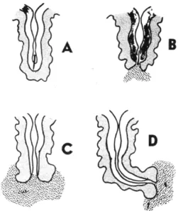
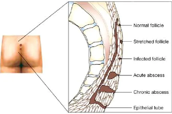
[image:37.595.120.506.300.529.2]STAGES OF PILONIDAL DISEASE
Fig.-11 Stages of pilonidal disease
· STAGE I - Normal follicle.
· STAGE II - Follicle becomes distended with keratin. It was the
Fibrous strands which suspend follicle and skin from sacrum.
closing the mouth. Because of the pressure increase inside the
infected follicle, rupture occurs which invades into fat causing acute
collection of pus called Acute abscess.
· STAGE IV - Chronic pilonidal abscess. The ruptured follicle which
is open at both ends forms the mouth of the abscess.
· STAGE V – Stage of Epithelialisation. The normal epithelium grows
lining the sinus tract forming epithelial tube.
CLINICAL FEATURES
Pilonidal sinus disease has a wide range of clinical presentations
ranging from asymptomatic pits to chronic draining sinus tracts which are
more common in the sacro coccygeal regions and in persons who have
abundant hairs in that region and who succumb to chair most of their
lifetime.
Several reports show that pilonidal sinus has a tendency to occur in
interdigital spaces in barbers, sheep shearers and dog groomers. Rarer
reports mentioned its occurrences in finger tip pulp and penis.
Pilonidal sinus occurs in either sex except that it occurs a little earlier
Studies on the routine physical examination of Minnesota college
students showed 364 out of 31597 males (1.1%) and 22 of 21467 females
(0.11%) had pilonidal sinus.
The inference being the pilonidal sinus predominates in the males
during their second and third decades but however in the children, the
disease predominates in the female with a female to male ratio being
4 : 1.
The most common presentation in patients with pilonidal disease
being the pits or holes in the gluteal cleft. And studies on the Iraq
soldiers of 1000 members showed, Out of 88 members who had pilonidal
sinus, 48 were asymptomatic and 40 presented with symptoms.
Another common presentation is chronic or recurrent discharging
sinus.
Fig.-13 Recurrent pilonidal sinus showing bridging and fibrosis
Sondenaa et al. noted discharge in 66%, swelling in 50% and pain
in 35% of chronic pilonidal disease presentations. 50% of patients
present with abscess and with discomfort or pain following physical
activity or after a long drive due to prolonged sitting. They also present
with acute purulent discharging sinus, with inflammatory changes like
pain and swelling.
[image:40.595.169.494.505.725.2]The patients usually have a waxing and waning phase because of
spontaneous drainage from the secondary sinus and again get reinfected
and spontaneous rupture occurs accounting for waxing and waning
phase. The most common organisms cultured from the abscess are
anaerobic organisms like bacteroides and anaerobic cocci accounting for
77% and aerobic organisms of about 4% and both are cultured in about
17%.
The sinus usually tracks into cephalad direction in majority of
cases but in some cases like in about 7% track toward the anus and
presents as perianal sepsis.
Most dreaded and rare complication of the pilonidal sinus being
the carcinomatous change from the long standing sinus of squamous cell
or spinocellular variety have been reported. They carry a very high
recurrence rate and usually have a poorer prognosis than a regular non
melanoma skin cancer.
Although the pilonidal sinus manifests as an abscess or pilonidal
sinus or a recurrent or chronic pilonidal sinus or a perianal pilonidal
sinus, the most common manifestation being a fluctuant and painful mass
in the sacro coccygeal region. It occurs most commonly in the young
PRESENTATION:
Most common symptoms being the pain and the purulent
discharge from the sinus opening in about 70 – 80% of the time.
And 50% of the patients initially presents with acute abscess
which is cephalad to the hair and the sinus infection.
Earlier stages being a cellulitis or folliculitis stage. It is when the
folliculitis expansion into the subcutaneous tissue, an abscess is formed
or when a pre-existing foreign body granuloma becomes infected. Those
cavity and the sinus tract and laterally oriented sinus tract openings are
generally lined with granulation tissue whereas the midline sinus pit is
[image:42.595.140.442.490.690.2]lined by epithelium.
DIAGNOSIS :
Palpation of a deep indurated region beneath the skin in the sacral
region and the presence of an epithelialized follicle opening. When you
palpate the tracts, these usually track in the cephalad direction. Wherein
in a case where perianal sepsis is noted, the tract must have run in the
caudal direction.
Patients with a more benign cause, present with chronic
discharging sinus with intermittent exacerbations and remissions after
[image:43.595.218.399.414.598.2]incision and drainage.
RECURRENT / CHRONIC PILONIDAL DISEASE
DEFINITION:
Patients with chronic discharging sinus without an acute
exacerbation tend to have a recurrent or chronic pilonidal disease.
PATHOGENESIS:
· Hair follicle is not the inciting agent in chronic or recurrent
pilonidal disease.
· It refers to patients with a pilonidal sinus that is associated with a
chronic discharge without an acute abscess and it is seen after
incision and drainage of a pilonidal abscess.
· So in such circumstances, the sinus would not have been excised
and it is still there after the abscess cavity heals and recurs later.
· Pathology behind the chronicity or recurring ability of the sinus
is that Unhealed surgical wound becomes filled with granulation
tissue, hair and skin debris which acts as a nidus for the ongoing
foreign body reaction and further inflammation. This along with
midline cleft hairs responsible for the chronicity and recurring
Fig.-17 Recurrent pilonidal disease
ENDOANAL PILONIDAL SINUS
Rare variety
Involves the perianal skin directly or circumferentially around the
anus, involving the anal verge skin.
THREE MOST IMPORTANT CAUSES OF ENDOANAL
PILONIDAL DISESASE :
· Perianal fissure of fistula which communicates with
the anal canal was created by the pilonidal sinus which tract down
caudally.
· Post Operatively, in a fistula in ano patient, hair enter
· Hair may normally enter and penetrate the anoderm
eliciting a foreign body reaction in the sacrococcygeal region.
INFECTION
Anaerobes predominates aerobes in causing the reinfection and
wound breakdown following surgery. The most common anaerobic
bacterias responsible as Bacteroides and Enterococci and among the
aerobes staphylococci and haemolytic streptococci are most common.
COMPLICATIONS [10, 18]
The most common complications of pilonidal disease are:
Ø Recurrence of the abscess is by far the most common
complication. Most literature reviewed quote a rate between 40% and
50%.
Ø The next complication is the recurrence of p i l o n i d a l disease.
Recurrence can be early recurrence and late recurrence
o Early recurrence – due to failure of identification of
one or more sinuses during surgery
o Late recurrence is commonly due to secondary
bacterial infection of the residual debris which was
not completely removed during the surgery, improper
Ø Wound infection after primary incision and drainage is rare but
described.
Ø Squamous cell carcinoma after recurrence of pilonidal disease has
been described; it is in incidence but, when diagnosed, requires en
bloc surgical resection and appropriate oncologic care with local
radiation and possibly chemotherapy.
PROGNOSIS
Long-term prognosis for pilonidal disease is excellent and
mortality is practically nil, unless squamous cell carcinoma develops,
though abscess recurrence is common as described.
DIFFERENTIAL DIAGNOSIS [3]
The distinctions among pilonidal disease are:
ANAL FISTULA
Several study of surgical literature mention the difficulty in
differentiating pilonidal disease from anal fistula and hidradenitis
suppurativa. Pilonidal disease may result in sinuses that reach the
perianal region and simulate an anal fistula. A valuable clinical
observation in establishing diagnosis of anal fistula is palpation of the
secondary opening is observed and no tract is palpable, the possibility of
an extra-anal source of the infection must be considered.
HIRADENITIS SUPPURATIVA
A chronic inflammatory disease of the apocrine sweat glands in
which folliculitis and local friction also play a role, in patients aged
30years or older, especially with co-morbidities such as diabetes and
obesity.
CONGENITAL ABNORMALITIES
In some congenital lesions (meningocoele), a continuous tract with
the central cord of the spinal cord may occur, and discharge of CSF may
be present
PERIRECTAL ABSCESS
Location of the lesion is the best means to differentiate this entity
from pilonidal disease. Perirectal abscesses frequently require emergency
surgical consultation for formal drainage in the operating room.
· Primary presacral or sacro-coccygeal sinus
· Furuncle or carbuncle
· Pyoderma gangrenosum is an ulcerative lesion also
generally seen in the fourth decade of life with other
co-morbidities.
· Syphilitic granuloma
· Tuberculous granuloma
· Sebaceous cyst & Dermoid cyst
MANAGEMENT
The management of the pilonidal sinus depends on the
presentation of the disease. The common clinical presentations of the
pilonidal sinus disease are categorized into 3 categories. These are:
1. Acute pilonidal abscess,
2. Chronic pilonidal disease, and
3. Complex or recurrent pilonidal disease.
The surgical management is then tailored to the above
classification category. The goals of the ideal procedure for the treatment
of this disease should be:
1. Low recurrence rate with better wound healing.
2. Lesser hospital stay
3. Convenience to the patient
4. Less complications with fewer morbidity
5. Early return to normal activities as early as possible.
Various non-surgical and surgical modalities of treatment
NON-SURGICAL
· Injection of sclerosing agent
· Fibrin Glue
· Cryo-surgery
· Electro-cauterization
· Repeated shaving or use of depilation creams
SURGICAL
· Drainage with/without excision
· Marsupialization
· Excision with healing by secondary intention
· Excision with primary closure
To prevent the recurrence rate and chronicity, various other
techniques are brought into action. They are
· Karydaki’s flap
· Bascom procedure
And other procedures which use the technique of transposition
flaps have been described. They are:
· Z plasty
· V-Y fascio-cutaneous advancement flap
· Crossed Triangular Flaps
· Gluteus maximus musculo-cutaneous flap
· Rhomboid flap of Limberg
The above mentioned surgeries have low recurrence rate by
reducing the depth of the natal cleft and place the suture line away from
the midline natal cleft and with low tension at the suture lines.
CONSERVATIVE TREATMENT
Conservative treatment needs longer period to achieve positive
results and there is low incidence of cases being proceeded to excisional
procedures following this.
The treatment includes
· Hair control by shaving the natal cleft
· Removal and scraping of the granulation tissue
· Cleaning out the natal cleft and removing all hairs
· Frequent washing of the parts with detergent and water and
with a solution containing equal parts of witch hazel and
alcohol
· Avoidance of prolonged sitting.
INDICATIONS :
First time presentation with mild symptoms
Armstrong and Barcia studied the role of conservative,
non-operative treatment of pilonidal sinus disease at an army hospital
for a period of 17years and only a few required excisional treatments.
[40]
[image:53.595.109.511.472.682.2]A-Natal cleft with pilonidal sinus B- after 3 treatments C-after 7 treatments
TREATMENT OF ACUTE EXACERBATION (ABSCESS)
A pilonidal abscess is managed by incision, drainage, and
curettage of the abscess cavity to remove hair nests and skin debris.
This can be accomplished in the surgical theatre or in the
emergency department under local anesthesia. If possible, make the
drainage incision laterally, away from the midline, opened through a
small incision. All hairs and granulation tissues removed. Track can
be destroyed by careful instillation of pure phenol solution.
Wounds heal poorly in the deep intergluteal natal cleft, which
allows frictional movement of one buttock over the other. The wound
should be cleansed daily in the shower or with a sitz bath.
Paying close attention to hygiene and hair shaving of the
surrounding area are important to prevent hair from penetrating the
healing scar. This meticulous treatment of the diseased area should
continue for approximately 3 months, even after the wound has
completely healed.
In more than 90% of cases, the wound heals completely in
approximately 1 month. Incision and drainage, without curettage results
these patients, 40% develop a recurrent pilonidal sinus, requiring further
treatment.
Inform patients that drainage of the abscess is not a curative
procedure. Some studies have shown that as many as 85% of patients
require further surgical treatment. Excising the pilonidal pit at the time of
abscess drainage reduces the recurrence rate to 15%. The difficulty
with doing this is that the pilonidal pit initially cannot be identified
during the first drainage procedure of the abscess. Approximately 5days
later, when the oedema is reduced, the pit can be identified. Often,
having the patient return 5-7days after abscess drainage to identify the pit
and to excise it with a small incision is possible.
A-Pilonidal Abscess B-Aspiration of Pus
C-Incision and Drainage D-Packing of wound
Fig-20 Pilonidal abscess incision and drainage
MEDICAL THERAPY
1.
INJECTION OF SCLEROSING AGENT [36, 37, 84]
Phenol injections used as treatment of the pilonidal sinus are more
common in Europe than in the United States. Both chronic pilonidal
disease and acute pilonidal abscess (after drainage) may be managed by
phenol injection. Eighty percent phenol is injected into the sinus, left
The sinus is then curetted. This may be repeated as many as 3 times
for a total of 3 minutes of phenol exposure at one treatment. The
treatments may be repeated every 4-6weeks as necessary as wound
healing progresses. Paraffin jelly may be used to protect the skin from the
phenol, which destroys the epithelium.
Phenol sterilizes the sinus tract and removes embedded hair. Phenol
injections may be combined with local excision of the sinus. Wound
healing usually requires 4-8weeks.
The incidence of recurrence is reported to be approximately 9–27%,
which is similar to the incidence following simple excision and packing
open the wound. Because of the intense local inflammatory response after
the phenol injection, patients usually stay in the hospital overnight.
Thereafter, the patient
is allowed to return home with instructions to bathe daily and keep the
area shaved. Dressings are used for comfort.
2.
FIBRIN GLUE [45]
Curettage followed by injection of fibrin glue. Promising early
results with improved pain scores and earlier return to normal daily
A-Delineating the sinus tracts B-Curetting the sinus
C-Injection of fibrin glue into D-The fibrin glue seen coming
[image:58.595.131.283.83.245.2]the tracts out of the tracts
Fig.21Procedure of injection of fibrin glue
3. CRYOSURGERY [3, 46]
The sinus tracts are laid open and curetted and the open wound is
then sprayed with liquid nitrogen for about 5minutes. It can be done as an
out–patient procedure. But there is delayed healing especially at the skin
4.
ELECTROCAUTERISATION [3, 47]
This is done under local anesthesia. The sinus tracts are opened
with a diathermy knife. Track curetted and hair removed and the track is
cauterized. Patient is sent home with a mild analgesic. The procedure
can be repeated depending up on the healing. The rate of recurrence is
[image:59.595.207.408.306.564.2]11%.
Fig-22 Electrocauterisation
The surgical options for management of a non-complicated chronic
pilonidal sinus include:
1. excision with primary closure.
3. wide and deep excision to the sacrum.
4. incision and marsupialization.
All the surgical procedures are carried out under general or spinal
anesthesia with the patient in a prone or jackknife position. After
shaving off the hair and cleaning the area, methylene blue dye mixed
with hydrogen peroxide is injected in the external openings to give a
guideline for the tract and branching.
1. EXCISION WITH PRIMARY CLOSURE [35, 57]
Excision of a pilonidal sinus entails excision of the midline pits and
lateral openings down to the presacral fascia, with removal of minimal
surrounding skin. In general, removing more than 0.5cm of skin
surrounding the sinus opening is not necessary. Curetting the wound to
remove the hair, granulation tissue, and skin debris is essential to promote
adequate wound healing. Although performing this procedure under local
anesthesia alone is possible, mild sedation in addition to local anesthesia
allows for a more complete excision and a more comfortable patient.
Lord and Millar in 1965 described and popularized their
technique of coring out the midline epithelial follicles under local
anesthesia, but they also included a brush in their procedure to cleanse
oriented granulation-lined tract. [34] The brushing of the tracts continues
in the outpatient setting in the postoperative period until the tract heals
completely and closes. At about the same time 50% phenol was used after
curettage to destroy the epithelial component of the track.
Schneider et al. in 1994 reported that phenol injection gave
similar results to surgery with patients staying in hospital for 1–2 days
and returning to work within weeks; 60% of sinuses showed complete
healing with an average healing time of .2weeks. [36] The follicle
excision sites may be closed primarily but are usually packed and
[image:61.595.213.404.423.591.2]dressed to heal by secondary intention.
A-Simple excision of pilonidal sinus -Primary closure of wound
C-Excision of sinus Primary closure with drain
E-Excision F-Primary closure with drain
MODIFIED BASCOM TECHNIQUE [8, 18, 19, 48, 73]
More recently, Bascom described using a lateral incision for entry
into the pilonidal cavity. Curettage of the cavity is accomplished through
this lateral incision, which is not excised. The midline pits are excised
separately, including the epithelialized tube. The midline incisions are
closed, while the lateral wound may be either left open to drain and heal
by secondary intention or closed primarily.
The advantages of a primary closure are small wounds, quicker
healing time, usually within 3weeks, minimal wound care, earlier return
back to work and no need for daily scheduled dressing changes. The
obvious disadvantages are wound infection and wound dehiscence.
Rather than primarily closing a midline or lateral vertical incision,
some physicians advocate the use of asymmetrical or oblique elliptical
incisions in an attempt to keep incisions out of the natal cleft where
wound healing is poor and to prevent unnecessary tension on the closure
of the wound. The goal of the asymmetric incision is to reduce the depth
of the gluteal fold, thereby eliminating the frictional forces between the 2
opposing skin edges. Although the use of an incision that crosses the
vertical gluteal fold to excise the pilonidal cavity does eliminate a vertical
suture line within the gluteal fold, healing times may remain
Skin flaps have also been described to cover a sacral defect
after wide excision. Similarly, this keeps the scar off the midline and
flattens the natal cleft. The potential complications include loss of skin
sensation in the flap, which is observed in more than 50% of patients, and
necrosis of the flap edges. Again, primary healing is achieved in 90% of
cases.
Fig.-26 In the Bascom operation, midline pilonidal pits or follicles are excised. One to 10 follicles can be removed, leaving wounds 2–4 mm in diameter. The sinuses or cavity are opened through an incision 2 cm lateral and parallel to the natal cleft. The lateral incision undermines the midline and gauze is pushed through the cavity to ‘‘scrub out’’ hair and granulation tissue
2. EXCISION WITH LAYING OPEN OF TRACT [10, 18, 31,39]
Excision of the pilonidal sinus and laying the tract open to allow
healing by secondary intention has been described as an option to ensure
that the cavity has adequate drainage. This avoids a wound infection after
primary closure. Consider laying the tract open when the primary closure
is not free of tension. Even after excision of the pilonidal sinus down to
healthy presacral fascia, the wound is still considered contaminated. Both
aerobic and anaerobic organisms are found in 50-70% of wounds. The
patient, with frequent dressing changes, and close observation of the
wound to ensure proper wound healing and avoid premature closure of
the edges. The average time for wound healing to occur is approximately
6weeks.
Laying the tract open is always appropriate when there
is cellulitis surrounding the pilonidal sinus. Primary wound closure
versus wound healing by secondary intention are the 2 principal surgical
options for a chronic pilonidal sinus. This is associated with a low
recurrence rate but slow healing.
In a group of 146 patients with pilonidal sinus, Isbister and Prasad
felt the procedure could be done safely on an outpatient basis. Differences
remain between these 2 techniques in terms of wound healing and
recurrence. Although primary closure has the potential for earlier wound
healing if infection does not occur, it does require that the patient restrict
many activities until wound healing is complete. This is because a
primary closure is rarely completely free of tension and the wound is
considered contaminated despite excision and debridement.
Recurrence rates after primary closure may be as high as 38%. Not
uncommonly, wounds may require 4-6months to heal, but on average,
is felt to be due to the more broad based, flattened and hairless scar
produced by secondary intention. This prevents buttocks friction, hair
penetration and hair follicle infection. Although advantages exist, these
open wounds require aggressive management with frequent dressing
changes and close observation by both the patient and the surgeon.
A-On table picture of laying B-9days post excision C-12days Status
D-16days post excision E-20days post excision F-22days post excision
Fig-27 Excision with laying open of tracts
3. EXCISION WITH MARSUPIALIZATION [10, 18, 66]
Marsupialization as a treatment option of a pilonidal sinus was first
introduced in 1937. Marsupialization is a compromise between
primary wound closure and wound healing by secondary intention. The
rationale is to avoid wound infection and dehiscence after primary closure
wound is sutured open. After excision of the pilonidal sinus cavity, and
lateral tracts, the cavity is then scrubbed and curetted to remove hair and
granulation tissue. The skin edges of the wound are then sutured to
the presacral fascia. The wound is then loosely packed and requires daily
dressing changes.
Marsupialization provides the patient with a smaller wound
compared to wounds that are left open to granulate. By suturing the
wound open, wound infection is prevented and the subcutaneous tissue is
covered, resulting in reduced healing time. Healing is usually complete
by 6weeks, and the recurrence rate has been reported to be 4-8%.
Many authors consider marsupialization the preferred method of
treatment for chronic pilonidal disease because it avoids closure of a
contaminated wound and combines shorter healing times with a lower
recurrence rate. The patient still needs to pay meticulous attention to
personal hygiene, with daily wound cleansing and frequent hair
shaving and removal.
D-During marsupialization E -After marsupialization F-5weeks later
Fig.-28 Excision with marsupialization
4. WIDE EXCISION AND PRIMARY DRAINAGE [10, 18, 83]
In this method, wide local excision is carried out around the
pilonidal sinus and healing occurs by secondary intention which takes a
longer time but has a lower recurrence rate. Mean hospital stay is about 4
weeks. The healing usually gets delayed by secondary infection by
anaerobic bacteria.
Patients with recurrent pilonidal disease or complex unhealed
pilonidal wounds present a challenge to the surgeon. Tissue loss from
previous attempts at excision further complicates the surgical
management and limits options. The causes of recurrence are thought to
be due to
· Well known Midline cleft with inward and outward forces promoting
hair to grow into the scar and cause recurrence.
· Most susceptible site for the recurrent pilonidal disease is the midline
cleft with scar and due to poor wound healing.
· Generally a flap procedure will be required for the treatment of
recurrent disease, wherein wide excision will be done and primary
closure can be achieved with flap procedures and prevent the suture
lines in the midline and the dead space is completely obliterated. Such
techniques prevents the fricitional forces from causing the pilonidal
sinus disease.
· Reserve the use of a flap closure for complex or recurrent pilonidal
disease that has failed to respond to the simple conservative operative
techniques initially used to treat chronic pilonidal disease. A wound
that has failed initial therapy must be re-excised down to the
sacro-coccygeal fascia. The re- excision must include the unhealed wound,
A-Plan for wide excision B-2 weeks after wide excision
[image:71.595.314.487.84.206.2]
C-6 weeks post excision D-10weeks post excision
CLOSURE OF THE DEFECTS
Following wide excision, Primary wound closure is achieved by
doing a Flap closure.
The following techniques are available
1. PRIMARY CLOSURE OF THE CLEFT
2. KARYADAKIS ADVANCEMENT FLAP PROCEDURE
3. ADVANCEMENT FLAP
a. Z–PLASTY
b. LIMBERG’S RHOMBOID FLAP
c. V–Y ADVANCEMENT FLAP
d. CROSSED TRIANGULAR FLAPS
1. CLEFT CLOSURE
Primary closure of the Cleft surgery was first devised by Bascom,
in that the mobilization and excision of the fat is not needed.
Technique begins with excision of the wound by placing the apex
of the incision lateral to the apex of the natal cleft and forming a
triangular incision.
Full thickness skin flaps are raised.
Debris is removed and the sinus cavity is made free allowing the
gluteal fat to appose . The inferior margin becomes crescent shaped,
with its point positioned towards the anus.
A skin flap involving only the dermis is created on the convex side
of the lower wound margin. Excess skin is excised from one side, and
the wound is closed. This reshapes the cleft, making it shallower with the
suture line displaced out of the fold.
Before beginning the procedure, the lateral edge of the raised skin
flap should be defined by marking the line of contact of the buttocks.
Then the edges of the skin are overlapped, and the excess skin is
excised. This creates a primary closure that is off midline and
obliterates the intergluteal cleft. The wound is closed in multiple layers
by keeping a closed suction drain underneath. The recurrence rate is
A – Pilonidal sinus
B – The natal cleft is approximated to mark the line of cleft closure
C – The area of cleft closure marked
E – The sinus tract excised and the incision extended and undermined to involve the site of cleft closure on one side.
F – The undermined flap is bought to suture to one side of the planned site of incision of cleft closure.
G – The subcutaneous tissue and the skin are approximated after placing drain.
I-6 months post operative picture of cleft closure
Fig.-30 Cleft closure procedure
2. KARYDAKI’S LOCAL ADVANCEMENT FLAP [10, 18, 20, 32,
65]
The Karyadakis advancement flap procedure starts with excision of
the wound with en bloc removal of the sinus with an elliptical overlying
skin specimen. The incision will be made off Midline.
After excision of the wound, a full thickness flap is raised on the
contralateral side of the semilateral incision which allows the
contralateral side of the skin to be mobilized to cross the midline and
thereby allowing primary wound closure, avoiding midline closure. The
midline sinus is excised elliptically and the wound closed lateral to the
ADVANTAGE :
The natal cleft will be flattened
The suture line will be in a lateral position but not in the midline.
DISADVANTAGE:
Has a 1.3% recurrence rate
Requires extensive dissection and so cannot be done as OPD
procedure.
PROCEDURE :
Thick flap created and advanced across the midline and primary
closure done. Wound is closed in several layers after keeping the suction
drain underneath.
Karyadakis local advancement flap technique is considered as a
primary procedure for pilonidal sinus.
ADVANCEMENT FLAPS
The various types of Local advancement flaps are
· Z–plasty
· Rhomboid flap
· V-Y advancement flap
These are indeed methods of covering defects which results from
recurrent pilonidal disease.
ALTERNATIVES:
So Because of the above mentioned disadvantages, whenever an
advancement flap is needed, myocutaneous flap should be considered.
· Myocutaneous flaps helps to reconstruct Complex wounds as these
flaps heal well and helps to cover large area of skin loss.
· They are less susceptible to infection
· They have a predictable vascular supply that promotes better
wound healing.
DISADVANTAGES:
· Technically demanding techniques
· Prolonged hospitalization
· Lengthier operating time
· Failed flap is another great problem, which creates a
additional extensive skin loss which is difficult to manage.
So these techniques are only reserved for the Management of
Complex Recurrent Wounds where other procedures have failed.
3. Z–PLASTY [18, 32, 75]
PRINCIPLE :
Obliterating the natal cleft and increasing the transverse length by
recruiting the lateral tissue.
PROCEDURE :
· Excision of the midline sinus
· From the ends of the midline wound, the limbs of the “Z” are cut · Subcutaneous flaps are raised upto the level of fascia
· Transposition of the flaps carried out · Skin is closed.
Mansoory and Dickson used this technique on 120 patients and
follow up of 9years. The patients were discharged on post operative
day 1 and they returned to work 2 weeks later.
Fig-32 Z-plasty
a. marking of Z limbs A and C
4. V-Y ADVANCEMENT FLAP [18, 20, 76]
The V-Y advancement flaps can be raised unilaterally or
bilaterally.
· Unilateral flap will cover a defect 8-10cms in diameter.
· Bilateral flaps cover defects greater than 10cm.
· The flaps are raised upto the level of fascia and thereby
composed of skin, fat and underlying gluteal fascia.
ADVANTAGES:
· Closure of the primary area can be achieved without tension
· Dead space can be obliterated easily
· Complete removal of all midline pits and necrotic tissue can be
achieved.
· Mean hospital stay was around 10 days
· Wound complication occurs at a rate of 8% and 17% for unilateral
and bilateral flaps respectively
Fig.-33 V-Y advancement flap.
Fig 1 and 3 – Unilateral advancement flaps
Fig 2 and 4 – bilateral advancement flap
5. CROSSED TRIANGULAR FLAPS [60]
ADVANTAGES :
· Easy technique
· Early return to work and lesser hospital stay
· Cosmetically acceptable appearance postoperatively.
· Wound complication rate of 5%
· Recurrence rate 1.73%
PROCEDURE :
· A semicircular midline incision is made including all the
sinus openings and excision of the sinus tracts are done.
· Zig zag incision across the wound in done to form multiple
triangles, with apical part on one side of the wound and basal
part on the other side.
· All apical parts of each triangle are excised, basal parts
undermine, and haemostasis achieved.
· The wound is closed in a zigzag line, with the basal flaps
Fig-34 Diagram of crossed triangular flaps technique.
Fig A shows the vertical and zi