Functional outcome of extra-articular
distal tibia fractures treated with
Minimally Invasive Plate
Osteosynthesis
A DISSERTATION SUBMITTED IN PARTIAL FULFILLMENT OF
THE REQUIREMENT OF THE TAMILNADU
DR. M. G. R.
MEDICAL UNIVERSITY,
TAMIL NADU, CHENNAI FOR THE
I
Functional outcome of
extra-articular distal tibia fractures treated
with Minimally Invasive Plate
Osteosynthesis
A DISSERTATION SUBMITTED IN PARTIAL FULFILLMENT
OF THE REQUIREMENT OF THE TAMILNADU
DR. M. G. R.
MEDICAL UNIVERSITY,
TAMIL NADU, CHENNAI FOR THE
DEGREE OF
M.S. ORTHOPEDICS
TO BE HELD IN APRIL
II
ENDORSEMENT BY HEAD OF DEPARTMENT AND
PRINCIPAL OF INSTITUTION
This is to certify that this dissertation entitled “
FUNCTIONAL OUTCOME
OF EXTRA ARTICULAR DISTAL TIBIA FRACTURES TREATED
WITH MINIMALLY INVASIVE PLATE OSTEOSYNTHESIS”
is a
bonafide research work done by
Dr. Smruti Ranjan Panda,
under the
guidance of
Dr. Thilak Jepegnaman,
Professor, Department of orthopaedics
Unit 3, Christian Medical College Hospital, Vellore.
Prof Vernon N.Lee
Dr. Alfred Daniel Job
Head of Department of Orthopaedics Principal
Christian Medical College, Hospital Christian Medical College, Hospital Tamil Nadu, India Tamil Nadu, India.
Dated: Dated:
III
CERTIFICATE
This is to certify that the dissertation entitled “
FUNCTIONAL OUTCOME
OF EXTRA ARTICULAR DISTAL TIBIA FRACTURES TREATED
WITH MINIMALLY INVASIVE PLATE OSTEOSYNTHESIS”
is the
bonafide work by
Dr. Smruti Ranjan Panda
in the partial fulfillment of the
requirement for the M.S. Degree (Orthopedics) of the Tamil Nadu Dr. M.G.R.
Medical University ,Chennai to be held in April 2014.
Guide: Co- Guide:
Dr Thilak Jepegnaman, Dr Viju Daniel V.,
Professor, Asst Professor,
IV
ACKNOWLEDGEMENT
A volume of this magnitude could not have been possible without the contributions and guidance of many, for this reason, I would like to recognize few specific individuals whose contributions have been especially helpful.
First and foremost, I thank Lord Jesus who is able to do immeasurably more then we can ask or imagine. This thesis would never have been accomplished without the blessings of my God Almighty.
I owe my deepest gratitude to my teacher and guide Prof. Thilak Jepegnaman, Orthopaedic Unit 3 for his supervision, advice and guidance from the initial to the final level of this study. His constant oasis of ideas and constructive comments has exceptionally inspired me and enriched my growth as a student.
I am obliged to Dr. Viju Daniel Varghese, Asst. professor and co guide for his untiring effort in going through all the studies, timely suggestions and words of encouragement.
I thank Prof. Vernon N. Lee, Head of orthopaedics for his continuous encouragement and support.
V
I am also grateful to other faculty members of the Department and my post-graduate colleagues who helped me in all possible ways in this study.
I would like to thank my wife and family who has been instrumental in giving me ideas in framing and writing the thesis, and standing by me in all the difficult times.
I thank all my patients who were a part of the study. Though I had to do house visits for most of my patients, the warmth with which they welcomed me and cooperated in getting the x rays done and allowing me to take clinical photos, really touched my heart. I wish all of them good health.
I thank Gowri S from the Department of Biostatistics for helping me in my analysis.
Finally, I offer my regards to all those who were important to the successful realization of this thesis, as well as expressing my apology that I could not mention personally one by one.
VI
ABSTRACT
TITLE OF THESIS: Functional outcome of extra articular distal tibia fractures treated with minimally invasive plate osteosynthesis.
DEPARTMENT: Department of Orthopaedics Unit 3, C.M.C. Hospital, Vellore
NAME OF CANDIDATE: Dr. Smruti Ranjan Panda
DEGREE AND SUBJECT: Masters in Orthopaedics (Two year, Post Diploma)
NAME OF GUIDE: Dr. Thilak Jepegnaman
OBJECTIVES:
The purpose of this research is to study whether minimally invasive plate osteosynthesis leads to consistent union and early return to work. The specific objectives included :
1) Correlation between the functional scores and radiological parameters of distal thirds diaphyseo - metaphyseal tibial fractures treated by minimally invasive plate
osteosynthesis along with the time to bony union and
2) Documentation of the major and minor complications associated with this treatment modality.
VII
The functional outcome was measured by using standard questionnaires and consisted of the AOFAS (American Orthopaedic Foot and Ankle Society score & Olerud and Molander scores and were then graded as excellent/ good/ fair and poor and compared to similar studies by other authors.
Radiographic assessment was done comparing the anteroposterior and lateral views of the affected and the normal leg with both the knee and ankle joint included and the time to union, malunion and shortening were documented.
All major and minor complications were documented at follow up.
RESULTS :
22 patients (84.7%) had uneventful healing of the fractures. Delayed union and infection occurred in two patients each (7.5%). All fractures healed without the need of any secondary
procedures. There was no noticeable mal- alignment or non-union. Functional outcome was good to excellent in all patients. Average time to bony union was 24 weeks.
CONCLUSIONS :
VIII
Contents
INTRODUCTION 1
AIMS AND OBJECTIVES 4
REVIEW OF LITERATURE 5
MATERIALS AND METHODS 32
RESULTS 42
DISCUSSION 66
CONCLUSIONS 70
BIBLIOGRAPHY 71
1
Introduction
The process of rapid and unplanned urbanization has resulted in an unprecedented revolution in the growth of motor vehicles worldwide. The alarming increase in morbidity and mortality owing to road traffic accidents over the past few decades is a matter of great concern globally. Currently motor vehicle accidents ranks ninth in order of disease burden and are projected to be ranked third in the year 2020(1). Worldwide, every year almost 1.2 million people killed in road traffic, while the number of people that are injured could be as high as 50 million(1).
In India, over 80,000 persons die in the traffic crashes annually, over 1.2 million are injured seriously and about 300000 disabled permanently. For individuals more than 4 years of age, more life years are lost due to traffic crashes than due to cardiovascular diseases or neoplasms. The highest number of deaths due to road accidents during the years were reported in Tamil Nadu (11.6%) followed by Uttar Pradesh (10.9%), Andhra Pradesh (10.8%) and Maharashtra (10.0%). The wage-earning age group comprised of more than half of the road traffic casualties(2).
Fractures are the commonest injury among the victims of non-fatal road traffic accidents and it commonly involves the bones of the lower extremity. This can be due to the interplay of gravitational force and velocity of the vehicle at the time of accidents.
2
contribute to a relatively high incidence of post traumatic complications following tibia fractures(4). These complex open fractures which are produced by high energyforces,
threaten to pose a challenge to orthopaedic surgeons. Considerable advances in the methods and concepts of internal fixation along with newer innovations in implants help to meet such challenging tasks.
3
In biological osteosynthesis, the fracture hematoma and soft tissue attachment of the comminuted fragments are not disturbed, thereby preserving the osteogenic capacity and vascularity of the fragments. The fracture site is stabilized by fixing the plate to the proximal and distal major fragments by minimal soft tissue dissection. Rotational and angular alignment and limb length are restored by indirect reduction, thereby improving the functional outcome. In biological internal fixation recognition of the optimum requirements for bone healing now takes precedence, with mechanical stabilisation being less rigid while still allowing painless function and reliable healing.
4
Aims and objectives
Objective : To study the functional outcome of patients with extra-articular distal thirds diaphyseo-metaphyseal tibial fractures treated by minimally invasive plate osteosynthesis .
AIM 1: To document time to bony union.
AIM 2: To document and correlate the functional scores and radiological parameters of distal thirds diaphyseo-metaphyseal tibial fractures treated by minimally invasive plate osteosynthesis.
AIM 3: To document the major and minor complications associated with this treatment modality.
5
Review of literature
HISTORICAL REVIEW
Plate osteosynthesis was first reported more than a century ago by C. Hansmann(12) , a German surgeon from Hamburg. The development of asepsis and antiseptic operating theatre rooms’ conditions led to further development of fixation methods.
The initial plates were too weak to provide sufficient stabilization. The implant design and composition were further improved to augment its strength. When AO/IF first introduced the concept on internal fixation of fractures their tenets included anatomic reduction and rigid fixation. By adhering to these techniques and developing a unique set of instruments and implants, the AO/ASIF group showed good union rates in simple fractures(13). However the improved implant design and material properties neglected the biological aspects and reaction of the cortical bone adjacent to the plate. In 1988 Stephan Perren stated that the porosis which was initially attributed to stress protection, was found to be the result of the accelerated remodelling of necrotic cortical bone (14).
6
Further evidence of fracture healing without absolute stability came from the fact that fractures with flexible fixation like splints, external fixators and bridge plating also lead to bony healing with callus formation. In fact indirect healing often leads to faster healing and better bony union. Even multifragmentary fractures held by bridge plating demonstrated high union rates without the need for bone grafting.
This was explained by the concept of Interfragmentary strain(15) .
Concept of interfragmentary strain : Single narrow gaps are intolerant of even minute amount of displacement due to displacement of repair tissues while multifragmentary fractures can tolerate greater degree of instability as overall displacement is shared between many fracture gaps.
Thus the stage was set for the progression to more biological methods of fracture fixation namely Minimally Invasive Osteosynthesis.
Minimally invasive fracture fixation was first introduced with external fixator by the Belgian surgeon Albin Lomboette(16) at the beginning of the 20th century. Intramedullary nailing by Gerhardt Kuntscher(17) during World War II , improvised small skin incisions and indirect reduction techniques that lead to indirect bone healing with callus formation.
In 1990 Jeff W. Mast and Reinhold Ganz created the term “ Biological plating ” to describe indirect reduction techniques in applying blade plates around the epiphysiometaphyseal areas as extramedullary splints (18). Thus a biomechanically stable construct with the individual bone fragments left untouched.
7
supply of the fractured bones. The LISS is an internal fixator taking advantage of locked full-length metaphyseal screws, and a combined plate allowing for compression fixation and/or locked internal fixation (21). LISS could be considered the first plate that was specifically designed and instrumented for application using a minimally invasive sub-muscular approach as it has its own insertion handle which facilitated the introduction of the implant sub-muscularly and at the same time acts as a drill guide for accurate insertion of the screws through separate small stab wounds.
In 1997 Christian Krettek (22) while using minimal invasive techniques in applying D.C.S plates for the distal femur coined the term Minimally Invasive Plate Osteosynthesis ( M.I.P.O), which resulted in fewer complications as compared to the traditional open access surgery. He also stated that it was technically demanding and that limb alignment must be properly handled.
In 1997, Helfet (23) performed M.I.P.O. for the first time in the distal tibia region and described it as a feasible method of stabilization, while avoiding severe complications.
The advent of LCP with combination holes, variable angle locked plates(24) and various anatomical plates for different anatomical regions made MIPO more reliable and successful.
The current AO recommendation for minimally invasive osteosynthesis (25) includes :
- Small window to allow implant insertion remote from the fracture site.
- Indirect reduction of fracture with careful and minimal soft tissue handling.
8
The main problem of MIPO technique is the reduction of the fracture (no direct manipulation is possible) and the intra operative assessment of the fracture reduction (no direct visualization). Most of the complications that occur in the MIPO technique are mal-alignment, either malrotation or angulation, and limb length discrepancy. They occur resulting from technical errors that are preventable.
In the last decade many clinical non randomized case series from different anatomical regions have been published to illustrate that MIPO technique has higher fracture healing rates, smaller complications as well as low amounts of malreductions and malalignments
(26-40)
.
As a conclusion of the above studies, MIPO technique has been proven to be reliable and satisfactory results are achieved in terms of soft tissue healing, fracture union rates as well as functional outcome.
Thus summarizing the principles of minimally invasive plate osteosynthesis:
1) Minimize iatrogenic soft tissue disruption.
2) Utilize indirect reduction techniques(align the two major
or parent fracture fragments in a functional position without precise anatomical reconstructing of the individual fracture fragments).
3) Provide stable fixation.
4) Promote the early return to limb function
9
The plate osteosynthesis gives sufficient stability, so that early mobilization can be started. Early mobilization imparts functional load and strain within critical limits which in turn promotes callus formation. This also contributes to improved functional outcome.
10
BIOLOGICAL ASPECTS OF HEALING :
Fracture healing is a sequence of inflammation, repair and remodelling. Immediately after fracture, hematoma forms between the fragments and beneath the elevated periosteum. Inflammatory mediators released from the platelets and injured tissues induce neoangiogenesis. As this phase ends the necrotic tissue is removed and fibroblasts appear and produce new matrix.
The repair phase is started by organisation of fracture hematoma. Experimental works have shown that loss of hematoma slows fracture healing. The hematoma, intake periosteum and soft tissue envelope form a tube which facilitates fracture healing. Often reduction particularly anatomical reduction may disturb this, thereby retarding the healing process.
The inflammatory mediators recruit pluripotent mesenchymal cells and induce them of differentiate into fibrous, cartilaginous and osseous lineage. The source of the mesenchymal cells is the injured tissue and new blood vessels. The osteoblasts from the endosteal surface also contribute to callus formation. These facts emphasize the protection of the intact periosteum and the new blood vessels.
Thus the new bone formation results in the formation of fracture callus. This callus is less stiff and hence deforms under load.
11
BIO-MECHANICS OF FRACTURE HEALING IN COMMINUTED
FRACTURES
Fracture healing is a biological process, which results in healing of the bone injury as in other living tissues. Fracture of cancellous bone heals by creeping substitution. Fracture of cortical bone heals by two mechanisms. The first one is primary bone healing. Here there will be no evidence of callus formation. When there is direct across the fracture site, parallel to the long axis of the bone, by direct extension of osteons. This type of healing is known as contact healing. In small gaps of 150-200μm which are practically invisible, the cells form lamellar bone at right angles to the axis of the bone. This is followed by Haversian remodelling. This type of healing is known as gap healing.
In comminuted fractures where there are multiple fragments and large gaps, the union takes place by the formation of abundant callus. This type of healing is known as secondary bone healing.
12
STRESS :
[image:21.595.174.510.183.525.2]Force (N) acting upon a material results in a state of internal stress. The unit of stress is force area i.e. N/m². This force deforms a material on which it acts
13
STRAIN
This represents a change in the length of a material by the acting force. This is percentage change of the original dimension. Σ = δL/L. Thus it is unit less. (refer Figure 1)
These stress and strain determine stability (or) instability which ultimately determines bone healing. The degree of instability is best expressed as magnitude of strain. There should be a balance in strain. It should be adequate for mechanical induction of tissue differentiation and at the same time it should be below the critical level for that repair tissue. The critical level varies from tissue to tissue.
Elongation at rupture of different tissues Granulation tissue 100% Dense fibrous tissue 20% Cartilage 20%
Cancellous bone 2% Lamellar bone 2%
Strain characterizes the condition of deformation of the tissue elements taking into consideration the degree of displacement and the gap with (δL/L).
The deformation of the cells (or) tissues is critical. It depends on two factors viz. initial width of the fracture (L) and degree of displacement (δL) of the fragments.
14
Figure 2. fracture gap in simple pattern of fractures
15
Figure 3. fracture gap in comminuted fractures
To put it simply, no gap and almost no strain produces primary bone healing which is seen in a simple fracture stabilized by rigid fixation.
16
The union in comminuted fractures depends on the formation of bridging callus. This type of callus formation is particularly advantageous and can be explained biomechanically. The strength or stiffness of any structured depends on the product origin, Geometric factor and the strength or stiffness of the material within.
Strength of the material is directly proportional to the: Geometric factor x stiffness of the material
If the geometric factor is large, even a weaker material can be strong. This is true in the case of bridging callus.
Cross sectional moment of inertia for a rod = Cross section area X square of distance from the central axis
π /4 x r2 x r2 = π /4 x r4
Cross sectional moment of inertia for a tube (E.g. Bone) π/4 x (R4 - r4) The section modulus equal = CSMI /R
Strength of the bone in callus formation in comparison (refer to figure 4)
17
Figure 4. strength of bridging callus
18
VARIABLES AFFECTING FRACTURE HEALING IN COMMINUTED
FRACTURES
Comminuted fractures occur as a result of high violence. Hence they are associated with considerable damage to the soft tissue envelope due to dissipation of the energy, displacement and comminution of long fragments. Secondary to this, there is local disruption of blood supply which results in more necrotic tissues. This impedes new angiogenesis as well as decreases the viability of the mesenchymal cells. Because of the severe violence, this fracture may be of compound nature. This leads to even more necrosis and by predisposing to infection, it further increases the risk of non-union.
A unique type of comminuted fracture is segmental fracture. Here the medullary blood supply of the middle segment is entirely cut off. The viability of this middle segment is entirely dependent on periosteal and soft tissue envelope. If this envelope is damaged either by the initial violence or by the surgical technique the viability is greatly decreased. This is particularly true in tibial segmental fractures.
When the fracture gap is less, the amount of reparative tissue needed to fill the gap is less. But when the surrounding soft tissue is intact, lack of apposition may not compromise the healing potential of the fracture.
19
loading at the fracture site, increased vascularity, micro motion at fracture site all leading to fracture healing. Early rehabilitation also leads to improved joint function.
For controlled loading to occur without excessive motion at fracture site adequate fracture stabilization is important. Excessive loading and motion at the fracture site increase the risks of delayed union and non–union.
In many a situation, the comminuted fragments with intact soft tissue attachment serve as vascularised bone graft. Hence primary bone grafting is not needed. It may also violate the hematoma and intact soft tissue envelope at the fracture site.
20
ROLE OF SOFT TISSUE IN THE PROCESS OF FRACTURE HEALING:
An ideal healing process for bone fractures requires harmony between optimal biology and optimal fixation(44). However fractures are mostly associated with a certain degree of soft tissue injury, which influences the treatment strategy of fractures and consequently their outcome. “Biological osteosynthesis” and “Less invasive surgery” emphasize the importance of adequate perfusion at fracture site and thus integrity of the surrounding tissues. With severe soft tissue trauma and apparent prominent oedema, conventional open approach to the fracture site with a wide dissection of soft tissues, including division of perforating vessels and exposure of the fracture zone, may lead to major complications like infection, prolonged fracture healing, non-union or a higher incidence of bone grafting(45). Some of the clinical studies also give evidence for early soft tissue coverage of such denuded fracture areas, especially in predisposed anatomical areas such as tibial shaft with benefits from plastic reconstructive procedures(46).
Between 1960 and 1980, several groups published and emphasized the importance and key role of soft tissue on fracture healing: the periosteum as well as the surrounding soft tissue(47). They were mainly focussed on preservation of the blood supply, the development of an extraosseous blood supply(48) and the cellular activity within the processes of osteogenic induction(49). With further research and the detection of the molecular interactions and pathway during the healing process, it became even more evident that it is necessary to protect and support the biological environment of fractures(50).
21
CURRENT REVIEW
Management of unstable distal tibia fractures are an interesting challenge. The mechanism of injury and the prognosis of these fractures are different from pilon fractures, but their proximity to the ankle makes the surgical treatment more complicated than the treatment tibial midshaft fractures. Various treatment modalities have been suggested for these injuries, including conservative management, external fixation, intramedullary nailing, and plate fixation.
However, each of these treatment modalities is associated with certain advantages and disadvantages. Conservative management may be complicated by loss of reduction and subsequent malunion(51)(52). Similarly, external fixation of distal tibia fractures may result in insufficient reduction, malunion, and pin tract infections(53). Intramedullary nailing is considered the ‘‘gold standard’’ for the treatment of tibial midshaft fractures, but there are concerns about their use in distal tibia fractures. This is because of technical difficulties with a stable distal nail fixation ( discrepancy between the narrow triangular diaphyseal diameter and wide circular metaphyseal diameter of the intramedullary canal) and the risk of propagating an existing distal tibia fracture into the ankle joint(54)(55). Open reduction and internal plate fixation results in extensive soft tissue dissection/devitalisation and may be associated with wound complications and infections(56). The optimal treatment of unstable distal tibia without articular involvement still remains controversial till date.
22
Most studies are small case series and form the bulk of literature (level 4 evidence) and therefore treatment superiority for any one mode cannot be established. There is limited information on which to make evidence-based treatment recommendations for treatment of distal tibia fractures.
23
OUTCOME STUDIES OF MIPO IN DISTAL TIBIA FRACTURES :
In 1997, Helfet et al(23)were one of the first to introduce MIPO in distal tibia fractures. They treated the fracture in 2 stages. In the first stage they fixed the fibular fracture, if present, and applied an external fixator to the tibia. In the second stage they did a limited ORIF of the pilon fracture, and introduced subcutaneously a
semitubular plate that they contoured manually to the shape of the distal tibia. They applied this protocol to 20 patients with 8 intra-articular and 12 open extra-articular distal tibial fractures. All their fractures united. Two fractures healed with more than 5º varus alignment and 2 fractures healed with more than l0º recurvatum. None of the patients had deep infection. The average range of motion in the ankle for dorsiflexion was 14º and plantar flexion averaged 42º.They concluded that with a longer follow up and larger number of patients, MIPO will be a good option keeping in mind the low incidence of complications.
24
failure, and reflex sympathetic dystrophy. However, their study did not comment on the functional results of the patients.
Borg et al (57) treated closed distal tibia fractures in 21 patients with titanium L.C.P. and reported fracture healing in 17 patients within 6 months. There were 2 cases of non-union and delayed union each along with 2 cases of deep infection and 4 malunions. 2 patients had to be re-operated due to initial fracture malreduction.
Hasenboehler et al(42) did a retrospective study of 32 patients who underwent MIPO with L.C.P. and found that prolonged healing was observed in simple fracture patterns which were treated with bridge plating. 27 patients healed at 9 months with his criteria for radiological union being callus at any one cortex, both anteroposterior and lateral views.
Redfern et al (39) studied 20 patients who were treated by MIPO for closed fractures of their distal metaphyseal tibia and found bony union in all patients with a mean period of 23 weeks (range: 18–29 weeks), without need for further surgery. There was one malunion, but no cases of deep infections or failures of fixation.
Lueng and law et al(59) did a retrospective study on 62 patients who underwent MIPO with LCP for their distal tibial fractures (both extra and intra-articular) and reported satisfactory ankle scores with a mean time of bony union of around 19.5 weeks.
25
that though union was achieved in all but 1 patient, the level of physical activity was permanently reduced in most patients.
Collinge et al(61) studied 26 patients who underwent MIPO in high energy
metaphyseal distal tibia fractures and found the mean fracture healing time was 35 weeks (12–112 weeks) with acceptable alignment in all but 1 case. Two patients (7%) had loss of fixation and 9 (35%) underwent secondary surgeries to achieve union. Risk factors for healing problems included high grades of fracture comminution, bone loss, and high-grade open injuries.
Some of the above and other MIPO studies are condensed in table below:
AUTHOR NO. OF CASES TECHNIQUE OUTCOMES
Gao et al. 32 distal tibia (17 extra-articular, 9 open)
21 MIPO – polyaxial medial
locking plate 11 ORIF – polyaxial medial
locking plate
Mean time to union–13 weeks (MIPO)
Lau et al.(51) 48 distal tibia (43 extra-articular, 9 open)
MIPO – precontoured LCP
Mean time to union – 18.7 weeks
1 acute infection (open fracture)
7 late infection (5 closed, 2 open) 5 delayed union 25 metalwork removal (mainly for irritation) Collinge et al.(38) 26 high-energy
extrarticular or simple intra-articular distal
tibia fractures
MIPO – precontoured LCP or DCP
Mean time to union – 35 weeks
26
delayed/non-union
Bahari et al. 42 distal tibia and pilon (8 open, 15 extra-articular)
MIPO – precontoured LCP
Mean time to union – 22.4 weeks
1 deep and 2 superficial infection
Pai et al. 23 closed distal tibia MIPO – precontoured DCP
Mean time to union – 19.5 weeks
1 superficial infection 1 revision due to fixation failure Hazarika et al. 20 distal tibia (8 open) MIPO – LCP Mean time to full
weight bearing – 18.1 weeks
(closed) 19.3 weeks (open)
2 non-union (both open fractures)
3 wound
infection/breakdown (all closed fractures) 1 implant failure 3 removal of metalwork Krackhardt et al. 69 distal tibial fractures
(41 extra-articular)
MIPO – LC-DCP 1 malunion 1 revision due to instability
3 cases of infection 5 delayed union requiring bone grafting Redfern et al. 20 distal tibia
(extra-articular)
MIPO – precontoured DCP
Mean time to union – 23 weeks
1 malunion
1 superficial infection 3 cases of metalwork irritation
1 screw impingement on distal tibia–fibula joint
1 complex regional pain syndrome Maffulli et al. 20 distal tibia
(15 extra-articular)
MIPO – 1/3 tubular, cloverleaf or DCP
7 malunion (angular deformity of 7–108) 1 non-union requiring bone grafting
1 screw breakage requiring re-operation Borg et al. 21 distal tibia
(extra-articular)
MIPO – LC-DCP 2 non-union 2 delayed union 2 deep infection 4 malunions
2 re-operations due to malreduction
Oh et al. 24 tibial fracture (12 distal tibia)
MIPO – LC-DCP Mean time to union – 13.7 weeks (for distal fractures)
27
fractures
Oh et al. 21 distal tibia MIPO – LC-DCP Mean time to union – 15.2 weeks
1 rotational malunion Khoury et al. 24 distal tibia (4 open) MIPO – DCP Mean time to union –
12.3 weeks 2 malunion
1 superficial infection Helfet et al. 20 distal tibia
(12 extra-articular, 2 open)
MIPO – semitubular plate
Mean time to full weight bearing – 10.7 weeks
4 malunion
Ronga M et al. 19 MIPO Union: 18 (22.3 wks, range 12-24) Nonunion:1 No malunion ( ≥7° deformity or ≥1 cm LLD) Deep infection:3 Ahmad MA et al. 18 MIPO Union: 15 (21.2 wks)
Delayed union: 3 Superficial wound infarction: 1 Chronic wound infection: 1 Implant failure: 1 Hasenbohehler E et al. 32 (open fracture: 8) MIPO Union: 29 ( 27.7 wks,
range 24–60) Nonunion: 2 No malunion (≥ 5° deformity or ≥ 1 cm LLD)
Plate bending (18°): 1 Pseudoarthrosis: 2 Hazarika S et al. 20 (open fracture: 8) MIPO Union: 18 ( 28.5 wks,
range, 9–68) Nonunion: 2
Delayed wound break down: 2
Wound infection: 1 Implant failure: 1 Secondary procedure: 2 Bahari S et al. 42 (open fracture: 8) MIPO Union: 42 (22.4 wks)
No malunion Superficial wound infection: 2 Deep infection: 1 Implant failure: 1 Collinge C et al. 38 (open fracture: 8) MIPO Union: 38 (21 wks,
range 9–48) Malunion ( ≥ 5° deformity) : 1
Secondary procedure: 3 Mushtaq A et al. 21 (open fracture: 4) MIPO Union: 21( 5.5 months,
28
Secondary procedure: 2 Lau TW et al. 48 (open fracture: 9) MIPO Delayed union: 5
Wound infection: 8 Secondary procedure:1 Gupta RK et al. 80 (open fracture:19) MIPO : 71, Open: 9 Union: 77 (19 wks,
range 16-32) Delayed union :7 Non union: 3 Malunion (≥ 5° deformity or ≥ 1 cm LLD): 2
Wound infection:1 Wound breakdown: 2 Secondary procedure: 2 Srestha et al 20 MIPO Union: 20 (18.5 wks,
range 14-28) Delayed union :1 No malunion (≥ 5° deformity or ≥ 1 cm LLD)
Superficial wound infection: 2 Deep infection: 1 Secondary procedure: 1 Present Study 26 MIPO Union: 6.06 months ±
1.7
[image:37.595.67.496.70.415.2]Malunion: 0 Infection: 2 Non-union: 0
29
COMPARISION OF MIPO WITH OTHER TECHNIQUES:
In a retrospective study, Cheng et al (63) compared minimally invasive plate osteosynthesis with plate fixation and demonstrated a significantly higher incidence of hardware irritation complaints among patients treated with minimally invasive plating than in those treated with the conventional open reduction and internal fixation (9 of 28 Vs 2 of 30;
P = 0.008). There was no significant difference in healing time or functional result between the two techniques though.
A systematic review of plate fixation versus intramedullary nailing for displaced extra-articular distal tibia(defined as 4cm to 11cm from the tibial plafond) fractures from Jan 1965 to July 2012 done by Li and Yang et al (2013)(62) revealed that only 8 studies were significant. Most of the studies were retrospective or large case series. Out of a total of 424 fractures, 207 patients underwent intramedullary nailing and 217 patients underwent plate fixation.
30
They concluded that plate fixation; especially MIPO technique should be preferred for extra-articular distal tibia fractures because of its low complication rate, whereas in fractures with serious soft tissue injuries, intramedullary fixation should take priority.
In a diagonally opposite prospective randomized study of 85 distal tibia fractures managed with either IM nailing or minimally invasive plate osteosynthesis (44 nailed, 41 plated), Guo et al(64) found that all fractures united with no statistically significant difference in pain, function, or alignment based on American Orthopaedic Foot and Ankle Society scores. Their study had stricter inclusion criteria (purely extra articular and closed fractures).Co morbid conditions of patients relevant to fracture healing were excluded. Patients were also excluded if they required fibular plating. They found a wound complication rate of 14.6% in MIPO as compared to 6.8% in the IM nailing group.
A systematic review of 1125 non articular distal tibia fractures (from 1975 to 2005) done by Boris et al in 2006 (56) revealed that only 16 studies were significant. Most of the studies were retrospective or large case series. 521 patients were treated conservatively, 489 patients underwent intramedullary nailing, and 115 patients underwent internal plate fixation. 12.4% of the fractures were open.
31 Their final conclusions were :
• Non union and malunion appeared more frequently in the IM nailing group than in the plating group.
• The risk of infection trended lower in the plating group (2.6%) Vs ( 4.3%) IM nailing group [difference not statistically significant]
They however did not have many patients treated with MIPO plating.
COMPLICATIONS OF MIPO :
Pierre Joveniaux & Xavier Ohl et al studied the management and complications of
Distal tibia fractures in 101 cases(65) and stated that surgical complications occurred in 30 patients (30%). Nonunion was found in 35% of comminuted fractures (p<0.001), in 38%
of open fractures (against 8% closed fractures and p<0.007) and in 29% of cases of external
fixation (against 6% for other treatments and p<0.003).
Lau et al(58) evaluated the clinical outcome of 48 cases of MIPO with special attention to infection rates and found a 15% incidence of late infections. The presence of late
infection had no obvious effect on the time to bony union. 52% had implant removal and
the most common reason was skin impingement by the implant.
32
Materials and Methods
From January 2008 to December 2011 the Ortho 3 unit of Christian Medical College and Hospital, Vellore, South India, treated 107 distal tibia fractures(both open and closed).46 patients underwent IM nailing , 24 patients underwent conventional open reduction and internal fixation with plate fixation and 37 underwent percutaneous plating with MIPO technique.
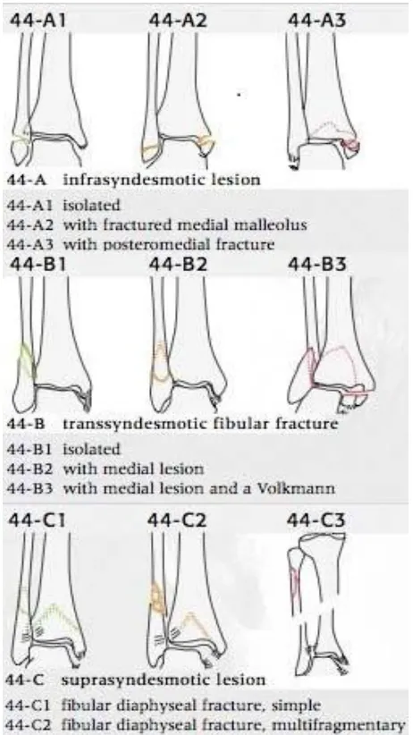
All consecutive 37 patients who were treated by minimally invasive plate osteosynthesis for their extra articular distal thirds tibial fracture in this period were included. The criteria for inclusion in our study were (1) All distal thirds extra articular tibial fractures treated with MIPO (AO type A1,A2 & A3 only) {Distal third will range from 11 cm to 1 cm above the ankle joint}(66)with or without concurrent fibula fractures (AO type A1,B1,C1,C2,C3 only) , (2) both open and closed fractures , (3) available follow up results in terms of 24 months or more; and (4) informed consent from the patient to take part in the study.
Our exclusion criteria included: Stress fractures, Paediatric age group fractures, Pathological fractures/metabolic bone diseases and distal tibia fractures with intra-articular involvement (Pilon fractures)
We excluded 3 patients with pathological fractures and 2 patients in the paediatric age group (less than 15 years).
33
The hospital numbers of these patients were retrieved from the operation theatre register. All these patients were then contacted by Email / phone or posts and asked to come for a follow up in OPD. Five patients were lost to follow up and could not be traced even on a house visit. One patient who had followed up for the initial 1 year after the trauma and surgical treatment, died of stroke related complications.
Finally 26 patients were included in our study.
Consent was obtained from the respective patients and a detailed clinical assessment was done by the two examiners independently followed by specific functional scores and measurement of radiograph parameters.
Outcome measurement
The functional outcome was measured by using standard questionnaires and consisted of the AOFAS & OLERUD and MOLANDER Scores.
The American Orthopaedic Foot and Ankle Society Score ( AOFAS ) introduced in 1994 by Kitaoka et al(67) and has nine questions related to three components:
Pain (1 Question with 40 points), Function (7 Questions with 50 points) and Alignment (1 Question with 10 points) leading to a total possible score of 100 points. The questions related to alignment and range of motion (measured by an orthopaedic goniometer) was completed by the examiner based upon clinical assessment and radiographs; the other questions were completed by the individual patients. The individual scores were then added together to obtain an overall functional score, which then was expressed as a percentage of the normal (100 points).
34
Work/Activities of daily living. Scores of 91-100 were graded as excellent, 61-90 as good, 31-60 as fair and 0-30 as poor results.
Radiographic assessment was done comparing the ateroposterior and lateral views of the affected and the normal leg with both the knee and ankle joints included.
[image:43.595.120.381.214.626.2]
35
If fracture union was not achieved by the sixth month after surgery, the situation was graded as delayed union and by the ninth month as nonunion. We assessed deformities in sagittal and frontal planes and shortening on standard long-leg radiographs. The joint orientation angles were used to access axial deviation in frontal and sagittal planes ( lateral distal tibial angle 89 ± 3 , anterior distal tibial angle 79.8 ± 1.6 degrees)(69)
[image:44.595.144.375.236.647.2]
36
Malunion (more than 5 degrees of varus & valgus angulation, more than 10 degrees of Anteroposterior angulation ), shortening (more than 10mm – significant)(70) and the time to radiographic union (callus on three out of four cortices). Change of alignment from immediate postoperative period till final follow up was also documented.
All major and minor complications were documented at follow up.
37
[image:46.595.196.373.83.336.2]Figure 7. Pre bending of the plate
[image:46.595.238.364.407.685.2]38
Figure 9. Sliding of plate through sub-cutaneous tunnel
39
Figure 11. Applying of positioning screw
[image:48.595.201.404.422.703.2]40
41
Antibiotic prophylaxis in the perioperative period consisted of intravenous Cloxacillin and Gentamycin for closed fractures. Intravenous Crystalline Penicillin was added if it was an open fracture. All intravenous antibiotics were continued for a maximum of 2 days in case of closed fractures and 5 days in case of open fractures.
The patients were discharged from the hospital once the wounds looked healthy and were asked to ambulate with a pair of crutches (non weight bearing) and were followed up at regular intervals in the OPD. Progressive weight bearing was advised depending on fracture union.
42
Results
26 patients who underwent minimally invasive plate osteosynthesis of their distal tibial extra articular fractures were analysed.
Mean age of patients at the time of injury was 42.5 ± 13.12 years (range : 21 to 74 years) ; there were 18 male and 8 female patients. The minimum follow up period was 24 months (mean of 32.7 months; range: 24 to 70 months).
Diagram 1. Pie Chart showing distribution of sex
Diagram 2. Pie Chart showing distribution of comorbidities
69%
31%
SEX
MALE
FEMALE
77%
23%
COMORBIDITIES
43
Diagram 3. Pie Chart showing distribution of associated polytrauma
Isolated lower leg injuries were seen in 16 patients and another 10 had additional injuries/fractures of other segments.
In the 26 patients, 24 patients had high energy injuries (23 had road traffic accidents and 1 patient had fall from a height of 15 feet) and 2 patients had low energy injuries (both had twist and fall from a chair).
Diagram 4. Pie Chart showing distribution of mechanism of injury
ABSENT - 92%
PRESENT - 8%
ASSOCIATED POLYTRAUMA
ABSENT
PRESENT
92%
8%
MECHANISM OF INJURY
HIGH ENERGY
44
Fracture distribution according to AO-Muller classification is shown (figure 5). Most fractures were of A2 pattern. The average distance of the fracture was 5.88cms from the tibial plafond (range : 2.4 to 10.9 cms). In most, the center of fracture was at the level of metadiaphyseal junction.
Diagram 5. Bar Diagram showing distribution of pattern of fracture
Open fractures were seen in 10 patients and included Gustilo and Andersen type I fractures in two patients; Type II in two; Type IIIA in four; and Type IIIB in two patients. Closed soft tissue injuries were seen in 2 patients (Tscherne grade I in one patient and Tscherne grade II in one patient). The remaining patients had no major soft tissue injuries.
6 (23.08%)
13 (50%)
7 (26.92%)
0 2 4 6 8 10 12 14
A1 A2 A3
PATTERN OF TIBIA FRACTURE
45
Diagram 6. Pie Chart showing distribution of type of fracture
Diagram 7. Bar Diagram showing distribution of type of open fracture
62%
38%
TYPE OF FRACTURE
CLOSED OPEN
0 1 2 3 4 5 6
Type1 Open Type2 Open Type3 Open 2 (7.69%) 2 (7.69%)
6 (23.08%)
TYPE OF OPEN FRACTURE
46
Diagram 8. Bar Diagram showing distribution of soft tissue injury
Diagram 9. Pie Chart showing distribution of concurrent fibula fracture
0 2 4 6 8 10 12 14
Grade 0 Grade 1 Grade 2
14 (53.84%)
1 (3.85%) 1 (3.85%)
SOFT TISSUE INJURY
SOFT TISSUE INJURY (TSCHERNE GRADING)
15% 85%
CONCURRENT FIBULA FRACTURE
ABSENT
47
Diagram 10. Bar Diagram showing distribution of pattern of fibula fracture
The mean interval between injury and definitive surgery was 7.92 ± 23 days (range: 1 to 120 days).One patient had underwent native splinting elsewhere and had presented to us 4 months post injury. If we consider him as an outlier then our time to definitive surgery would become 3.44 days.
The average hospital stay for all patients was 10.2 days and was slightly less for patients with closed fractures (average of 7 days). Most of the patients were advised toe touch weight bearing of the operated limb with the help of axillary crutches at suture removal, excluding 2 polytrauma patients and 4 patients with the opposite side upper limb or lower limb fracture.
0 2 4 6 8 10 12 14
A1 B1 C1 C2 C3
0
6 (23.07%)
13 (50%)
2 (7.7%) 3 (11.53%)
PATTERN OF FIBULA FRACTURE
48
Diagram 11. Pie Chart showing distribution of same limb fracture
Diagram 12. Pie Chart showing distribution of concurrent fixation of fibula
69% 31%
SAME LIMB FRACTURE
ABSENT
PRESENT
31% 69%
CONCURRENT FIXATION OF FIBULA
NOT DONE
49
Diagram 13. Pie Chart showing distribution of implant
The average time to partial weight bearing was 2.87 months which was usually decided after radiographic evidence of some callus on routine x rays.Time to full weight bearing was on an average of 4.79 months.
The time to radiological union was slightly more for patients with associated fractures (average of 6.65 months) and open fractures (average of 6.67 months) as compared to closed ones (average of 5.6 months). Excluding 2 delayed unions the time to radiological union in our study was 5.68 months which is comparable to most western literature and studies.
None of the healed fractures had a varus/valgus angulation of more than 5 degrees or an anteroposterior angulation of more than 10 degrees.
Immediate post operative additional procedures were done in two patients which included two planned reverse sural flaps as cover for the soft tissue defect in open fractures. There were 2 instances of culture positive infection. First one was in the immediate post operative period which was treated with appropriate antibiotics and an implant exit once bony
DCP - 11%
LCP - 85%
OTHERS - 4%
IMPLANT
DCP
LCP
50
healing was evident. The second one was a delayed infection (cellulitis) where the patient was managed conservatively with appropriate antibiotics and had implant removal on a later date.
On a total 7 patients underwent implant exit.
The average time taken to return to work was 6.31 ± 2.17 months. All patients went back to their previous jobs except for one patient who had to modify her job.
The mean AOFAS functional scores was 93 ± 7.3 ( range : 69 to 100) and Olerud and Molander functional scores was 89.42 ± 7.79 (range : 70 to 100). The mean AOFAS functional scores were slightly better in males ( mean : 94.16 ± 7.6) as compared to females ( mean : 90.37 ± 8.4) , though not statistically significant.
Diagram 14. Bar Diagram showing distribution of olerud and molander score
0 2 4 6 8 10 12 14 16
EXCELLENT GOOD FAIR BAD 11 (42.3%)
15 (57.7%)
0 0
OLERUD AND MOLANDER SCORE
51
variable group n %
AOFAS 0-30 0 0
31-60 0 0
61-90 7 26.92
>90 19 73.08
OAMS 0-30 0 0
31-60 0 0
61-90 15 57.69
[image:60.595.113.516.437.679.2]>90 11 42.31
Table 2.AOFAS and OAMS
Diagram 15. Bar Diagram showing distribution of AOFAS score
0 2 4 6 8 10 12 14 16 18 20
EXCELLENT GOOD FAIR BAD 19 (73.07%)
7 (26.93%)
0 0
AOFAS SCORE
52
OLERUD AND MOLANDER SCORE
BAD(0-30) 0
[image:61.595.138.476.98.173.2]FAIR(31-60) 0 GOOD(61-90) 15 EXCELLENT(91-100) 11
Table 3.Olerud and molander score
Statistical Analysis showed that the fracture pattern and the time to surgery are the only significant factors affecting the functional score. Rest of the variables like age, sex, comorbidities, concurrent fibula fixation etc... did not show any significant correlation.
53
Correlation of difference in radiological angulation with the variables :
coefficient p-value
AOFAS -0.26 0.18
OAMS -0.15 0.45
Time to RU 0.42 0.03
Table 4.Correlation of difference in radiological angulation
Diagram 16. Scattered plot of difference in angulation vs time to union
The change in the amount of varus or valgus angulation in the post operative period seems to be associated with the factor that the fracture is taking a longer time to unite.
4
6
8
10
12
ti
me
t
o
re
u
n
io
n
0 1 2 3 4
54
The agreement between AOFAS and OAMS (Bland-ALtman plot ) :
Variable Pearson's correlation bias(SD) agreement limits
AOFAS vs OAMS 0.71 -3.57(5.96) [-15.25, 8.11]
The OAMS underestimates the result compared to AOFAS. The pearson’s correlation between the functional scores are not very high.
Diagram 17. Bland-ALtman plot for OAMS vs AOFAS
-1
5
-1
0
-5
0
5
10
D
if
fe
re
n
ce
75 80 85 90 95 100
55
The average loss of ankle range of movement as compared to the opposite side was more than 30 degrees in only 3 patients.
All patients had fracture union with MIPO as the primary procedure. None of them required secondary bone grafting.
56
ILLUSTRATIVE CASES
CASE - 1
This 36 years old gentleman, met with Road Traffic Accident and had AO type A3 distal Tibia fracture of Right Lower Limb. The fracture was stabilized by using locked plate for the distal Tibia and distal Femur. Radiologically the callus was evident by 3 months. It progressed to good consolidation. His ankle range of movement was 20 degrees of dorsiflexion and 30 degrees of plantarflexion at the last followup (22months). He did not have any limb length discrepancy. Functional result of the patient was good.
[image:65.595.325.521.410.686.2]
57
[image:66.595.73.245.70.312.2]Figure 16. IMMEDIATE POST OP AP Figure 17. IMMEDIATE POST OP LAT
[image:66.595.325.497.70.313.2]58
59
CASE - 2
57 years old lady sustained an open fracture of her left distal tibia ( AO type A2) following Road Traffic Accident. The fracture was stabilized by using distal tibia L.C.P. and the fibula with a D.C.P. Post – operative period was uneventful. Radiologically the callus was evident by 12 weeks. She was walking full weight bearing by 4 months. At the last follow up (2½ years), she had normal ankle range of movement and no limb length discrepancy.
Functional result of the patient was Excellent.
[image:68.595.356.517.383.644.2]
60
Figure 23. IMMEDIATE POST OP (AP) Figure 24. IMMEDIATE POST OP (LAT)
[image:69.595.325.494.384.695.2]
61
62
63
64
65
66
Discussion
Innovative surgical techniques have developed over the years with improved understanding of biomechanics, biology and biomaterials, which have ultimately lead to better functional outcome for patients. Managing severely comminuted distal tibia fractures is a challenging task. The aim of this study was to assess the efficacy of minimally invasive plate osteosynthesis in the management of these injuries.
Fracture fixation using plate osteosynthesis is a demanding procedure and the success is related to the surgical technique used(71). A decade ago, more importance was placed on anatomical reduction and rigid fixation to achieve stability. The results were not so encouraging (i.e. increased incidence of delayed union and non union) due to violation of the soft tissue envelope around the fracture site. This led to evolution of newer technique which gave more importance to biology of optimal rather than maximal stability.
MIPO has gained wide application in the treatment of periarticular fractures of the tibia. Despite wide acceptance and assurance in possibilities of the procedure, most reports are based on a small number of patients and the investigators report differing rates of wound complications, time to union, malalignment, and function (Table 2), thus posing questions about whether the theoretical advantages are achieved. Our aims were to estimate the rate of union in tibial fractures treated by MIPO in our institution alongwith, the major and minor complications and functional outcome.
67
terms of surgery, or experience of the treating surgeon. We believe this would be a better representation of the results for this treatment modality by the average orthopaedic surgeon.
Our study group included only extra-articular fractures (1 to 11 cms from the tibial plafond ). Similar studies are scarce(66) because the mechanism of injury and prognosis is different in intra articular and diaphyseometaphyseal extra articular fractures.
Variables Vs Scores
AOFAS OAMS
correlation
coefficient p-val
correlation
coefficient p-val
age -0.16 0.44 -0.21 0.30
bmi 0.14 0.50 0.06 0.78
time indays( from injury to
surgery) -0.56 <0.001 -0.21 0.31
hospital stay 0.02 0.93 0.29 0.16
time partial weight bearing -0.46 0.02 -0.22 0.27
time full weight bearing -0.57 0.00 -0.23 0.27
time of radiological union -0.09 0.66 -0.03 0.90
[image:76.595.71.530.236.430.2]Average time to return work -0.44 0.20 -0.24 0.24
68
The limitations of the study include the following. Firstly, ours is a retrospective study and has no controls. Second, a large portion of the patients (16 cases) was examined at 2 or more years after the injury, and patient answers about terms of restoring weight bearing or resumption of working capacity would be subject to recall bias and may be not accurate. Third, function in some patients could be studied at 24 to 48 months after surgery, whereas in other patients, the last examination was performed 1 year after surgery. Because function might improve after 1 year, our findings might underestimate mean function.
Another weakness is the fact that only the AOFAS scoring system was used to show functional outcome and no general health survey questionnaire (e.g., the SF-36) was incorporated into the study. The primary reason for not including a general health survey questionnaire was that at the beginning of this study none were being used to any degree in the trauma literature; another inherent weakness of the study is the small number of patients; this is an uncommon problem, making it difficult to build a large study population.
MIPO allowed uneventful healing in 84.7 % of our cases. Consistent with the literature, a high percentage of our patients resumed to their preinjury level of working activities with general restoration of lower leg function. However, complications occurred in a substantial portion of patients, which may be divided into three groups: disturbances of fracture healing (7.5 %), infection (both immediate post operative and late) (7.5 %), and hardware problems (26.9%).
High energy and comminuted fracture patterns took a long time to heal which is consistent with results of Collinge et al(38).
Milner et al(72) studied 164 fracture tibias with a long-term follow-up of 30 years and
concluded that there were no significant univariate associations between malunions of the
69
series did we observe valgus or varus malalignment more than 5°.
One grey area of confusion in comminuted fractures is whether primary bone grafting is indicated or not. Primary bone grafting is contraindicated if soft tissue dissection has to be done to place the graft(18). We have not done primary lone grafting in any of the cases. Bone grafting may be indicated if the healing is not progressive as assessed radiologically.
70
Conclusions
71
Bibliography
1. WHO : World report on road traffic injury prevention. Geneva: WHO 2004. Geneva: WHO; 2004. p. 3-29 p.
2. Ruikar M. National statistics of road traffic accidents in India. J Orthop Traumatol Rehabil 2013;6:1-6.
3. Court-Brown CM, Rimmer S, Prakash U, McQueen MM. The epidemiology of open long bone fractures. Injury. 1998 Sep;29(7):529–34.
4. Trueta J. Blood supply and the rate of healing of tibial fractures. Clin Orthop. 1974 Dec;(105):11–26.
5. Ruedi TP, Allgower M. Fractures of the lower end of the tibia into the ankle joint. Injury. 1969; 1(2): 92-99. 1969;
6. Møller BN, Krebs B. Intra-articular fractures of the distal tibia. Acta Orthop Scand. 1982 Dec;53(6):991–6.
7. McFerran MA, Smith SW, Boulas HJ, Schwartz HS. Complications encountered in the treatment of pilon fractures. J Orthop Trauma. 1992;6(2):195–200.
8. Rüedi T. Fractures of the lower end of the tibia into the ankle joint: results 9 years after open reduction and internal fixation. Injury. 1973 Nov;5(2):130–4.
9. Rüedi TP, Allgöwer M. The operative treatment of intra-articular fractures of the lower end of the tibia. Clin Orthop. 1979 Feb;(138):105–10.
10. Watson JT, Moed BR, Karges DE, Cramer KE. Pilon fractures. Treatment protocol based on severity of soft tissue injury. Clin Orthop. 2000 Jun;(375):78–90.
11. Pugh KJ, Wolinsky PR, McAndrew MP, Johnson KD. Tibial pilon fractures: a comparison of treatment methods. J Trauma. 1999 Nov;47(5):937–41.
12. Hansmann C. Eine neue Methode der Fixierung der Fragmente bei complicierten Fracturen. Verh Dtsch Ges Chir 15:134; 1886;
13. Perren SM. Fracture healing. The evolution of our understanding. Acta Chir Orthop Traumatol Cech. 2008 Aug;75(4):241–6.
14. Perren SM, Cordey J, Rahn BA, Gautier E, Schneider E. Early temporary porosis of bone induced by internal fixation implants. A reaction to necrosis, not to stress protection? Clin Orthop. 1988 Jul;(232):139–51.
72
16. Lambotte A. L’intervention operatoire dans les fractures récentes et anciennes, Paris. Maloine,1907 .
17. Küntscher G. Die Marknagelung von Knochenbrüchen. Arch Klin Chir 200:443–455; 1940;
18. Gerber C, Mast JW, Ganz R. Biological internal fixation of fractures. Arch Orthop Trauma Surg. 1990;109(6):295–303.
19. Tepic S, Perren SM. Tepic S, Perren SM (1995) The biomechanics of the PC-Fix internal fixator. Injury; 26(Suppl 2):5–10. 1995;
20. Frigg R, Appenzeller A, Christensen R, Frenk A, Gilbert S, Schavan R. The development of the distal femur Less Invasive Stabilization System (LISS). Injury. 2001 Dec;32 Suppl 3:SC24–31.
21. Marti A, Fankhauser C, Frenk A, Cordey J, Gasser B. Biomechanical evaluation of the less invasive stabilization system for the internal fixation of distal femur fractures. J Orthop Trauma. 2001 Oct;15(7):482–7.
22. Krettek C. Foreword: concepts of minimally invasive plate osteosynthesis. Injury. 1997;28 Suppl 1:A1–2.
23. Helfet DL, Shonnard PY, Levine D, Borrelli J Jr. Minimally invasive plate osteosynthesis of distal fractures of the tibia. Injury. 1997;28 Suppl 1:A42–47; discussion A47–48. 24. Sommer C, Gautier E. [Relevance and advantages of new angular stable screw-plate
systems for diaphyseal fractures (locking compression plate versus intramedullary nail]. Ther Umsch Rev Thérapeutique. 2003 Dec;60(12):751–6.
25. Helfet DL, Haas NP, Schatzker J, Matter P, Moser R, Hanson B. AO philosophy and principles of fracture management-its evolution and evaluation. J Bone Joint Surg Am. 2003 Jun;85-A(6):1156–60.
26. Lau TW, Leung F, Chan CF, Chow SP. Minimally invasive plate osteosynthesis in the treatment of proximal humeral fracture. Int Orthop. 2007 Oct;31(5):657–64.
27. Aksu N, Karaca S, Kara AN, Işiklar ZU. Minimally invasive plate osteosynthesis (MIPO) in diaphyseal humerus and proximal humerus fractures. Acta Orthop Traumatol Turc. 2012;46(3):154–60.
28. Zhiquan A, Bingfang Z, Yeming W, Chi Z, Peiyan H. Minimally invasive plating osteosynthesis (MIPO) of middle and distal third humeral shaft fractures. J Orthop Trauma. 2007 Oct;21(9):628–33.
29. Ji F, Tong D, Tang H, Cai X, Zhang Q, Li J, et al. Minimally invasive percutaneous plate osteosynthesis (MIPPO) technique applied in the treatment of humeral shaft distal fractures through a lateral approach. Int Orthop. 2009 Apr;33(2):543–7.
73
31. Krettek C, Schandelmaier P, Miclau T, Tscherne H. Minimally invasive percutaneous plate osteosynthesis (MIPPO) using the DCS in proximal and distal femoral fractures. Injury. 1997;28 Suppl 1:A20–30.
32. Schütz M, Müller M, Kääb M, Haas N. Less invasive stabilization system (LISS) in the treatment of distal femoral fractures. Acta Chir Orthop Traumatol Cech. 2003;70(2):74– 82.
33. Ricci WM, Loftus T, Cox C, Borrelli J. Locked plates combined with minimally invasive insertion technique for the treatment of periprosthetic supracondylar femur fractures above a total knee arthroplasty. J Orthop Trauma. 2006 Mar;20(3):190–6.
34. Kolb W, Guhlmann H, Windisch C, Marx F, Kolb K, Koller H. Fixation of distal femoral fractures with the Less Invasive Stabilization System: a minimally invasive treatment with locked fixed-angle screws. J Trauma. 2008 Dec;65(6):1425–34.
35. Apivatthakakul T, Chiewcharntanakit S. Minimally invasive plate osteosynthesis (MIPO) in the treatment of the femoral shaft fracture where intramedullary nailing is not indicated. Int Orthop. 2009 Aug;33(4):1119–26.
36. Cole PA, Zlowodzki M, Kregor PJ. Treatment of proximal tibia fractures using the less invasive stabilization system: surgical experience and early clinical results in 77 fractures. J Orthop Trauma. 2004 Sep;18(8):528–35.
37. Oh C-W, Oh J-K, Kyung H-S, Jeon I-H, Park B-C, Min W-K, et al. Double plating of unstable proximal tibial fractures using minimally invasive percutaneous osteosynthesis technique. Acta Orthop. 2006 Jun;77(3):524–30.
38. Collinge C, Sanders R, DiPasquale T. Treatment of complex tibial periarticular fractures using percutaneous techniques. Clin Orthop. 2000 Jun;(375):69–77.
39. Redfern DJ, Syed SU, Davies SJM. Fractures of the distal tibia: minimally invasive plate osteosynthesis. Injury. 2004 Jun;35(6):615–20.
40. Krackhardt T, Dilger J, Flesch I, Höntzsch D, Eingartner C, Weise K. Fractures of the distal tibia treated with closed reduction and minimally invasive plating. Arch Orthop Trauma Surg. 2005 Mar;125(2):87–94.
41. Hazarika S, Chakravarthy J, Cooper J. Minimally invasive locking plate osteosynthesis for fractures of the distal tibia--results in 20 patients. Injury. 2006 Sep;37(9):877–87. 42. Hasenboehler E, Rikli D, Babst R. Locking compression plate with minimally invasive
plate osteosynthesis in diaphyseal and distal tibial f