DIFFERENTIAL EXPRESSION OF ANGIOGENIC FACTORS
IN SKIN OF PATIENTS WITH PSORIASIS VULGARIS
Dissertation submitted in
Partial fulfillment of the regulations required for the award of
M.D. DEGREE
In
PATHOLOGY – BRANCH III
THE TAMILNADU
DR. M.G.R. MEDICAL UNIVERSITY
CHENNAI
DECLARATION
I hereby declare that the dissertation entitled “DIFFERENTIAL EXPRESSION OF ANGIOGENIC FACTORS IN SKIN OF PATIENTS WITH PSORIASIS VULGARIS” is a bonafide research work done by me in the Department of Pathology, Coimbatore Medical College during the period from April 2013 to July 2014 under the guidance and supervision of Dr. A. DHANALAKSHMI, M.D., Associate Professor, Department of Pathology, Coimbatore Medical College.
This dissertation is submitted to The Tamilnadu Dr.MGR Medical University, Chennai towards the partial fulfilment of the requirement for the award of M.D., Degree (Branch III) in Pathology. I have not submitted this dissertation on any previous occasion to any University for the award of any Degree.
Place: Coimbatore
CERTIFICATE
This is to certify that the dissertation entitled “DIFFERENTIAL EXPRESSION OF ANGIOGENIC FACTORS IN SKIN OF
PATIENTS WITH PSORIASIS VULGARIS” is a record of bonafide work done by Dr.S.LAKSHNA in the Department of Pathology, Coimbatore Medical
College, Coimbatore under the guidance and supervision of
Dr. A. DHANALAKSHMI, M.D., Associate Professor, Department of Pathology, Coimbatore Medical College and submitted in partial fulfilment of the requirements
for the award of M.D. Degree (Branch III) in Pathology by The Tamilnadu Dr. MGR
Medical University, Chennai.
Guide Head of the Department Dr.A. DHANALAKSHMI, M.D., Dr.C.LALITHA, M.D., Associate Professor, Professor,
Department of Pathology, Department of Pathology, Coimbatore medical college, Coimbatore medical college,
Coimbatore. Coimbatore.
Dr.S.REVWATHY, M.D.,D.G.O,DNB (OG) The Dean,
ACKNOWLEDGEMENT
To begin with, I thank the Almighty GOD in making this project a successful one.
I express my deep gratitude to Dr.S.REVWATHY, M.D., D.G.O.,DNB (OG) Dean, Coimbatore Medical College, for permitting me to undertake this study.
I express my sincere gratitude to Dr.C.LALITHA, M.D., Professor and Head, Department of Pathology, Coimbatore Medical College, for having suggested this topic for dissertation and for having rendered her valuable support and encouragement without which this project work would not have been feasible.
I am extremely grateful to Associate Professor Dr. A. DHANALAKSHMI, M.D., for her invaluable suggestions,
constant encouragement and support during this endurable work.
I thank all the technical staffs in the Department of Pathology, Coimbatore Medical College, for their sincere and timely technical assistance.
I express my heartfelt thanks to Department of Dermatology, Coimbatore Medical College, for their constant support throughout the course of this study.
I express my heartfelt thanks and gratitude to my parents, my husband, my in laws and my lovable sister for their extreme patience,constant support, encouraging words and source of strength all the way through this endeavour.
Submission author: Assignment title:
Submission title: File name:
File size: Page count:
Word count: Character count:
Submission date: Submission ID:
Digital Receipt
This receipt acknowledges that Turnitin received your paper. Below you will find the receipt information regarding your submission.
The first page of your submissions is displayed below.
201213253.md Pathology LAKSHNA… TNMGRMU EXAMINATIONS
Differential expression of Angiogenic … thesis_medical.docx
103.74K 107
15,275 90,187
19-Sep-2014 08:08AM 449811710
CONTENTS
SI.NO. PARTICULARS PAGE NO.
1. INTRODUCTION 1
2. AIM & OBJECTIVES 4
3. REVIEW OF LITERATURE 5
4. MATERIALS AND METHODS 65
5. OBSERVATION AND RESULTS 75
6. DISCUSSION 99
7. SUMMARY AND CONCLUSION 106
BIBLIOGRAPHY
ANNEXURES
ANNEXURE I – PROFORMA AND CONSENT
ANNEXURE II – MASTER CHART
LIST OF TABLES
TABLE
NO TITLE
PAGE NO
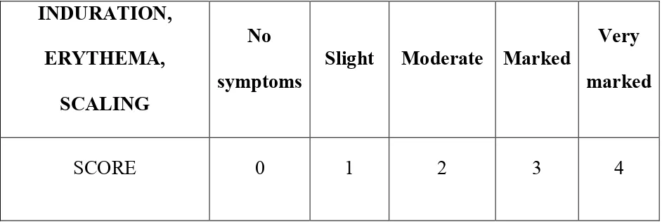
1 PASI SCORE- SEVERITY OF LESION 66
2 PASI SCORE – AREA OF DISTRIBUTION 66
3
PASI SCORE –METHOD OF
CALCULATION 67
4
AGE WISE DISTRIBUTION OF
PSORIASIS 76
5
MEAN AGE WITH GENDER
DISTRIBUTION 78
6
GENDER WISE DISTRIBUTION OF
SAMPLES 79
7
MICROVESSEL DENSITY -
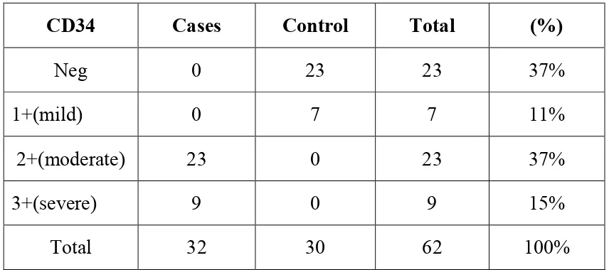
EVALUATION OF CD 34 STAINING 81
8
IMMUNOHISTOCHEMICAL STAINING
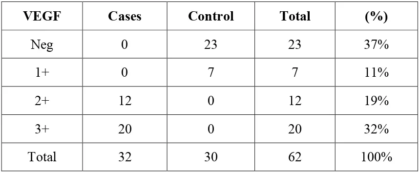
OF VEGF 84
9
IMMUNOHISTOCHEMICAL
EVALUATION OF VWFR 87
10
MEAN OF ANGIOGENIC FACTOR
EXPRESSION 90
11 AGE VS PASI SCORE 92
12
CORRELATION BETWEEN PASI SCORE
LIST OF CHARTS
CHART
NO. TITLE
PAGE NO
1 PRIMARY ANTIBODY WITH
SECONDARY KIT 72
2a AGE WISE DISTRIBUTION OF CASES
AND CONTROLS 77
2b AGE WISE DISTRIBUTION OF CASES
AND CONTROLS 77
3 GENDER WISE DISTRIBUTION OF
PSORIASIS 80
4a INTENSITY OF CD34 STAINING AMONG
CASES 82
4b
COMPARISON OF IMMUNOREACTIVITY OF CD 34 BETWEEN CASES AND
CONTROL
83
5a IMMUNOREACTIVITY OF VEGF AMONG
CASES 85
5b COMPARISON OF VEGF EXPRESSION
BETWEEN CASES AND CONTROL 86
6a INTENSITY OF VWFr STAINING AMONG
CASES 88
6b COMPARISON OF VWFr EXPRESSION
BETWEEN CASES AND CONTROL 89
7
PASI SCORE – IN CASES
PSORIATIC AREA AND SEVERITY INDEX
8 AGE Vs PASI SCORE 93
9 CORRELATION BETWEEN PASI SCORE
AND CD34 95
10 CORRELATION BETWEEN PASI SCORE
AND VEGF 96
11 CORRELATION BETWEEN PASI SCORE
AND VWFr 97
LIST OF COLOUR PLATES
FIG.NO.
TITLE
1. HISTOLOGY OF SKIN
2. HISTOPATHOLOGY OF PSORIASIS- LOW POWER
VIEW
3. DILATED DERMAL CAPILLARIES IN PSORIASIS-
HIGH POWER VIEW
4. IHC SHOWS LOW POWER - VEGF MODERATE
EPIDERMAL POSITIVITY (2+) IN CASES
5. IHC HIGH POWER SHOWS MODERATE DEGREE
OF EPIDERMAL STAINING (2+) IN CASES
6. IHC HIGH POWER SHOWS DIFFUSE EPIDERMAL
STAINING OF VEGF (3+ POSITIVITY) IN CASES
7.
IHC HIGH POWER SHOWS CYTOPLASMIC AND MEMBRANOUS POSITIVITY - HIGHER DEGREE (3+) IN CASES
8. IHC LOW POWER - SHOWS VEGF WEAK
EPIDERMAL STAINING (1+) IN CONTROLS
9. IHC HIGH POWER SHOWS WEAK EPIDERMAL
POSITIVITY OF VEGF (1+) IN CONTROLS
10. IHC LOW POWER VIEW SHOWS CD34 POSITIVITY
IN CASES
11. IHC HIGH POWER VIEW SHOWS CD 34 MILD
POSITIVITY (1+) IN CASES
12.
IHC HIGH POWER VIEW SHOWS CD34
13. IHC HIGH POWER VIEW SHOWS CD34 HIGHER DEGREE OF POSITIVITY (3+) IN CASES
14.
LOW POWER SHOWS WEAK EPIDERMAL STAINING OF VON WILLE BRAND FACTOR IN CASES
15.
ABSTRACT
INTRODUCTION
Psoriasis is a chronic inflammatory skin disease characterized by
hyper-proliferation, abnormal differentiation, and inflammatory infiltration in
epidermis and dermis. Angiogenesis or neovascularization refers to the
formation of new capillary vessels from the existing vascular bed .Dermal
microvascular expansion with abnormal orientation and dilatation of capillaries
in the biopsies of the psoriatic skin revealed that the disease was angiogenesis
dependent .
AIM AND OBJECTIVES
• to analyse immunohistochemical expression of angiogenic factors Vascular endothelial growth factor, Von wille brand factor and CD 34 in
skin biopsy of patients with psoriasis vulgaris and controls.
• To compare the neovascularisation score of CD 34, VEGF and vWFr in skin biopsy of psoriasis vulgaris cases and controls.
• To correlate the expression of angiogenic factors and psoriasis area and severity clinical index(PASI SCORE) which is a measure of clinical
MATERIALS AND METHODS
This is a case control study carried over a period of 15 months from
April 2013 to July 2014.Thirty two psoriasis cases and thirty control healthy
skin were studied.Biopsy specimen is taken from skin of newly diagnosed
psoriasis and patients who have not taken any treatment two months prior to
study. Histopathological examination of psoriasis vulgaris was confirmed.
Immunohistochemical expression for VEGF(vascular endothelial growth
factor), vonwillebrand factor and CD 34 was studied.
RESULTS
VEGF expression in epidermis was significantly higher in icases when
compared to control skin (p=<o.o1).CD 34 expression was significantly
upregulated in cases when compared to controls.(p<0.01). Whereas only weak
expression of vonwillebrand factor was observed in both cases and controls.
Significant correlation between the expression of VEGF and PASI
score(r=0.944;p<0.05), and expression of CD 34 and PASI score was
observed(r=0.942;p<0.05).
CONCLUSION
Significant overexpression of VEGF and CD 34 was noted in cases when
compared to controls.The keratinocytes in the psoriatic skin lesions were
namely the vascular endothelial growth factor (VEGF) and other growth factors
which promotes micrangiopathic modifications in psoriatic plaque.
Angiogenesis plays an important role in genesis and development of psoriasis
vulgaris. Therefore development of targeted anti- angiogenic therapy might
be beneficial for this chronic disabling dermatological disease.
1
INTRODUCTION
Skin is the largest organ in the body composed of many interdependent cell structures.1 Direct visual examination of skin lesions, gross description and histopathology are necessary for diagnosis of dermatological lesions.
Psoriasis is a chronic papulosquamous dermatitis with varying incidence between 0.5% to 1.5% of population.2 Psoriasis is one of the common type of dermatological condition in India with an epidemiological and prevalence characteristics being similar to Western countries.3 The mean age of onset is around 25 years although milder form is seen in older persons.4
2
The characteristic histopathological features are acanthosis, regular downward elongation of rete ridges, spongiform pustules and munro’smicroabscesses. Papillary dermis shows edema and tortuous dilated capillaries. These dilated tortuous capillaries are the source of bleeding points when the scales are scrapped off from the plaques.5
Psoriasis has a complex pathogenesis6 characterized by altered keratinocyte proliferation and differentiation, immune mediated inflammation , dysregulated angiogenesis and vascular remodelling.7 Various factors which play a central and pivotal role in the pathomechanism of psoriasis are Th1type of cell, Th17 cell, antigen presenting cell , Langerhan type of cells, natural killer cell , keratinocytes and macrophages and various cytokines of Th1 type.6
New blood vessel formation is seen in early stages of psoriatic lesions and neovascularisation disappears with clearance of skin disease.8 Various angiogenic mediators such as vascular endothelial growth factor, hypoxia inducible factor, and several proangiogenic cytokines like tumour necrosis factor , interleukin 17 and interleukin 8, are increased in lesional skin of psoriasis.8
3
In present study neovascularisation score in normal skin biopsy as compared to skin biopsy of psoriasis vulgaris patients is done using CD 34. Also the expression of VEGF-vascular endothelial growth factor and vonwillebrand factor in epidermis of normal skin biopsy compared to skin biopsy of psoriatic patients is done. This attempts to prove that vascular changes appears to be significant in pathomechanism of psoriasis. Thus angiogenesis appears to contribute to the pathogenesis of psoriatic lesions.9
Anti angiogenic agents may be potential targeted therapy in the future.
4
AIMS AND OBJECTIVES
• to analyse immunohistochemical expression of angiogenic factors Vascular endothelial growth factor, Von wille brand factor and CD 34 in skin biopsy of patients with psoriasis vulgaris and controls.
• To compare the neovascularisation score of CD 34, VEGF and vWFr in skin biopsy of psoriasis vulgaris cases and controls.
• To correlate the expression of angiogenic factors with psoriasis area severity index(PASI SCORE) which is a measure of clinical severity .
5
REVIEW OF LITERATURE
ANATOMY AND HISTOLOGY OF SKIN
Understanding normal skin histology is of prime importance to identify cutaneous pathology. It has two histologically and anatomically separate layers namely the epidermis and the dermis. But epidermis and dermis are functionally interdependent. There is subcutaneous adipose tissue below the dermis.
The skin has many functions including mechanical protection, sun protection, temperature regulation, immune related functions, sensory perception and nutrient metabolism.10 The epidermal keratinocyte has been recognised as a potent source of immunogenic molecules such as interleukins, colony stimulating factors , interferons, transforming growth factors, tumour necrosis factor and growth factors11
EPIDERMIS
6
The keratinocytes are organised into four layers – stratum basale , spinosum, granulosum, lucidum and corneum.
STRATUM BASALE (basal layer)
The basal cell layer is the lowermost layer of epidermis. It has single row of cells of cuboidal type. These cells have round to ovoid nuclei with basophilic cytoplasm and often contain melanin pigmentation from adjacent melanocytes.10 The basal layer rests on a basement membrane which separates epidermis from dermis. Basal layer is composed of mitotically active cells which give rise to other keratinocytes.
Individual cells are attached to each other by desmosomes and attachment to basement membrane by hemidesmosomes.10 Cells in the basal layer contain intermediate filaments and they increase in number as they move upward.
STRATUM SPINOSUM (squamous layer)
7
tonofilaments provide tensile strength against abrasion of epidermis. The intercellular spaces contain acid mucopolysaccharides and neutral mucopolysaccharides. Hyaluronic acid an important component of acid mucopolysaccarides, is abundant in intercellular spaces of stratum spinosum.12
STRATUM GRANULOSUM (granular layer)
This third layer consists of three to five rows of flattened cells. This layer is seen just above stratum spinosum. Cells in this stratum granulosum are filled with dense keratohyaline granules. The granular cell layer is the mature keratin forming transition zone of epidermis.10 Dissolution of nucleus and cell organelles is starts from this granular cell layer. Diffusely staining lysosomal enzymes in this layer plays an important role in autolytic changes in this layer.13
STRATUM LUCIDUM
Stratum lucidum consists of row of tightly packed flattened cells that lack nuclei or organelles. This layer is seen only in thick skin. Cells in this layer contains densely packed keratin filaments.
STRATUM CORNEUM (horny layer)
8
soft keratin. The superficial keratinised cells are shed or desquamated continuously and replenished by cells arising from deeper most basal layer or stratum basale. The horny cytoplasm of these cells contain cystine disulfide bonds, these shrink on formalin fixation and form a shell along cell membrane resulting in basket weave appearance in routine histologic sections.14
DERMIS
Dermis is seen just below epidermis and consists of connective tissue fibers and cellular components of epidermis. The cellular components are fibroblasts,dermal dendritic cells ,macrophages and mast cells. The extracellular components are collagen,elastic fibers and ground substance.10
The pale staining narrow zone of connective tissue just below the dermis is papillary dermis. The papillary dermis indents the basement membrane of epidermis to form dermal papillae. The reticular layer of dermis comprises the rest of dermis. The reticular dermis forms the bulk of dermis. The reticular dermis predominantly consists of dense connective tissue.
9
are numerous sensory receptor which are meissner’s corpuscles located closer to dermal papillae and pacinian corpuscles which are found deeper in connective tissue of dermis . Though skin appendages such as hair follicle and sweat glands develop from epidermis ,they are located in dermis.
HISTORICAL PERSPECTIVE OF PSORIASIS
Psoriasis is one of the common dermatological disease . Thus it is justified to have some knowledge in history of psoriasis.
The history of psoriasis, remained speculative for the time before Willan (1757-1812), and reliably assigned only for the last 200 years. Robert Willan (1757- 1812) described different types of psoriasis. Hebra (1806-1880) gave a morphological definition, in which histopathology feature was also taken into account.
10
PSORIASIS
Psoriasis is a one of the common , recurrent chronic inflammatory dermatological disease which affects about 2 % of the caucasian population16 and results in severe impairment of quality of life.
PREVALENCE
The prevalence of psoriasis differs in various parts of the world ranging from 0% to 11.8%.17,18,19,20. The prevalence of psoriasis in India ranges from 0.44% to 2.8%. The disease is two times commonly seen in males when compared with females.
GENETIC BASIS OF PSORIASIS
The molecular genetics of psoriasis is complex and multiple genes are involved. Genetic transmission plays an important role in etiopathogenesis of psoriasis. There are seven major susceptibility loci being reported for psoriasis. Many studies have shown that the major susceptibility locus is in chromosome 6p21, known as PSORS1 and is
represented in most of the populations. 21-26An association with various
loci was also reported on chromosomes 1p (PSORS7)25, 1q
(PSORS4)27, 3q (PSORS5)28, 4q (PSORS3)29, 17q (PSORS2)30, and 19p
11
incidence of psoriasis is higher in childhood psoriasis than adult onset psoriasis.32,33,34
Psoriasis vulgaris is associated with certain HLA antigens.3 Psoriasis is associated with HLA A1, B17 and Cw6.35 Association with HLABw57 and DR7 is seen in South India.36 HLA Cw *0602 is the main allele with higher frequency in North India.37
AGE DISTRIBUTION IN PSORIASIS
Psoriasis tends to occur more commonly in third or fourth decade.3
The age of onset for psoriasis is bimodal distribution was recognised in
many studies. The age for the first clinical presentation of psoriasis
ranges from 15 to 20 years of age,and a second peak of onset which
occurs at age of 55 to 60 years.38-41 Two types of psoriasis are seen, type
one and two, that can be distinguished by a bimodal age distribution.
Type 1 psoriasis has its onset by the age of 40 years; Type II psoriasis
has its onset after 40 years of age. Type I disease accounts for more than
75% of cases.41 Patients with earlier age of onset that is the type I
psoriasis have a severe disease and many relatives are affected compared
to patients with later onset disease or the type II psoriasis. In psoriasis
cases with early onset of skin lesions, strong associations are being
12 TRIGGER FACTORS
Various modifiable risk factors predispose an individual to
develop psoriasis or exacerbate the already existing psoriatic disease.
The modifiable risk factors includes smoking, intake of alcohol,
obesity, dietary habits, several types of infection ,drug intake and
stress.42-51 The actual mechanism which aggravates psoriatic skin
lesions is yet to be elucidated;
Acute bacterial and viral infections are associated with onset of
psoriasis or flaring up of the disease. Streptococcal infection is also an
aggravating factor for guttate psoriasis, more commonly in childhood
age group and younger adults. Human immunodeficiency virus is also
associated with an onset of severe plaque type of psoriasis and does not
respond to standard therapy or medications.
Various drugs which includes beta blockers, lithium, antimalarial
drugs, tetracycline, nonsteroidal antiinflammatory drugs and withdrawal
of steroids are also associated with the onset of psoriasis or flaring up
of the disease. Drug induced psoriasis is likely to occur in patients
with no past history of psoriatic skin lesions which regresses and clears
after withdrawal of the causative medication. Drug aggravated psoriasis
occurs in patients who already have a history of psoriatic skin lesions
13
mechanism of action by which a drug aggravates psoriasis is not clearly
understood. Studies suggest that the mechanism by which beta blocker
induced psoriatic skin lesion occurs is due to blocking of epidermal beta
2 receptor that leads to reduced cyclic adenosine monophosphate in the
epidermis and hyperproliferation of keratinocytes .
The current thought in lithium aggravating psoriasis is by inhibition of inositol monophosphate, resulting in decrease of intra cellular calcium levels and enhanced proliferation of epidermal keratinocytes. Also the lithium increases tumor necrosis factor alpha production and IFN - gamma production in the epidermal cells and they play a major role in psoriasis.
Antimalarial drugs exacerbate already existing psoriasis in 40% of patients. It acts by inhibition of the enzyme transglutaminase and causes proliferation of keratinocytes. The relevance of antibiotic induced psoriatic skin lesions is still a controversy. The tetracycline class of antibiotics exacerbates psoriasis through decrease in intracellular cyclic adenosine monophosphate. Finally, NSAIDs inhibit the arachidonic acid metabolism that leads to accumulation of leukotrienes and these leukotrienes aggravate psoriasis.
14
disease. Psychological stress disorder is also a trigger factor for psoriasis42,52-55.
PATHOGENESIS OF PSORIASIS
Cells like T lymphocyte cell, macrophages, (APC) antigen-presenting cells, natural killer cells, epidermal keratinocytes, Langerhans' cell, , various types of Th1 cytokines and various growth factors such as (VEGF), vascular endothelial growth factor, (KGF) keratinocyte growth factor play a major role in pathomechanism of psoriasis6. There is need to know current concepts and pathomechanisms of psoriasis
CELLULAR BASIS - PSORIASIS
15
EGF,interleukin-8, interleukin-12, interleukin-23, interleukin-17, interleukin-1, interleukin-6, Fractalkine, tumor necrosis factor-α and others are produced by these activated T lymphoctes. Due to the effect of the above cytokines there is proliferation of epidermis and hyperplasia of epidermis, migration of neutrophils , increase in response of Th-1 cell type, upregulation of adhesion molecules and angiogenesis,6 .
ROLE OF KERATINOCYTE
The type of cell which is responsible for the onset of psoriasis is still a controversy. Various investigations concentrated on epidermal keratinocytes. Aberrant activation of keratinocytes and metabolism of keratinocytes in the epidermis, leads to increased proliferation of keratinocytes and they are the characteristic features in lesional psoriatic skin56. Psoriatic skin has eight times shortened turnover for keratinocytes due to enhanced keratinocyte proliferation 57.
16
the epidermal cells. The cell cycle time for hyperproliferating keratinocytes in psoriasis is of shorter duration. The maturation and shedding of epidermal keratinocyte takes atleast 26 days in normal skin, whereas it takes only 4 days for the epidermial keratinocyte of psoriatic skin lesions59. Growth factors produced by various types of cell, are known to control the marked keratinocyte proliferation.
KERATINOCYTE AND IMMUNE SYSTEM CROSS TALK
17
cells and Tcells infiltrates into the epidermis, and they come in contact directly with epidermal keratinocytes.
The mononuclear cells which infiltrates the dermis secretes chemical mediators that induces the proliferation of keratinocytes and endothelium. The dermal tissue of psoriasis skin lesion is infiltrated by CD4 + T helper type of cells and these T helper cells secretes proinflammatory cytokines such as interferon-IFN-γ,interleukin-17 and tumor necrosis factor alpha. 66,67 . Also the increased levels of cytokines such as IL-8, IL-6 and keratinocyte growth factor [transforming growth factor-alpha] are seen in psoriasis skin lesions60,65,68. There is an intense cross-talk between cells of immune system and keratinocytes that establishes an interactive cytokine heirachy,which is responsible for the development of psoriasis.
ROLE OF T CELL AND T CELL ACTIVATION
18
haematological malignancy, who are cleared of disease or obtained remission after bone marrow transplantation from healthy donor without any history of psoriasis73. Some of the patients developed psoriatic skin lesions initially after the bone marrow transplantation from donors suffering from psoriatic skin lesions74.
Therapy for psoriatic lesions with monoclonal antibody targeted against CD 4 molecules improves skin lesion, whereas therapy with monoclonal antibody against the CD 8 molecules, does not improve psoriasis75-77. Also skin xenograft models on Severe Combined Immuno Deficiency mice shows that populations which includes autologous Interferon -γ- producing CD 4 T helper cells can produce psoriatic skin lesions in healthy grafts from patients with psoriatic skin lesion but autologous transfer of CD8 T helper cells from same patients with history psoriasis could not induce the disease78. IFN-γ-producing Th1 cells and IL-17-producing Th 17 cell plays pivotal role for causing psoriasis67,71,79,80 . The expression of both the Th17 cell promoting cytokine Interleukin 23 and the cytokine IL-22 which is associated with Th17 cell in psoriatic skin, supports that both Th1cell and Th 17 cell are responsible for the manifestation of psoriatic skin lesions71,81,82.
19
psoriatic skin lesion, the disease inducing Th1 cells and/or Th17 cells either proliferates in situ or migrates to dermis – the target organ from the peripheral site . The process also depends upon close interaction between the inflammatory Thelper 1/Thelper 17 cells within the dermal microvascular bed. There is an interaction between the lymphocyte function associated antigen - LFA-1 on the lymphocytes and the intercellular adhesion molecule - ICAM-1 on the endothelial cell which mediates adhesion of leukocytes to the endothelial cell which is supposed to be prerequisite for extravasation of leucocytes. In inflammatory diseases, ICAM-1 is markedly expressed on the endothelial cells of vascular structures.
20
Studies by Chang et al85 demonstrates that the cytokines secreted by keratinocytes of psoriasis increases activation of T lymphocyte to a significant level than the cytokines secreted from keratinocytes of normal skin. One school of thought is that only keratinocytes of psoriasis skin lesions respond to information from activated T lymphocyte with hyperproliferation, because of their specific type of receptors or specific signal transduction mechanisms84. Several studies shows that there is alteration in basement membrane structures and a heirachy of cytokines especially Thelper 1 type were involved in different stages of pathogenesis of psoriasis.83,86,87.
ANGIOGENESIS
21
during development of psoriasis. The epidermal keratinocytes isolated from psoriasis skin lesions shows decreased expression of thrombospondin-1 (TSP-1), an endogenous inhibitor of angiogenesis. Thrombospondin inhibits endothelial Cell migration and proliferation, new vessel formation and proliferation of tumour cells92,93,94. In normal healthy skin, production of thrombospondin -1 by basal epidermal cells maintains the separation between the avascular epidermis and vascular dermis95,96.
These findings put together suggests that the involvement of angiogenesis in pathogenesis of psoriasis. Physiological angiogenesis is seen transiently during healing of wounds, pregnancy or the menstrual cycle. Pathological angiogenesis is seen in conditions like neoplastic growth of tumour and chronic inflammatory conditions, which are seen in diseases like rheumatoid arthritis or psoriasis89,97-100.
22
neo vascular structure. 102 Activated and swollen endothelial cells leads to widened inter-cellular spaces and dilatation of dermal blood capillaries.
The lesional skin capillaries adopts a venous type, which includes bridging of fenestrations, and expression of E selectin, this helps for easier migration of leucocytes into the epidermis and dermis. 88
DEFINITION OF ANGIOGENESIS
Angiogenesis means new vessel formation from the preexisting blood capillaries. It is seen during embryogenesis whereas it is absent in many of the adult tissues. Angiogenesis takes place in two different manners : (a) the sprouting type – in which newer blood vessels sprouts from preexisting blood capillaries and (b) the non sprouting type angiogenesis or also known as intussusception,in which there is division of preexisting capillaries by trans-capillary pillars103,104.
SPROUTING OF NEW VESSELS
23
• Dilatation of blood vessels
• increased vascular permeability
• destabilization of blood capillaries already existing,
• degradation of the extracellular matrix
• Endothelial Cell migration and proliferation
• Formation of vascular lumen and maturation of vessels by recruitment of peri-vascular supporting cells105,106.
Increased vascular permeability results in leaking of plasma proteins that provides a temporary matrix for migration of endothelial Cells , also it requires the destruction of the extracellular matrix by protease enzymes like matrix metallo-proteinases and plasminogen activators. This also requires temporary destabilization of blood capillaries by dissolution of inter-endothelial and peri-endothelial cell contacts. Extra Cellular Matrix degradation results in release of proangiogenic factors vascular endothelial growth factor, insulin-like growth factor and basic fibroblast growth factor and are stored in the extracellular matrix and hence promotes angiogenesis.
24
with extracellular matrix components. This newly formed immature vascular structures acquires a vascular lumen and matures by the recruiting the supporting cells like pericytes or the smooth muscle cell. In a mature and stable blood vessel, the endothelial cells can survive for many years.
MICROVESSEL CHANGES IN PAPILLARY DERMIS IN
PSORIASIS
Psoriasis skin lesions begins with neoangiogenesis in superficial dermis. Dermal papillary capillaries shows prominent dilatation, increased tortuosity, permeability, and also shows elongation which is prominent.107-109.These morphological changes occur before epidermal hyperplasia becomes evident107,110.The microvascular changes in early stages of psoriatic lesions correlates with increased cutaneous vascular flow also in the neighbouring perilesional areas111. Electron Microscopy reveals ultrastructural changes in the capillaries in the dermis.
25
epidermis109. Apart from the morphological changes, the dermal microvasculature in psoriatic skin shows an enhanced expression of inflammatory associated adhesion molecules such as E-selectin, intercellular adhesion molecule 1 and vascular cell adhesion molecule 1. The adhesion molecules allows firm binding of leukocytes to the endothelial cell 113, which is of prime importance for lymphocyte extravasation and inflammatory response. Migration and proliferation of endothelial cells seem to be important characteristic features of angiogenic endothelial Cells. Endothelial cells show increased proliferation in psoriatic plaques109,114 proved by autoradiography and immunohistochemistry 115. For migration endothelial cells utilizes temporary adhesion to constituents of the extracellularmatrix, and is mediated by the integrins which are present on the surface of endothelial cells.
The Integrins are heterodimeric type of transmembrane proteins and it activates intracellular signalling cascades on adhering to the corresponding ligands. Many of the integrins modulates the proangiogenic responses 116. Among these type of integrins, αVβ3 is
expressed at lower levels on inactive and quiet vessels. The αVβ3 integrin
26
expression is markedly increased which may be due to inflammatory conditions or from neoplastic tumour growth.98,99,118-119. The supression of angiogenesis in vivo by peptides or monoclonal antibody antagonists of αVβ3 and this indicates its important role in neovascularization
118,119
. In psoriasis, enhanced αVβ3 expression on endothelial cell is noted. The
superficial microvessels of psoriasis skin lesions show increased αVβ3
levels compared with healthy normal skin120,121 Angiogenesis could be a reason for the proliferation of the superficial dermal microvessels in psoriatic skin lesions.
PROANGIOGENIC FACTORS IN PSORIASIS
27
proangiogenic response. There is a large array of proangiogenic factors, which includes vascular endothelial growth factor, hypoxia inducible factor, angiopoietin, Tumor necrosis factor alpha, Transforming growth factor α, Interleukin 8 and interleukin 17 .
VASCULAR ENDOTHELIAL GROWTH FACTOR (VEGF)
Vascular endothelial growth factor and their high affinity tyrosine kinase receptor vascular endothelial growth factor receptor -1 and- 2 are primarily involved in vessel embryogenesis and adult new vessel formation. VEGF was initially known as vascular permeability factor 123. . The active form is a homodimeric type of glycoprotein. VEGFR 1 and VEGFR 2 are expressed by endothelial cell. Vascular endothelial growth factor binding to VEGFR 1 or 2 receptors results in activation of receptors and intracellular signal transduction124-126. Vascular endothelial growth factor induced cell proliferation, survival, migration, and increased vascular permeability are essentially transduced by vascular endothelial growth factor receptor 2127.
28
increased expression of VEGFR 1 and VEGFR 2 on endothelial cells present in the dermis 128. As Transforming growth factor α induces secretion and expression of vascular endothelial growth factor by epidermal cells in vitro130 and is overexpressed by suprabasal epidermal cells of psoriasis skin lesions, transforming growth factor α may be a reason for the up-regulation of epidermal VEGF in psoriasis skin lesions.
Serum from patients with psoriatic skin lesions showed increased vascular endothelial growth factor levels. Also serum vascular endothelial growth factor levels correlates with disease severity129-131. Also single nucleotide polymorphisms of the vascular endothelial growth factor gene correlates with pathogenesis of psoriasis 132,133 and this suggests that VEGF represents a modified gene in the aetiopathogenesis of psoriatic skin lesions.
29
spontaneously developed an skin lesion with many features simulating psoriatic skin lesion which includes inflammatory infiltrates of mixed population of CD 4 T lymphocytes, macrophages, mast cells and changes in the microvasculature in the superficial dermis135. Transgenic mice treated with the vascular endothelial growth factor antagonist known as VEGF trap remained healthy ,which supports the pivotal role involved by VEGF in causing inflammatory skin disease.
Apart from its central role which causes aberrant angiogenesis in upper portion of the dermis, vascular endothelial growth factor also contributes to keratinocyte proliferation and homeostasis of epidermal barrier136,137. Also vascular endothelial growth factor receptor 1 and 2 are detectable in psoriasis skin lesions136. As VEGF induces increased expression of VEGFR by keratinocytes in vitro and expression of VEGF is upregulated by epidermal keratinocytes, and thus vascular endothelial growth factor contributes to proliferation of keratinocyte in an autocrine fashion.
30
recover , decreased density of dermal blood vessel and lacks keratinocyte hyperprloiferation and also angiogenic activity in response to barrier disruption137. This elucidates the physiological production of vascular endothelial growth factor plays a role in normal proliferation of epidermal keratinocytes, differentiation and functioning of the normal epidermal keratinocytes.
HYPOXIA INDUCIBLE FACTOR
31
inducible factor α and proteasomal destruction. Under hypoxia, prolyl hydroxylases are not active. So consequently, hypoxia inducible factor α subunit is not degraded and the increasing hypoxia inducible factor concentrations leads to translocation in the nucleus. The Hypoxia inducible factor target genes that regulates angiogenesis are VEGF
141-143
, VEGFR-1144 , VEGFR-2 145, IL-8146 and Tie-2147.
In psoriasis skin lesions, HIF 1α and HIF 2α expressions are increased148. In epidermal keratinocytes, HIF 1α co locates with VEGF expression, whereas hypoxia inducible factor 2α is expressed in the epidermal keratinocytes and in dermal vasculature. Hypoxia of epidermis and enhanced hypoxia inducible factor expression could result from the robust keratinocyte proliferation and the increased metabolic demands. Also expression of Von Hippel Lindau mRNA and protein are decreased149.
ANGIOPOIETINS
32
of intracellular signal transduction pathways, which leads to stabilization of blood vessels and maintenance during vascular embryogenesis 151. In adults, the low level Tie 2 activation maintains the resting status of the endothelium153. In contrast, the Ang-2 antagonizes the activation of Tie-2 , this causes vessel destabilization 152 and sensitizes preexisting blood capillaries for survival and growth signals. If the proangiogenic signals are absent , Ang 2 results in vessel regression, whereas in the presence of proangiogenic stimuli Angiopoietin 2 results in angiogenic activity.
The Ang–Tie-2 system is upregulated in psoriatic skin lesions154,155. Angiopoietin 1 and 2 and Tie-2 are increased in the dermal connective tissue of psoriatic lesional skin154. Angiopoietin 1 is also expressed in fibroblasts, mononuclear cells or dendritic Cells, whereas angiopoietin 2 expression appears to be restricted to Endothelial Cell. The predominant decrease of angiopoietin 2 expression following successful treatment suggests a central role of angiopoietin 2 during angiogenesis in psoriasis154.
33
infiltration composed of lymphocyte, mast cells, macrophage and enhanced dermal vascularization. Repression of transgenic Tie 2 expression reversed the inflammatory skin lesion entirely.
Apart from the role in angiogenesis, angiopoietin 2 also sensitizes endothelial Cell to these inflammatory signalling cascades like tumor necrosis factor alpha by influencing TNF induced expression of adhesion molecules like intercellular adhesion molecule 1 and vascular cell adhesion molecule 1 on endothelial cells in an autocrine manner, hence facilitates adhesion of leucocytes as well as leucocyte infiltration156. Thus, Ang-2 contributes to the inflammatory reaction in the development of psoriatic skin lesion.
CYTOKINES
34
TUMOUR NECROSIS FACTOR (TNF)
TNF is the first member of the tumor necrosis factor cytokines family and it is produced as a transmembrane precursor protein. TNF is cleaved proteolytically into a soluble type of protein. It stimulates intracellular signalling by either adhering to p55 tumor necrosis factor receptor with ubiquitous type of expression or p75 tumor necrosis factor receptor 2 with a constrained type of expression by cells of immune sytem and endothelial Cell. Tumor necrosis factor results in endothelial cell activation and it results in an enhanced expression of the adhesion molecules and chemokines157. The impact of tumor necrosis factor on angiogenic activity is dose dependent and also time reliant. It is also influenced by other tumor necrosis factor dependent mediators like vascular endothelial growth factor or platelet activating factor157-159
35
appropriate stimulation. Consequently increased levels tumor necrosis factor mRNA and its protein could be detected in psoriatic skin lesions
162
. Therapies that blocks the activity of tumor necrosis factor leads to clinical improvement of psoriatic skin lesions and also decrease in expression of the proangiogenic factors. Thus tumor necrosis factor takes part in angiogenesis which is associated with psoriatic skin lesions . It is still debatable whether TNF causes angiogenesis directly or indirectly through the stimulation of proinflammatory cytokines or angiogenic factors.
INTERLEUKIN 8(IL-8)
36
as well stimulate angiogenesis even when the inflammation is absent167,168. Interleukin -8 has also been shown to stimulate endothelial Cell proliferation, survival ,migration and expression of matrix metallo proteinases. Therefore, Interleukin-8 was shown to promote endothelial cell migration and endothelial cell proliferation and tube formation of endothelial cell in vitro166,169,170. Also, Interleukin -8 promotes endothelial cell survival by the inhibiting apoptosis of EC by inducing antiapoptotic proteins and down regulation of pro apoptotic proteins like Bax in endothelial cells 170. It can induce the activity of matrix metallo proteinases 2 and 9. The in vitro studies demonstrates proangiogenic properties of interleukin-8 and established by various in vivo assays166-168.
37
INTERLEUKIN 17(IL-17)
The proinflammatory cytokine interleukin 17 was termed formerly as cytotoxic T lymphocyte associated antigen 8 and now known as interleukin -17 A 176. The Interleukin-17 cytokine is in a family of six members, interleukin-17A to F, which plays a role in inflammation and autoimmune diseases like psoriasis and cancer177. IL-17A also stimulates the secretion of chemokines, various growth factors and several adhesion molecules by epithelial cells, fibroblast and endothelial cells, which includes Interleukin-6, Interleukin-8, Interleukin-1, GM-CSF, G-GM-CSF, and intercellular adhesion molecule 1. Therefore, interleukin-17 potentiates accumulation of neutrophils as well as granulopoiesis. Also , Interleukin -17A triggers the expression of tumor necrosis factor and interleukin 1β by the macrophages 178. The stimulation and production of Interleukin -17A during CD 4 or CD 8 memory T cell differentiation is regulated by an array of intimately related cytokines, which includes Transforming growth factor β, Interleukin-6, Interleukin 21 and Interleukin -23.
IL-38
17A stimulates endothelial cell migration and cord formation. Interleukin 17A induces expression of proangiogenic mediators, which includes vascular endothelial growth factor that is responsible for the proangiogenic effects of Interleukin-17A.
Though an array of cytokines was found to be involved in pathomechanism of psoriasis, but these cytokines alone cannot be considered causing psoriasis.179 The key cytokines involved in the pathobiology of psoriatic skin lesions are:
TUMOR NECROSIS FACTOR ALPHA- causes stimulation of epidermal keratinocytes to produce ICAM-1,IL-8, ,TGF ALPHA, BETA Defensins, GM CSF and plasminogen activator inhibitor 2. Increase the capacity of macrophage to secrete proinflammatory cytokines. It also stimulates endothelial cell to secrete VEGF and increases keratinocyte proliferation.
INTERFERON GAMMA – it induces intercellular adhesion molecule 1 expression on epidermal keratinocyte and vascular endothelial cell and thus influences the migration of lymphocytes into lesional skin. It also stimulates APC activity and TNF aipha release from phagocytes.
39
INTERLEUKIN-1 – induces expression of E SELECTIN,ICAM 1,VCAM 1 on epidermal keratinocytes and expression of keratinocyte growth factor and GM-CSF on fibroblasts which promotes keratinocyte proliferation and differentiation.
INTERLEUKIN-2 – It is a growth factor and chemoattractant for T cells as well. It also stimulates NK cell activity and induces T cell cytotoxicity. INTERLEUKIN 6 – increase the proliferation, activation and chemotaxis of T lymphocyte in dermis. Activation and proliferation of B lymphocytes and macrophages.
INTERLEUKIN 8- migration of Tcells and neutrophils to the epidermis. It also activates and helps in proliferation of Tlymphocytes along with stimulation of angiogenesis
INTERLEUKIN 12- stimulates type 1 Tcell maturation pathway
EPIDERMAL GROWTH FACTOR family – increased EGF/TGF alpha receptors in psoriatic epidermis
40
NERVE GROWTH FACTOR- overexpressed in psoriasis. It stimulates keratinocyte proliferation,endothelial cell proliferation and adhesion molecules expression.
INTERLEUKIN 23- It increases the levels of Interleukin-17 and Interleukin-22. It induces T helper17 cells and activates nuclear STAT3 transcription. It causes acanthosis and mixed inflammatory infiltration. INTERLEUKIN 22- In association with IL-17 ,it induces defensins,MMP and molecules like s100A7 which induces keratinocyte mobility.
41
wherein it interacts with a single G Protein Coupled Receptor and CX3CR1 induces chemotaxis and adherence of CX3C R1 expressing cells which includes neutrophils, Th-1 cells,natural killer cells and monocytes. 182
SUMMARY OF PATHOGENESIS OF PSORIASIS
The inflammatory infiltrate seems to be the prime reason for the whole pathogenesis of psoriasis. There is a significant correlation of epidermal hyperplasia with the inflammatory infiltrate, spongiform pustules of Kogoj, capillary proliferation and parakeratoses .
Studies have shown that aberrant regulation of T lymphocytes along with interaction between epidermal keratinocytes and a composite array of cytokines are involved in the pathomechanism of psoriasis. An injury to these defective epidermal keratinocytes results in activation of synthesis and release of several cytokines. These cytokines enhances T lymphocyte activation.This leads to secretion of several cytokines and various growth factors by the T cells, in addition to further proliferation of keratinocytes and thus a vicious cycle of events. This cycle explains there is a significant correlation between the degree of epidermal
hyperplasia and the inflammatory infiltrate. One school of thought is that neutrophils are recruited by the neutrophil-attracting chemokine
42
subset of T cells. There appears to be a strong correlation between the inflammatory infiltrate and the grade of capillary proliferation and also a significant correlation between the epidermal hyperplasia and the grade of capillary proliferation . It is also believed that Vascular Endothelial Growth Factor and IL -8 released from epidermal keratinocytes contributes to the neovascularization in psoriasis. Several studies suggests that lymphocytes releases angiogenic factors which induces capillary proliferation and vasodilatation7. A series of experiments by Mor et al., proved that T cells can synthesize and secrete VEGF183. Immune processes and inflammatory cascades are well known inducers of angiogenesis and also angiogenesis promotes and maintains immune responses and inflammatory cascades98
HISTOPATHOLOGICAL FEATURES OF PSORIASIS
Psoriasis is a dynamic process and hence the histopathological features varies during the evolution and subsequent resolution of individual lesions.
EARLY LESIONS
43
overlying the vessels and this is usually associated with mild spongiosis . The epidermis is normal. This is soon followed by the formation of mounds of parakeratosis, with migration of neutrophils through the epidermis to reach the peaks of these parakeratotic . There is overlying orthokeratosisof normal basket-weave type and loss of the underlying granular layer4.
PAPULAR LESIONS
At the papular stage, there is increased mitotic activity can be seen in the basal layer of the epidermis associated with a psoriasiform acanthosis . Keratinocytes in the upper epidermis shows cytoplasmic pallor. Blood vessels in the papillary dermis are dilated and somewhat tortuous, and their lumen might contain neutrophils. Lymphatic channels are also increased. Few neutrophils are ever present in the perivascular infiltrate: this consists of lymphocytes, Langerhans cells, and indeterminate cells. A few extravasated erythrocytes may also be seen. These changes are also seen in guttate psoriasis although the mild epidermal hyperplasia is usually seen in this variant of psoriasis.
PLAQUE LESIONS (EARLY AND LATE LESIONS)
44
containing neutrophils, with intervening layers of orthokeratosis, are present. Intracorneal collections of neutrophils known as Munro microabscesses are common, similar collections in the spinous layer known as spongiform pustules of Kogoj are less common. They are also much smaller than in pustular psoriasis4. These pustules contain lymphocytes in addition to neutrophils. The epidermis shows psoriasiform (regular) hyperplasia, with suprapapillary plate thinning overlying the dilated vessels of the papillary dermis . Increased expression of Ki 67 is noted . A few mononuclear cells are usually present in the lower layers of the suprapapillary epidermis.
The dermal inflammatory cell infiltrate is usually a heavier than in earlier lesions. The dermal inflammatory infiltrate includes activated T lymphocytes fewer Langerhans cells as compared to earlier lesions, and occasional neutrophils. A subset of spindle-shaped macrophages are situated along the basement membrane, and it is described as a characteristic feature.
They are known as ‘lining cells’ that are positive for CD11c4. Plasma cells and eosinophils are usually absent, but eosinophil cationic protein is identified, particularly in the upper third of the epidermis in psoriastic lesions.
45
coalescence of rete pegs in some areas. Later lesion shows orthokeratosis, an intact granular layer, and thickening of the suprapapillary plates. Migration of inflammatory cells is usually mild. The finding of many fatty vacuoles in the papillary dermis – pseudolipomatosis cutis is of less significance.
Differentiation of late plaque lesions of psoriasis from lichen simplex chronicus might be difficult, but in contrast in lichen simplex chronicus , the suprapapillary plates and granular layer are more prominent and vertically oriented collagen bundles are seen in the dermis. The histopathological features of the psoriasis lesions may be obscured by superimposed changes due to rubbing or scratching
PSORIATIC NEURODERMATITIS
The terminology psoriatic neurodermatitis is used for pruritic, lichenified plaques seen on the elbows and/or knees4. Lesions are numerous in number, smaller in size , more keratotic, and less excoriated than lichen simplex chronicus.
HISTOPATHOLOGY OF PSORIATIC NEURODERMATITIS
46
thinning in epidermis. One school of thought is that these cases represent psoriasis with superimposed lichen simplex chronicus.
TREATED PLAQUE LESIONS OF PSORIASIS
In resolving or treated plaques of psoriasis there is a marked decrease in the inflammatory infiltrate, a decrease in the amount of hyperproliferation of epidermal keratinocytes, and normalisation of the granular cell layer. Vessels in the papillary dermis are dilated, albeit by this stage there is an increase in number of fibroblasts in this region with only mild fibrosis. After a period of 10–14 weeks of treatment , the histological appearances return to normal.
HISTOPATHOLOGICAL CHANGES IN SCALP REGION
Changes reported in psoriasis of the scalp are minor changes only and includes sebaceous gland atrophy, a decrease in size of hair follicle and thinner hair shafts. Other features of scalp psoriasis includes infundibula dilatation with parakeratosis at the lips of the ostium of infundibula, papillomatosis, and scattered apoptotic keratinocytes. Munro microabscesses are not common in this region.
47
terminology spongiotic psoriasis is given for those cases with significant spongiosis in early stages , but with time evolves into classic psoriasis. The initial biopsies show spongiosis, mounds of parakeratosis containing neutrophils, dilated vessels in the papillary dermis, and a mild, superficial perivascular infiltrate of lymphocytes.
NAIL CHANGES IN PSORIASIS
The nail plate in nail lesions of psoriasis shows hyperkeratosis, focal parakeratosis, and variable number of neutrophil exocytosis into the parakeratotic layer. Spongiosis is one of the common features of nail psoriasis. Examination of periodic acid Schiff ‘s stained sections is required before making a diagnosis of nail psoriasis because of its similar histological features to onychomycosis.
HISTOPATHOLOGIC FEATURES OF ERYTHRODERMIC
PSORIASIS
48
HISTOPATHOLOGICAL FEATURES OF FOLLICULAR
PSORIASIS
The histopathological features in follicular psoriasis are follicular plugging with marked parakeratosis in the mid-zone of the ostium. Both perivascular and perifollicular dermal inflammatory infiltrate is noted.
HISTOPATHOLOGICAL FEATURES OF ANNULAR
VERRUCOUS PSORIASIS
The features are exaggerated papillomatosis resulting in finger-like projections of the epidermis. The papillomatosis and bowing of the peripheral rete ridges toward the center of the psoriatic lesion mimics the appearance of verruca vulgaris.
HISTOPATHOLOGY OF GENERALISED PUSTULAR
PSORIASIS
49
degenerated and degraded epidermal keratinocyte. In a large pustule, the epidermal keratinocyte in the middle of the lesion undergoes cytological destruction resulting in formation of a large cavity, however in the periphery of the lesion the thinned out epidermis persists. When the neutrophils in the pustular lesion migrates up into the horny layer, they become pyknotic forming a large munro abscess. Other features are parakeratosis, elongation of rete ridges. Dermis reveals lymphocytic infiltrate4.
HISTOPATHOLOGY OF LOCALISED PUSTULAR TYPE OF
PSORIASIS
Early lesion shows spongiosis and extravasation of lymphocytes in the lower epidermis overlying papillae of dermis. This is followed by formation of small intra epidermal vesicle which contains only lymphocytes . There is massive extravasation of neutrophils which penetrates the intercellular spaces in the wall of the vesicular lesion, forming spongiform pustules. In the acute type, pustular bacterid, leukocytoclastic vasculitis is noted.
DIFFERENTIAL DIAGNOSIS OF PSORIASIS
50
3. Nummular dermatitis
4. Pityriasis lichenoides chronica 5. Pityriasis rubra pilaris
6. Secondary syphilis 7. Bowen’s disease
8. Acute generalized exanthematous pustulosis 9. Hypertrophic lichen planus
10.Sneddon–Wilkinson disease 11.Small plaque parapsoriasis 12.Intertrigo
13.Langerhans cell histiocytosis 14.Dyshidrotic dermatitis
15.Tinea manuum/pedum/capitis 16.Seborrheic dermatitis
Chronic plaque type of psoriasis should be differentiated from mycosis fungoides type of cutaneous T cell lymphoma. Mycosis fungoides shows signs of atrophy of epidermis or poikiloderma which is a differentiating feature from plaque type psoriasis; but a biopsy of skin is required to differentiate between the two184.
51
keratoderma, keratotic follicular papules and classically spares the trunk. Histopathologic features are alternate horizontal , vertical orthokeratosis and parakeratosis with follicular plugging that helps to differentiates PRP from psoriasis.
Bowen’s disease and Nummular dermatitis are differential diagnosis of plaque type psoriatic lesions.
Nummular dermatitis is more pruritic than psoriasis and linked to previous history of atopy. Histological study is more dependable to distinguish nummular dermatitis from psoriasis plaque and Bowen’s disease. If psoriasis occurs in the tibial shin, hypertrophic lichen planus should be thought of, however a typical lichen planus lesion occurs elsewhere on the body in addition to involvement of mucosa helps to distinguish between the two184.
52
Small plaque type parapsoriasis presents with various types of erythematous plaques and roofed by fine scales.Infrequently, it might present with lengthened, finger like patches that are symmetrically dispersed on the flanks, and it is called as “digitate dermatosis.”
Pityriasis rosea is differentiated from psoriasis by the presence of herald patch and remission of the disease in a duration of few months.
Secondary syphilis should be clinically differentiated based on palmoplantar involvement, that is not seen in guttate psoriasis, and on the basis of histopathological features and serological studies.
Pityriasis lichenoides chonica is characterized by clinical appearance of recurring crops of spontaneously regressing, reddish-brown papules with scales. Histopathologically, there is an interface dermatitis consisting of monoclonal T lymphocytic population predominantly and keratinocytes which are necrotic184.
53
SneddonWilkinson disease can be clinically differentiated by its annular type of plaques with flexor surfaces being more prone for lesions. The diagnosis of Ig A pemphigus can be given by using direct immunofluorescence studies which is positive,and is absent in both psoriatic skin lesion and SneddonWilkinson disease. The annular type of pustular psoriasis mimics SneddonWilkinson disease.
The important differential diagnosis of inverse psoriasis includes intertrignous lesions and if infants- Langerhans cell histiocytosis(LCH) . Patients with LCH can have scales and crusts on the scalp, as well as affecting internal organs such as liver enlargement , lytic bony lesions. Histopathological examination of skin should be diagnostic.
The differential diagnostic consideration for palmoplantar type of psoriasis includes dyshidrotic dermatitis and tinea manuum or pedum. Yellowishbrown macular lesions mixed with sterile pustular lesions favors palmoplantar type of psoriasis. The KOH preparation of scales helps to diagnose a dermatophytic infection184.
54
Nail pits are larger, deeper and distributed in an irregular manner in psoriasis whereas in alopecia areata nail pits are smaller in size, superficial and distributed in a regular manner. Oil drops, splinter hemorrhages and distal onycholysis helps to differentiate between the two disorders. Thinning of nails laterally , linear ridges , fissures and dorsal pterygium are some of the features that favors the diagnosis of lichen planus. Trachyonychia or also known as twenty nail dystrophy can be owing to psoriasis, alopecia areata, or lichen planus,. In case of lack of cutaneous features, biopsy of nails helps to arrive at a diagnosis. Scalp psoriasis resembles tinea capitis and seborrheic dermatitis. Psoriasis could be clinically differentiated from tinea capitis, laboratory tests are required to confirm the diagnosis of dermatophytosis.
Laboratory tests includes a KOH examination, microbiological culture studies for fungi or histopathological examination of a skin biopsy .
55
Histopathological examination is the gold standard for distinguishing between seborrheic dermatitis and psoriasis. Lastly psoriatic erythroderma has a larger differential diagnosis, and there are several etiologies for erythrodermic psoriatic lesions which includes seborrheic dermatitis, Sezary syndrome ,atopic dermatitis,pityriais rubra pilaris, drug reactions, and graft versus host disease. A skin biopsy establishes the underlying etiology. Classic type of psoriatic plaques precedes psoriatic erythroderma but typical psoriatic features are lost when there is development of generalized erythema. Nail changes like pitting of nails , oil drop and onycholysis might be seen and provides a hint to the diagnosis of psoriasis erythroderma184.
CLINICAL FEATURES OF PSORIASIS
Psoriasis is a papulosquamous disorder with varied
morphologic features, different distribution , severity, and variable
course. It classically presents as circular and well circumscribed
erythematous papular lesions or plaque leions with a greyish or
silverywhite dry scales. The psoriatic lesions are symmetrically
dispersed on the scalp body fold region, elbow, knee and lumbosacral
area,. Psoriasis can develops at the sites of physical damage or chemical
damage which is known as Koebner phenomenon. Uncontrolled
56
erythroderma. Involvement of nails can be present, especially when
psoriatic arthritis is present.
Psoriatic lesions might occur in the oral mucosal region or
tongue. If it involves the dorsal surface of tongue , lesions will have
sharp circumscribed gyrate reddish patch with a whitish yellow border.
The patches might extend , changing on a regular basis, forms specific
annular pattern and resembles a map, so it known as geographic tongue.
Psoriasis has a varied morphologic features, distribution, and
intensity . Albeit the classic clinical presentation as above, the
morphology can ranges from smaller papules- guttate psoriasis to
pustular lesions -pustular psoriasis and generalised erythematous lesions
with scaling – psoriatic erythroderma. Also these different types of
psoriasis might be limited to small area or generalised. Psoriasis has a
varied course presenting as chronic plaque or presents in an acute form
that progresses rapidly with widespread involvement185. Psoriasis can be
symptomatic with complaints of severe itching or burning sensation.
57
palms, soles and nails are also involved in one third of the psoriasis patients. Flexural psoriasis was not common. None of their patients had oral mucosa affected.
Bedi 187 analyzed data of 530 cases of psoriasis for duration of 5 years . Chronic plaque psoriasis seeme