COMPARATIVE STUDY OF FUNCTIONAL OUTCOME
ANALYSIS AND EXTENT OF PARASPINAL MUSCLE
DAMAGE BETWEEN LUMBAR SPINOUS PROCESS
SPLITTING DECOMPRESSION AND CONVENTIONAL
MIDLINE DECOMPRESSION FOR LUMBAR CANAL
STENOSIS
Dissertation submitted to
THE TAMILNADU DR.M.G.R. MEDICAL UNIVERSITY in partial fulfillment of the regulation for the award of
M.S. DEGREE IN ORTHOPAEDIC SURGERY BRANCH II
TIRUNELVELI MEDICAL COLLEGE
THE TAMILNADU Dr. M. G. R. MEDICAL UNIVERSITY
CHENNAI – 600 032
ii
CERTIFICATE
This is to certify that the work entitled “COMPARATIVE STUDY OF FUNCTIONAL OUTCOME ANALYSIS AND EXTENT OF PARASPINAL MUSCLE
DAMAGE BETWEEN LUMBAR SPINOUS PROCESS SPLITTING
DECOMPRESSION AND CONVENTIONAL MIDLINE DECOMPRESSION FOR
LUMBAR CANAL STENOSIS” which is being submitted for M.S Orthopaedics, is a bonafide work of Dr.S.NALLA KUMAR, Post Graduate student in the department of Orthopaedics, Tirunelveli Medical College, Tirunelveli.
Dean
Tirunelveli Medical College, Tirunelveli.
iii
CERTIFICATE
This is to certify that the work entitled “COMPARATIVE STUDY OF FUNCTIONAL OUTCOME ANALYSIS AND EXTENT OF PARASPINAL MUSCLE
DAMAGE BETWEEN LUMBAR SPINOUS PROCESS SPLITTING
DECOMPRESSION AND CONVENTIONAL MIDLINE DECOMPRESSION FOR
LUMBAR CANAL STENOSIS” which is being submitted for M.S Orthopaedics, is a bonafide work of Dr.S.NALLA KUMAR, Post Graduate student in the department of Orthopaedics, Tirunelveli Medical College, Tirunelveli.
He has completed the necessary period of stay in the Department and has fulfilled the conditions required for submission of this thesis according to the university regulations. The study was undertaken by the candidate himself and observations recorded have been periodically checked by us.
Recommended and forwarded
Prof. ELANGOVAN CHELLAPPA,
Prof. & HOD, Department of Orthopaedics, Tirunelveli Medical College,
iv
DECLARATION
I solemnly declare that the dissertation titled “COMPARATIVE STUDY OF FUNCTIONAL OUTCOME ANALYSIS AND EXTENT OF PARASPINAL MUSCLE
DAMAGE BETWEEN LUMBAR SPINOUS PROCESS SPLITTING
DECOMPRESSION AND CONVENTIONAL MIDLINE DECOMPRESSION FOR
LUMBAR CANAL STENOSIS” was done by me from JUNE 2013 onwards under
guidance and supervision of Prof. ELANGOVAN CHELLAPPA, M.S Ortho., D.Ortho.
This dissertation is submitted to the Tamilnadu Dr. MGR Medical University , Chennai towards the partial fulfillment of the requirement for the award of M.S. Degree in Orthopaedics (Branch II) to be held in April 2015.
Place:
v
ACKNOWLEDGEMENT
The most pleasant part of writing a thesis is acknowledging one‟s gratitude to all those who have helped in its completion.
It is my proud privilege to express my sincere thanks to
Dr. L.D. Thulasiraman, M.S.ortho, Dean, Tirunelveli Medical College for permitting me to utilize the clinical materials of this hospital.
I take this opportunity to express my deep sense of gratitude, although I
find words inadequate to express the greatness of my teacher
Prof. Elangovan Chellappa, M.S. Ortho., D.Ortho., Professor and Head of the Department of Orthopaedics, Tirunelveli Medical College for permitting me to use all the clinical materials and for his valuable advice and encouragement in conducting the study. He has been a pillar of discipline, courage and immense kindness and who was instrumental in guiding me throughout the course of this thesis. I consider myself fortunate and privileged to work under his affectionate guidance, excellent supervision and sustained support.
I express my sincere and hearty gratitude to
vi
My sincere thanks and gratitude to Prof.Thanigaimani, M.S.Ortho: Prof. Thannappan., M.S.Ortho., for his excellent support and advice.
My profound and immense thanks to Dr .S. Mageswaran , Asst. Prof of Orthopaedics, who has been my guide, provided constant advice and excellent support for the successful completion of this study.
I humbly acknowledge and express my sincere thanks to Dr.M.SenthilKumar, Dr. M.S. Abul Kasim, Dr .A. Suresh Kumar, Dr. R. Sundarapandian ,
Dr. Arokia amalan for their excellent encouragement in the completion of this study.
My sincere thanks to Dr.V. Chandrasekar, Dr. Abraham James and Dr. Babu aloy, for their suggestion and help during my study.
I thank my colleagues; CRRI‟s and Staff nurses who have been a source of constant help. I am very much indebted to all my patients who lent themselves for this study.
vii
TABLE OF CONTENTS
S.NO
SECTION A
PAGE NO
1
INTRODUCTION1
2
REVIEW OF LITERATURE4
3
NATURAL HISTROY8
4
DEFINITION AND APPLIED ANATOMY10
5
CLASSIFICATION19
6
CLINICAL EVALUATION25
7
RADIOLOGICAL EVALUATION30
8
MANAGEMENT OF LUMBAR CANALSTENOSIS
38
viii
S.NO
SECTION B
PAGE NO
1
PREAMBLE62
2
AIM OF THE STUDY63
3
MATERIALS AND METHODS63
4
SURGICAL TECHNIQUE65
5
COMPLICATIONS74
6
FOLLOW UP & RESULTS75
7
DISCUSSION93
8
CONCLUSION100
9
BIBLIOGRAPHY101
1
INTRODUCTION
Lumbar spinal canal stenosis is a clinical syndrome of back or leg pain with characteristic provocative and palliative features, which occurs due to narrowing of spinal canal, nerve root canal and the intervertebral foramen. Lumbar spinal canal stenosis has been regarded as “the forgotten spinal disease” for more than 100 years. This neglect occurred because of the association between herniated intervertebral discs and sciatica received most of the attention after it was discovered by Mixter and Barr (1) in 1934. However, Lumbar spinal canal stenosis was not widely understood until Verbiest (2) in 1954 described the classic finding of this syndrome. It occurs in middle aged and older adults with back pain and lower extremity pain precipitated by standing and walking and aggravated by hyperextension. The secondary degenerative changes that further narrow the lumbar spinal canal precipitated symptoms. Lumbar spinal canal stenosis now is an accepted clinical entity. The degenerative lumbar spinal canal stenosis is due to thickening of interspinous ligament, ligamentum flavum and facet joint hypertrophy.
2
At present, various surgical options are available. The surgical options include midline decompression by laminectomy , different kinds of unilateral and bilateral fenestrations and partial or full hemilaminectomies. Nowadays, it is not very clear which of the techniques is the most favourable and their long term results are inconclusive.
Since the patients suffering from degenerative lumbar spinal canal stenosis are elderly patients and its incidence increases considerably. And the elderly patients have associated co-morbid conditions compared to younger generatio problems regarding various surgical procedures need to be addressed.
Such choices of proedure are important because greater invasiveness associated with higher mortality, greater complications but generally similar clinical benefits use. So risk versus benefit ratio carefully weighed before choosing surgical procedure.
3
Lumbar spinal stenosis decompression by spinous process splitting laminectomy method thought to avoid paraspinal muscle damage and extensor weakness by preserving muscle and ligamentous attachments to the spinous processes(8). We present the prospective randomized control study comparing the outcome of Lumbar spinous process splitting decompression and conventional midline decompression (CMD) by laminectomy in 20 patients who underwent surgery for lumbar spinal canal stenosis.
4
REVIEW OF LITERATURE
In the 18th century the concept of lumbar canal stenosis was describrd by portal in the year1803. And the awareness of lumbar canal stenosis bought by various authors. Finally in the year 1954 Dutch surgeon Verbiest gives the classical description of Lumbar canal stenosis , he understands the clinical significance and need for surgical treatment. After that various surgical treatment modalities evolved. Since the classical description of Lumbar canal stenosis (LCS) by Verbiest in 1954 (Fig-1), our understanding of the disease and its treatment modalities have evolved tremendously(2). In general the initial treatment of Lumbar canal stenosis is non-operative, surgical decompression remains the mainstay of treatment in patients refractory to conservative treatment methods. Standard midline decompression by conventional laminectomies are regarded as an effective surgical treatment for degenerative lumbar spinal stenosis.
5
In a randomized controlled study by Amundsen et al, of 100 patients with symptomatic spinal canal stenosis, 19 patients with severe symptoms were treated operatively, 50 patients with moderate symptoms were treated conservatively and 31 patients were randomly separated to receive operative and non-operative treatment. Regardless the treatment pain relief was noted after 3months in most of the patients, but took 12 months in a few patients. Results in non-operatively treated patients deteriorate over time. 80% patients treated operatively still had good results at the end of 4 years(23).
Weinstein et al. showed significant improvement in patients treated operatively when compared to those treated conservatively(23).
These authors concluded that non-operative treatment is appropriate for patients with moderate pain and those who had 50% pain relief in less than 3 months duration, but operative treatment is probably indicated in whom conservative treatment fails and in patients with severe pain .
Kalbarczyk et al. performed a midline decompression by conventional laminectomy in 70% of their patients with degenerative lumbar spinal stenosis(15).
6
Turner and colleagues(18) reported only a 65% a success rate after wide laminectomies. Due local tissue trauma and postoperative spinal instability, resulting from a wide decompression, have been frequently attributed to the unsatisfying results of this technique.
It is important in treatment of degenerative lumbar spinal canal stenosis to achieve adequate decompression with maintaining the spinal integrity. The preservation of posterior ligamentous and muscle complex associated with less invasive surgery could minimize the risk of developing post operative changes in the spinal alignment and acceleration of disc and facet joint degeneration.
The best alternative would be an adequate decompression without disturbing the stability of the spine. Various methods have been developed to decrease the incidence of paraspinal muscle atrophy and preservation of posterior musculoligamentous structures.
Weiner et al described spinous process osteotomy decompression for degenerative lumbar spinal canal stenosis, where the spinous process is osteotomised at its base and retracted to opposite side with unilateral elevation of paraspinal muscle (24, 25)
7
Cho et al. described a technique called „Marmot operation‟ where the spinous process splitting done and interspinous ligaments are retracted, exicision of the hypertrophied ligamentum flavum and facetal undercutting is done. Reports of safety and efficacy are lacking(28).
.
Watanabe et al. in described a new technique called spinous process splitting laminectomy. In this method spinous process burred till their base and osteotomised, followed by minimal dissection of muscles from the lamina and then laminectomy. The supraspinous and interspinous ligaments incised longitudinally are resutured later(28).
8
NATURAL HISTROY
Although symptoms of lumbar canal stenosis may arise from narrowing of the spinal canal, not all patients with narrowing will develop symptoms. The natural history of most forms of spinal canal stenosis is insidious development of symptoms. But , there can be an acute onset of symptoms precipitated by trauma or heavy activity. Many patients have significant radiological findings with minimal complaints or clinical findings. Johnsson, Rosén, and Udén reported 19 (70%) of 27 patients with moderate, untreated spinal stenosis (≥11 mm anteroposterior canal diameter) remained unchanged after 4 years of continuous observation; four (15%) improved, and four deteriorated. Johnsson et al. found that 11 of 19 (58%) untreated patients were remain unchanged at 31-month follow-up, six were improved, and only two went worse. In a prospective study design to compare operative and conservative treatment of stenosis, Atlas et al. found that 50% of patients treated conservatively reported improved back and leg pain after 8 to 10 years.
9
patients. Results in non operatively treated patients deteriorated over time, at 4 years were excellent or fair in 50% of patients treated nonoperatively; 80% of patients treated operatively had good results. Results were not worse if surgery was done 3 years after failed conservative line of management, and significant deterioration did not occur during the 6 years of follow-up in any of the three groups. These authors concluded that conservative treatment is appropriate for patients with moderate pain, but operative treatment is indicated for patients with severe pain and patients in whom conservative treatment fails(22).
10 DEFINITION:
Lumbar Canal Stenosis is defined as narrowing of the lumbar spinal canal, nerve root canal or intervertebral foramina with decrease in the cross sectional area of dural sac <75 mm2 or < 10mm in sagittal plane of spinal cord and causing clinical symptoms of pain in the back, buttock and leg with provocative and palliative features(8).
APPLIED ANATOMY:
The lumbar spine is consists of five vertebra. Each vertebra is made up of vertebral body, pedicles, superior and inferior facets, pars interarticularis, laminae, spinous process and the transverse process.
JOINTS:
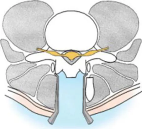
11 THE BONY LUMBAR CANAL:
The conus medullaris ends at the level of lower border of L1 vertebra. Beyond which the dural sheath contains only the cauda equine.three basic shapes in the lumbar spinal canal round,trefoil and oval(fig-2). The shape of bony lumbar canal varies from L1 to L5. At the level of L1 it is almost round. At L5 level it is trifoliate. The well developed lateral recesses is due to this transformation at L4 & L5 vertebrae. Any pathology in the Lateral recess can be maximally seen in these two vertebrae. The normal sagittal diameter of this canal varies from 15 to 25 mm. A canal of 20 mm is capacious and canal diameter between 12 and 15mm are suggestive of small canal and below 12 mm the canal is narrow causing spinal canal stenosis. Acquired stenosis is more at the level of L4 -L5 and L3 –L4 .
As the nerve root leaves the dural sac, it passes through the lateral recess or nerve root canal. Each nerve root is intimately related to the medial and the inferior aspect of the corresponding pedicle.
12 THE NERVE CANAL:
The spinal nerve root leaves the dural sac through the lateral part of spinal canal by an oblique passage called nerve canal. The nerve canal ends where the nerve root emerges from the intervertebral foramen.
Fig-2: Lumbar canal
A – Bony lumbar canal
B – Nerve canal
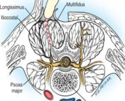
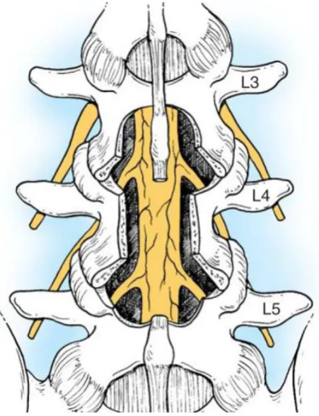
13 PARASPINAL MUSCLES:
On either side of the spinous process is the convex column of muscle known as paraspinal muscles, which are collectively called as erector spinae or sacrospinalis(fig-3). It consists of three muscles namely multifidus, longissimus and iliocostalis muscles. The main function of sacrospinalis muscle is to maintain the spine erect . Subperiosteal resection of the paraspinal muscles should be carried out in order to maintain the blood supply and its musculature to be handled to the minimum.
[image:21.595.93.296.534.697.2]Lazzennec had done extensive MRI studies on cross section of spinal musculature after surgery and has demonstrated weakening in the muscles following fibrosis due to operative procedures(32).
Fig -3: Paraspinal muscles
14
LIGAMENTS:
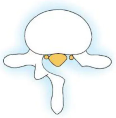
A)LIGAMENTUM FLAVUNM
These are strong, yellow, elastic ligaments which unites the adjacent lamina(fig-4). They are short and limited on either side by articular process. Along with the lamina it forms the smooth posterior surface. With aging these ligaments losses its elastic nature and the collagen hypertrophies which buckle and encroach the thecal sac and cause spinal canal stenosis
Fig-4: Ligamentum flavum
B) INTERSPINOUS LIGAMENTS
15
C) ANTERIOR LONGITUDINAL LIGAMENT
It stretches from atlas to sacrum. It firmly attaches to the vertebral bodies and intervertebral disc.the lateral margin fades into the periosteum.
D)POSTERIOR LONGITUDINAL LIGAMENT
It lies within the vertebral canal. It attaches to the posterior margin of intervertebral disc and adjacent margins of vertebra. The ligaments are narrowed to allow basivertebral veins (Fig-5).
16
The pathology of Lumbar canal stenosis can be caused by dynamic as well as a structural component. Degeneration of the intervertebral disc occurs with narrowing of disc space and subsequent ligamentous redundancy which compromises spinal canal. Instability may occur. This relative hypermobility leads to the formation of facet overgrowth and ligamentous hypertrophy. The ligamentum flavum may be markedly thickened into the lateral recess where it attaches to facet capsule causing nerve root compression.
Spinal canal stenosis in this region usually caused by protruded disc, bulging annulus, osteophyte, thickened or buckled ligamentum flavaum.resulting in narrowing of both the central and lateral canals. This can occur alone or in combination to create the symptom complex characteristic of spinal stenosis.
17
from internal disruption and disc resorption. Later osteophytes form at the back of the vertebral bodies. Disc may also losses its height as a result of infection, excision or herniation.
The combination of changes in the inter vertebral disc and posterior joints results in subluxation of the facetal joints , as a result the superior articular process moves upward and forward to encroach on the nerve canal. The nerve canal also narrowed by bulging annulus, osteophytes at the back of the vertebra, and subluxated superior articular process. Central spinal stenosis results from enlargement of inferior articular process.
Junghan‟s in 1932 introduced concept of motion segment.
THE MOTION SEGMENT
Intervertebral disc Intervertebral foramen Superior and inferior facets Interlaminar space
Ligamentum flavum
Inter and supraspinous ligaments
18
Motion segment should be preserved to a maximum extent in any operative procedure.
The changes in the disc space are not the only factors that would change the function of the motion segment. The effect of each component in the motion segment can be influenced by the other. If the supraspinous or interspinous ligament is removed, then there is an increased range of motion in flexion and to a lesser extent in extension of the lumbar spine. This would produce alteration of the loading point.
19 CLASSIFICATION:
Based on etiology and anatomic location.
Van Akkerveeken has classified canal stenosis(12).
Primary stenosis:
It is rare, it is approximately 9%, due to congenital malformations or developemental defects( achondroplasia).
Secondarystenosis:
Mainly due to acquired conditions.
1.Degenerative
Central canal
Lateral recess, foramen
20 2.Iatrogenic
Postlaminectomy Postfusion
Spondylolytic Posttraumatic
3.MISCELLANEOUS
Paget‟s disease Fluorosis
Diffuse idiopathic skeletal hyperostosis(DISH ) Hyperostotic lumbar spinal stenosis( Forestier disease) Pseudogout
21
DEGENERATIVE SPINAL STENOSIS:
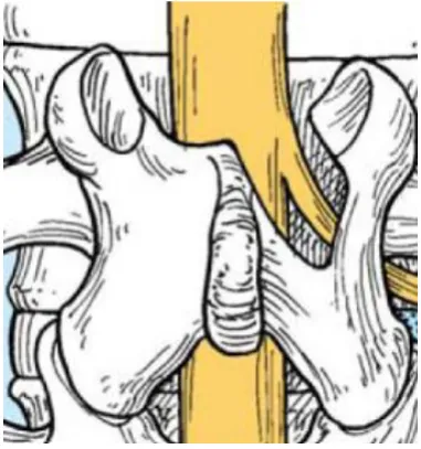
The most common type of type of spinal canal stenosis is degenerative arthritis of spine. The disc degenerates and losses its elasticity and height, the annulus bulges into the canal. Similarly the vertebral body and facet approach each other with the formation of osteophytes at the margin. The nerve root emerges through the intervertebral foramen got caught between the facet and pedicle(Fig-6). The degenerative process mostly localised to the facet joints and ligamentum flavum.
22
CONGENITAL OR DEVELOPMENTAL STENOSIS:
There is uniform narrowing of the canal, usually it is central canal stenosis. Idiopathic congenital narrowing will have decreased AP diameter due to short pedicles. In achondroplasia, the neural canal got compressed due to diminish in the interpedicular distance.
IATROGENIC STENOSIS:
In iatrogenic stenosis the mechanism is unclear the probable reasons are,
- Incomplete treatment of stenosis - Hypertrophy of posterior bone graft
23 ANATOMIC CLASSIFICATION:
Central spinal stenosis:
It denotes the involvement of area between the facet joints, which is occupied by the dura & its contents.
Causes:
1. Protrusion of disc 2. Bulging annulus 3. Osteophytes
4. Buckled or thickened ligamentum flavum.
Most common symptom: neurogenic claudication
Lateral canal stenosis:
24
Most common symptom by root compression is the radiculopathy. Fig-7:Zones of nerve canal
Lee‟s entrance zone ( lateral recess or lateral canal stenosis)
Lee‟s mid zone (foraminal stenosis)
Lee‟s exit zone
Borders:
Medial-central canal. Lateral-pedicle. Dorsal -superior facet. Ventral - disc &
posterior ligamentous complex .
Borders:
Medial-lateral recess. Lateral-lateral border of pedicle.
Ventral- Posterior vertebral body & disc. Dorsal –pars
interarticularis.
25 Causes:
Facet arthritis, vertebral body spur,protruded disc,etc.,
Causes:
pars fracture with proliferative fibro cartilage, lateral disc herniation.
Cause:
[image:33.595.66.530.73.343.2]“far lateral” disc, spondylolisthesis,facet arthritis.
Table-1: Zones of nerve canal
CLINICAL PRESENTATION OF LUMBAR CANAL STENOSIS:
1. Common in older individuals.
2. Back pain predominant in 95% of the cases.
3. Leg pain approximately in 90% of the cases which maybe unilateral or bilateral.
4. Neurogenic claudication defined as poorly localized pain, numbness and cramping pain in one or both lower extremities of a neurologic origin, which is more on walking and relieved by sitting, frequently accompanies Lumbar canal stenosis.
26
6. In rare instances, patients may present as acute cauda equine syndrome with the involvement of bowel and bladder disturbance.
7. In patients with central canal stenosis, symptoms usually are bilateral which involve the buttocks and posterior thighs in a nondermatomal fashion.
8. Lateral canal stenosis, symptoms usually are dermatomal due to the compression of specific nerves.
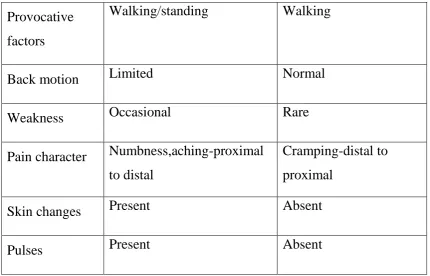
9. Patients with lateral canal stenosis may have more pain during night and at rest, but more walking tolerance than patients with central stenosis. 10.Neurogenic claudication should be distinguished from vascular
claudication,which has a different etiology and clinical features and the differences are given below
Evaluation Neurogenic claudication Vascular claudication
Back pain Common Occasional Walking
distance
Variable Fixed
Walking uphill Painless Painful
Bicycle test Negative Positive(painful) Palliative
factors
27
Provocative factors
Walking/standing Walking
Back motion Limited Normal Weakness Occasional Rare Pain character Numbness,aching-proximal
to distal
Cramping-distal to proximal
[image:35.595.101.532.70.346.2]Skin changes Present Absent Pulses Present Absent
Table-2: Difference between Neurogenic claudication and vascular claudication 11) The differences between the spinal canal stenosis and lumbar disc herniation are given below
Lumbar canal stenosis Lumbar disc herniation
Age >50 <50
Onset Insidious Acute
Pain Referred/diffuse Radicular/dermatomal Provocative factors Standing/walking Sitting
28
Neurological findings Rare Present
Table-3: Differences between the spinal canal stenosis and lumbar disc herniation
CLINICAL EXAMINATION:
1. Straight-leg raise test is positive in approximately 50% of patients.
2. Symptoms are aggravated by extension and weight bearing, relieved by flexion and non weight bearing postures.
3. Associated sensory and motor deficit may be present.
In most of the cases, patients seeks medical attention when the walking distance progressively diminished due to Neurogenic claudication.
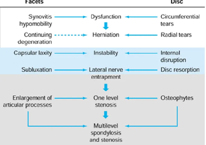
PATHOPHYSIOLOGY OF SPINAL STENOSIS:
Stage of transient dysfunction:
29 Stage of instability:
As the disease progresses, there is abnormal movement at the facet joints due to laxity in the ligaments. There is also abnormal motion at the level of intervertebral disc. Following subluxation the superior facet moves upwards and forwards and causes stenosis. Complaints became more severe and more frequent. X ray shows abmormality. Conservative treatment usually helps but symptoms persist. Surgical decompression and fusion is indicated.
Stage of fixed deformity:
30
Table-4: Pathophysiology Of Spinal Stenosis
RADIOLOGICAL EVALUATION:
The imaging modalities available for diagnosing Lumbar Canal Stenosis are
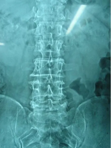
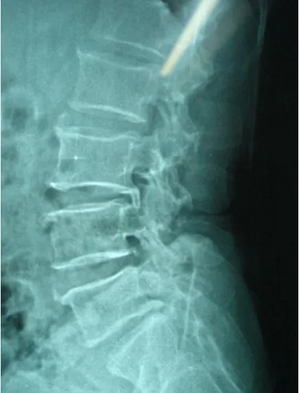
a)Plain X-ray lumbo-sacral spine shows, 1. Fecetal joint hypertrophy.
31
3. Reduced distance between the posterior border of the vertebral body and anterior border of the superior facet.
4. Short, stout spinous process and laminae with reduced distance between the pedicles of adjoining vertebrae.
5. Associated features narrowing of intervertebral disc space, posterior osteophytes.
6. Decrease in inter laminar space.
7. Irregular and laterally arranged facets.
[image:39.595.189.409.374.668.2]
32
Fig.9: lateral view
MYELOGRAPHY:
Myelography was the gold standard one for diagnosis of Lumbar canal stenosis in earlier days. The anterior-posterior (AP) diameter of spinal canal on myelography was used as a reference(13).
AP diameter Normal = 15 mm Relative stenosis = 10 to 12 mm Spinal stenosis = < 10 mm
Advantages :
33
2. To know the narrowing during movement of spine.
Disadvantages:
1. Adverse effects to contrast agents.
2. Difficulty in identifying lateral stenosis pathology.
34
CT CHANGES IN SPINAL STENOSIS Herniated disc
Disc protrusion Vacuum disc sign
Hypertrophy of posterior articular processes Osteoarthritis of apophyseal joints
Osseous proliferations of nonarticular aspects of superior apophyseal joint
Osseous proliferations of nonarticular aspects of inferior apophyseal joint
C/O of posterior longitudinal ligament C/O of yellow ligament
C/O of supraspinal ligament
Anterior C/O of posterior articular capsule Posterior C/O of posterior articular capsule Anteroposterior diameter of spinal canal Transverse diameters of spinal canal
35 Advantage:
1. Central and lateral canal can be directly visualised and measured. 2. Soft tissue pathology can be identified.
MRI FEATURES:
1. Waist-like narrowing of dural tube at the level of facet joint . 2. Indentation of the dural tube by prolapsed disc.
3. Axial CT OR MRI cuts demonstrates hypertrophy. 4. Lateral recess narrowing in lateral cuts of MRI . 5. Reduced mid sagittal distance in saggittal cuts
MRI :
36
37
MEASUREMENT OF LATERAL RECESS (CIRIC INDEX)
Normal - 5mm Suggestive - 3mm
Diagnostic - 2mm
38 OTHER DIAGNOSTIC STUDIES:
Electrodiagnostic studies should be used if the diagnosis of neuropathy is uncertain, especially in patient with diabetes mellitus.
Electromyography (EMG) and nerve conduction velocity (NCV) have been recommended as useful adjuncts to diagnosis in patients with peripheral neuropathy from Lumbar canal stenosis.
39 NONOPERATIVE TREATMENT:
Symptoms of spinal canal stenosis usually respond well to conservative management.
Conservative measures should include 1. Bed rest not exceeding for 2 days.
2. Pain management with NSAIDS and acetaminophen.
3. Trunk stabilization with braces and exercise program along with good aerobic fitness and abdominal muscle strengthening exercise
4. Traction has no proven benefit in adult lumbar spinal stenosis. 5. Stationary cycling
Onel and colleagues proposed a program of flexion exercises and infrared heating modalities to reduce pain and spasm and improve flexibility, because of the structural narrowing of the spinal canals produced by extension.
40
1) Spinal canal stenosis cause mechanical compression of neural elements, which leads to structural and chemical injury to the nerve roots.
2) Edema and venous congestion of nerve roots can lead to further compression and ischemic neuritis.
3) This result in the leakage of neurotoxins, such as phospholipase and leukotriene B, which lead to increased inflammation and edema. 4) Corticosteroids are potent antiinflammatory agents, which decreases
the leukocyte migration, the inhibition of cytokines and decrease edema.
5) These actions provide the rationale for the use of epidural steroid injections in spinal canal stenosis.
6) Although epidural steroid injections have been used for many years, no scientifically validated long-term outcomes have been reported to substantiate their use..
The technique of placement- Caudal, translaminar and transforaminal with fluoroscopy.
41
Epidural hematoma, temporary paralysis, retinal hemorrhage, epidural abscess, chemical meningitis, dural puncture and headache.
Suitable candidates:
Patients with acute radicular symptoms or neurogenic claudication unresponsive to traditional analgesics and rest.
SURGICAL TREATMENT:
1. The primary indication for surgery in patients with spinal canal stenosis is increasing pain that is resistant to conservative methods.
2. Patients with severe back and leg pain with significant limitation in walking tolerance
3. Acute cauda equina syndrome
4. Rapidly deteriorating neurological deficits.
In general, surgeries give good relief of claudicatory leg pain with variable response to back pain.
42
1. Disc herniation
2. Stenosis at single level
3. Weakness < 6 weeks duration 4. Monoradiculopathy
5. Age < 65 yr
PRINCIPLES OF SPINAL STENOSIS SURGERY:
Decompression is the treatment of choice for lumbar canal stenosis. Fusion is required if excessive bony resection compromises spine stability or if isthmic or degenerative spondylosis , scoliosis or kyphosis is present.
Laminectomy is preferred in older individuals with multiple level stenosis and fenestration procedures in younger patients with intact disc especially done through a minimally invasive approach.
43
The lateral recess and foramen dissection may require a small, sharp osteotome, which allows the surgeon to thin the bone sufficiently to allow removal with angled curets. In contrast to disc surgery, decompression the lateral recess is best seen from the opposite side of the table. The operating surgeon may switch sides during the operation to view the nerve roots better. Blunt probes with increasing diameters are useful for determining adequate foraminal enlargement.
A good approach is to start with decompression at the point of lesser stenosis and work towards the area of more severe stenosis. This frees the neural structure enough to make the final decompression easier and decrease the risk of damage to duramater and the nerve root.
44
LAMINECTOMY
The gold standard surgical procedure for lumbar canal stenosis is conventional midline laminectomy. This procedure involve the removal of lamina and ligamentum flavum on both sides of the stenotic level and the lateral recess. Decompression starts from distal extent of neural compression and proceeds in a proximal direction. Perform decompression sequentially, from medial to lateral.
[image:52.595.203.429.346.640.2]
45
HEMILAMINECTOMY
[image:53.595.111.308.472.673.2]Hemilaminectomy involves unilateral removal of lamina and ligamentum flavum. The spinous processes, interspinous ligaments, and supraspinous ligaments are preserved. So, less risk of development of postoperative instability. Preserve the pars interarticularis laterally in order to minimize risk of postoperative instability . Hemilaminectomy is appropriate for patients with unilateral symptoms from stenosis. A disadvantage of this procedure is the difficulty of performing contralateral decompression
46
HEMILAMINOTOMY:
Hemilaminotomy involves removal of only the ligamentum flavum and adjacent portions of two hemilaminae responsible for neural compression. This procedure is more commonly performed in younger patients. Extensive laminectomy carries the risk of instability.
Fig-14: Hemilaminotomy WIDE FENESTRATION:
[image:54.595.180.371.542.746.2]Wide fenestration is done for central stenosis in which only the medial portion of the inferior facets and adjacent ligamentum flavum is removed . Preserve the interspinous or supraspinous ligament complex and spinous processes, which form the midline stabilizing structures.
47
SPINOUS PROCESS OSTEOTONY DECOMPRESSION
[image:55.595.181.408.443.649.2]The spinous process is ostetomised at its base and retracted to opposite side. Removal of lamina and ligamentum flavum are done. Complete laminectomy is recommended for severe stenosis or congenital stenosis involving the anatomical zones (central, lateral recess, and foraminal zones). A minimally invasive technique allows decompression of compressing anatomy, while preserving paraspinal muscles, the spinous processes, and intervening supraspinous and interspinous ligaments.
48
LUMBAR SPINOUS PROCESS SPLITTING DECOMPRESSION:
The spinous process was identified and burred down until its base. The interspinous ligaments and supraspinous ligaments were cut longitudinally in line with the spinous processes. Using osteotome the spinous process split into two halves, the split halves of the spinous processes were osteotomized at the base and separated it from the lamina. The split halves of the spinous process along with the paraspinal muscles were then retracted on either side to expose the laminae. Decompression then done according to the conventional laminectomy method. The spinous process and paraspinal muscles are reapproximated with each other.
49 COMPLICATIONS:
1) Intra operative and
2) Post operative complications
These complications include inadequate neural decompression, recurrent stenosis, incidental dural tear, neural injury, epidural hematoma, neural compression from either fat grafts or other barriers to scar formation, vascular injury, and late instability.
INADEQUATE NEURAL DECOMPRESSION
50
the neural segment located between the two sites of compression resulting in a compartment syndrome like condition of the intervening segment,which may leads to inadequate neural decompression.
RECURRENT STENOSIS
Distinguishing between neural compression and scar formation in recurrent symptoms is difficult. It requires a precise history and high-quality radiographic imaging. Failure to obtain even temporary pain relief following decompression suggests either inadequate neural decompression, irreversible neural damage at the time of surgery, or a nonspinal cause for the pain. A pain-free interval of less than 6 months suggests development of scar formation as the cause of recurrent pain. Recurrence of pain following a free interval of more than 6 to 12 months suggests a new process such as a recurrent disc herniation or recurrent stenosis.
DUROTOMY
51
Repair of Durotomy
The patient is placed in a slightly head-down (Trendelenberg) position to minimize the amount of CSF in the field. This provides a drier operative field and minimizes the tendency for the individual roots of the cauda equina to float on the surface, which can result in injury during dural repair.
For large tears, place a small cottonoid patty over the exposed nerve roots for the initial portion of the repair and then remove it just before dural closure
For tears associated with loss of tissue, or tears in difficult locations, an autologous fat graft, a piece of autograft fascia (thoracolumbar fascia or fascia lata) may be required to close the defect
Perform a watertight closure.
Drain is often not employed in order to minimize risk of development of a CSF fistula.
Keep the patient on bed rest for 3 to 5 days.
52
Fig-18: Dural repair
NERVE ROOT INJURY
53
a pre-existing laminectomy defect on the preoperative radiograph in order to minimize risk of damage to underlying dura and nerve roots. It is important to visualize the lateral edge of the nerve root during surgical decompression to aviod inadvertent exposure in the axilla of a nerve root where accidental dural laceration and neural injury could occur . In revision lumbar surgery dissection performed usually lateral to the root along the lateral edge of the bony canal to avoid a potentially dangerous midline scar. While performing lateral nerve root decompressions,work parallel, rather than perpendicular, to the long axis of the nerve root in order to minimize risk of cutting across a root.
SCAR TISSUE
It has been implicated as a potential cause for continued pain following spinal surgery. Postoperative scar tissue may be located either intradurally (arachnoiditis) or extradurally (epidural fibrosis). Its etiology is often unclear, but it has been associated with oil-based myelographic contrast agents, prior surgery or dural laceration in which blood gains entry into the dural sac and mixes with neural elements.. When postoperative pain exists, the primary differential diagnosis is between scar and recurrent disc herniation. Radiographic distinction is best made with gadolinium-enhanced MRI or post-contrast CT .
54
membrane. These barriers include a thin layer of fat or synthetic agents such as an absorbable gelatin sponge. The use of a free fat graft has been considered the gold standard interposition membrane.
EPIDURAL ABSCESS
Epidural abscess, is one of the most feared complications of spinal surgery because of its risk of paresis or frank paralysis. It is a rare occurrence. Patients have significant fever, back pain, and often present with neurologic findings such as nuchal rigidity and weakness or paralysis of the lower
extremities. Both the WBC and acute phase reactants are elevated. MRI is the diagnostic imaging modality of choice and clearly visualizes the abscess . Treatment of an epidural abscess: surgical evacuation of the abscess and any adjacent necrotic tissue, followed by parenteral antibiotics.
EPIDURAL HEMATOMA
55
Once the diagnosis is confirmed immediately return the patient to the operating room for decompression and drainage of the hematoma.
The risk of this complication can be minimized by meticulous attention to preoperative, intraoperative, and postoperative detail. check the prothrombin time (PT), partial thromboplastin time (PTT), bleeding time, platelet count, and platelet function. Intraoperatively, position the patient with the abdomen hanging freely in order to minimize epidural venous congestion. Keep the blood pressure below 100 mm Hg systolic, in order to minimize bleeding. Control epidural bleeding with bipolar electrocoagulation. At the end of the surgery, after removing the deep paraspinal muscle retractors check the muscle walls for persistent bleeding, because prolonged retraction may temporarily occlude but significant muscle bleeders that could begin bleeding after muscle layer closure.
SUPERFICIAL WOUND INFECTION
56
milder cases consisting only of cellulitis these may be absent. Patients may be febrile. Laboratory data show elevation of the erythrocyte sedimentation rate (ESR) and C-reactive protein (CRP).
Treatment of superficial wound infections consists of surgical debridement of necrotic tissue , local wound care with short-term parenteral antibiotics.
DEEP WOUND INFECTIONS
Symptoms include disproportionate back pain or leg pain. The patient look ill and may exhibit generalized malaise. Fever is often present but may be deceptively low grade. The ESR and CRP are usually elevated. MRI provides the best and most useful information by revealing both the presence and extent of a deep abscess. If MRI is not available, diagnosis may be confirmed radiographically by the presence of a circumscribed area of fluid density visualized by CT. If a deep abscess is suspected, diagnosis may be confirmed by aspiration, with culture and sensitivity of any fluid obtained.
57
POSTOPERATIVE INSTABILITY
58 FUNCTIONAL EVALUATION:
Various scoring systems are available to assess low back function. Japanese Orthopaedic Association Score (JOA Score) has two components subjective symptoms assessed in first section (maximum 9 points) and the clinical signs in second section (maximum 6points). The Scores from the two sections are added to form a JOA Score. The Score of -6 represents poorest function, and negative points being incorporated for bladder symptoms. A score of 15 represents an asymptomatic and fully functional subject.
59
60
Neurogenic claudication Outcome score(NCOS):
[image:68.595.80.517.349.599.2]Neurogenic claudication Outcome score(NCOS) is a questionnaire designed mainly for assessing the severity of neurogenic claudication symptoms. The questionnaire consists seven sections of questions pertaining to activities of daily living and a section of visual analog scale for pain. These scores are added to form the NCOS score. A score of 0 represents poorest function while a score of 100 represents full function.
61 Visual Analog Scale(VAS):
The typical visual analog scale is a rectangular strip with pictorial representations of human faces with varying degrees of pain which are numbered from zero (smiling face, no pain) to 10(worst imaginable pain). Patient is asked to choose the face that most closely represents his/her pain. This can be used to assess the back pain as well as the claudication pain.
Paraspinal muscle injury
62
damage to the paraspinal muscles are more likely to have a good functional outcome.
1) Creatine Phosphokinase
Creatine Phosphokinase muscular isoenzyme(CPK-MM) originates from skeletal muscle and which is a reliable indicator of skeletal muscle damage during surgery. CPK-MM normal value ranges from 45- 230 U/L. CPK-MM levels in blood rises immediately after surgery and plateaus off after first postoperative day. The enzyme level starts to decrease one to two weeks after surgery. Measurement of CPK-MM levels in the postoperative period can be used to determine the amount of paraspinal muscle damage.
2) C- Reactive Protein
C- Reactive Protein also related to skeletal muscle damage and inflammation in many studies.
Comparison between the preoperative and postoperative levels of these protein is an indicator of the invasiveness of the procedure and the extent of paraspinal muscle injury .
63
In this era of modernisation and sophisticated investigation technologies, degenerative lumbar canal stenosis has come to the fore as a cause of low back pain in the elderly with more cases diagnosed and more surgeries done, it is imperative to find a comparison between surgical modalities. In this study, we have made an attempt to identify the best surgical modality for lumbar canal stenosis.
64
This prospective Randomised Control Study compares the the functional outcome and extent of paraspinal muscle damage between Lumbar spinous
process splitting decompression (LSPSD) and Conventional Midline
Decompression(CMD) by laminectomy surgical approaches in degenerative
lumbar canal stenosis and their aim was whether
1) Lumbar spinous process splitting decompression (LSPSD ) approach provide sufficient decompression.
2) Preserve posterior musculoligamentous complex and reduces associated morbidity.
STUDY DESIGN:
Prospective randomised Control study
MATERIALS AND METHODS
This randomized prospective control study was approved by the medical ethics committee of the Institutional Review Board in our hospital. Patients meeting the following inclusion criteria were enrolled for the study after
65 INCLUSUION CRITERIA:
- Degenerative LCS affecting 3 or less levels, -Typical neurogenic claudication symptoms,
- Magnetic resonance image demonstrating good clinical correlation
- Failure of conservative methods of treatment for a minimum period of 6 months.
EXCLUSION CRITERIA:
-Spondylolisthesis with slip grade 2 or greater (Meyerding grade).
- Instability at the level of stenosis (as defined by >3-mm translation or >10° angular change on flexion extension lateral radiographs) - Associated symptomatic cervical or thoracic stenosis.
- Multiple level canal stenosis.
-Spinal canal stenosis due to congenital, traumatic , iatrogenic causes. - Presence of spinal disorders( ankylosing spondylitis, neoplasm ) - Comorbidities ( such as cardiopulmonary insufficiency, peripheral neuropathy, peripheral vascular disease, prior lumbar spine surgery, and severe hip or knee disease).
66
-Patient history and neurological examination
- Preoperative clinical evaluation of the patients was made by 1) Japanese Orthopaedic Association (JOA) score 2) Neurogenic claudication outcome score (NCOS).
3) Visual analogue score for back pain and neurogenic claudication (VAS)
- Baseline C -reactive protein (CRP) and CPK-MM levels.
Radiography of lumbosacral spine
a) Antero –posterior view b) Lateral view
c) Dynamic flexion-extension lateral view d) MRI of the lumbo sacral spine.
Surgical Technique
67
Fig-19: Level identification with C-arm
68
Fig-21: Skin incision
CONVENTIONAL MIDLINE DECOMPRESSION
- About 5to8 cm skin incision was made in the midline centered over the level of stenosis - The deep fascia incised in the midline and the either side paraspinal muscles were elevated subperiosteally from the spinous process and lamina.
- Identify and remove the spinous process, the interspinous and the supraspinous ligaments of the level to be decompressed.
-Remove the lamina upto the insertion of ligamentum flavum.
- Once the of ligamentum flavum has been identified it can be removed from the lamina.
- Remove the lamina upto medial border of the pedicle, it can be helpful in decompressing thelateral canal.
- Adequate decompression of the dura and nerve root by probing the foramen .
69
-The paraspinal muscles were approximated in the midline using absorbable sutures and the subcutaneous tissue and skin were closed.
Fig-22: Removal of spinous process & interspinous ligaments
70
Fig-24: Root decompression
LUMBAR SPINOUS PROCESS SPLITTING DECOMPRESSION(LSPSD)
- The positioning of the patient and anaesthesia techniques are similar to the standard midline decompression technique.
- A 3 to 5 cm long incision was made over the proximal spinous process (e.g., L3 spinous process in case of L3–L4 decompression
- The posterior surface of the L3 spinous process was identified and using a high-speed 2-mm burr, the spinous process was burred down until its base. -The proximal and distal interspinous ligaments and supraspinous ligaments were cut longitudinally in line with the spinous processes.
71
-The split halves of the spinous process along with the paraspinal muscles were then retracted on either side to expose the laminae. Decompression then done according to the conventional laminectomy method.
-Holes were made in the center of the split spinous processes to facilitate easy closure
[image:79.595.86.510.371.685.2]- After adequate decompression of the dural sac and the roots, the split halves of the spinous process were sutured with no.1 Vicryl .
72
73
74
Fig-28: Decompression of spinal canal
Fig-29: Suturing of spinous process
Intra operative assessment:
- Operative time
- Blood loss and transfusion
75
POSTOPERATIVE PROTOCOL
-Patients allowed to turn to sides in the bed on the day of surgery and were encouraged to sit as early as possible once pain subsides.
-Patients were encouraged to walk as soon as is comfortable.
-Postoperative day on which patient is able to ambulate was noted, the day of the surgery being counted as day 0. Patients were discharged on the tenth day or when comfortable.
- Serum CRP and CPK-MM values were assessed on postoperative day 1 and day 3
- Patients were put on a program of spinal flexion exercises 3 weeks after surgery.
76
COMPLICATIONS
77
FOLLOWUP AND RESULTS
78 PRE OPERATIVE PARAMETERS:
S.NO CONTENTS
CONVENTIONAL MIDLINE DECOMPRESSION (LAMINECTOMY) LUMBAR SPINOUS PROCESS SPLITTING DECOMPRESSION
1. No. of patients 10 10
2. Average age 60.4 58.9
3. Male: Female 4:6 5:5
4.
Mean No. of decompressed
levels
1.30 1.20
5.
Associated protruded disc
removal
6 7
6.
Average duration of follow up
[image:86.595.60.537.131.655.2]11.6 months 11.2 months
79 INTRA OPERATIVE PARAMETERS:
S.no Contents
Conventional Midline Decompression (Laminectomy)
Lumbar spinous process splitting decompression
1. Average duration of
procedure 71.23 min 80.25 min
2. Average blood loss 126 ml 130 ml
3. No.of blood
transfusion 2 1
4. Dural Tear 1 1
5. Iatrogenic Neurologic
[image:87.595.63.536.153.596.2]Deficit _ _
80
POST OPERATIVE PARAMETERS:
S.no Contents
Conventional Midline Decompression (Laminectomy)
Lumbar spinous process splitting decompression
1.
Average ambulation Time
81 Table - 9
2. Wound
complications 1 1
3. Urinary Tract
Infections 2 -
4.
Lower Respiratory Tract Infection
2 _
82 FUNCTIONAL OUTCOME SCORES:
Japanese Orthopaedic Association Score (JOA Score):
In the Lumbar spinous process splitting decompression group JOA score improved from preoperative mean 5.4 to 12.50 at the last follow up. In the Conventional Midline Decompression the score improved from preop mean 5.3 to 11.3 at the last follow up. The mean JOA recovery rate was 73.96% for the Lumbar spinous process Decompression group and 61.86% for the Conventional Midline Decompression group. There was no statistically significant difference between the two groups.
Parameter Lumbar spinous process splitting decompression (LSPSD) Conventional Midline Decompression (Laminectomy) Significance (P<0.05) Preop JOA
score 5.4 5.3 P>0.05
JOA score at
last follow up 12.5 11.9 P>0.05
Change in
JOA Score 7.1 6.6 P>0.05
JOA Recovery
rate (%) 73.96 68.05 P>0.05
83 Table - 10
Notably 70% of Conventional Midline Decompression group had good or excellent outcome while 100% of Unilateral Decompression group had good or excellent outcome.
[image:91.595.118.480.367.633.2]
Table - 11
Outcome(JOA score Recovery rate) at final
follow up Lumbar spinous process splitting decompression (LSPSD) Conventional Midline Decompression (Laminectomy))
Excellent(≥ 75%) 4 3
Good(50-74%) 5 5
Fair (25-49%) 1 2
Poor(≤24%) 0 0
84
Fig-30: JOA RECOVERY RATE Score
0 1 2 3 4 5 6 7 8 9 10
LSPSD
85
Neurogenic Claudication Outcome Score(NCOS)
NCOS score improved from a mean preoperative score of 28.30 to 66.10 at last follow up in the Lumbar spinous process Decompression group, and from 27.60 to 65.10 in the Conventional Midline Decompression group . Statistical analysis did not reveal any significant difference between groups.
Lumbar spinous process splitting decompression
(LSPSD)
Conventional Midline Decompression (Laminectomy)
Significance (P<0.05)
Preop NCOS
score 28.30 27.60 (P>0.05)
NCOS score at
last follow up 66.10 65.10 (P>0.05)
Change in
NCOS Score 37.80 37.50
86 Table - 12
Fig-31:Neurogenic Claudication Outcome Score(NCOS)
0 10 20 30 40 50 60 70
PREOP NCOS NCOS SCORE AT LAST
FOLLOW UP
LSPSD
CMD(CONVENTIONAL)
[image:94.595.71.507.365.622.2]87
Visual Analog Scale for Back Pain (BPVAS):
At the last follow up the mean BPVAS score for the Lumbar spinous process Decompression group was 2.7 and for Conventional Midline Decompression group it was 3.70. Statistical analysis revealed significant difference between two groups.
Parameter
Lumbar spinous process splitting decompression
(LSPSD)
Conventional Midline Decompression (Laminectomy)
Significance (P<0.05)
Preop BPVAS 7.8 7.7 (P<0.05)
88 last follow up
Change in
BPVAS 5.1 4.0 (P<0.05)
[image:96.595.67.526.72.217.2]N = 10 10 (P<0.05)
Table - 13
Fig-32:Visual Analog Scale for Back Pain (BPVAS):
89 Neurogenic Claudication(NCVAS):
Mean NCVAS score at last follow up was 2.10 for Lumbar spinous process Decompression group and 2.0 for Conventional Midline Decompression group. There was no significant difference between the two groups
0 1 2 3 4 5 6 7 8
LSPSD
CONVENTIONAL(CMD)
PREOP BPVAS
90 Parameter
Lumbar spinous process splitting decompression
(LSPSD)
Conventional Midline Decompression (Laminectomy)
Significance (P<0.05)
Preop NCVAS 7.70 7.70 (P>0.05)
NCVAS S score
at last follow up 2.10 2.0 (P>0.05)
Change in
NCVAS 5.60 6.70 (P>0.05)
[image:98.595.67.531.109.360.2]N = 10 10
Table - 14
91
Biochemical markers for paraspinal muscle damage
1) Creatine Phosphokinase (CPK-MM)
0 1 2 3 4 5 6 7 8
LSPSD CONVENTIONAL(CMD)
PREOP NCVAS
92
Rise of CPK-MM on postoperative day 1 and day 3 were estimated. CPK MM-1 (POD1-Preop)mean(U/L) for LSPSD was 225U/Land for CMD was 253U/L.CPK-MM-3 (POD3-Preop)mean (U/L) for LSPSD was 212 and for CMD was 240.Although the mean values on POD1 and POD3 for the CMD group were higher than the LSPSD group signifying more muscle damage, the difference was not statistically significant(p>0.05)
Parameter LSPSD Conventional
(CMD)
Significance (P<0.05)
CPK MM-POD1 225 253 (P>0.05)
CPK MM-POD3 212 240 (P>0.05)
[image:100.595.66.531.297.458.2]N = 10 10
93
Fig-34: Creatine Phosphokinase (CPK-MM)
190 200 210 220 230 240 250 260
CPK MM-POD1 CPK MM-PO3
LSPSD
94 2) C-Reactive Protein (CRP)
Rise of CRP(mg/dL) on postoperative day 1 and day 3 were estimated. CRP-1 (POD1-Preop)mean(mg/dl) for LSPSD was 30.9 and for CMD was 35.7. CRP-3 (POD3-Preop)mean (mg/dl) for LSPSD was 35.3 and for CMD was 43.4.There was no statistically significant difference between the groups.
Parameter LSPSD Conventional
(CMD)
Significance (P<0.05)
CRP-1(POD1-Preop) mean(mg/dl)
30.9 35.7
(P>0.05)
CRP-2 (POD3-Preop) mean(mg/dl)
35.3 43.4
(P>0.05)
[image:102.595.66.531.297.508.2]N= 10 10
95
Fig-35: C-Reactive Protein (CRP)
0 5 10 15 20 25 30 35 40 45 50
CRP-1(POD1-Preop) mean(mg/dl CRP-2 (POD3-Preop) mean(mg/dl)
LSPSD
96
ANALYSIS OF DATA
The data was analysed by an independent observer to find any statistical difference between two groups in terms of Japanese Orthopedic
Association Score and Neurogenic Claudication Outcome Scores,. Student‟s t test and chi-square tests were used.
At 1 year follow up the results were classified according to JOA score recovery rate as
Excellent - >75% or more Good - 50- 74%
97
COMPARISON OF PILOT STUDY AND MY STUDY:
S.NO CONTENTS
CMD
LSPSD
Pilot study My study Pilot study My study 1. No. of patients 16 10 18 10
2. Average age (years) 69 60.4 71 58.9
3. Male: Female 8:8 4:6 10:8 5:5
4. Mean No. of decompressed levels
1.5 1.30 1.4 1.20
5. Associated protruded disc removal
10 6 12 7
6. Blood loss (ml) 103 126 119 130
7. Time (min) 69 71.23 82 80.25
8. JOA Recovery rate 74% 63.69% 75% 69.52%
98
DISCUSSION
With 20 patients, we have presented the prospective randomized control study comparing short term functional outcome of Lumbar Spinous Process Splitting Decompression with Conventional midline decompression by laminectomy.
The two groups of our study were comparable to each other in terms of patient characteristics like age and sex. The patients in our study (mean age 59.65 years) which parallels the average life expectancy in India (65 years), degenerative canal stenosis affects more females than males.
99
.
In our study the complications were few and were comparable between groups.
Other complications like dural tear (one patient 10%) and wound dehiscence (one patient 10%) were observed equal in frequency in both the groups. Conventional Midline Decompression by laminectomy group, as also the post operative morbidity like UTI, LRI (14.3%) etc .The average ambulation time in Lumbar Spinous Process Splitting Decompression (4.45 days) is less when compared to Conventional Midline Decompression by laminectomy (6.52 days). Post operative radiological evaluation to assess the instability was not routinely performed and when the clinical symptoms and signs of back pain and claudication persist, X-rays of lateral view, flexion and extension view was taken to rule out post operative instability. One patient developed instability in the last follow up in Conventional Midline Decompression group, later posterior fusion and pedicle screw instrumentation were done. .
100
mobilisation and decreased back pain VAS due to preservation of posterior musculoligamentous complex.
Japanese Orthopaedic Association (JOA) Score and recovery rate:
The two (LSPSD and CMD) groups were comparable in terms of the preoperative JOA scores (5.4 and 5.3). The postoperative JOA scores at last follow up (12.5 and 11.5 respectively) and change in JOA score (7.1 and 6.0 respectively) did not show any statistically significant difference.
However a