Copyright © 2002, American Society for Microbiology. All Rights Reserved.
Evolution of Porcine Reproductive and Respiratory Syndrome Virus
during Sequential Passages in Pigs
C.-C. Chang,
1K.-J. Yoon,
1* J. J. Zimmerman,
1K. M. Harmon,
1P. M. Dixon,
2C. M. T. Dvorak,
3and M. P. Murtaugh
3Department of Veterinary Diagnostic and Production Animal Medicine
1and Department of Statistics,
2Iowa State University,
Ames, Iowa 50010, and Department of Veterinary PathoBiology, University of Minnesota, St. Paul, Minnesota 55108
3Received 6 November 2001/Accepted 12 February 2002
Porcine reproductive and respiratory syndrome (PRRS) viruses are recognized as possessing a high degree
of genetic and antigenic variability. Viral diversity has led to questions regarding the association of virus
mutation and persistent infection in the host and has raised concerns vis-à-vis protective immunity, the ability
of diagnostic assays to detect novel variants, and the possible emergence of virulent strains. The purpose of this
study was to describe ongoing changes in PRRS virus during replication in pigs under experimental conditions.
Animals were inoculated with a plaque-cloned virus derived from VR-2332, the North American PRRS virus
prototype. Three independent lines of in vivo replication were maintained for 367 days by pig-to-pig passage
of virus at 60-day intervals. A total of 315 plaque-cloned viruses were recovered from 21 pigs over the 367-day
observation period and compared to the original plaque-cloned virus by virus neutralization assay, monoclonal
antibody analysis, and sequencing of open reading frames (ORFs) 1b (replicase), 5 (major envelope protein),
and 7 (nucleocapsid) of the genome. Variants were detected by day 7 postinoculation, and multiple variants
were present concurrently in every pig sampled over the observation period. Sequence analysis showed ORFs
1b and 7 to be highly conserved. In contrast, sequencing of ORF 5 disclosed 48 nucleotide variants which
corresponded to 22 amino acid variants. Although no epitopic changes were detected under the conditions of
this experiment, PRRS virus was shown to evolve continuously in infected pigs, with different genes of the viral
genome undergoing various degrees of change.
Porcine reproductive and respiratory syndrome (PRRS) first
appeared as catastrophic, uncontrollable, clinical outbreaks in
swine herds in North America, Europe, and Asia in the late
1980s and early 1990s (10, 13, 26, 57). The etiology of PRRS
was established in 1991 when a previously unrecognized virus
was identified as the causal agent (57).
PRRS virus (PRRSV) is a member of the family
Arteriviri-dae
, along with equine arteritis virus, lactate
dehydrogenase-elevating virus of mice, and simian hemorrhagic fever virus (9,
48). PRRSV is enveloped and has a polyadenylated,
single-stranded, nonsegmented, positive-sense RNA genome 15 kb in
size (7, 14, 34, 35). The genome consists of eight open reading
frames (ORFs) that are expressed through the production of a
nested set of subgenomic 3
⬘
-coterminal mRNAs (14, 34, 35).
ORF 1, which comprises two-thirds of the genome, encodes
the viral RNA-dependent RNA polymerase (14, 37). ORFs 2
to 7 are postulated to encode structural proteins, but only three
proteins have been consistently identified in virions and/or
lysates of virus-infected cells. These three are the 15-kDa
nu-cleocapsid (N), 19-kDa matrix (M), and 25-kDa envelope (E)
glycoproteins that are encoded by ORFs 7, 6, and 5,
respec-tively (41, 42). Proteins encoded by ORFs 2 to 4 are designated
GP2, GP3, and GP4, where GP indicates glycoprotein and the
number designates the ORF from which it is derived (36, 56).
PRRSV infection is prevalent in swine-producing regions
throughout the world. Infection with PRRSV produces viremia
with subsequent dissemination to and viral replication in
mul-tiple organs (50, 52). The virus replicates predominantly in
cells of the monocyte/macrophage lineage (47, 50, 52, 57).
Clinically, infection may range from totally inapparent to
caus-ing severe disease. Generally, clinical signs involve
reproduc-tive disorders in pregnant animals and/or respiratory disease in
pigs of all ages (11, 20, 21, 23, 47, 51, 63). Reproductive disease
in pregnant animals is manifested as late-term abortions or
premature farrowings. Affected litters have higher proportions
of stillbirths and piglets born weak and increased preweaning
mortality (11, 23, 63).
PRRSV produces a persistent infection despite an active
immune response (1, 6, 20, 27, 60). The PRRSV carrier state
was first recognized in an experiment that documented the
transmission of virus from animals infected 99 days earlier to
commingled sentinel pigs (66). Subsequently, Wills et al. (60)
reported the isolation of virus at up to 157 days postinoculation
(p.i.). Persistence is an important epidemiological feature
be-cause it provides a ready means for PRRSV to perpetuate
itself through a cycle of transmission from carrier to
suscepti-ble animals. As a consequence, the elimination of PRRSV
from herds is difficult, and cyclic bouts of PRRSV-associated
health problems are commonplace.
The mechanism(s) by which the virus persists in the host is
not known, but PRRSV isolates are characterized by a high
degree of genetic and antigenic variability. Persistence in the
host and viral diversity could be two manifestations of the same
function. Genetic analyses have shown the existence of at least
two major virus genotypes, the European and the North
Amer-ican, with extensive genetic variability both within and between
* Corresponding author. Mailing address: Department of
nary Diagnostic and Production Animal Medicine, College of
Veteri-nary Medicine, Iowa State University, Ames, IA 50011. Phone: (515)
294-1083. Fax: (515) 294-6619. E-mail: kyoon@iastate.edu.
4750
on November 8, 2019 by guest
http://jvi.asm.org/
these genotypes. Nelsen et al. (40) found important differences
between the prototypic North American (VR-2332) and
Eu-ropean (Lelystad) viruses in the 5
⬘
leader sequence and parts
of ORF 1a. Marked differences were also found between
Eu-ropean and North American isolates in some structural genes
(24, 39). ORF 7 is highly conserved among North American
isolates, with 95 to 100% amino acid homology, but a
compar-ison of North American viruses and the Lelystad virus revealed
only 57 to 59% amino acid homology (32, 39). ORF 6 is the
most conserved gene among North American isolates, with up
to 100% amino acid identity, and is the most conserved gene
between North American and European isolates, with 70 to
81% identity (24, 33, 39). The amino acid sequence homology
for ORF 5 varies from 88 to 97% among North American
isolates and from 51 to 59% when North American viruses are
compared to the Lelystad virus (4, 24, 31, 39). A comparison of
the Lelystad virus with isolate ATCC VR-2332, the North
American prototype virus, revealed amino acid identities of 63,
58, and 68% for ORFs 2, 3, and 4, respectively (39). Similar
results were reported when the Lelystad virus was compared
with U.S. isolate VR-2385 (38).
Genetic diversity is mirrored in antigenic diversity among
PRRSV isolates. Antigenic variation was initially
demon-strated in a comparison of European and North American
isolates. Using an immunoperoxidase monolayer assay,
Wens-voort et al. (58) evaluated the reactivity of porcine polyclonal
antibodies raised against either the Lelystad virus or North
American isolate ATCC VR-2332 with PRRSV isolates from
around the world. The investigators were able to differentiate
European from North American isolates on the basis of
dif-ferences in immunoperoxidase monolayer assay antibody
ti-ters. That is, significantly higher antibody titers were obtained
in the homologous assay system. Later studies found even
greater antigenic diversity among PRRSV isolates than initially
suspected (5, 15, 17, 28, 64). Yoon et al. (64) examined 22
PRRSV isolates recovered from samples collected between
1989 and 1993 in eight different U.S. states. With a panel of
five monoclonal antibodies (MAbs) specific for the N protein,
the 22 virus isolates fell into one of three groups based on their
reactivity patterns. Yang et al. (61) expanded this study to 70
North American isolates from samples collected between 1989
and 1995 and a panel of 23 MAbs against the N protein. Their
results showed five antigenic groups, with the European
Lelys-tad virus representing an antigenic group distinct from any of
the North American groups identified. Furthermore, with
an-tibodies against discontinuous epitopes of the N and M
pro-teins and continuous epitopes of the E protein and GP3, the 65
North American isolates in the first and second antigenic
groups (I
15and II
15) were further subdivided into nine and
four antigenic subgroups, respectively (62).
That field isolates of PRRSV show a remarkable degree of
genetic and antigenic variability has become abundantly
evi-dent. Understanding the source and degree of virus diversity is
important because of its potential impact on viral virulence,
persistence in the host, protective immunity, and even on the
ability to diagnose the infection. The assumption is that these
changes occur during the course of in vivo replication in swine
and arise, in large part, because of errors that occur during
RNA replication. However, the degree and rate of mutation of
PRRSV in infected pigs over time are not known. To address
this question, we examined the genetic and antigenic changes
that occurred in a well-characterized PRRSV isolate during
the course of multiple pig passages over a period of 367 days.
MATERIALS AND METHODS
Experimental design.The objective of the study was to characterize PRRSV mutations during prolonged in vivo replication. The experiment began by inoc-ulating pigs free of PRRSV and virus-specific antibody with a highly homologous inoculum of a well-characterized PRRSV. Three independent lines (A, B, and C) of in vivo virus replication were established and monitored over seven animal passages (passage 1 [P1] to P7). Pigs in a fourth line (D) served both as mock-infected negative controls and as environmental sentinels (Fig. 1). Virus was periodically recovered from individual pigs in lines A, B, and C over a period of 367 days and compared genetically and antigenically to the original PRRSV.
In P1, three 14- to 21-day-old PRRSV-free pigs (1A, 1B, and 1C) were inoculated intranasally (1 ml per naris) and intramuscularly (8 ml per pig) with
a 3⫻-plaque-cloned PRRSV designated VR-2332-CC-01 (see below) at a titer of
10650% tissue culture infective doses (TCID
50) per ml. A fourth pig (1D) was
inoculated with cell culture medium. Animals were housed in HEPA-filtered isolation units (Barrier Systems, Inc., Tom River, N.J.) to prevent exposure to extraneous viruses. For 60 days p.i., pigs were observed daily and serum samples were collected periodically for virological and serological assays. At 60 days p.i., the animals were euthanatized and whole blood in EDTA, samples of tissues (tonsils, lungs, spleen, and tracheobronchial and medial iliac lymph nodes), and bronchoalveolar lavage fluid were collected. For each subsequent passage after P1, four PRRSV-free pigs were randomly assigned to the two groups and inoc-ulated as described above, except that tissue homogenate filtrate prepared from the postmortem samples collected from the pig in the previous passage was used in place of the cell culture-derived PRRSV inoculum. The use of tissue homog-enate made it possible to expose animals directly to pig-adapted PRRSV and avoid any selective pressures arising from isolation and replication of the virus in cell cultures.
[image:2.587.309.532.73.291.2]Viruses, cells, and media.The North American prototype PRRSV ATCC VR-2332 (American Type Culture Collection, Manassas, Va.) was used in the study. VR-2332 was selected because the complete genomic sequence has been published (GenBank accession no. PRU87392) (40) and the epitopic profile has been described (61, 62). The virus was propagated on MARC-145 cells, a clone of the African monkey kidney cell line MA-104 that is considered highly per-missive to PRRSV (25). The cells were cultured and maintained in culture
FIG. 1. Experimental design. The diagram illustrates three
inde-pendent lines (A, B, and C) and seven pig passages (P1 to P7) of
PRRSV. Pigs 1A, 1B, and 1C were inoculated with plaque-cloned
PRRSV. Pigs in P2 to P7 were inoculated with tissue filtrates from the
corresponding pig in the previous passage.
on November 8, 2019 by guest
http://jvi.asm.org/
medium composed of Dulbecco’s modified Eagle’s medium (DMEM; Sigma Chemical Co., St Louis, Mo.) supplemented with 10% fetal bovine serum
(Hy-Clone Laboratories, Inc., Logan, Utah), 50g of gentamicin (Sigma)/ml, and
0.25g of amphotericin B (Fungizone; Sigma)/ml.
To produce a highly homologous challenge virus, the stock virus was subjected to three rounds of plaquing on MARC-145 cells. Briefly, the stock virus was
10-fold serially diluted (i.e., 10⫺1to 10⫺6) with culture medium. Two milliliters
of each dilution was inoculated into each well of a six-well plate (Corning Inc., Corning, N.Y.) containing 24-h-old confluent MARC-145 cells. Inoculated cells
were incubated for 2 h at 37°C in a humidified 5% CO2incubator. At the end of
the incubation period, the inocula were discarded and the cells were rinsed twice with culture medium and then covered with 4 ml of overlay medium composed of 0.2% agarose (FMC Bioproducts, Rockland, Maine), 10% fetal bovine serum,
50g of gentamicin/ml, and 0.25g of amphotericin B/ml in DMEM. The cells
were incubated for 3 to 7 days at 37°C in a humidified 5% CO2atmosphere.
During the incubation period, well-demarcated plaques were selected, sus-pended in 2 ml of culture medium, and then propagated once on MARC-145 cells. This process was repeated three times. Following the third round of pla-quing, one virus clone, designated VR-2332-CC-01 (hereafter referred to as CC-01), was selected and propagated once on MARC-145 cells to obtain an amount sufficient for inoculating the pigs in P1. Surplus CC-01 was divided into
aliquots and stored at⫺80°C for future use.
The same procedure was used to recover plaque-cloned virus isolates from pigs. That is, 15 plaque-cloned virus isolates were recovered from each of the three pigs in each of the seven passages by directly diluting the serum samples collected on day 7 p.i. with the procedure described above. The use of viremic serum samples made it possible to recover viruses directly from pigs and elimi-nated the requirement for virus isolation in cell cultures prior to plaque cloning. This process also served to reduce the selection effect of cell culturing on the recovery of plaque-cloned viruses. It was not expected that sequencing 15 clones from each pig and passage would identify all the unique nucleotide and amino acid sequences present in each pig. A rare sequence was unlikely to be included in the sample. However, if the true prevalence of a sequence exceeded 18%, then it was presumed with 95% or higher probability that we would be able to observe at least one clone of that sequence in our sample. Selected plaque-cloned viruses were propagated once on MARC-145 cells, divided into aliquots, and stored at
⫺80°C until used. Fifteen plaque-cloned viruses were also obtained from the
original virus inoculum to document the genetic and antigenic characteristics of CC-01, assess the variability of the virus population within the inoculum, and provide a baseline for comparisons.
Animals and animal care.Throughout the study, animals were housed and
cared for in compliance with the requirements given inGuide for the Care and
Use of Agricultural Animals in Agricultural Research and Teaching(17a). For the study, crossbred pigs were obtained from a closed specific-pathogen-free herd known to be specific-pathogen-free of PRRSV. Pigs were weaned at 10 to 14 days of age, ear tagged, and randomly assigned to a treatment. Weaned pigs were group housed in HEPA-filtered isolation units for several days prior to exposure to virus and then individually housed in the isolation units from the time of expo-sure until the end of the observation period. The isolation units were equipped with internal flush mechanisms that allowed for disposal of waste products while maintaining a biosecure environment. Likewise, a sealed system permitted feed-ing without jeopardizfeed-ing biosecurity. Contact with the external environment and other animals was eliminated to the greatest degree possible. Inoculations and sample collections were scheduled so that only one pig was removed from its isolation unit in any 24-h period. Immediately following inoculation or sample collection, the environment was cleaned and then disinfected with chlorhexidine diacetate disinfectant (Nolvasan Solution; Fort Dodge Laboratories, Fort Dodge, Iowa).
Biological samples.Blood samples were collected from all pigs on days 0, 7, 14, 21, 28, 35, and 60 p.i. with a single-use blood collection system (Vacutainer; Becton Dickinson, Franklin Lakes, N.J.). After 30 min at room temperature, the
serum was harvested by centrifugation of the sample at 1,000⫻gfor 10 min. At
day 60 p.i., pigs were euthanatized and necropsied. Whole blood, tonsils, tra-cheobronchial and medial iliac lymph nodes, spleen, lungs, and bronchoalveolar lavage fluid were collected from each animal. Whole blood samples were col-lected with a single-use blood collection system containing EDTA (Vacutainer) and processed immediately after sampling to obtain peripheral blood leukocytes (PBLs) as previously described (43). Briefly, whole blood in EDTA was
centri-fuged at 800⫻gand 4°C for 30 min. The PBLs at the interface of the plasma and
red blood cells were collected and resuspended in 3 ml of Hanks’ balanced salt solution (HBSS; Sigma). The cell suspension was then layered over an equal
volume of Histopaque-1077 (Sigma) and centrifuged at 1,200⫻gand 4°C for 20
min. The PBLs at the interface of the upper medium layer and the lower
erythrocyte layer were collected and then washed twice by resuspension in 10 ml
of HBSS and centrifugation at 200⫻gfor 10 min. All samples were stored at
⫺80°C.
To prepare inocula for P2 through P7, biological samples (tonsils, lymph nodes, spleen, lungs, PBLs, and lung lavage fluid) were suspended in cold HBSS at approximately 20% (wt/vol). The pool was homogenized in a Stomacher 80
(Seward, London, United Kingdom) for 2 min and then centrifuged at 4,000⫻
gfor 30 min at 4°C. The supernatant was passed through a 0.2-m-pore-size
nonpyrogenic nitrocellulose membrane filter (Corning) and used to inoculate the next pig.
ELISA. A commercial enzyme-linked immunosorbent assay (ELISA) kit (IDEXX Laboratories, Inc., Westbrook, Mass.) for the detection of antibody specific for PRRSV was used by following the directions supplied by the man-ufacturer. According to the manufacturer, samples for which the ratio of net optical density of test sample to net optical density of positive control (S/P) is
ⱖ0.4 are positive for antibody against PRRSV.
Virus titration.A microtitration infectivity assay was performed to assess the virus titer of the original inoculum, as well as the levels of PRRSV in serum samples collected over time from pigs in P1 through P7. Briefly, samples were
10-fold serially diluted (i.e., 100to 10⫺6) in culture medium. One hundred
microliters of each dilution was added to each of 3 wells of a 96-well microti-tration plate (Corning) containing 24-h-old confluent MARC-145 cell
monolay-ers. Inoculated cells were incubated at 37°C in a humidified 5% CO2incubator.
Each sample was run in duplicate. The cells were monitored daily for cytopathic effects (CPE) for up to 7 days. If CPE were not evident, the cells were fixed with aqueous 80% acetone solution, dried, stained with fluorescein isothiocyanate-conjugated MAb SDOW17 (Rural Technologies, Brookings, S.Dak.), which is specific for the N protein of PRRSV, and visualized with fluorescence micros-copy. The presence of PRRSV was determined based on the observation of virus-specific CPE and/or fluorescence reaction. Virus titers were determined by using the method described by Reed and Muench (49) and were expressed as
TCID50per milliliter.
VN assay.A one-way virus neutralization (VN) assay with constant antibody and various virus concentrations was performed as described previously (62) to compare the relative susceptibilities of pig-derived plaque-cloned virus isolates to the neutralizing activity of serum and to screen for potential escape mutant viruses. A total of 105 plaque-cloned viruses were examined in the one-way VN assay, i.e., 15 clones from the CC-01 inoculum, 45 clones from P1 (15 clones from each of three pigs), and 45 clones from P7 (15 clones from each of three pigs). The P1 clones represented 7 days and the P7 clones represented 367 days of in vivo replication.
To perform the assay, virus clones were 10-fold serially diluted in DMEM
supplemented with 50g of gentamicin/ml and 0.25g of amphotericin B/ml.
Serum collected from each of three pigs (1A, 1B, and 1C) at the termination of P1 (day 60 p.i.) served as the source of antibody. The assay was performed in duplicate for each combination of antibody source and virus clone. Serum sam-ples were heat inactivated at 56°C for 45 min and then diluted 1:2. Fifty micro-liters of each diluted virus clone was mixed with an equal volume of diluted
serum. An additional 50l of each diluted virus clone mixed with an equal
volume of cell culture medium instead of antiserum served as an untreated
control. All mixtures were incubated for 1 h at 37°C, and then 100l of each
mixture was added to 96-well plates containing 24-h-old confluent MARC-145 cell monolayers in the same manner as that described above for virus titration. Inoculated cells were incubated for up to 7 days. At the end of the 7-day incubation period, the virus titer of each clone was calculated for each plaque
clone in the presence and absence of antiserum and was expressed as TCID50in
log10units. The relative susceptibility of plaque-cloned viruses was calculated
with the following equation and was expressed as the VN index: [(TCID50
without antiserum⫺TCID50with antiserum)/TCID50without antiserum]⫻100.
MAb analysis.The epitopic profile of each of 150 plaque-cloned viruses, i.e., 15 clones from the CC-01 inoculum, 45 clones from P1 (day 7 of in vivo repli-cation), 45 clones from P2 (day 67 of in vivo replirepli-cation), and 45 clones from P7 (day 367 of in vivo replication) was determined with a panel of MAbs in an indirect immunofluorescence assay (61). The panel consisted of five MAbs against the N protein and two against the E protein. The production and char-acterization of the MAbs have been described elsewhere (61, 62).
To prepare PRRSV-infected cell monolayers, 24-h-old confluent MARC-145 cells in 96-well plates were inoculated with optimally diluted virus. Procedures for control wells were identical except that the cells were not inoculated with virus. At 24, 48, or 72 h, plates were fixed by immersion in aqueous 80% acetone
solution for 10 min, air dried, and stored at⫺20°C until used. MAbs used in the
assay were optimally diluted with 0.01 M phosphate-buffered saline (PBS, pH
7.2). Individual wells received 40l of each MAb dilution, after which plates
on November 8, 2019 by guest
http://jvi.asm.org/
were incubated at 37°C for 45 min in a humid environment. After the MAb solution was discarded, plates were washed three times with PBS. To visualize
the presence of antigen-antibody complexes, each well received 40l of
1:100-diluted goat anti-mouse immunoglobulin G conjugated to fluorescein isothio-cyanate (Kirkegaard & Perry Laboratories, Gaithersburg, Md.), after which plates were incubated for 45 min in a humid environment at 37°C. Plates were then washed three times with PBS and air dried. The reactivity of each MAb with each virus clone was evaluated by using fluorescence microscopy.
RT-PCR and sequencing.A total of 150 plaque-cloned viruses were sequenced for ORFs 1b and 7, i.e., 15 clones from the CC-01 inoculum, 45 clones from P1 (day 7 of in vivo replication), 45 clones from P2 (day 67 of in vivo replication), and 45 clones from P7 (day 367 of in vivo replication). A total of 330 virus clones were sequenced for ORF 5, including 15 clones from the CC-01 inoculum and 45
plaque-cloned viruses collected from each of the seven passages (n⫽315). Fifty
plaque-cloned viruses from the CC-01 inoculum were included in each sequenc-ing reaction to assess the homogeneity of the inoculum and any random muta-tion(s) which could be introduced during reverse transcription (RT)-PCRs or sequencing reactions.
Viral RNA for RT-PCR amplification and sequencing was extracted from each plaque-cloned virus with a QIAamp viral RNA minikit (Qiagen Inc., Valencia, Calif.) by following the protocols recommended by the manufacturer. Viral RNA
was collected and stored at⫺80°C until used.
The RT-PCR of ORF 1b was performed with 5l of extracted RNA by using
a Qiagen OneStep RT-PCR kit according to the manufacturer’s protocol and primers 8100 and VRBP2 (Table 1). RT was performed for 30 min at 50°C.
Reverse transcriptase was inactivated andTaqpolymerase was activated by
raising the temperature to 95°C for 15 min. PCR amplification was achieved with 35 cycles of annealing at 57°C for 30 s, extension at 72°C for 45 s, and denatur-ation at 94°C for 45 s. Products were polished by incubdenatur-ation at 72°C for 7 min.
For ORF 5 RT-PCR, 10l of extracted RNA template was added to a tube
containing 50l of 2⫻reaction mixture (0.4 mM each dNTP and 2.4 nM
MgSO4), 2l of RT-Taqmixture, and 34l of RNase-free H2O (SuperScript
one-step RT-PCR with PlatinumTaq; Life Technologies, Grand Island, N.Y.); 2
l of Prime RNase inhibitor (Brinkmann, Westburg, N.Y.); 1l (5 pmol) of
primer P5F; and 1l (5 pmol) of primer P5R (Table 1). The RT-PCR was
performed with a thermocycler (GeneAmp PCR system 2400; PE Biosystems, Foster City, Calif.) at 50°C for 30 min and 92°C for 2 min; then, 35 cycles of denaturation at 92°C for 15 s, annealing at 50°C for 30 s, and extension at 72°C for 30 s followed. After the last cycle, the extension period was maintained at 72°C for another 2 min, and then all final products were stored at 4°C until used.
To confirm the positive reaction, 2l of the final products was separated by
electrophoresis in a 1% agarose gel (Amresco Inc., Solon, Ohio) containing 0.1% ethidium bromide in Tris-borate-EDTA buffer. The positive products were vi-sualized and photographed under UV light, and the remaining products were stored at 4°C until used.
The ORF 7 RT-PCR procedure was identical to that described above for ORF 5 except for the use of a pair of sense (P7F) and antisense (P7R) primers (Table 1), designed as previously reported (12), and 58°C instead of 50°C for the annealing process.
In every round of RT-PCR, RNA extracted from reference PRRSV was used as a positive control and mock-infected cell culture fluid was used as a negative control. The final RT-PCR products were purified with either QIAquick or 96-well PCR purification kits (Qiagen) by following the procedures recom-mended by the manufacturer. The final purified product was collected and stored at 4°C until sequenced.
Sequencing reactions for ORF 1b were carried out at the University of
Min-nesota Advanced Genetic Analysis Center (St. Paul) by using 4l of purified
PCR product and 3.2 pmol each of primers RESP1BP.1 and VRBP2 (Table 1). Sequencing of ORF 5 and ORF 7 was performed at the Iowa State University Nucleic Acid Facility (Ames) by using the appropriate amount of purified PCR
product (30g/l) and 5 pmol of each primer. Primers P5F, P5R, PS1, and PS1R
were used for ORF 5 and primers P7F and P7R were used for ORF 7 (Table 1). For all three ORFs, pairs of primers were primed from the positions before and after the target sequences. However, for ORF 5, PS1 and PS1R were also used to increase sequence accuracy and were primed from the middle of ORF 5 in both directions.
Analysis of data.ELISA S/P values were analyzed by fitting a regression model to longitudinal data (16). An appropriate model for the time course in each pig was chosen by using plots of the results and observations of antibody responses.
A simple model that fit the data from day 7 through day 60 was ELISA S/P⫽ ␣
⫹ log (day)⫹ε. Differences among lines and generations were tested
sepa-rately for each parameter of the model.
Viremia data from days 7 to 35 were analyzed by analysis of variance and by longitudinal regression methods. Data for days 0 and 60 were excluded because no virus was detected at those times. All analyses were done with the average of
two replicate determinations of log10TCID50for each pig and day. The analysis
of variance was done with a split-plot analysis to account for the repeated measures across days. However, because there was a significant three-way inter-action among passage, lines (A, B, and C), and day p.i., longitudinal data analysis (16) was used to find a simpler model for the time course of viremia in each pig.
A quadratic regression equation, log10TCID50⫽ ␣ ⫹ day⫹ ␥day2⫹ε, was
fit to data from each pig. In the model,␣was an intercept that was the same for
all groups,was the slope of the line,εwas the error associated with each
observation, and␥was the quadratic coefficient. The day of peak viremia (Dpeak)
was estimated for each pig from the quadratic regression coefficients as follows:
Dpeak⫽ ⫺/2␥. Differences among lines and generations were tested separately
for each parameter.
Data from the VN assay were summarized by using the mean VN index for each combination of isolate and P1 antiserum. An escape mutant was considered to be a virus clone with a significantly smaller mean VN index.
Sequence alignment, comparisons, and phylogenetic analyses were carried out by using computer software from DNASTAR Inc., Madison, Wis. Nucleotide sequences of VR-2332 (40) and the RespPRRS vaccine strain (GenBank acces-sion no. AF159149) (2) were obtained and included in the sequence analyses for comparative purposes. Like the RespPRRS vaccine strain, CC-01 is derived from VR-2332. Nucleotide mutation rates and amino acid sequence changes over the course of the experiment were compared to those of CC-01. The mutation rates were calculated as the proportion of substitutions and were expressed as mean percent change separately for ORFs 1b, 5, and 7 per plaque-cloned virus. Phy-logenetic trees representing amino acid variations in the E protein (ORF 5) and the polymerase (ORF 1b), GP5, and the N protein combined were computed by using the Hein method with the PAM250 weight table (22).
RESULTS
Clinical observations, viremia, and antibody responses.
HEPA-filtered housing units, necessary to protect pigs from
exposure to extraneous viruses, barred the investigators from
contact with animals, except as required for sample collection.
For that reason, clinical observations were based on
percepti-TABLE 1. Primers used for PCR and sequencing
Primer Sequence Location (nucleotides)a
8100
5
⬘
-CTGACTGCCCTAAACAGCTGAC-3
⬘
8346–8367
VRBP2
5
⬘
-CAGATGTTCAACCCACCAGT-3
⬘
9259–9239
RESP1BP.1
5
⬘
-CATCGCACTAGCCCACCGAGCAGTG-3
⬘
8713–8737
P5F
5
⬘
-CCTGAGACCATGAGGTGGG-3
⬘
13696–13714
P5R
5
⬘
-TTTAGGGCATATATCATCACTGG-3
⬘
14459–14437
PS1
5
⬘
-AGTAGCATCTACGCGGTCTGTGCC-3
⬘
14093–14116
PS1R
5
⬘
-CACAGACCGCGTAGATGCTACT-3
⬘
14114–14093
P7F
5
⬘
-TCGTGTTGGGTGGCAGAAAAGC-3
⬘
14816–14837
P7R
5
⬘
-GCCATTCACCACACATTCTTCC-3
⬘
15300–15279
aFrom GenBank accession no. PRU87392 (40).
on November 8, 2019 by guest
http://jvi.asm.org/
[image:4.587.50.543.84.187.2]ble changes in behavior, rather than direct examination.
Fol-lowing inoculation with cell culture-derived virus or tissue
ho-mogenate filtrates from inoculated animals, all pigs in the
experimental group exhibited mild to moderate lethargy and
anorexia, occasionally with dyspnea, beginning on day 2 and
occasionally lasting up to day 10 p.i. All pigs appeared clinically
normal thereafter. No remarkable or consistent difference in
the severity of disease was observed between passages.
ELISA-detectable antibody responses and PRRS viremia in
successive passages (P2 to P7) showed that inoculation of pigs
with tissue homogenate filtrates resulted in transmission of the
infection. All pigs in lines A, B, and C showed similar humoral
immune responses, as measured by the ELISA (Fig. 2). No
statistically significant differences were found in the ELISA
responses among lines A, B, and C for any sampling time.
All pigs in lines A, B, and C became viremic following
inoculation (Fig. 3). The data for individual pigs were fit to a
quadratic equation, and the day of peak viremia was estimated
for each passage and line (Table 2). Statistical analyses showed
that the responses were not the same among all lines or
pas-sages. Comparisons of the slopes of lines A, B, and C showed
a significant difference in the viremia pattern between lines A
and B (
P
⫽
0.0057) but no difference between lines A and C (
P
⫽
0.19) or lines B and C (
P
⫽
0.13). Specific comparisons
between passages also showed a highly significant difference (
P
⬍
0.0001) between the slope of the line for P1 and the mean
slope for P2 through P7. A higher level of PRRS viremia
quickly developed in pigs that received tissue homogenate
from P1, whereas viremia at a lower level but for a much longer
duration was observed in pigs inoculated with CC-01. This
result suggested an adaptive difference between cell
culture-derived PRRSV and pig-passaged PRRSV.
All pigs in line D, i.e., negative controls and environmental
sentinels, remained free of PRRSV and virus-specific antibody
throughout the experiment. These results provided evidence
that biosecurity procedures effectively prevented the
inadver-tent transmission of PRRSV among pigs.
Genetic characterization of the CC-01 inoculum.
Partial
ORF 1b sequences (435 bases), complete ORF 5 nucleotide
sequences (603 bases), and complete ORF 7 nucleotide
se-quences (372 bases) were obtained for the 15 plaque-cloned
viruses derived from the CC-01 inoculum. The ORF 1b, 5, and
7 sequences of all 15 clones were identical during repeated
sequencing attempts. This result demonstrated the
homogene-ity of the original virus population, proved that no random
mutation was introduced during PCR and sequencing, and
provided a baseline for comparing and contrasting virus clones
recovered in later passages.
Relative to VR-2332, the ORF 5 nucleotide sequence of
CC-01 differed at three positions: 38 (G to A), 252 (C to T),
and 451 (A to G). These changes resulted in amino acid
sub-stitutions at two residues: 13 (arginine to glutamine) and 151
(arginine to glycine). Compared to the RespPRRS vaccine
virus, the ORF 5 sequence of CC-01 had one synonymous
nucleotide change at position 252 (C to T). In contrast, the
ORF 1b nucleotide sequence of CC-01 was identical to those
of VR-2332 and the RespPRRS vaccine virus. The ORF 7
nucleotide sequences of VR-2332 and the RespPRRS vaccine
virus were identical, but CC-01 differed in two synonymous
mutations at positions 30 (G to T) and 345 (C to T).
Assessment of genetic changes. (i) Sequence analysis of
PRRSV ORF 5.
Complete ORF 5 nucleotide sequences were
obtained for 315 swine-derived virus clones (15 clones per pig,
three pigs per passage, and seven passages). These clones
rep-resented 7, 67, 127, 187, 247, 307, and 367 days of in vivo
[image:5.587.43.284.72.241.2]FIG. 2. Antibody response to PRRSV infection. Values represent
means and standard deviations of ELISA S/P ratios in line A (
F
), line
B (
Œ
), and line C (
■
) over all passages (P1 to P7).
FIG. 3. Level and duration of viremia. Values represent means and
standard deviations in pigs (P1) inoculated with cell culture-derived
PRRSV (
E
) and pigs (P2 to P7) inoculated with pig-passaged virus in
line A (
F
), line B (
Œ
), and line C (
■
).
TABLE 2. Estimated day of peak viremia by line and passage
Passage Day p.i. at which peak viremia occurred in line:
A B C
1
18.8
23.4
20.9
2
4.1
8.7
6.2
3
6.4
10.9
8.5
4
6.9
11.4
9.0
5
4.3
8.9
6.4
6
9.0
13.5
11.1
7
8.2
12.7
10.3
on November 8, 2019 by guest
http://jvi.asm.org/
[image:5.587.45.284.522.689.2]replication in P1 through P7, respectively. As shown in Table 3,
three or more ORF 5 nucleotide variants (5NVs) were
de-tected in every pig beginning as early as day 7 p.i. Six 5NVs
were recovered from one pig. Among the 315 virus clones, 291
differed from CC-01 in the ORF 5 nucleotide sequence. In fact,
no clone with 100% homology to the CC-01 ORF 5 sequence
was detected after P1. In total, 48 5NVs, each containing from
one to eight nucleotide substitutions, were observed. No
nu-cleotide insertions or deletions were detected.
Nucleotide mutations producing amino acid substitutions
were observed in 289 of the 315 clones (Table 4). The amino
acid sequences of the remaining 26 virus clones were identical
to that of CC-01. No virus clone with the original CC-01 amino
acid sequence was detected after P1. A total of 22 ORF 5
amino acid variants (5AVs) were observed, with as many as
five 5AVs being detected in a single pig.
[image:6.587.48.542.83.577.2]At both the nucleotide and the amino acid levels, ORF 5
continued to mutate over the course of the 367-day
observa-tion period. The majority of variants were transient, i.e.,
ap-peared and then disapap-peared from later passages. The most
TABLE 3. Chronological appearance of 48 5NVs within three independent lines and over seven pig passages
Genotype
No. of plaque-cloned viruses identical to each genotype within the following passage and linea:
P1 P2 P3 P4 P5 P6 P7
A B C A B C A B C A B C A B C A B C A B C
CC-01
b5
12
7
—
—
—
—
—
—
—
—
—
—
—
—
—
—
—
—
—
—
5NV-01
7
—
5
10
—
13
—
—
—
—
—
—
—
—
—
—
—
—
—
—
—
5NV-02
1
—
—
—
—
—
—
—
—
—
—
—
—
—
—
—
—
—
—
—
—
5NV-03
1
—
1
—
—
—
—
—
—
—
—
—
—
—
—
—
—
—
—
—
—
5NV-04
1
—
—
—
—
—
—
—
—
—
—
—
—
—
—
—
—
—
—
—
—
5NV-05
—
1
—
—
—
—
—
—
—
—
—
—
—
—
—
—
—
—
—
—
—
5NV-06
—
1
1
—
—
—
—
—
—
—
—
—
—
—
—
—
—
—
—
—
—
5NV-07
—
1
—
—
—
—
—
—
—
—
—
—
—
—
—
—
—
—
—
—
—
5NV-08
—
—
1
—
—
—
—
—
—
—
—
—
—
—
—
—
—
—
—
—
—
5NV-09
—
—
—
2
—
—
—
—
—
—
—
—
—
—
—
—
—
—
—
—
—
5NV-10
—
—
—
1
—
—
—
—
—
—
—
—
—
—
—
—
—
—
—
—
—
5NV-11
—
—
—
1
—
—
—
—
—
—
—
—
—
—
—
—
—
—
—
—
—
5NV-12
—
—
—
1
—
—
—
—
—
—
—
—
—
—
—
—
—
—
—
—
—
5NV-13
—
—
—
—
9
1
—
—
—
—
—
—
—
—
—
—
—
—
—
—
—
5NV-14
—
—
—
—
6
—
—
—
—
—
—
—
—
—
—
—
—
—
—
—
—
5NV-15
—
—
—
—
—
1
—
—
—
—
—
—
—
—
—
—
—
—
—
—
—
5NV-16
—
—
—
—
—
—
14
—
—
—
—
—
—
—
—
—
—
—
—
—
—
5NV-17
—
—
—
—
—
—
1
—
—
—
—
—
—
—
—
—
—
—
—
—
—
5NV-18
—
—
—
—
—
—
—
13
—
—
—
—
—
—
—
—
—
—
—
—
—
5NV-19
—
—
—
—
—
—
—
1
—
—
—
—
—
—
—
—
—
—
—
—
—
5NV-20
—
—
—
—
—
—
—
1
—
—
—
—
—
—
—
—
—
—
—
—
—
5NV-21
—
—
—
—
—
—
—
—
8
—
—
—
—
—
—
—
—
—
—
—
—
5NV-22
—
—
—
—
—
—
—
—
7
—
—
—
—
—
—
—
—
—
—
—
—
5NV-23
—
—
—
—
—
—
—
—
—
10
—
—
13
—
—
14
—
—
10
—
—
5NV-24
—
—
—
—
—
—
—
—
—
1
—
—
—
—
—
—
—
—
—
—
—
5NV-25
—
—
—
—
—
—
—
—
—
1
—
—
—
—
—
—
—
—
—
—
—
5NV-26
—
—
—
—
—
—
—
—
—
1
—
—
—
—
—
—
—
—
—
—
—
5NV-27
—
—
—
—
—
—
—
—
—
1
—
—
—
—
—
—
—
—
—
—
—
5NV-28
—
—
—
—
—
—
—
—
—
1
—
—
—
—
—
—
—
—
—
—
—
5NV-29
—
—
—
—
—
—
—
—
—
—
12
—
—
—
—
—
—
—
—
—
—
5NV-30
—
—
—
—
—
—
—
—
—
—
1
—
—
—
—
—
—
—
—
—
—
5NV-31
—
—
—
—
—
—
—
—
—
—
1
—
—
—
—
—
—
—
—
—
—
5NV-32
—
—
—
—
—
—
—
—
—
—
1
—
—
—
—
—
—
—
—
—
—
5NV-33
—
—
—
—
—
—
—
—
—
—
—
13
—
—
—
—
—
—
—
—
—
5NV-34
—
—
—
—
—
—
—
—
—
—
—
2
—
—
15
—
—
—
—
—
—
5NV-35
—
—
—
—
—
—
—
—
—
—
—
—
1
—
—
—
—
—
—
—
—
5NV-36
—
—
—
—
—
—
—
—
—
—
—
—
1
—
—
—
—
—
—
—
—
5NV-37
—
—
—
—
—
—
—
—
—
—
—
—
—
15
—
—
14
—
—
13
—
5NV-38
—
—
—
—
—
—
—
—
—
—
—
—
—
—
—
1
—
—
—
—
—
5NV-39
—
—
—
—
—
—
—
—
—
—
—
—
—
—
—
—
1
—
—
—
—
5NV-40
—
—
—
—
—
—
—
—
—
—
—
—
—
—
—
—
—
14
—
—
—
5NV-41
—
—
—
—
—
—
—
—
—
—
—
—
—
—
—
—
—
1
—
—
—
5NV-42
—
—
—
—
—
—
—
—
—
—
—
—
—
—
—
—
—
—
3
—
—
5NV-43
—
—
—
—
—
—
—
—
—
—
—
—
—
—
—
—
—
—
1
—
—
5NV-44
—
—
—
—
—
—
—
—
—
—
—
—
—
—
—
—
—
—
1
—
—
5NV-45
—
—
—
—
—
—
—
—
—
—
—
—
—
—
—
—
—
—
—
1
—
5NV-46
—
—
—
—
—
—
—
—
—
—
—
—
—
—
—
—
—
—
—
1
—
5NV-47
—
—
—
—
—
—
—
—
—
—
—
—
—
—
—
—
—
—
—
—
14
5NV-48
—
—
—
—
—
—
—
—
—
—
—
—
—
—
—
—
—
—
—
—
1
aWithin passages, A, B, and C each represent one pig from which 15 plaque-cloned viruses were collected. —, no identical virus was found.
bCC-01 is the original virus inoculum in P1.
on November 8, 2019 by guest
http://jvi.asm.org/
notable exception to this trend was 5AV-01, which was
de-tected in line A at 7, 67, 127, 187, 247, 307, and 367 days of
passage. No variant “skipped” a passage(s), i.e., appeared,
went undetected in a subsequent passage(s), and appeared in a
later passage(s).
The ORF 5 sequences of CC-01, VR-2332, the RespPRRS
vaccine virus, and the 22 amino acid variants were aligned and
compared (Fig. 4). Among the 22 variants, no specific “hot
[image:7.587.47.543.84.345.2]spots” were identified, except at position 151 (glycine to
argi-nine). Most amino acid substitutions due to nucleotide changes
were observed in amino acid residues located between residues
10 and 34. Eleven of the 22 5AVs had an amino acid
substi-tution at either residue 33 (asparagine to serine) or residue 34
(asparatic acid to asparagine or serine), which is part of the
ectodomain of GP5. An amino acid substitution at residue 33
abolished a postulated variable N-linked glycosylation site.
[image:7.587.45.538.508.707.2]FIG. 4. GP5 amino acid sequence comparisons of CC-01, 22 amino acid variants, VR-2332, and the RespPRRS vaccine virus. Dots indicate
bases identical to those in CC-01, and letters indicate amino acid substitutions. Positions 41 to 90 and 101 to 120 have been truncated.
TABLE 4. Chronological appearance of 22 5AVs within three independent lines and over seven pig passages
Phenotype
No. of plaque-cloned viruses identical to each phenotype within the following passage and linea:
P1 P2 P3 P4 P5 P6 P7
A B C A B C A B C A B C A B C A B C A B C
CC-01
b5
14
7
—
—
—
—
—
—
—
—
—
—
—
—
—
—
—
—
—
—
5AV-01
8
—
5
13
—
14
15
—
—
14
—
—
14
—
—
15
—
—
10
—
—
5AV-02
1
—
—
—
—
—
—
—
—
—
—
—
—
—
—
—
—
—
—
—
—
5AV-03
1
—
1
—
—
—
—
—
—
—
—
—
—
—
—
—
—
—
—
—
—
5AV-04
—
1
1
—
—
—
—
—
—
—
—
—
—
—
—
—
—
—
—
—
—
5AV-05
—
—
1
—
—
—
—
—
—
—
—
—
—
—
—
—
—
—
—
—
—
5AV-06
—
—
—
2
—
—
—
—
—
—
—
—
—
—
—
—
—
—
—
—
—
5AV-07
—
—
—
—
9
1
—
—
—
—
—
—
—
—
—
—
—
—
—
—
—
5AV-08
—
—
—
—
6
—
—
—
—
—
—
—
—
—
—
—
—
—
—
—
—
5AV-09
—
—
—
—
—
—
—
14
—
—
1
—
—
—
—
—
—
—
—
—
—
5AV-10
—
—
—
—
—
—
—
1
—
—
—
—
—
—
—
—
—
—
—
—
—
5AV-11
—
—
—
—
—
—
—
—
15
—
—
—
—
—
—
—
—
—
—
—
—
5AV-12
—
—
—
—
—
—
—
—
—
1
—
2
—
—
15
—
—
—
—
—
—
5AV-13
—
—
—
—
—
—
—
—
—
—
12
—
—
15
—
—
15
—
—
13
—
5AV-14
—
—
—
—
—
—
—
—
—
—
2
—
—
—
—
—
—
—
—
—
—
5AV-15
—
—
—
—
—
—
—
—
—
—
—
13
—
—
—
—
—
—
—
—
—
5AV-16
—
—
—
—
—
—
—
—
—
—
—
—
1
—
—
—
—
—
—
—
—
5AV-17
—
—
—
—
—
—
—
—
—
—
—
—
—
—
—
—
—
15
—
—
—
5AV-18
—
—
—
—
—
—
—
—
—
—
—
—
—
—
—
—
—
—
4
—
—
5AV-19
—
—
—
—
—
—
—
—
—
—
—
—
—
—
—
—
—
—
1
—
—
5AV-20
—
—
—
—
—
—
—
—
—
—
—
—
—
—
—
—
—
—
—
1
—
5AV-21
—
—
—
—
—
—
—
—
—
—
—
—
—
—
—
—
—
—
—
1
—
5AV-22
—
—
—
—
—
—
—
—
—
—
—
—
—
—
—
—
—
—
—
—
15
aSee Table 3, footnotea.
bSee Table 3, footnoteb.
on November 8, 2019 by guest
http://jvi.asm.org/
Phylogenetic analysis (Fig. 5) revealed two distinct branches of
evolution among the 22 variants, with 8 of 9 variants from line
B on one branch and 13 of 14 variants from lines A and C on
the other branch.
(ii) Sequence analysis of PRRSV ORFs 1b and 7.
ORF 1b
(polymerase domain) and ORF 7 (N protein) sequencing was
attempted with a total of 135 plaque-cloned viruses, i.e., 15
clones from each of three pigs in P1 (day 7 of in vivo
replica-tion), P2 (day 67 of in vivo replicareplica-tion), and P7 (day 367 of in
vivo replication). Partial sequences (435 bases) of ORF 1b
were obtained for 149 clones, including 15 clones from CC-01.
ORF 1b sequencing was not successful for one clone from line
A in P2. ORF 7 nucleotide sequences were obtained for all 135
plaque-cloned viruses.
Relative to CC-01, a total of 16 ORF 1b nucleotide variants
(1NVs) (Table 5) were observed, each containing from one to
four nucleotide changes. In P1, only one variant was detected
among 45 virus clones, but all 45 virus clones from P7 were
variants. All nucleotide changes were randomly distributed,
and only one ORF 1b amino acid variant (1AV) was found
(Table 6). Specifically, the mutation at position 207 (T to C) in
one virus clone recovered from line A in P7 resulted in an
amino acid substitution (valine to alanine). The remaining
nucleotide mutations were synonymous.
[image:8.587.127.462.74.274.2]ORF 7 nucleotide sequencing revealed no changes in 107 of
the 135 pig-derived virus clones compared to CC-01. Nine
ORF 7 nucleotide variants (7NVs) were detected among the 28
remaining clones (Table 7), with each 7NV containing one or
two nucleotide changes at various positions. Analysis of the
sequences revealed that amino acid substitutions
correspond-ing to nucleotide mutations in 8 of the 28 clones resulted in
three ORF 7 amino acid variants (7AVs) (Table 8). Amino
acid substitutions in the three amino acid variants occurred at
residue 15 (asparagine to lysine), 128 (glutamine to arginine),
[image:8.587.42.285.495.704.2]FIG. 5. Phylogenetic relationship of 22 PRRSV 5AVs detected during 367 days of in vivo replication. Letters in parentheses represent the lines
(A, B, and C) of pig passages in which the variant was recovered. Lines represent the distances of sequence divergence from the ancestor (CC-01).
TABLE 5. Appearance of 16 1NVs within three independent lines
and in three pig passages
Genotype
No. of plaque-cloned viruses identical to each genotype
within the following passage and linea:
P1 P2 P7
A B C Ab B C A B C
CC-01
c14
15
15
2
13
6
—
—
—
1NV-01
1
—
—
—
—
—
—
—
—
1NV-02
—
—
—
9
—
—
—
—
—
1NV-03
—
—
—
1
—
—
—
—
—
1NV-04
—
—
—
2
—
—
—
—
—
1NV-05
—
—
—
—
1
—
—
—
—
1NV-06
—
—
—
—
1
—
—
—
—
1NV-07
—
—
—
—
—
6
—
—
—
1NV-08
—
—
—
—
—
1
—
—
—
1NV-09
—
—
—
—
—
2
—
—
—
1NV-10
—
—
—
—
—
—
6
—
—
1NV-11
—
—
—
—
—
—
3
—
—
1NV-12
—
—
—
—
—
—
5
—
—
1NV-13
—
—
—
—
—
—
1
—
—
1NV-14
—
—
—
—
—
—
—
14
—
1NV-15
—
—
—
—
—
—
—
1
—
1NV-16
—
—
—
—
—
—
—
—
15
aSee Table 3, footnotea.
bSequencing was successful for 14 of the 15 clones.
cSee Table 3, footnoteb.
TABLE 6. Appearance of one 1AV within three independent lines
and in three pig passages
Phenotype
No. of plaque-cloned viruses identical to each phenotype
within the following passage and linea:
P1 P2 P7
A B C Ab B C A B C
CC-01
c15
15
15
14
15
15
14
15
15
1AV-01
—
—
—
—
—
—
1
—
—
aSee Table 3, footnotea.
bSequencing was successful for 14 of the 15 clones.
cSee Table 3, footnoteb.
on November 8, 2019 by guest
http://jvi.asm.org/
or 239 (lysine to arginine). The disappearance of 7AV-01 and
7AV-02 from subsequent passages suggests that these variants
underwent negative selection.
(iii) Collective sequence analysis.
The genetic variation and
evolution of CC-01 during animal passages were further
char-acterized by combining the sequence data for ORFs 1b, 5, and
7. Among the 45 virus clones recovered from P1, 44 from P2,
and 45 from P7, a total of 40 combination nucleotide variants
(CNVs) and 19 combination amino acid variants (CAVs) were
identified (Tables 9 and 10). Thirteen CNVs and 9 CAVs were
detected among the 45 virus clones recovered at P7, i.e., after
a total of 367 days of in vivo replication. Most variants
under-went negative selection and disappeared from succeeding
pas-sages, but CAV-01 was detected in each line A pig in P1, P2,
and P7. Phylogenetic analysis (Fig. 6) showed that these CAVs
followed two distinct evolutionary directions. Similar to what
was observed for ORF 5, of the 19 CAVs, 5 of 6 variants from
line B were on one branch, while 12 of 13 variants from lines
A and C were on the other.
(iv) Rates and types of mutations.
All sequences were
com-pared to the CC-01 sequence, and the mutation rates for ORFs
1b, 5, and 7 were calculated (Fig. 7). A very gradual increase in
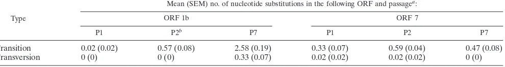
nucleotide changes occurred in ORF 1b (435 bases) of PRRSV
over seven animal passages (Fig. 7, top). At the termination of
the study, the mean percentage of nucleotide changes among
45 plaque-cloned viruses of P7 was 0.67% compared to the
data for CC-01. However, most of the nucleotide changes were
synonymous mutations, i.e., no amino acid substitutions. The
ORF 7 gene (372 bases) of PRRSV was also conserved during
serial animal passages (Fig. 7, bottom). In comparison to the
data for CC-01, the mean percentages of changes among virus
clones were 0.01, 0.03, and 0.13% at the nucleotide level and
0.02, 0.02, and 0.11% at the amino acid level for P1, P2, and P7,
respectively.
The ORF 5 gene (603 bases) of PRRSV showed higher rates
of change during animal passages (Fig. 7, middle). The mean
percentages of changes in nucleotides were 0.09, 0.29, 0.53,
0.69, 0.61, 0.89, and 0.70% for P1 through P7, respectively.
Most of the nucleotide changes in ORF 5 were
nonsynony-mous mutations, resulting in substitutions of amino acid
resi-TABLE 7. Appearance of nine 7NVs within three independent
lines and in three pig passages
Genotype
No. of plaque-cloned viruses identical to each genotype
in the following passage and linea:
P1 P2 P7
A B C A B C A B C
CC-01
b15
15
13
13
14
13
9
15
—
7NV-01
—
—
1
—
—
—
—
—
—
7NV-02
—
—
1
—
—
—
—
—
—
7NV-03
—
—
—
1
—
—
—
—
—
7NV-04
—
—
—
1
—
—
—
—
—
7NV-05
—
—
—
—
1
—
—
—
—
7NV-06
—
—
—
—
—
1
—
—
—
7NV-07
—
—
—
—
—
1
—
—
—
7NV-08
—
—
—
—
—
—
6
—
—
7NV-09
—
—
—
—
—
—
—
—
15
aSee Table 3, footnotea.
[image:9.587.44.285.94.244.2]bSee Table 3, footnoteb.
TABLE 8. Appearance of three 7AVs within three independent
lines and in three pig passages
Genotype
No. of plaque-cloned viruses identical to each genotype
in the following passage and linea:
P1 P2 P7
A B C A B C A B C
CC-01
b15
15
14
15
15
14
9
15
15
7AV-01
—
—
1
—
—
—
—
—
—
7AV-02
—
—
—
—
—
1
—
—
—
7AV-03
—
—
—
—
—
—
6
—
—
aSee Table 3, footnotea.
bSee Table 3, footnoteb.
TABLE 9. Appearance of 40 CNVs within three independent lines
and in three pig passages
Genotype
No. of plaque-cloned viruses identical to each genotype
in the following passage and linea:
P1 P2 P7
A B C Ab B C A B C
CC-01
c5
12
6
—
—
—
—
—
—
CNV-01
6
—
4
1
—
5
—
—
—
CNV-02
1
—
—
—
—
—
—
—
—
CNV-03
1
—
1
—
—
—
—
—
—
CNV-04
1
—
—
—
—
—
—
—
—
CNV-05
1
—
—
—
—
—
—
—
—
CNV-06
—
1
—
—
—
—
—
—
—
CNV-07
—
1
1
—
—
—
—
—
—
CNV-08
—
1
—
—
—
—
—
—
—
CNV-09
—
—
1
—
—
—
—
—
—
CNV-10
—
—
1
—
—
—
—
—
—
CNV-11
—
—
1
—
—
—
—
—
—
CNV-12
—
—
—
8
—
—
—
—
—
CNV-13
—
—
—
1
—
—
—
—
—
CNV-14
—
—
—
1
—
—
—
—
—
CNV-15
—
—
—
1
—
—
—
—
—
CNV-16
—
—
—
1
—
—
—
—
—
CNV-17
—
—
—
1
—
—
—
—
—
CNV-18
—
—
—
—
6
—
—
—
—
CNV-19
—
—
—
—
6
1
—
—
—
CNV-20
—
—
—
—
1
—
—
—
—
CNV-21
—
—
—
—
1
—
—
—
—
CNV-22
—
—
—
—
1
—
—
—
—
CNV-23
—
—
—
—
—
5
—
—
—
CNV-24
—
—
—
—
—
1
—
—
—
CNV-25
—
—
—
—
—
1
—
—
—
CNV-26
—
—
—
—
—
1
—
—
—
CNV-27
—
—
—
—
—
1
—
—
—
CNV-28
—
—
—
—
—
—
4
—
—
CNV-29
—
—
—
—
—
—
1
—
—
CNV-30
—
—
—
—
—
—
3
—
—
CNV-31
—
—
—
—
—
—
1
—
—
CNV-32
—
—
—
—
—
—
1
—
—
CNV-33
—
—
—
—
—
—
1
—
—
CNV-34
—
—
—
—
—
—
4
—
—
CNV-35
—
—
—
—
—
—
—
3
—
CNV-36
—