Alternative Splicing Regulation in
Programmed Cell Death and Neurological
Disorders: A Systems Biology Approach
The Harvard community has made this
article openly available.
Please share
how
this access benefits you. Your story matters
Citation
Wang, Qingqing. 2013. Alternative Splicing Regulation in
Programmed Cell Death and Neurological Disorders: A Systems
Biology Approach. Doctoral dissertation, Harvard University.
Citable link
http://nrs.harvard.edu/urn-3:HUL.InstRepos:11129152
Terms of Use
This article was downloaded from Harvard University’s DASH
repository, and is made available under the terms and conditions
applicable to Other Posted Material, as set forth at
http://
ALTERNATIVE SPLICING REGULATION IN PROGRAMMED CELL DEATH AND
NEUROLOGICAL DISORDERS: A SYSTEMS BIOLOGY APPROACH
A dissertation presented
by
Qingqing Wang
to
The Committee on Higher Degrees in Systems Biology
in partial fulfillment of the requirements
for the degree of
Doctor of Philosophy
in the subject of
Systems Biology
Harvard University
Cambridge, Massachusetts
iii
Dissertation Advisor: Professor Pamela A. Silver Qingqing Wang
ALTERNATIVE SPLICING REGULATION IN PROGRAMMED CELL DEATH AND
NEUROLOGICAL DISORDERS: A SYSTEMS BIOLOGY APPROACH
Abstract
Alternative splicing (AS) is a major source of biological diversity and a crucial
determinant of cell fate and identity. Characterizing the role of AS regulatory networks in
physiological and pathological processes remains challenging. The work presented here
addresses this challenge using systems biology analyses of AS regulatory networks in
programmed cell death and neurological disorders.
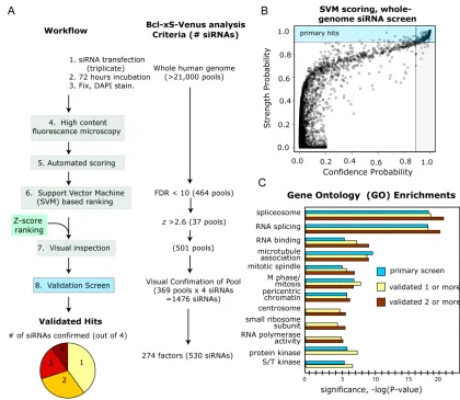
The first study describes a genome-wide screen based on splicing-sensitive
reporters to identify factors that affect the AS of apoptosis regulators
Bclx
and
Mcl1
. The
screen identified over 150 factors that affect apoptosis through modulating the pro- and
anti-apoptotic splicing variants of these apoptosis regulators. This screen revealed a new
functional connection between apoptosis regulation and cell-cycle control through an AS
network. It also unearthed many disease-associated factors as AS effectors.
The second study describes the functions of the Polyglutamine-binding protein 1
(PQBP1)-mediated AS regulatory network in neurological disorders. PQBP1 is a factor
linked to intellectual disability and was unexpectedly identified as an AS effector from
the screen described above. We found that PQBP1 influences the splicing of many
mRNAs and is associated with a wide range of splicing factors. Depletion of PQBP1 in
mouse primary cortical neurons caused defects in neurite outgrowth and altered AS of
mRNAs enriched for functions in neuron projection regulation. Disease-mutants of
iv
patterns and neuron morphology defects in PQBP1 depleted-neurons. This study revealed
a novel function of PQBP1 in AS regulation associated with neurite outgrowth and
indicated that aberrant AS underlies the pathology of PQBP1-related neurological
disorders.
A final study examines the dynamics of the Drosophila Sex-lethal AS regulation
network using a combination of experimental tools and mathematical modeling. This
study demonstrates that the features of Sxl AS regulation have great potentials in building
synthetic memory circuits in mammalian cells to track cell fate.
v
TABLE OF CONTENTS
Abstract
...
III!
ACKNOWLEDGEMENT
...
VII!
ATTRIBUTIONS
...
X!
CHAPTER 1: INTRODUCTION... 1!
MESSENGER RNAS: A DYNAMIC READOUT OF GENETIC INFORMATION
... 2!
MECHANISM OF PRE-MRNA SPLICING... 3!
ALTERNATIVE SPLICING IN BIOLOGICAL DIVERSITY AND REGULATORY NETWORKS... 7!
A HIERARCHICAL CONTROL OF ALTERNATIVE SPLICING REGULATION
... 11!
ABERRANT SPLICING AND HUMAN DISEASES... 15!
Cis-acting splicing defects: disruption of the ‘splicing code’... 16!
Trans-acting splicing defects: disruption of the RNA splicing ‘regulon’ ... 17!
Pathological mutations in the core splicing machinery... 18!
ALTERNATIVE SPLICING REGULATION AND HUMAN DISEASES: A SYSTEMS BIOLOGY APPROACH
... 19!
High-throughput tools for systematic AS studies ... 19!
Computational challenges in evaluating global alternative splicing... 21!
Screening for unidentified links between alternative splicing regulation and human diseases
... 21!
Dynamics of the alternative splicing regulatory network and applications in synthetic
biological circuits... 22!
THIS WORK
... 24!
REFERENCE
... 28!
CHAPTER 2: AN ALTERNATIVE SPLICING NETWORK LINKS CELL-CYCLE
CONTROL TO APOPTOSIS ... 37!
ABSTRACT... 38!
INTRODUCTION
... 39!
RESULTS
... 41!
DISCUSSION... 63!
MATERIALS AND METHODS
... 68!
ACKNOWLEDGMENTS
... 71!
REFERENCES
... 72!
CHAPTER 3: PQBP1, A FACTOR LINKED TO INTELLECTUAL DISABILITY,
AFFECTS ALTERNATIVE SPLICING ASSOCIATED WITH NEURITE OUTGROWTH
... 76!
ABSTRACT... 77!
INTRODUCTION
... 78!
RESULTS
... 80!
DISCUSSION... 106!
MATERIALS AND METHODS
... 113!
ACKNOWLEDGMENTS
... 117!
REFERENCES
... 118!
CHAPTER 4: A JUNCTION-BASED METHOD FOR DIFFERENTIAL ANALYSIS OF
GLOBAL ALTERNATIVE SPLICING FROM RNA-SEQ DATA ... 124!
ABSTRACT... 125!
INTRODUCTION
... 126!
RESULTS
... 133!
vi
MATERIALS AND METHODS
... 142!
ACKNOWLEDGEMENT
... 143!
REFERENCES
... 144!
CHAPTER 5: A SYNTHETIC ALTERNATIVE SPLICING NETWORK TO CONFER
MEMORY OF EXTRACELLULAR STIMULI IN MAMMALIAN CELLS... 148!
ABSTRACT... 149!
INTRODUCTION
... 150!
RESULTS
... 154!
DISCUSSION... 170!
MATERIALS AND METHODS
... 173!
ACKNOWLEDGEMENTS
... 174!
REFERENCES
... 175!
APPENDIX A: AN ALTERNATIVE SPLICING NETWORK LINKS CELL-CYCLE
CONTROL TO APOPTOSIS ... 179!
APPENDIX B: GENOME-WIDE RNAI SCREEN DISCOVERS FUNCTIONAL
COUPLING OF ALTERNATIVE SPLICING AND CELL CYCLE CONTROL TO
APOPTOSIS REGULATION... 192!
APPENDIX C: PQBP1, A FACTOR LINKED TO INTELLECTUAL DISABILITY,
AFFECTS ALTERNATIVE SPLICING ASSOCIATED WITH NEURITE OUTGROWTH
... 196!
APPENDIX D: SUPPLEMENTAL MATERIALS... 210!
SUPPLEMENTAL MATERIALS FOR CHAPTER 2 ... 211!
SUPPLEMENTAL MATERIALS FOR CHAPTER 3 ... 212!
SUPPLEMENTAL MATERIALS FOR CHAPTER 4 ... 224!
vii
ACKNOWLEDGEMENT
I would like to express my sincere thanks to the many colleagues, advisors, family
and friends who supported me through graduate school and witnessed my transformation
from a pure mathematics major to a systems biologist.
First and foremost, I thank my advisor Pam Silver, without whom there is no way
I could have gone this far. I greatly appreciate her faith in her students, her
encouragement of us to think “deep” and “big” in biology, and her support of us to take
on ambitious even risky approaches towards scientific questions. Her tutoring of these
many years significantly influenced and shaped the way I think about biology and
conduct research. I also want to thank her for her caring about her students. Being an
international student, from the moment I joined the lab I have been encouraged strongly
by her to work hard on my English, and to watch, feel, and enjoy the American culture. I
also owe special thanks to her for the tremendous support I received from her during a
difficult time early in graduate school when I was not sure how to pursue my research in
biology. Finally, I want to thank her for running such a fantastic lab from where I found
my American “family.”
viii
I have benefited from the wisdom and kindness from many colleagues and
collaborators. In the Silver lab I owe special thanks to my “big brother” Michael Moore,
from whom I received much of my basic biological training: an intense and ferocious
lesson in “Biological experiments for dummies” when I popped in lab knowing nothing
but doing mathematical proofs. I was also lucky to know Natalie Gilks-Farny, Jessica
Hurt and Bill Senapedis from whom together with Michael, I learned a tremendous
amount about RNA biology and I thank them for their expertise and patience. I also want
to thank Karmella Haynes, Jeffrey Way and Daniel Ducat for all the insights and
thoughtful advice I received from them in learning and doing research in synthetic
biology.
Outside of the Silver lab, I am especially indebted to Michael Greenberg’s lab
for the generous sharing of the precious embryonic mouse neurons and their expertise in
neuron culture. Their kindness made it possible for me to conduct experiments in neurons
that much of my work relied on. I also want to thank Guillaume Adelmant for his
expertise in proteomic analysis using mass-spectrometry, and Jennifer Waters and Lauren
Piedmont from the Harvard Nikon Imaging Center who made neuron imaging far less
painful for me.
ix
sure everything worked and tolerated my constant bothering of “URGENT please ASAP”
orders.
I want to thank all the wonderful friends who populated my journey through
graduate school and made the experience not only enjoyable but also a transformative
one. I am especially grateful for the big, weird and probably (but properly) dysfunctional
fun family of the “Silverinos,” members of whom are too numerous to name. Time passes
and face changes, but the essence of this family stays. The characteristics and
personalities from three generations of Silverinos during my time here embraced me
deeply and shaped my personality greatly.
x
ATTRIBUTIONS
For Chapter 3, Chapter 4 and Chapter 5, I performed all the work presented in this
dissertation with exceptions detailed here. Guillaume Adelmant analyzed the proteins
associated with PQBP1 and its two disease mutants with mass spectrometry in Chapter 3,
and contributed to the written explanation of the method in the supplemental data of
Chapter 3.
1
2
MESSENGER RNAS: A DYNAMIC READOUT OF GENETIC INFORMATION
The central dogma of molecular biology states that the genetic information of life
is stored in DNA and transferred to messenger RNA (mRNA) for expression as proteins
(Crick 1970). mRNA however, is more than a passive intermediate in the expression of
genetic information. Instead, multiple mechanisms on mRNA processing enable mRNA
to selectively extract and re-organize information from DNA. mRNA provides a dynamic
readout of the genetic information stored in DNA in a tissue- and cell type-specific way.
It is now appreciated that the number of protein-coding genes in a genome is not
proportional to the degree of complexity of that organism (Lodish et al. 2008). For
example, the human genome encodes around 25,000 protein-coding genes, only four
times as many as the yeast Saccharomyces and comparable to that of the plant
Arabidopsis thaliana, the nematode C. elegans and the fruit fly Drosophila. Furthermore,
human and chimpanzee genomes show a 98% sequence identity, even though humans
exhibit far more complicated cognitive behaviors such as the use of language (Lander et
al. 2001; Olson and Varki 2004). These examples illustrate that the phenotypic
complexity of an organism is not solely determined by the number of protein-coding
genes it possesses. Rather, it has been proposed that the diversity and sophistication of
the eukaryotic proteome is augmented by mRNAs via regulatory mechanisms at the
transcriptional and post-transcriptional levels.
3
(introns). The maturation of an mRNA transcript requires the excision of introns and the
concomitant joining of exons (Gilbert 1978; Matlin et al. 2005). In many cases of
multi-exon mRNA transcript maturation there is more than one way to remove introns and join
exons, enabling a gene to generate multiple mRNA and protein variants (Wang et al.
2008; Pan et al. 2008) in a process termed alternative splicing. In addition to pre-mRNA
splicing, mRNA transcripts are subject to many other regulatory processes, such as
control of mRNA stabilization, transport and RNA editing. These mRNA processing
steps greatly increase the genome’s capacity to generate different mRNA profiles and
expand the number of functionally distinct proteins that can be encoded by the genome
(Nilsen and Graveley 2010).
Multicellular organisms are typically composed of hundreds of distinct cell types
that are specialized in a variety of functions. These cells share largely identical genetic
information, but are capable of generating diversified gene expression patterns in
response to tissue- and development stage-specific needs. The mRNA profiles are thus of
special importance as a dynamic reflection of how genes are expressed and regulated.
Moreover, aberrant mRNA profiles are associated with human diseases and could provide
valuable information on pathology and potential treatment strategies.
MECHANISM OF PRE-MRNA SPLICING
4
nuclear RNP (snRNP) elements (U1, U2, U4, U5 and U6) and is dynamically associated
with up to 300 additional proteins (Rappsilber et al. 2002; Wahl et al. 2009).
The basic splice sites in eukaryotic pre-mRNAs are generally defined by short
consensus sequences (Figure 1-1A) and the spliceosome is assembled onto the
pre-mRNA in a step-wise manner on those sites. During initiation of the spliceosome
assembly (spliceosomal E complex), the 5’ splice site (5’SS) is recognized by the U1
snRNP, the branch point sequence (BPS) by SF1 (splicing factor 1) and 3’ splice site (3’
SS) by U2AF (U2 auxiliary factor) (Figure 1-1B). Following the formation of the E
complex, U2 snRNP docks to the BPS and replaces SF1, forming the pre-spliceosome—
complex A. Subsequently, U4/U5/U6 pre-assembled tri-snRNPs are recruited to the A
complex, forming the pre-catalytic spliceosome—complex B. The spliceosome then
undergoes major conformational changes in which U1 and U4 snRNPs are released,
giving rise to the activated spliceosome—the B* complex. The spliceosomal B* complex
performs the first catalytic step of pre-mRNA splicing: the 2’-hydroxyl group of the
adenine base at the BPS attacks the phosphodiester bond at the 5’SS, generating a free 5’
exon and an intron lariat-3’ exon. This intermediate together with the remaining snRNPs
forms the C complex. A second round of spliceosome conformational change takes place
here and the second catalytic step of pre-mRNA splicing is performed: the
5
Figure 1-1: Consensus sequences of eukaryotic splice sites and step-wise spliceosome
assembly on splice sites.
(A) The consensus sequences of splice sites in metazoans and yeasts. Schematics of a pre-mRNA
are shown, with green rectangles representing exons and black lines between exons representing
introns. 5’ splice site is represented by the consensus sequence “GUAAGU” in yeast and “GU” in
metazoans; branch point sequence by the sequence “UACUAAC” in yeast and “A” in metazoans;
3’ splice site by “AG” in both metazoans and yeasts. The consensus sequences of the 5’ splice
site, branch point sequence and 3’ splice site in yeast are highly conserved but in metazoans are
degenerate.
6
Exon
Exon
U5
U6
A
5' splice site
Exon
GU
A
AG
Exon
GUAAGU
UACUAAC
AG
3' splice site
Branch point sequence
Metazoan
Yeast
Intron
B
Spliceosomal E complex
GU A AGU1
SF1
U2AF
5' SS BPS 3' SS
U1
GU A AGSpliceosome
complex A
U6
U4
U5
U1
GU A AGU6
U4
U5
U2
Spliceosome
complex B
U4
U1
U2
GU A-OH AGSpliceosome
complex B*
First transesterification reaction
U5
U6
U2
-OH A-O AGSpliceosome
complex C
Second transesterification reaction
U5
U6
U2
A-O+
Exon Exon Exon Exon Exon Exon Exon Exon Exon ExonU2AF
U2
[image:17.612.142.517.75.728.2]7
ALTERNATIVE SPLICING IN BIOLOGICAL DIVERSITY AND REGULATORY NETWORKS
Alternative splicing (AS) depicts the phenomenon in higher eukaryotes that
introns can be removed and exons are pieced together in different patterns to produce
functionally distinct mRNA and protein variants from one gene. AS is a major source of
biological diversity in eukaryotic transcriptomes and proteomes. The contribution of AS
to eukaryotic complexity is likely even greater than previously recognized, as
high-throughput studies showed that >90% of mammalian genes undergo AS and generate
polymorphic mRNA transcripts (Pan et al. 2008; Wang et al 2008). The number of
mRNA transcripts that a multi-exon gene is able to encode varies from several to
thousands, a staggering level of diversity. As an example, the Drosophila gene Down
syndrome cell adhesion molecule (Dscam) is able to make approximately 38,000 mRNA
isoforms, far surpassing the number of total protein-coding genes (~14,500) in the
organism (Schmucker et al. 2000).
There are seven basic patterns that pre-mRNAs can follow during AS (Figure 1-2;
Black 2003). An exon can be alternatively included or excluded in the transcript, and is
defined as a cassette exon (Figure 1-2A). Multiple cassette exons can be mutually
[image:19.612.139.507.70.455.2]
(20)8
Figure 1-2: Seven basic AS patterns in multicellular eukaryotes.
Schematics of hypothetic pre-mRNAs are shown under different AS patterns, with green
rectangles representing exons and gray rectangles represent alternatively spliced exons or exonic
regions. Arrows show transcription start site. Arrowheads show poly-A sites.
In addition to serving as a major source of biological diversity, AS also plays
important roles in regulatory networks. There are many mechanisms of AS in biological
regulation. As one mechanism, AS enables the generation of functionally distinct protein
cassette exon
mutually exclusive exon
alternative 5' splice
site
alternative promoter
site
poly-A
poly-A
alternative poly-A
site
alternative 3' splice
site
intron retention
A
B
C
D
E
F
9
products from the same gene in response to different stimuli, and can work as a switch in
cell fate determination (Figure 1-3A). For example, the gene encoding Bcl-2-like protein
1 (Bcl2l1 or Bcl-x), a member of the Bcl-2 family of apoptosis regulators, produces two
major splicing isoforms—anti-apoptotic Bclx-L and pro-apoptotic Bclx-S due to the usage
of an alternative 5’ splice site in exon 2 (Figure 1-3A; Akgul et al. 2004). The switch of
the two splicing isoforms of Bcl-x affects the cell’s decision on whether to commit
apoptosis. Besides Bcl-x, several other apoptosis regulators also generate pro- and
anti-apoptosis isoforms (Akgul et al. 2004). As another mechanism, AS can introduce
premature termination codons in an mRNA, leading to degradation of the mRNA through
nonsense-mediated mRNA decay (AS-NMD) (Lewis et al. 2003) (Figure 1-3B). Here,
AS directly regulates the total abundance of a target, rather than the expression of
specific isoforms. As an example, the Drosophila sex determination master gene
Sex-lethal (Sxl) can produce the male-specific isoform or female-specific isoform based on
the inclusion or exclusion of exon 3, which contains a premature stop codon (Figure
1-3B; Baker 1989). AS-NMD causes the degradation of the male-specific isoform and lack
of functional Sxl protein in male flies, leading to male-related phenotypes. On the other
hand, the female-specific isoform lacking exon 3 is translated into functional Sxl proteins
in female flies. Sxl is itself a splicing regulator, and alters the splicing of a cascade of
downstream mRNA transcripts, resulting in female-related phenotypes (Baker 1989). The
two example AS mechanisms above can be found in many biological regulatory
[image:21.612.128.521.104.427.2]
(22)10
Figure 1-3: Two examples AS mechanisms in biological regulatory networks.
(A) AS enables Bcl-x to generate functionally distinct mRNA isoforms and protein variants to
affect cells’ decision in committing apoptosis. A schematic of Bcl-x is shown. Green rectangles
represent exons and black lines between exons are introns. Gray rectangle represents the
alternatively spliced exonic region. Arrows show translation start site.
(B) AS regulates the abundance of Sxl through AS-NMD in Drosophila. A schematic of Sxl is
shown. Gray rectangles represent exons and black lines between exons are introns. Dark rectangle
represents the alternatively spliced exon 3, with the stop sign representing a premature
termination codon.
A
Bcl-xL
anti-apoptotic
Bcl-xS
pro-apoptotic
Regulatory switch using
functionally distinct isoforms
1 2
3
4 5 6 7 8
1 2 4 5 6 7 8
1 2 3 4 5 6 7 8
Degradation
SXL protein
B
Quantitative gene expression
control with AS-NMD
male
11
A HIERARCHICAL CONTROL OF ALTERNATIVE SPLICING REGULATION
The selection of splice sites by the spliceosome determines AS decisions. In yeast
where constitutive splicing is dominant and AS is rare, the sequence elements for 5’SS,
BPS and 3’SS are conserved and well recognized by U1 snRNP, SP1 and U2AF of the
spliceosome machinery (Figure 1-1A; Izquierdo et al. 2006). However in higher
eukaryotes where AS is more a rule than exception, the splice site sequence elements are
degenerate and not strong enough to direct the spliceosome on their own (Figure 1-1A).
AS in multicellular eukaryotes is under the regulation of a large repertoire of
trans-acting non-snRNP RNA-binding proteins (RBPs) and additional cis-elements in the
pre-mRNA sequences (Wahl et al. 2009). Two classical classes of these RBPs are
serine-arginine repeat proteins (SR proteins) and heterogeneous nuclear ribonucleoproteins
(hnRNPs) (Matlin et al. 2005). RBPs recognize cis-elements within the pre-mRNA
sequences and help to recruit or dissociate the spliceosome machinery to/away from a
particular splice site (Figure 1-4A; Singh and Valcárcel 2005). These cis-elements
associated with RBPs are short regulatory sequences flanking splice sites, including
exonic or intronic splicing enhancers or silencers (Figure 1-4A). Collectively, AS
decision is the combinatorial result from the ‘splicing code’ embedded in the pre-mRNA
sequences and a large repertoire of proteins devoted to interpreting the code (Matlin et al.
2005).
The splicing code in AS is analogous to transcription factor binding sites in DNA
sequences in the transcription network. Much effort has been dedicated to deciphering the
splicing code (Wang and Burge 2008; Hartmann and Valcárcel 2009; Nilsen and
12
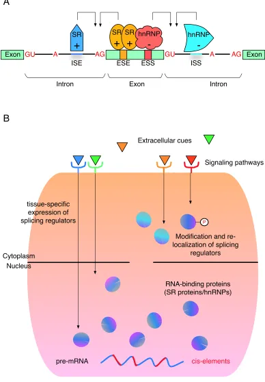
Figure 1-4: A hierarchical control of AS regulation.
(A) The interplay of cis-elements within the pre-mRNA sequence and trans-acting RBPs in AS
regulation. A schematic of a hypothetic pre-mRNA is shown. Green rectangles represent exons
and black lines between exons are introns. 5’ splice sites are marked by “GU”, branch sites by A
and 3’ splice sites by “AG”. Orange and red bands within the middle exon represent an exonic
splicing enhancer (ESE) and an exonic splicing silencer (ESS). Blue short lines within introns
represent an intronic splicing enhancers (ISE) and an intronic splicing silencers (ISS). SR proteins
and hnRNPs can bind to these cis-elements and cast positive (+) or negative (-) effects on the
selection of the nearby splice sites.
(B) A hierarchical control of AS regulation in multicellular eukaryotes. AS decision is under the
regulation of cis-elements (red short lines) within pre-mRNA sequences and a large repertoire of
RNA-binding proteins (RBPs, purple and blue circles). These RBPs are further controlled by
many regulatory mechanisms that can cause tissue-specific synthesis, re-localization or
13
Figure 1-4 (Continued).
Exon
GU
A
AG
GU
ESE
ESS
ISE
ISS
SR
+
hnRNP
-Exon
SR
+
hnRNP
-AG
A
Exon
Intron
Intron
A
B
cis-elements
pre-mRNA
RNA-binding proteins
(SR proteins/hnRNPs)
Nucleus
Cytoplasm
Extracellular cues
Modification and
re-localization of splicing
regulators
tissue-specific
expression of
splicing regulators
Signaling pathways
P
[image:24.612.130.518.79.637.2]