0022-538X/84/010066-06$02.00/0
Copyright©1984, AmericanSociety for Microbiology
Mouse
Mammary Tumor
Virus: Specific Methylation
Patterns
of
Proviral DNA in Normal Mouse Tissues
WEI-SHAU HU, THOMAS G. FANNING, ANDROBERT D. CARDIFF*Department of Pathology, School of Medicine, University of California, Davis, California95616 Received 18 July 1983/Accepted 4 October 1983
The methylation state of endogenous mouse mammary tumor virus (MuMTV) proviral DNA was
examined innormalmousetissues. DNAs from various tissueswerecleaved withthe methylation-sensitive enzymes HhaI and HpaII and analyzed by Southern blotting. Tissue-specific MuMTV proviral DNA methylation patterns were found in the BALB/c, C3H, C57BL, GR/A, and GR-Mtv-2- mouse strains.
MuMTVproviral DNAwashypomethylated in DNAs from the spleens andtestesof all strains examined. The GR/A mouse strain, which was most thoroughly studied, also showed hypomethylation of MuMTV proviral DNA in bone marrow and placental tissues. Analysis of RNAs extracted from GR/A liver, mammarytumor, testes,placenta, and spleen tissues demonstrated thatMuMTVproviralhypomethylation neednotreflect significant proviral transcription.
The timing and extent ofgene expression in eucaryotes
maybecontrolled, atleast in somedegree, bythe presence
orabsenceofmodifiedbasesinDNA.Specifically,anumber
of investigators have postulated that cytosine methylation
may play a significant role in determining which cellular
geneswill be transcribed andatwhat times(4, 11,26, 28).
5-Methylcytosine may control gene transcription by one or
more mechanisms: the base may modulate protein-DNA
interaction (4, 11, 26, 28) or induce (or stabilize) localized
conformational alterations of the DNA (1). The strong
correlation between DNA hypomethylation and gene
expression which has been demonstrated in a number of
systems (4, 11,26, 28)lends credencetothe concept that
5-methylcytosine plays an important role in transcription.
However, the correlation between DNA hypomethylation
andtranscriptional activityhasnotbeendemonstrated in all
cases(15, 20, 25).
Endogenous and acquired mouse mammary tumor virus (MuMTV) proviral DNA is less methylated in mammary tumors than theendogenous proviruses in mouse liver (3, 6, 10, 12, 14).SinceMuMTV-specificRNAiseasilydetected in mammary tumorsbutnot in livers(33), hypomethylationof MuMTV proviral DNA has been taken as prima facia
evidence for MuMTV transcriptional activity (3, 6, 10, 12,
14).
Althoughthecorrelationbetween MuMTVproviral
hypo-methylation and geneexpression is
demonstratively
true inmammary tissue, it does notappear tobe valid ingeneral.
Wehave foundhypomethylated endogenousMuMTV
provi-ral DNA inmousetissueswhich contain littleor noMuMTV RNA. However,ourdatadonotexclude thepossibilitythat
hypomethylation of MuMTV DNAis anecessary
prerequi-siteforproviralgene expression.
MATERIALS ANDMETHODS
Animals. BALB/c, C57BL, C3H, and GR/A mice were
originally obtained from the Cancer Research Laboratory,
Berkeley, Calif. or the Jackson Laboratory, Bar Harbor,
Maine. GR-Mtv-2- mice were originally provided by Jo
Hilgers, The Netherlands Cancer Institute, Amsterdam.
Nucleic acid extraction. DNA was extracted from mouse tissues by the proteinase K-sodiumdodecyl
sulfate-phenol-*Correspondingauthor.
chloroform method asdescribed previously (13). RNAwas
extracted as follows. Fresh tissue was homogenized in an
ice-jacketed Waring blender in a buffer containing 10 mM
Tris-hydrochloride(pH 8), 10 mM EDTA, 40 mMNaCl,and
100 to200
pig
ofproteinase K perml. The homogenatewaspassed through fourlayers of cheesecloth to remove large
debris, made0.5%in sodiumdodecyl
sulfate,
and extractedseveral times with phenol-chloroform. The partially
depro-teinized sample was ethanolprecipitated, dissolved in
buff-er,andagain extractedwithphenol-chloroform. Thesample
was ethanol precipitated and dissolved in 20 mM sodium acetate(pH 5-10mMMgSO4, and thenucleic acid
concen-tration was determined by measuringtheoptical density at
260 nm. The sample was thendigested with DNase(RNase
free; Miles Laboratories, Elkhorn, Ind.) under conditions
recommendedbythesupplier.The digestswere stopped by
freezing, and asamplewas subjectedtoelectrophoresis ina
2%agarosegelcontaining 1
jig
of ethidium bromideperml.Inaddition to themouse RNAs, at least onelane ofthegel
contained a known amount of purified Escherichia coli
rRNA. The gel was photographed under shortwave UV
light, and each lane of the photographic negative was
scannedwithaGilford2000spectrophotometer(13,14).The
densitometerscansof each lanewerecomparedwith thatof
theE.coli rRNA standardtoarriveat abestestimate ofthe
RNA concentration in each sample. Only those samples
showingnoRNAdegradationwereused inthe
experiments
reported here.
Blot analysis. Cellular DNAs were digested with
restric-tionenzymes(fromNewEnglandBiolabs, Boston, Mass.or
Bethesda
Research Laboratories, Bethesda, Md.) and Southern blotted as described previously (13, 14). Dot blot(18)analysisof cellular RNAswasperformed with
nitrocel-lulose filters soaked in lOx SSC(lx SSC is 0.15MNaClplus
0.015 Msodiumcitrate) on aDot Blot apparatus
(Bethesda
Research Laboratories). All blots, unless otherwise noted,
were probed with a mixture ofcloned MuMTV PstI
frag-ments (19) that had been labeled bynick translation
(27).
RESULTS
Methylation patterns ofMuMTVproviral DNA in normal GR/A tissues. Previous studies have
compared
themethyl-ation patterns of MuMTV
proviral
DNAinmousemammary tissues with the patterns exhibited in normal nonmammary 66on November 10, 2019 by guest
http://jvi.asm.org/
_ \_
~~ ~
4-5-o f
2 6*
1 2#
1 2 3 4 5 6 7 8
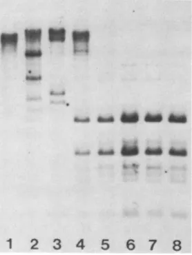
FIG. 1. Hypomethylated endogenous MuMTVproviral DNAin GR/A mouse tissues. Tissue DNAs(7 ,ug) weredigested with the
enzymeHhaI,electrophoresedinto 1%agarosegels, and Southern blotted. The blots were hybridized with labeled MuMTV-specific DNAandautoradiographed. Lane1,femaleliver;lane2,male and
female bonemarrows(pooled fromseveralanimals);lane3,female
spleen; lane4,testes;lane 5, placenta; lane 6,fetuses(pooledfroma
single litter); lane 7,mammarytumor;lane8,femalekidney.Arrows denotethe sizesof DNAfragmentsin kilobasepairs.
tissues (3, 6,10, 12, 14). The normal tissue mostcommonly used has been liver, although kidney, heart, spleen, and mammarygland tissues have also been investigated. In these
studies, onlytumorandspleen tissues contained
hypometh-ylated MuMTV proviral DNA.
We surveyed a number of normal nonmammary GR/A mouse tissues for the presence ofhypomethylated
endoge-nousMuMTVproviral DNA. Proviral DNA inGR/Aspleen,
placenta, andmammarytumortissuesappearedtobe signifi-cantly hypomethylated when digestedwiththe methylation-sensitiverestriction enzymeHhaI(Fig. 1, lanes3, 5, and 7). Bonemarrow (Fig. 1, lane 2) andtestes(Fig. 1, lane 4) also appearedtoharborhypomethylated MuMTV proviral DNA, although the degree of hypomethylation seemed to be less than forspleen and placenta. The patternsexhibitedby both the fetus and kidneysamples suggested that MuMTV provi-ral DNA was largely methylated in these tissues, although
somewhat less sothan proviral DNA inGR/A liver(Fig. 1;
comparelanes 6 and 8 with 1).
In addition to the quantitative differences in MuMTV proviral DNAmethylationseeninFig. 1, thereweremarked
qualitative differences. For example, the 2.6-kilobase pair (Kb) and 6.0-Kb MuMTV proviral bands seen in
HhaI-digested spleen DNA (Fig. 1, lane 3) were not present in HhaI-digestedtestesDNA(Fig. 1, lane 4). These qualitative differences suggest that different MuMTV proviruses are
hypomethylated inatissue-specific fashion,orthatdifferent
enzyme sites on a given provirus are hypomethylated in a
tissue-specific fashion,orboth. Atpresent,wehavenoway ofunambiguously differentiating between thesetwo possibil-ities.
Similarities amongthe profiles shown in Fig. 1 were also apparent. MuMTV proviral DNA fragments of 9 Kb were
generated by HhaI digestion ofmanytissue DNAs (Fig. 1, lanes 2, 3, 5, 6, and 7). Since integrated MuMTV proviral DNAis 10 Kb (7), this finding would suggestthat only two
restriction sites are available for cleavage within, ornear,
one ormoreproviralDNAs in these tissues. We have no way
ofdeciding, however, whetherthese 9-Kb fragments
origi-nated from one or more proviruses or whether both sites werelocated within aprovirus, as opposed to one site within
a provirus and the other in the flanking host DNA.
Paren-thetically, all bands seen in Fig. 1 (as well as those in all
subsequent figures) arose from enzymatic cleavage of
inte-grated proviral DNA since Southern blot analysis of un-cleaved tissue DNAs failed to reveal any MuMTV-specific
bands below that of the bulk DNA, which ran at a position of 30to50 Kb(datanotshown).
The bands seen below the 9-Kb band (Fig. 1, lanes 2 to 5
and 7) suggested the presence ofmultiple hypomethylated
enzyme sites within MuMTV proviral DNA. We have, in
fact, demonstrated that this is the case for several of the
lower-molecular-weight bands by performing restriction
mapping experiments and by using smallfragmentsof cloned MuMTV proviral DNA as probes (data not shown). Since
GR/Amice contain five nearly identical copies ofMuMTV proviralDNA (13, 17, 22), we have not been able to assign specificfragments to specific proviruses with oneexception: the 4.2-Kb proviral fragment present in GR/A mouse testes is missing in GR-Mtv-2- mouse testes and, thus, probably originated from the GR-MTV-2 provirus (see below).
The tissue-specific proviral restriction patterns in Fig. 1 were generated by the restriction enzyme HhaI. To corrob-orate thisfinding, we have digested GR/A tissue DNAs with the isoschizomer pair MspI (methylation insensitive) and HpaII(methylationsensitive). TheMspI-HpaIIdigests con-firmed the results obtained with HhaI (Fig. 2) and, in
addition, demonstratedthat although specific MuMTV
pro-viral DNA sequences were hypomethylated in spleen,
tes-tes, and tumortissues,manyotherproviral DNA sequences weremethylated inthese same tissues (Fig. 2; compare lane 2with 6, 3 with 7, and 4with 8).
Hypomethylated MuMTV proviral DNAs in spleens of different mouse strains. DNAs derived from GR/A spleens
appearedtocontainhypomethylated MuMTVproviralDNA
(Fig. 1, lane3;Fig. 2,lane2).To determine whether this was
[image:2.612.94.253.78.261.2]1
2
3
4
5
6
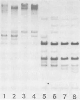
7 8FIG. 2. Hypomethylated endogenous MuMTV proviral DNA in GR/Amousetissues. Tissue DNAs were digested with eitherHpaIl (lanes1 to4) orMspI(lanes 5 to 8) and treated as described in the legend toFig. 1. Lanes 1 and 5, female liver; lanes 2 and 6, male spleen; lanes 3 and 7, testes; lanes4and 8, mammary tumor.
on November 10, 2019 by guest
http://jvi.asm.org/
[image:2.612.361.500.494.679.2]a general phenomenon, DNAs were extracted from the
spleensofBALB/c,C57BL, C3H, and GR-Mtv-2- miceand
examined for the degree of MuMTV proviral DNA
methyl-ation with the enzyme HhaI. In all cases, hypomethylated
proviralDNA wasdetected, although both quantitativeand
qualitativedifferences were apparent among the strains (Fig.
3).
Figure 3 shows both Sacl and HhaI digests of mouse spleenDNAs. Since Saclis not inhibited by DNA
methyl-ation, these patterns indicate the total amount of
MuMTV-specificDNAapplied to each lane of the gel (Fig. 3, lanes 6
to 10). Taking into account the overloading of the
GR-Mtv-2- lanes (Fig. 3, lanes 4 and 9), it was apparent that spleen DNAs from GR/A and GR-Mtv-2- mice contained more
hypomethylated MuMTV proviral DNA than did spleen
DNAsfromBALB/c, C57BL, and C3H mice (Fig. 3). Since theGR/A strain contains more endogenous MuMTV provi-rus than do the other strains (24), this finding was not
completely unexpected. Nevertheless, the fact that both
GR/A and GR-Mtv-2- mice contained more
lower-molecu-lar-weight MuMTV restriction fragments did suggest that more HhaIenzyme sites werehypomethylated inthese two
strains.
An alternative possibilitywas thatMuMTV proviruses in
GR/AandGR-Mtv-2- mice have moreHhaIsites per sethan
doproviruses in the otherstrains. Althoughwecannot rule
out this possibility, it seems unlikely in view of the high
sequencehomologies exhibitedbyMuMTVproviruses(33). Thus, enzymes with short recognition sequences (such as
HhaI and MspI-HhaII, which cleave at four base pair
sequences) would be expected to show conservative pat-terns; indeed, this appeared to be the case (see Fig. 2 and
Fig.5).
Significantly,nodifferences(either qualitative or
quantita-tive)weredetected between the GR/A mouse strain and the
congenic line GR-Mtv-2-(Fig. 3). These two lines differ only
in thepresence (GR/A) or absence(GR-Mtv-2-)of thehighly
oncogenic GR-MTV-2 provirus (13, 17, 22). Thus, it seems
12
3t
478
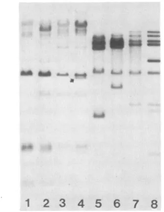
10FIG. 3. Hypomethylated endogenous MuMTV proviralDNA in spleens ofdifferentmousestrains.Tissue DNAsweredigestedwith eitherHhaI (lanes 1 to 5) or Sacl (lanes 6 to 10) and treated as described inthe legendto Fig. 1. Lanes 1 and6, female BALB/c; lanes 2 and7,femaleC57BL;lanes 3 and8,femaleC3H;lanes 4 and 9, maleGR-Mtv-2-;lanes 5 and10, femaleGR/A.
1
2
3
45
6
7 8FIG. 4. Hypomethylated endogenousMuMTV proviralDNA in testesofdifferentmouse strains. TissueDNAswere
digested
with either HhaI (lanes 1 to 4) or Sacl (lanes 5 to 8) and treated asdescribed in thelegendtoFig. 1. Lanes 1 and5, BALB/c;lanes2 and 6, C3H; lanes3 and7, GR-Mtv-2-;lanes 4and8,GR/A.The arrow identifies the 4.2-Kbfragment present inGR/Amice that is missing in GR-Mtv-2- mice.
reasonable to assume that all of the HhaI restriction
frag-ments seen in GR/A spleen samples (Fig. 1 and2) contain
sequences from
proviruses
other than GR-MTV-2.Hypomethylated MuMTV proviral
DNAs
intestesofdiffer-ent mouse strains. DNAs derived from the testes of male
micebelongingto-different strains contained
hypomethylat-ed MuMTV
proviral
DNA (Fig. 4). Whenthe differences intotal amounts of DNA loaded per lane were taken into
account, it appearedasthough roughly
equivalent
amountsof
hypomethylated
MuMTVproviral
DNAswerepresent in the testes of BALB/c,C3H,
GR-Mtv-2-, andGRIA
mice (Fig. 4).Interestingly, the MuMTV
proviral
patterns exhibitedafter HhaI cleavage of testes DNAs
appeared
to be verysimilarbetween thedifferentmousestrains
(Fig.
4,lanes 1 to4). To test thisfurther, the samples were digestedwith the
isoschizomerpair
MspI
(methylation insensitive) andHpaII
(methylation sensitive). The different strainsexhibitedvery
similar
MspI
profiles(Fig.
5, lanes 5 to 8)indicating
thatMspI
restriction sites are highly conserved between theMuMTVproviruses ineach strain.
The MuMTV proviral patterns
generated
by HpaII were alsovery similar(Fig.5, lanes 1 to 4). This indicates that themethylation patternsofMuMTVprovirusesin mousetestes
are strongly conserved among the different strains. The
patterns are less strongly conservedin mouse
spleens
(Fig.
3) or in thedifferenttissuesofa
single
individual(Fig.
1and 2). As yet, we have no explanation for this interstrainconservation ofMuMTV
proviral
DNAmethylation
patternsin mouse testes.
The profiles shown in Fig. 4 demonstrate the only
differ-ence in MuMTV
proviral
methylation
between GR/A and GR-Mtv-2- mice that we have encountered to date. GR/A(Fig.4, lane4)and GR-Mtv-2-(Fig. 4,lane3) differina
4.2-Kb MuMTV proviral DNA fragment that is present in the
GR/A testes sample but absent in the GR-Mtv-2-
sample.
to xmt
on November 10, 2019 by guest
http://jvi.asm.org/
[image:3.612.354.515.77.285.2] [image:3.612.114.265.476.669.2]1
2
3
4 56
7 8FIG. 5. Hypomethylated endogenousMuMTV proviral DNAin testesofdifferent mouse strains. TissueDNAs were digested with either HpaIl (lanes 1 to 4) orMspl (lanes 5 to 8) and treated as described in the legend toFig. 1. Lanes 1 and5, BALB/c: lanes 2 and6. C3H;lanes 3 and 7,
GR-Mti'-2-;
lanes4and8, GR/A.The twomouse lines differ, intermsof MuMTVproviruses,
only in respecttothe GR-MTV-2provirus,which is missing in the GR-Mtv-2- line.Thus,wehaveprovisionally assigned
the 4.2-Kb band to GR-MTV-2.
MuMTV proviral transcriptioninGR/Amousetissues. Are hypomethylated MuMTV proviral DNAs transcriptionally
active? RNA extracted from two GR/A mammary tumors contained significant quantities of MuMTV-specific RNA. Whencompared with controls(Fig. 6,rows1 to3),0.64% of the total RNA from tumor A (Fig. 6, row 6) was found to
hybridize with an MuMTV-specific probe,whereas 2.5% of
the total RNAfromtumor B(Fig. 6,row7) hybridized with the probe. In addition, 0.33% of the total RNA extracted from GR/A prelactating mammarygland tissue (Fig. 6, row
16) hybridized with the MuMTV-specific probe. These
re-sults are in agreement with the observations ofothers that
GR/Atumorsand prelactatingandlactatingmammarygland
tissues containsignificant amounts ofMuMTV RNA (33). Little or noMuMTV RNA could bedetected in the livers of either maleor female GR/A mice (Fig. 6, rows4 and 5),
again confirming resultspreviously obtainedbyother
inves-tigators (33). Similarly, little or no MuMTV RNA was detected in GR/A male spleens, female spleens, testes, placenta, apool offetuses,orinfemalethymustissues(Fig.
6, rows 9 to 15 respectively).
SpleensandtestesofGR/Amice contained
hypomethylat-ed MuMTV proviruses (Fig. 1 and 4). Since these same tissues lacked detectable quantities of MuMTV RNA (Fig.
6), we conclude that hypomethylation of MuMTV proviral
DNA does not indicate significant transcriptional
activity
per se (i.e., transcription on apar with that in tumors). DISCUSSION
Wehave shown that certain normal nonmammary mouse tissues contain hypomethylated endogenousMuMTV provi-ral DNA.Additionally, we haveestablishedthat hypomethy-latedproviruses in nonmammary tissuesareeithersparingly transcribed or not transcribed at all. This result is at odds with many (3, 6, 10, 12, 14, 21) but notall (15)observations
demonstrating a correlation between MuMTV DNA
hypo-methylation and gene expression in mammary tissues and cultured cells.
MuMTV proviruses are present in the genomes of all inbreedstrainsofmice andarealso foundinmanyferalmice (8, 24). The viruses are known to be a causative agent in mammary neoplasia, but nootherbiological functions have
been assigned to them (2). The recent discovery ofhealthy
feral mice lacking MuMTV sequences would suggest that viral genetic information is superfluous for normal mouse
development (5, 8, 9). Thus, why should apparently
nones-sential DNA sequences exhibit tissue-specific methylation
patterns? This question is especially cogent in view of the well-established correlation between hypomethylation and transcriptional activity (4, 11, 26, 28) and the lack of sucha correlation for MuMTV.
Baseduponthe results obtainedinothersystems, weoffer three proposals to explain our results with MuMTV provi-ruses: (i)genes nearMuMTV proviruses are hypomethylat-ed and transcribed, with MuMTV proviruses acting as
"hitch-hikers",(ii) hypomethylated MuMTV proviral DNA
cannot be transcribed because a "critical site" remains methylated: or(iii) hypomethylated MuMTV proviral DNA cannot be transcribed because atranscriptional "trigger" is not present.
The hitch-hiker proposal assumes that MuMTV provi-ruses are integrated near genes that are transcriptionally active. It is known that large regions of chromatin undergo conformational alterations around activegenes (16, 34) and that nontranscribed sequences within these active regions contain hypomethylated DNA (30). Endogenous MuMTV proviruses do not appear to be clustered in the mouse genome (31). Thus, for the hitch-hiker proposal to be cor-rect, one would need topostulate that at least one MuMTV provirusispresentnear an activegene(s)ineach mouse line. This hypothesis is reasonable if, at the time ofgerm line infection, MuMTV preferentially integrated in active
(ex-10
4
12
600
0*
o8**e.
8
14
16e
*FIG. 6. MuMTV-specific RNA in GR/A mouse tissues. Tissue RNAs were extracted and analyzed by the dot blot procedure as described in the text. Rows 1 to 3 are purified MuMTV RNA extracted from C3H virions produced by the Mm5mt/cl cell line. Each rowhas the most concentrated sample on the left (spot 1) and proceeds with serial twofold dilutions to the right. Thus, row 1 begins with 100 ng of MuMTV RNA for spot 1 and proceeds (left to right)to50, 25, 12.5, and 6.3ng; row 2begins with (spot 1) 5 ng of MuMTV RNA;and row 3 begins with (spot 1) 0.5 ng of MuMTV RNA.The remaining rows begin with (spot 1) 1.2 ,ug of total tissue RNA. Row 4, male liver; row 5. female liver; row 6, mammary
tumorA; row 7. mammary tumorB; row8, E.colirRNA; row 9.
malespleen;row 10,femalespleen:row 11, testes from mouse no. 1; row12, testes from mouse no.2;row13.placenta;row 14, a pool of fetuses; row 15, female thymus; row 16, prelactating mammary gland.
0
*
2
0
* it"..on November 10, 2019 by guest
http://jvi.asm.org/
[image:4.612.104.244.82.256.2] [image:4.612.314.549.456.583.2]pressed) regions of the genome and if these regions are
expressed ina tissue-specificfashion.
The hitch-hikerproposalmayexplainthe hypomethylated
state of endogenous proviruses in normal nonmammary
mouse tissues, but it does not explain ourfailure to detect
MuMTV RNAin thesametissues. The critical site proposal
assumesthat hypomethylated MuMTV proviral DNA isnot transcribed because one or more cytosine bases remain
methylated, thus blockingafunctionnecessaryfor transcrip-tion initiatranscrip-tion. Support for such a proposal comes from recentwork with both Adenovirus and MuMTV where it has been demonstrated that the methylation ofa select number
of cytosines markedly affected transcription (21, 32). Cer-tainly, our results with endogenous, nontranscribed
MuMTV proviral DNA demonstrate that although some
cytosinesarenotmethylated, manyothers remain methylat-ed(Fig. 2 and 5).
Thetrigger proposalassumesthatalthough hypomethylat-ed MuMTV proviruses are fully capable of being tran-scribed, they are not because some component of the transcriptional machinery is either missing or prevented
from operating. Since MuMTV transcription is knowntobe hormonally controlled (29), the trigger could be ahormone
or hormone receptor protein. If by some mechanism the trigger were prevented from exercising its effect, MuMTV
proviral DNA would remain transcriptionally silent despite its hypomethylated state.
We have found hypomethylated MuMTV proviruses in manytissues (e.g.,bonemarrow,thymus, andlymph nodes)
associated with the lymphatic system (Fig. 1 and unpub-lished data). If hypomethylation is a prerequisite for
MuMTV proviral gene expression, could it be that these provirusesareoccasionallyexpressed? Therecentfinding of MuMTV proviral amplification in some GR/A male mouse leukemias (23) is intriguing in this respect. Given the hypo-methylated state of MuMTV proviruses in GR/A lymphoid tissue, the inadvertanttranscription of these proviruses may result in MuMTV proviral amplification, a situation not unlike that found in mammary tumors (7, 10, 12-14, 17). Why this shouldoccuronly in male mice is unknown, but it mayreflectadifference in the quantityorquality of various hormones.
In summary, our current working hypothesis is that
en-dogenous MuMTV proviral DNA ishypomethylated insome
normalnonmammary mousetissues(e.g., spleen and testes) due to its close physical association with active cellular genes. These hypomethylated proviruses are not transcrip-tionally active, however, because one ormorecritical cyto-sine bases remain methylated, or a transcriptional trigger
(e.g., hormonereceptor protein) islacking, orboth. ACKNOWLEDGMENTS
We thank G. Heidecker, V. Pathek, L. J. T. Young, and D.
Morris forsuggestions and discussion.
This work was supported by Public Health Servicegrant
5RO1-CA21454 fromthe National Cancer Institute.
LITERATURE CITED
1. Behe, M., andG. Felsenfeld. 1981. Effects ofmethylation ona
synthetic polynucleotide: the B-Z transition in
poly(dG-m5dC) *poly(dG-m5dC). Proc. Natl. Acad. Sci. U.S.A.
78:1619-1623.
2. Bentvelzen, P., and J. Hilgers. 1980. Murine mammary tumor virus, p. 311-355. In G. Klein (ed.), Viral oncology. Raven Press,NewYork.
3. Breznik, T., and J. C. Cohen. 1982. Altered methylation of endogenous viralpromoter sequencesduringmammary carcino-genesis. Nature(London)295:255-257.
4. Brown, D. D. 1981. Gene expression in eukaryotes. Science 211:667-674.
5. Callahan, R., W. Drohan,D. Gallahan, L. D'Hoostelaere, and M. Potter. 1982. Novel class ofmouse mammarytumor virus-related DNA sequencesfound in all speciesofMus, including micelackingthe virusproviralgenome. Proc. Natl. Acad. Sci. U.S.A. 79:4113-4117.
6. Cohen, J.C. 1980. Methylation of milk-borne and genetically transmitted mouse mammary tumor virus proviralDNA. Cell 19:653-662.
7. Cohen,J.C.,P. R.Shank,V.L.Morris,R. D.Cardiff,and H. E. Varmus. 1979. Integration of the DNA of mouse mammary
tumor virus in virus-infected normal andneoplastic tissues of
themouse. Cell 16:333-345.
8. Cohen, J.C., and H. E. Varmus. 1979.Endogenous mammary
tumorvirus DNAvariesamongwildmice and segregatesduring
inbreeding. Nature(London)278:418-423.
9. Cohen, J. D., V. L. Traina, T. Breznik, and M. B. Gardner. 1982.Developmentofa mouse mammarytumorvirus-negative
strain:a newsystemfor thestudyofmammarycarcinogenesis. J. Virol.44:882-885.
10. Drohan,W. N.,L. E.Benade,D. E.Graham,andG. H. Smith. 1982.Mouse mammarytumorvirusproviralsequences congeni-taltoC3H/Sm micearedifferentially hypomethylated in chemi-cally induced, virus-induced, and spontaneous mammary
tu-mors.J. Virol. 43:876-884.
11. Ehrlich, M., and R. Y.-H. Wang. 1981. 5-Methylcytosine in eukaryotic DNA. Science 212:1350-1357.
12. Etkind, P. R., and N. H. Sarkar. 1983. Integration of new endogenous mouse mammary tumor virus proviral DNA at
commonsites inthe DNAofmammary tumorsofC3Hf mice
andhypomethylation of the endogenous mouse mammary tu-morvirusproviralDNAinC3Hfmammarytumorsandspleens. J.Virol. 45:114-123.
13. Fanning,T. G., J.P. Puma,and R. D. Cardiff.1980. Selective amplification of mouse mammary tumor virus in mammary
tumorsof GR mice.J. Virol. 36:109-114.
14. Fanning, T. G.,A. B. Vassos,and R.D. Cardiff.1982. Methyl-ation and amplification of mouse mammary tumor virus in normal, premalignant and malignant cells of GR/A mice. J. Virol. 41:1007-1013.
15. Feinstein, S. C., S. R. Ross, and K. R. Yamamoto. 1982. Chromosomal positioneffects determinetranscriptional poten-tial of integrated mammary tumor virus DNA. J. Mol. Biol. 156:549-565.
16. Garel, A., andR. Axel. 1976. Selectivedigestion of transcrip-tionallyactive ovalbumingenesfrom oviductnuclei. Proc.Natl. Acad. Sci. U.S.A. 73:3966-3970.
17. Hynes, N. E., B. Groner, H. Diggelmann,R. Van Nie, and R. Michalides. 1980. Genomic location ofmouse mammarytumor virus proviral DNA in normal mouse tissue and in mammary
tumors. ColdSpringHarborSymp. Quant.Biol.44:1161-1168.
18. Kafatos,F.C.,C. W.Jones,and A.Efstratiadis.1979. Determi-nation of nucleic acidsequencehomologiesand relative concen-trations bya dothybridization procedure. NucleicAcids Res. 7:1541-1552.
19. Majors, J. E.,and H. E. Varmus.1981.Nucleotidesequencesat host-proviral junctions for mouse mammary tumorvirus. Na-ture(London)289:253-258.
20. McKeon, C., H. Ohkubo, I. Pastan, and B. de Crombrugghe. 1982. Unusual methylation pattern of the alpha-2(1) collagen gene. Cell 29:203-210.
21. Mermod, J.-J., S. Bourgeois, N. Defer, and M. Crepin. 1983. Demethylation andexpressionofmurine mammarytumor pro-viruses in mouse thymoma cell lines. Proc. Natl. Acad. Sci. U.S.A.20:110-114.
22. Michalides, R., R. Van Nie, R. Nusse, N. E. Hynes, and B. Groner. 1981. Mammarytumorinduction loci in GRandDBAf mice containoneprovirusofthemousemammarytumorvirus. Cell 23:165-173.
on November 10, 2019 by guest
http://jvi.asm.org/
23. Michalides, R., E. Wagenaar, J. Hilkens, B. Groner, and N. E. Hynes. 1982. AcquisitionofproviralDNA ofmousemammary tumor virus inthymic leukemia cells from GR mice. J. Virol. 43:819-829.
24. Morris, V. L., E.Medeiros, G. H. Ringold, J. M. Bishop, and H. E. Varmus. 1977. Comparison of mouse mammary tumor virus-specific DNA in inbred, wild and Asian mice, and in
tumors and normal organs from inbred mice. J. Mol. Biol.
114:73-91.
25. Ott,M.-O., L. Sperling, D. Cassio,J.Levilliers, J. Sala-Trepat,
and M. C. Weiss. 1982. Undermethylation atthe 5' end of the albumingeneisnecessarybutnotsufficient for albumin produc-tion byrathepatoma cells in culture. Cell 30:825-833. 26. Razin, A., and J. Friedman. 1981. DNA methylation and its
possible biological roles. Progr. Nucleic Acid Res. Mol. Biol.
25:33-52.
27. Rigby, P. W., M. Dieckmann, C. Rhodes, and P. Berg. 1977. Labelling deoxyribonucleic acidtohigh specific activityin vitro by nick translation with DNA polymerase I. J. Mol. Biol. 113:237-251.
28. Riggs, A. D. 1975. X inactivation, differentiation and DNA methylation. Cytogenet. Cell Genet. 14:9-25.
29. Ringold, G. M., K. R. Yamamoto, J. M. Bishop, and H. E. Varmus. 1977. Glucocorticoid-stimulated accumulation of
mouse mammarytumorvirus RNA: increasedrateof synthesis of viral RNA. Proc. Natl. Acad. Sci. U.S.A. 74:2879-2883. 30. Shen, C.-K. J., and T. Maniatis. 1980. Tissue-specific DNA
methylation inaclusterof rabbit ,B-like globingenes.Proc. NatI. Acad. Sci. U.S.A. 77:6634-6638.
31. Traina, V. L., B. A. Taylor, and J. C. Cohen. 1981. Genetic mapping of endogenous mouse mammarytumorviruses: locus characterization, segregation, and chromosomal distribution. J. Virol. 40:735-744.
32. Vardimon, L., A. Kressmann, H. Cedar, M. Maechler, and W. Doerfler. 1982. Expression of a cloned adenovirus gene is
inhibited by in vitro methylation. Proc. Natl. Acad. Sci. U.S.A.
79:1073-1077.
33. Varmus, H. E., N.Quintrell, E. Medeiros, J. M. Bishop, R. C. Nowinski, and N. H. Sarkar. 1973. Transcription of mouse mammarytumorvirusgenesin tissues from highand lowtumor incidencemousestrains. J. Mol. Biol.79:663-679.
34. Weintraub, H., and H. Groudine. 1976. Chromosomal subunits inactivegeneshaveanaltered conformation. Science 193:848-856.