0022-538X/93/021050-08$02.00/0
Copyright © 1993,AmericanSocietyforMicrobiology
Isolation
and
Characterization
of
a
2.3-Kilobase-Pair cDNA
Fragment
Encoding
the
Binding
Domain of the
Bovine
Leukemia Virus
Cell
Receptor
JOZEFBAN,1 DANIELPORTETELLE,2 CESTMIRALTANER,' BENOITHORION,3DENIS MILAN,4 VIKTORKRCHNAK,5ARSENE BURNY,3 ANDRICHARD KE1T'MANN3*
Department of Molecular Virology, Cancer ResearchInstitute, SlovakAcademy of Sciences, 81232Bratislava,1and Research Institutefor Feed Supplements and Veterinary Drugs, 25449 JiloveuPrahy,5 Czechoslovakia;
Departments of MolecularBiology3 and Microbiology,2 Faculty ofAgronomy, B-5030Gembloux,
Belgium;
and LaboratoiredeGenetique Cellulaire, Institut National de la RechercheAgronomique, 31326 Castanet-Tolosan, France4 Received 6August 1992/Accepted10 November 1992
AnimmunoscreeningstrategywasusedtoisolateacDNA cloneencodingthebindingdomainfor the external glycoprotein gp5lof the bovine leukemia virus(BLV).Three recombinantphages demonstratingBLVbinding activityandcontaining 2.3-kbpcDNA inserts with identical nucleotidesequenceswereisolated fromalambda
gtllcDNAlibraryof bovinekidneycells(MDBK).Oneclone,BLVRcpl, hybridizedwitha4.8-kbmRNAfrom
cells of bovine origin and was also found to be conserved as a single-copy gene in murine, bovine, ovine, primate, canine, feline, and porcine DNAs. The same gene is amplified in caprine DNA isolated from a
BLV-inducedtumor.The longestopenreadingframe ofBLVRcplencodes aprotein fragmentof 729 amino acids withaputative receptorstructure.BLVRcplcDNAwasclonedintheeucaryotic expressionvectorpXT-1 and transfected into murine NIH 3T3 and human HEp-2cells. Cells expressing BLVRcpl mRNAbecame susceptible toBLV infection. BLVRcplhasnoknownphysiologicalfunction and hasnosignificant homology with sequences registered in the GenBank and EMBL data libraries (31 July 1992). Expression of deleted constructsofBLVRcplindicates that theBLVbinding regionis encoded at the 5' side of thereceptor clone.
Retrovirus hostrange and cell tropism aredetermined in
part by the presence of receptors at the cell surface. The
external envelope protein of the virus interacts with the receptor with a given affinity constant, a parameter that
governsthephenomenon known asvirus interference (32).
Several classes of viral receptors have been described. Receptors for human immunodeficiency virus (14, 22, 25) and other human viruses suchas poliovirus(27) and
rhino-virus (18) belong to the immunoglobulin superfamily. An-othergroupof retroviral receptors contains amino acid and phosphatepermeases. Forexample, Rec 1,the receptor for ecotropic murine leukemia virus, is a basic amino acid
transporter (1, 21, 36), and the receptor for gibbon ape
leukemiavirus, GLVR1, was foundtobe homologous to a
phosphate permeaseofNeurosporacrassa (19).
Bovineleukemia virus(BLV),anaturally occurring
exog-enous B-lymphotropic retrovirus, is the etiologic agent of
enzootic bovine leukosis (11). BLVinfectsavarietyof cells
in vitro andpropagates in various animalspecies(2, 5, 6, 7, 26, 28). BLV and human T-cell lymphotropicvirus types I and II(HTLV-IandHTLV-II)arerelated retroviruses.They
shareproperties such as genomeorganization, presence of
regulatory proteins Tax and Rex, nucleotide sequence
ho-mology,lack ofviremiainthe infected host, and integration
atmultiplesites in thehost cell DNA.BecauseBLVand the HTLVs do notinfect thesametargetcells, it is anticipated thatthey donotshare thesamereceptor(32).
In the present study, we isolated and characterized a
cDNAencodinga polypeptidetowhich the BLV envelope glycoprotein gpSl selectively binds.
*Correspondingauthor.
MATERIALS AND METHODS
Cells and viruses. Cells were cultivated in Dulbecco's modified Eagle's mediumsupplemented with 10% fetal calf
serum.The bovinekidneycell line(MDBK;ATCC CCL22)
and the humanlarynxcell line(HEp-2;ATCC CCL23)were
obtained from the AmericanType Culture Collection. Immunoscreening was carried out by using either the crude supernatant of BLV-producing cells (35) or a cell lysate obtained from Vero cells (31) infected byavaccinia
virus recombinant that expressed BLV envelope proteins gp5l andgp3O. Sensitivity to BLV infectionwas estimated by counting the number of cellsexpressinglacZ after infec-tion of the culturebyarecombinant BLVcarryingthe lacZ
gene. Such a recombinant is expressed by the cell line
FLK-BLV/BLVSSVnlsLacZ
as described by Milan andNicolas (28). The culture supernatant was filtered through
0.22-,um-porefilters and used for infection.
Escherichia coli N4830-1 cellsexpressing the carboxylic partofgpSl (amino acids[aa] 167to 268)and the complete gp3Owere used as asourceofrecombinant BLVenvelope proteins (8).
Isolation of BLV gpSl. Theexternal glycoproteingpSl was
purified by immunoaffinity chromatographyfrom BLV
viri-onsproduced bytheVP-1 subclone of FLKcells (3, 4). Northern (RNA) and Southern blot hybridization. RNA
was isolated from cells by the guanidinium
isothiocyanate-cesium chloride centrifugation method (13), and poly(A)+ RNAwas selected on oligo(dT)-cellulose columns. mRNA was treated with glyoxal and either 1 ,ug per sample was
dotted onto nitrocellulose membranes (34) or 5 p.g per
samplewaselectrophoresedfor Northern blotanalysis (17). Cellular DNAswereanalyzedunderSouthern blotstringent 1050
on November 9, 2019 by guest
http://jvi.asm.org/
BOVINE LEUKEMIA VIRUS RECEPTOR 1051
hybridization conditions (in 3 x SSC[1 x SSC is 0.15 M NaCl plus0.015 M Na citrate]).
Immunoscreening. After a 4-h incubation at 42°C, the plates containing lambda gtll plaques were covered with nylon membranes impregnated with 10 mM isopropyl-3-D-thiogalactopyranoside (IPTG) and transfer was allowed to proceed for 4 h at 37°C. The membrane lifts with immobi-lized plaques were then saturated in 10 mM Tris-HCl (pH 7.4)-150 mM NaCl (TBS) containing 10% skim milk at room temperature. They were subsequently immersed into a solu-tion containing the gpSl antigen (tissue culture fluid from virus-producing cells treated with 0.1% N-octylglucoside for 30 min on ice before use or 1,ug of immunoaffinity-purified
gpSlperml). After a 16-h incubation at 4°C, the membranes were washed in TBS containing 0.05% Tween 20 and incu-bated with a mixture of mouse monoclonal antibodies (MAbs) directed against BLVgp51 for 1 h at room temper-ature. Themixture contains MAbs directed against epitopes Ato H(10), each of them at a final concentration of 1
,ug/ml.
After being washed inTBS-0.05% Tween 20, the filters were incubated with an alkaline phosphatase conjugate of anti-mouse immunoglobulin G antibodies (diluted 7,500-fold; Promega) for 1 h at room temperature. After being washed, the plaques were visualized by using a mixture of nitroblue tetrazolium chloride and 5-bromo-4-chloro-3-indolylphos-phate p-toluidine salt.
DNA sequencing. The BLVRcpl cDNA was subcloned into the EcoRI site of plasmid pBluescript II SK+ (Strata-gene) and partially deleted by successive ExoIII exonucle-asedigestions with theErase-a-Base system (Promega). The nucleotide sequence was determined with a Sequenase 2.0
(U.S. Biochemical) sequencing kit according to the
manu-facturer's recommendations.
Construction of BLVRcpl deletion mutants. Insertion of a 2.3-kbp EcoRI fragment of BLVRcpl cDNA into plasmid
pUC8-2 (Pharmacia) places these coding sequences in frame
with the initiation ATG codon of the
0-galactosidase
gene. This recombinant plasmid was named p3-gal-BLVRcpl. Several mutants were derived from p3-gal-BLVRcpl by sequence deletions: (i) p3-gal-BLVRcpl/dell, which con-tained sequences from the 5' end ofBLVRcpl defined by the EcoRI-SmaIfragment, encoding the first 148 aa of BLVRcpl(deletion of the SmaI fragment in p,B-gal-BLVRcpl); (ii)
p,-gal-BLVRcp1/del2, defined by the EcoRI site and the first
Sacl site, encoding the first 262 aa of BLVRcpl; and (iii)
p3-gal-BLVRcp1/del3,
definedby
the EcoRI-HindIII frag-ment, encoding the first 331 aa ofBLVRcpl (deletion of the HindIII fragment in p,-gal-BLVRcpl). The constructsp,B-gal-BLVRcpl/del4 and
pp-gal-BLVRcpl/del5
were made inthe same way by deleting parts defined by the BglII and BamHI fragments andPstI fragments in p,B-gal-BLVRcpl, respectively. The construct
pp-gal-BLVRcpl/del6
was made by deleting theEcoRI-HindIIIfragment in p,B-gal-BLVRcpl, encoding the protein segment expanding between aa 332 andthecarboxylic end of the protein.
Construction of the pXT-1-BLVRcpl
eucaryotic
expression vectorand itstransfection into NIH 3T3 and HEp-2 cells. TheBamHI-XhoI fragment from pBluescript-BLVRcpl was
in-serted into BglII- and XhoI-digested pXT-1 (Stratagene),
generating the pXT1-BLVRcpl plasmid containing BLVR
cpl in the correct orientation forexpression.
NIH 3T3 murine fibroblasts or HEp-2 human cells were transfected with pXT-1-BLVRcplorpXT-1 (10 ,ug of DNA per 4 x 10⁴cells) by using the calcium phosphate procedure with 5 ,ug/ml of Polybrene followed by a dimethyl sulfoxide shock (10% dimethyl sulfoxide in Dulbecco's modified
Ea-gle's medium). When necessary, selection was performedin the presence ofG418 at a concentration of 800
ug/ml
for 2 weeks. Single G418-resistant colonies were picked and grown separately.Immunoprecipitation. Cultured cells were metabolically labeled with [35S]methionine and
[35S]cysteine
(48 TBq; 0.5 mCi of each per 60-mm dish; Amersham) for 8 h and then treated as previously described (3). The cell extracts were immunoprecipitated after an overnight incubation at4°C
with rabbit anti-peptide RP1 (aa 25 to 36 of the BLVRcplprotein, PENALPSDEDDK) serum at a 1:10 dilution fol-lowed by protein A-Sepharose affinity precipitation. The radiolabeled proteins recognized by this antibody were ana-lyzed by sodium dodecyl sulfate-polyacrylamide gel electro-phoresis (SDS-PAGE [10% polyacrylamide]) followed by autoradiography. Peptide RP1 was prepared by continuous-flow solid-phase multiple peptide synthesis (24), and rabbit antipeptide antibodies were prepared as previously de-scribed (12) by using glutaraldehyde as the coupling agent. The sequence of the peptide RP1 was derived from the nucleotide sequence data.
Nucleotide sequence accession number. The nucleotide sequence presented in Fig. 1A is available through the GenBank and EMBL data libraries (accession number M98430).
RESULTS
Isolation of a cDNA encoding a polypeptide binding to BLV gp5l. In order to isolate a cDNA potentially corresponding to the BLV receptor, a lambda gtll expression library was screened by using a solution containing BLV envelope glycoprotein gpSl as the probe. This strategy assumes that (i) the recombinant receptor protein is able to bind the virion external glycoprotein
gpSl
and that (ii) the ligand-receptor complex is detectable by subsequently binding to a mixture of MAbs directed against various epitopes ofgpSl.
There-fore, a cDNA library was constructed with mRNAs from bovine cells susceptible to BLV infection (MDBK cell line). The screening of 5 x 10⁵ recombinant phages from an amplified library yielded three recombinant phages having identical inserts of about 2.3 kbp each (confirmed by DNA sequencing). The cDNA used for further characterization is referred to as BLVRcpl.The binding of the BLVRcpl expression product to BLV
gpSl has been confirmed by using immunoaffinity-purified
gpSl, gpSl produced by recombinant vaccinia virus, or
recombinant gp5l produced in bacteria in the immuno-screening assay. No reaction occurred in the absence of BLV gp5l or when gp5l-unrelated MAbs (anti-BLV p24, bovine growth hormone, or pig phosphohexose isomerase) were used (data not shown).
Nucleotide sequence of BLVRcpl and its deduced amino acidsequence. The nucleotide sequence ofBLVRcpl and its deduced amino acid sequence were determined as follows.
The BLVRcpl insert was subcloned into pBluescript II
SK+. The nucleotide sequence of theBLVRcpl cDNA and the predicted amino acid sequence are shown in Fig.1A. The longest open reading frame shows a first ATG located at position 70 (position 1 is the first nucleotide after the 13-galactosidase sequences). The first TAG stop codon in the same frame is at position 2188 and is followed by a poly-adenylation signal (AATAAA) at position 2336. That open reading frame, in frame with ,B-galactosidase, encodes a protein of 729 aa with a calculated molecular mass of 80.3 kDa in the absence of posttranslational modifications. The VOL. 67,1993
on November 9, 2019 by guest
http://jvi.asm.org/
R S L H T E S D E D I A P A Q R V D I V T E E M P E N A L P S D E D D K D P N D
CGCTCGCTGCCACGAGAGCGACGAGGACATCGCACCTGCCCAGCGCGTGGACATCGTCACCGAGGAGTGaCCTGAGAACGCCCTGCCCAGTGACGAGGACGACAAAGATCCCAATGAC
P Y S A L D I D L D K P L A D S E K L P V Q K H R N A E T S K S P E K E D V P L
CCTTACAGGGCCCTGGACATCGACCTGGATAAGCCCCTAGCTGATAGTGAGAAGCTGCCTGTCCAGAAACACAGAAACGCTGAGACCTCCAAGTCCCCTGAGAAGGATGTCCCCCTT
V E K K S K K P K K K E K K H K E K E R E K K K K E V E K G E D L D F W L S T T
GTGGGAGAGAGCcAGGcAAACC CAGccAAGTaGAGTAGCTG TGGATCGTTGTCTAACTACG
P P A A T P A L E E L E V N T T V T V L K E G Q E E P R E R N R M P K R T G S R
CCGCCTGCTGCCACACCAGCCCTCvG AGAGCT GGAGGTGAACACCACAGTCACTGTCCTGACACGCGAGGCCCCGGGAAGACAGGATGCCGAGAGGACAl;GGAGCAGG
T W R R N P P S T R R R N T R K T R R S G P R T R G S P R R R C H Q R M R R L L
ACCTGGAGAAGAkCCCTCCAGA CCAAGGACAAGAGGAGTCCAAGAAGAAGGTGCCACGGATGGAGTGCTG
S P W R T A A L E E E P L P P M S S Y I L L A E N S Y I K M T Y D V Q G S L Q K
AGiCCCGTGGAG;AACGGCAGCACTCGAAGAGGAGCCCTGCCGCCCATGTCCAGCTACATTCTTCTGGCTGAAAATTCTTATATCAAGATGACGTACGATGTACAGGGCAGCCTACAGIUG.
D S Q V T V S V V L E N QS D T F L K S M E L N V L D S L N A R L A R P E G S S
GACAGCCAGGTCACAGTCTCTGTGGTCCTGGAGAACCAGGG CACGTTTCTAGGAGACTCAATGTGCTGGACTCGCTCAATGCCCCCGCCCGGCCCAGTCCTr,G
V H D G V P V P F Q L P P G I S N E A Q F V F T I Q S I V M A Q K L K G T L S P
GTCCACGACGGTGTCCCCGTGCCTTTCCAGCTGCCCCCTGGTATTTCCAACGAGGCCCAGTTTGTGTTCACCATTCAGAGCATCGTCATGGCCCAGACCAGGCACTGTCTTTC
I A K N D E G S T H E K L D F K L H F T C T S Y L V T T P C Y S D A F A K L L E
ATCGCCAAGAACGACGAAG3GCTCCACCCACARGAAGCTTGACTTCAAACTGCACTTCACCTGCACCTCGTACCTGGTCACCACACCGTGCTACAGCGATGCCTTTGCCAAGTTGCTGGAG
S G D L S M S S I K V D G I S M S F H N L L A K I C F H H R F S V V 2 R V D S C
TCTGGGGACCTGAGCATGAGCTCGATCAAGGTGGATGGCATTAGTATGTCCTTCCATAACCTCCTGCAATCTGTTTTCATCACCGTTTTTCTGTTGTGACGTGGACTC-Ts'
A S M Y S R S I Q G H H V C L L V K K G E K S V S V D G K C S D P T L L S N L L
GCCTCCATGTACAGCCGCTCCATCCAGGGCCACCACGTCTGCCTCCTGGTGAAGAGCGAAAAGTCAGTGTCGGTGGATGGGAAGTGCAGCGACCCAACGCTGCTGAGCAACC'rGCTG
E E M K E T S G H V L S A A S H R R A A V V C T H W V Q P G L T K S T I V H C V
GAGGAAATGAAGGAGACGTCTGGCCACGTGCTGAGTGCCGCCTCCCACAGGAGCCCGTGGTCTGCACACACTG5GTCCAGCCAGGCTTGACCAAGTCCACCATCGTCCACTGTGTA
D L A V H V A L I Y L V I F F Y F A S L Y S L A S S P C Q S P,S Q P G L F W L E
GACCTCGCAGTTCACGTTGCGTTGATTTACCTTGTCATCTTTTTTTACTTTGCTTCTCTCTACTCTCTAGCzGAQAGCCTTGCCAGTCCCCAAGCCAGCCAGGGCTCTTCTGGCTGGAG
R T V P V L W G L P E G E R D R Y R G L R T S G G W M G T T M L L A T S V K S S
CGGACTGTGCCTGTGCTATGGGGGCTGCCTGAGGGTGAGCGTGACCGCTATCGAGGTCTCAGGACCTCGGGGGGCTGGATGGGGACGACGATGCTGCTCGCCACTTCTGTCAAGTCGTCC
E S M S S R L L P L G L S R W R P W P A A W G D I P L H P T P R G T S S P G H T
GAGTCCATGAGTTCCAGATTGCTGCCGCTAGGCCTTAGTCGGTGGAGGCCATGGCCAGCTGCTTGAACATTCCTCTTCACCCCACGCCTAGGGGCACTTCCTCCCCAGCCC
M G Q S L A G C R P S H P V C S T T V S C S A E G S A Q R G P G P W P P C P A A
ATGGGGCAGAGTCTTGCAGGCTGCCGTCCATCACATCCTGTTTGTTCTACCACCGTGTCCTGTTCTGCTGAGGGGAGTGCCCAGCGGGGTCCAGGGCCTTGGCCTCCGTGCCCAGCTGCG
C C G E W W R A T A L A L L S S L D A L Q V C V C T C G R A W V P G L F P C W E
TGTTGCGGTGAGTGGTCGAPG=CTACAGCTCTGGCTCTCCTGTCATCACTGGATGCTCTGCAGGTCTGTGTCTGCACCTGTGAGGCTGGGTACCTGGCCTGTTTCCTTGCTGGGAA
A R G S G G G W A L R C T S G A G G E P T P R E T E L S S N M M V L N D I L T S
GCACGTGGGTCCGGGGGTGGCTGGGCCCTGAGATGCACCTCAGGGGCAGGGAACCGACCCCCCGTGAGACTGAACTTTCCTCAAACATGATGGTCCTCAATGACATTTTAACTTCT
F D E N C H F S M '**
TTTGATGAAAACTGTCACTTTAGCATGTAGAGTAACCTATTACAGAATCCTGTGCAGTGATTCTAGAATCTCTAAAATTGTATGATGTGTTATTAAGAATTTATTTGATATCGACATTCC
,2369
B
ConsensushudrophobicitW
-m
4J
.0
DL
:
L ,,
56 100 150 200 250
amino acids (100)
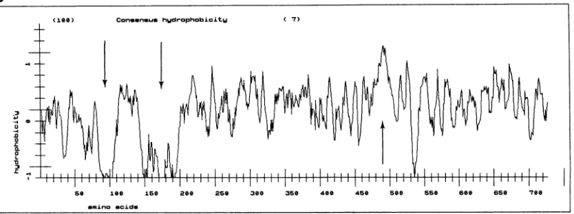
FIG. 1. (A) Nucleotide and deduced amino acid sequences of BLVRcpl cDNA. The putative starting ATG codon at position 25 is
indicatedwithboldfaceletters.TwoconsensusN-glycosylation sites(N-X-S/T;aa134to136and 252to254)areindicatedby asterisks.The
consensus site for phosphorylation by cAMP-dependent protein kinase (R-R/K-X-S/T; aa 171 to 174) is underlined. The putative transmembrane domain (aa 486 to 512) is underlined twice. The in-frame terminator TAG codon (nucleotide positions 2188 to 2190) is indicatedbyasterisks.The poly(A)+trackattheendofthecDNAbegins 13 nucleotidesafterthe AATAAAsequence,whichis underlined twice.(B)Hydropathy plotof thededucedBLVRcpl protein accordingtoEisenbergetal. (16).Numbers indicatethe aminoacid positions. Arrowsindicate thepresence ofhighlyhydrophobicorhydrophilic regions.
1052
1
41
121
81
241 121 361 161 481
201
601 241
721
281
841
321
961
361 1081 401 1201 441 1321
481 144 1
521 1561 561 1681 601 1801 641 1921
681
2041
721
2161
2281 CGTGTAAAGGAGAGACATATCATGCTGCTGTAATGATTTTGTGTCAAGATGATCCAATAAACTTGCGAAACAGGAAAAAAAAAAAAAAA
on November 9, 2019 by guest
http://jvi.asm.org/
[image:3.612.101.518.495.651.2]BOVINE LEUKEMIA VIRUS RECEPTOR 1053
hydropathy plot of the predicted BLVRcpl amino acid
sequence suggestsalong extracellular region encompassing two N-glycosylation sites (positions 134 to 136 and 252 to 254) and one consensus site for phosphorylation by the
cyclic AMP (cAMP)-dependent protein kinase
(R-R/K-X-S/T)
at positions 171 to 174, a transmembrane domain(positions 486 to 512), and probably an intracellular,
cyto-plasmic region (Fig. 1B). The ATG codon mentioned above isnotconsistent with the sequence AXX ATG G, defined as anefficient translation start site by Kozak(23). Searches (31
July 1992) in the GenBank and EMBL data bases for
homologywith known nucleotide and amino acid sequences
failed to identify any significant homology with previously reported sequences.
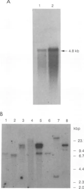
Northern analysis of bovine mRNAs. Todetermine the size of the mRNA transcript that encodes the BLV receptor in bovine cells, a Northern blot experiment was performed withpoly(A)+ RNAs isolated from MDBK cells and bovine
kidney tissue.The hybridization probe was BLVRcpl. After
the filters were washed under stringent conditions, the
presence of a single band of approximately 4.8 kb was
detectedin both MDBK cells and bovinekidney tissue RNA
(Fig. 2A). These data suggest that the BLVRcpl cDNA
represents only part of the genetic message for the BLV receptor.
Presence of sequences homologous to BLVRcpl in cells of differentspecies. The widehostrangeof BLV among species other than bovine indicates thatthe BLV receptoris
wide-spreadand is conserved among animalspecies. To testthe
conservation of the isolated sequencedescribedhere,
South-ernanalysiswasperformed withDNAobtained from
differ-ent species. Hybridization with a BLVRcpl probe under
stringentconditionspermitsthe detection ofalimited
num-ber of hybridizing fragments in EcoRI digests of murine,
bovine (DNAfrom MDBKcells), ovine, primate, caprine,
canine, feline, and porcine DNAs (Fig. 2B). These data
suggest the presence of a single-copy gene that is highly
conserved in DNA from various species. In caprine DNA isolated from a BLV-induced tumor (Fig. 2B, lane 5),
BLVRcpl sequences areprobably amplified.
Expression ofBLVRcpl cDNAin murine and human cells
conferssensitivitytoBLV infection. Toexamine theabilityof
BLVRcplproteinto render cellspermissivetoinfectionby
BLV,theBLVRcplcDNAwasplacedunder thethymidine
kinase promoter in thepXT-1 expressionvector,generating
pXT-1-BLVRcpl. pXT-1-BLVRcpl or
pXT-1
alone wastransfected into murine NIH 3T3 and humanHEp-2cells and tested for transient expression of BLVRcpl. Forty-eight
hours aftertransfection, cellsweretested fortheir
suscepti-bility to BLV infection by
using
a recombinant BLVex-pressing lacZ
(BLV,SVnls
LacZ) (28).The number ofBLV-sensitive cells was estimated by counting the number of
5-bromo-4-chloro-3-indolyl-f3-D-galactopyranoside
(X-Gal)-positive cells. In untransfected control
cells,
17(for
NIH3T3)and 85(for
HEp-2) positive
cellsweredetectedoutof 2 x 10⁵ cells. Cells transfected with the parental plasmidpXT-1
did notshow anyincreasein BLVinfectivity.
How-ever,asignificantincreaseofBLVsensitivitywasobserved aftertransfection withpXT-1-BLVRcpl,because about 150 and 300 BLV-sensitive cellswere detected in 2 x 10⁵NIH 3T3 andHEp-2 cells,respectively.
In orderto ensure that the increased
sensitivity
to BLV infection observed in thetransientexpression
experiment
is duetoexpression
ofBLVRcpl,
weperformed
thefollowing
experiment. pXT-1-BLVRcpl
orpXT-1
alone wastrans-fected into NIH 3T3
cells,
and cells were selected in theA
1 2
-*-4.8 kb
B
1 2 3 4 5 6 7 8
kbp
0. .I"'
*.
..
Ut.b
t. r, * *
- 23.1
- 9.4 - 6.7
- 4.4
-2.3
- & -2.0
FIG. 2. (A) ExpressionofBLVRcplmRNA in MDBK cells and bovine kidney tissue. Poly(A)+-selected RNAs isolated from MDBK cells (lane 1) and bovine kidney tissue (lane 2) were
fractionated on an agarose-formaldehydegel, blotted, and hybrid-ized with theBLVRcpl probe. (B) Southern blot hybridizationwith theBLVRcpl probeof cellularDNAsfrom different animalspecies. GenomicDNAs(10 ,ug) digestedwithEcoRIwereelectrophoresed
on a0.9% agarosegel,blottedandhybridized.DNAs(by lane)are
as follows: 1, murine; 2, bovine-MDBK; 3, ovine; 4, primate; 5, caprine (isolatedfrom aBLV-inducedtumor); 6, canine; 7, feline; and8, porcine.
presenceof G418. Twentycolonieswereisolated from NIH 3T3 cells transfected withpXT-1-BLVRcpl, and5 colonies were isolated from NIH 3T3 cells transfected with the control
pXT-1.
Thesensitivity
of these colonies to BLV infection was estimated as mentioned above. The data obtained for some of the NIH 3T3 transfectantclones,
aswell as
negative
andpositive
controls infected with the recombinantBLVexpressing
lacZ,
arepresented
in Table 1. Intheuntransfected control NIH 3T3cells,
20positive
cells among 2 x 10 were detected. The clones transfectedwith theparental
plasmid pXT-1
did not show any increase instaining efficiency,
asexamplified by
cloneNIH3T3XT-1/1.
Inone outof20clones
(clone
NIH3T33x6A6)
correspond-ing
to cells transfected withpXT-1-BLVRcpl,
asignificant
increase in BLV
sensitivity
was observed. This clone wasfound to be as sensitive to BLV infection as the BLV-VOL.67, 1993
on November 9, 2019 by guest
http://jvi.asm.org/
[image:4.612.357.520.75.440.2]TABLE 1. Susceptibility of various cell clones toBLVinfectivity
No.of1-galactosidase-positivecellsinfectedwith:
Cells DNAtransfected RV+MAbsc
RVb
F G H E
NIH3T3 None 20 10 10 10 20
NIH 3T3XT-1/1 pXT-1 23 8 10 8 20
NIH3T3 3x3A1 pXT-1-BLVRcpl 15 12 12 11 17
NIH 3T3 3x3A6 pXT-1-BLVRcpl 2,350 198 240 185 2,275
MDBK None 2,366 175 244 165 2,370
Cellswereinfectedwith1mlofsupernatantcontainingtheretroviralvector(RV)harvested fromFLK-BLV/BLV,SVnksLacZcells.Atapproximately24h postinfection,cells werefixed and stainedovernightwith5-bromo-4-chloro-3-indolyl-1-D-galactopyranoside.
b Numberofpositive cells after transfectionof 2x 10⁵cells with the recombinant BLVexpressinglacZ.
cPrior toinfection,therecombinantBLV(RV)wasincubated with MAbs of differentepitope specificities(F, G, H,andE)for1 honice.
permissive MDBK cell line. BLV infection of cells with
acquired sensitivity wasblocked
by
virus-neutralizing
anti-gpSl BLV MAbsofepitopeF, G, and H
specificity
butnot by the sequential (virusnonneutralizing)
MAb directed againstepitope E.Atvariance with thatspectacularincrease in BLVsensitivity, transfection ofpXT-1-BLVRcpldidnot enhancesusceptibilitytothe virus in clone NIH 3T3 3x3A1. Similarexperimentswere done with humanHEp-2
cells. After transfection of these cells withpXT-1-BLVRcpl,
1 G418-resistant cloneoutof18wasfoundsusceptible
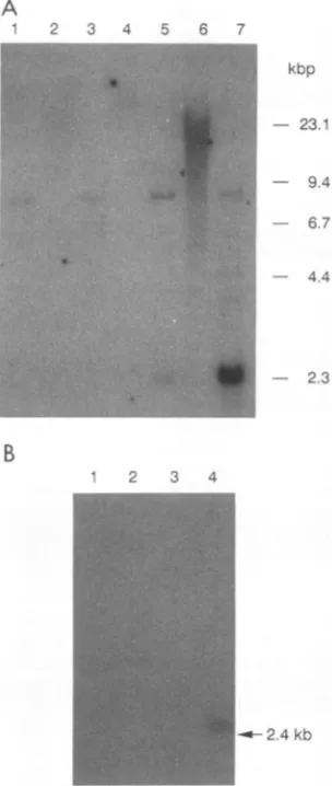
toBLV infection(data notshown).Southern blot hybridization of
undigested
andEcoRI-digested cellular DNAs from the NIH 3T3 3x6A6 cells
revealed an amplification of integrated 2.3-kbp BLVRcpl
DNAsequencesin addition to afragment
corresponding
tothe endogenous sequences
(Fig.
3A, lane7).
A similarobservation was made for the NIH 3T3 3x3A1 clone but withoutamplificationofthe additional copy(Fig. 3A,lane5).
Northern blot hybridization analysiswith the BLVRcpl
probe revealed the presence ofBLVRcpl
transcripts
in the NIH3T3 3x6A6 cloneonly(Fig. 3B,lane4). Specific
mRNAexpression was not found in NIH 3T3 cells that had
inte-gratedonlyoneadditional copy of theBLVRcpl sequences orin untransfectedcontrol cells(Fig. 3B,lanes1, 2, and3).
Similar levels ofBLVRcpl expressionwerefound in
BLV-permissiveMDBKcells and intheNIH3T33x6A6 cells that
hadacquiredBLV sensitivity (datanotshown).
Expression of the BLVRcpl gene product in NIH 3T3 3x6A6 cells was demonstrated by immunoprecipitation of cell extracts with a rabbit antipeptide RP1 serum reactive against the sequence aa25to36(PENALPSDEDDK) inthe BLVRcpl.Aprotein withamolecularmassofabout 70 kDa wasprecipitated from the transfected NIH3T3 cells (clone
3x6A6) (Fig.4, lane3)butnotfrom normal murine cells(lane
1).Thesameantipeptide serumimmunoprecipitated a
prod-uctwithanapparentmolecularmassof 90 kDa(Fig.4, lanes 5 and6) fromMDBKcells. Thisprotein probably represents the entire BLV receptor in itsnative structure.
Localization of the binding domain of BLV gpSl on the BLVRcpl product. The hydropathy profile of the BLVRcpl product revealed the existence of two hydrophilic regions (aa80 to 112 and140 to 200)flanking a hydrophobic region at theamino side of BLVRcpl (16) (Fig. 1B).
To determine whether these regions are involved in the
bindingof
gpSl,
severaldeletionmutantsofBLVRcpl (Fig.5) were prepared and expressed in pUC8-2. Their
lysates
weretestedby dot blot immune assays for their capacity to bind
gpSl.
The reactions were visualized by using the mixture ofantiglycoprotein MAbs and nitroblue tetrazoliumA
B
2 3 4 5 6 7
1 2 3 4
kbp -23.1
9.4
6.7
4.4
:. - 2.3
F-24kb
FIG. 3. (A) Southern analysis of NIH 3T3 transfectant clones with the BLVRcpl cDNA as probe. Cell types (by lane) are as follows: 1,untransfected NIH 3T3; 2 and 3, NIH 3T3XT-1/1;4 and 5,NIH3T33x3A1;and 6 and 7,NIH 3T3 3x6A6. Lanes 1, 3, 5, and 7contained DNAsdigested with EcoRI; lanes 2, 4, and 6 contained undigested DNAs. (B)Northernblot analysis of NIH 3T3
transfec-tantclones with theBLVRcplprobe. Each lane contains 30 ,ug of total RNA. Cell types(by lane)are asfollows: 1, untransfected NIH 3T3; 2,NIH 3T3XT-1/1;3, NIH 3T33x3A1;and 4, NIH 3T3 3x6A6.
on November 9, 2019 by guest
http://jvi.asm.org/
BOVINE LEUKEMIA VIRUS RECEPTOR 1055
1 2 3 4 5 6 7 8
FIG. 4. Detectionof the BLV receptor products by immunopre-cipitation. NIH 3T3 (lanes 1 and 2), transfected NIH 3T3 (clone 3x6A6) (lanes 3 and 4), and MDBK (lanes 5 to 8) cells were metabolically labeled with [35S]methionine and [35S]cysteine as described in Materials and Methods. Cell extractswere immunopre-cipitated withanormal rabbit serum (lanes 2, 4,and7,dilution of 1:10; lane 8, dilution of 1:15)or with the rabbit anti-peptide RP1 serum(lanes 1, 3, and5, dilution of1:10; lane6,dilution of 1:15) before SDS-PAGE andautoradiography (24-h exposureforlanes 1 to 4and5days for lanes5 to8).
chloride-5-bromo-4-chloro-3-indolylphosphate p-toluidine
salt reagents as substrate for an anti-mouse antibody conju-gatedwith alkaline phosphatase. When the first 993 bp from the 5' end of BLVRcpl DNA (fragments EcoRI to HindIII, the first 331 aa) was deleted, the binding activity was abolished (p,-gal-BLVRcp1/del6). When the 789-bp frag-ment defined by sites EcoRI to Sacl (construct
pp-gal-BLVRcpl/del2) was present, the binding activity was unal-tered. Positive binding activities were also observed for other constructscarrying different portionsofthe BLVRcpl
gene (constructs
pp-gal-BLVRcpl/del3,
-del4 and -delS).Reduced binding activity was observed with the
pp-gal-BLVRcpl/dellconstructthat encodes the first 148 aa of the
protein, thus suggesting that the protein segment encoded by the SmaI-SacI DNA fragment plays a major role in gpSl
binding.
DISCUSSION
Inthis article, we report the identification and character-ization of a cDNA, BLVRcpl, that encodes a polypeptide
behaving as a receptorfor BLV. The BLVRcpl product was firstidentifiedinthree recombinant lambda gtll phages that
displayed specific binding activity for BLV gpSl. Two cell
lines(murineNIH3T3 andhumanHEp-2), foundtobequite resistant toBLVinfection compared with other cells
(i.e.,
bovine cells) (28), were used in transfection experiments
with the BLVRcpl-expressing plasmid. Expression
of
BLVRcpl inthese cellsconferred sensitivityto
BLV
infec-tion.This acquired sensitivity to BLV infection occurred
de-spite
the fact that the encodedpolypeptide (expressed as aproteinwith amolecularmassof about70kDa intransfected
NIH 3T3 cells) (Fig. 4, lane 3) might not be complete.
Indeed, (i) theputativeATGcodonlacks aclassicalKozak
environment, (ii)noleaderpeptideis
identifiable
attheNH2end of
BLVRcpl
protein, (iii) the cloned 2.3-kbp cDNA is shorter thanthe native mRNA(4.8 kb), and(iv)
BLVRcplexpressed from transfected cDNA in NIH 3T3 cells is
Sm Sc H Bg
I
Construct
pB3-gal-BLVRcp1
Binding
Activity
a..K.4tax,
R- _
*
=ATG=f
=ATG=I _=
pB-gal-BLVRcpl /dell
pB3-gal-BLVRcpl/del2 pB-gal-BLVRcpl/deI3
pB-gal-BLVRcpl /del4
pl3-gal-BLVRcp1/deI5
pB-gal-BLVRcpl/del6
=ATG=[
=ATG=I
=ATG=
FIG. 5. SchematicrepresentationoftheBLVRcpldeletion constructs and theirbindingactivities toBLV gpSl.E,EcoRI;Sm,SmaI;Sc,
SacI; H, HindIII; Bg, BglII; Ps, PstI; Bm,BamHI.ThepUC8-2ATG initiationcodon isindicated; =represents plasmid sequences;stippled boxes indicateBLVRcplsequences. gpSlbindingactivitiesofcellularlysateswererevealedondotsbyimmunoreaction, asperformedfor theimmunoscreening(seeMaterials andMethods).
kbp o 5,
E
0.5
=ATG= ---- I
1.0 1.5 2.0
Ps
3.
E SSm Bm Ps H
VOL. 67,1993
on November 9, 2019 by guest
http://jvi.asm.org/
[image:6.612.70.293.71.330.2] [image:6.612.64.563.478.680.2]smaller than theprotein precipitated from MDBK cells with the same serum. Obviously, however, the missing protein
segmentdidnotprevent correctprocessing and transport of BLVRcpl proteins to the cell membrane. The lack of a leader peptide sequence has been observed for the gibbon
apeleukemia virus receptor. In this case also, transfection of the cloned receptor sequence and expression in NIH 3T3 cells conferred sensitivity to gibbon ape leukemia virus (29).
Thegibbonape leukemia virus receptor, like all of the other
members of thepermease-like protein family, wouldnot be
expectedto havea signal peptide.
Analysisof theBLVRcpl product reveals a protein witha
receptor structure. It contains an extracellular domain
in-cluding aBLVgp51 binding region, a single transmembrane
region, and a putative intracellular, cytoplasmic domain.
The presence ofthese domains suggests that the BLVRcpl
protein is an integral membrane protein, as expected for a
receptor. No homologous nucleotide or amino acid se-quences could be identified in the presently available data bases. This observation again illustrates the notion that retroviral receptors are largely unrelated molecules, despite the fact that a limited number of interference groups have
been identified (20, 32, 33). It seems very likely that
retro-viral receptors have a primary but variable physiological
function, such as those of permeases, transporters, and
aminopeptidases, and coincidently act as attachment points
for retroviralenvelope glycoproteinsthat are responsible for virus entry.
The widespread distribution of BLVRcpl among
mam-mals and its highly conserved sequence suggest that the molecule recognized by BLV plays a major role in the
physiology of many cell types. Elucidation of the reasons
and mechanismsbywhich the virusis confined to a few cell types in vivo must await a much more detailed analysis.
However, from the limited data so far available concerning
in vitro susceptibility of infection of MDBK cells, NIH 3T3
cells,andoneclone of NIH 3T3 cells transfected with pXT-1
BLVRcpl (Table 1), a positive correlation between the
receptorexpression levels (Fig. 2Aand 3B) and the
suscep-tibilityto BLVinfection seems to exist.
It is worth mentioning that the gpSl binding ability of
BLVRcpl is conserved when the protein is expressed in
procaryotes, indicating that glycosylation is not required for
bindingtoBLVgpSl.Therefore,BLVRcpl deletion mutants
expressed in procaryotes were used to localizethe receptor
binding region for BLV gpSl to the NH2 part of the
BLVRcpl recombinant protein. Computer-assisted
se-quence analysis according to Eisenberg et al. (16) and De Loof et al.
(15)
reveals the presence of three putative receptorbinding domains at positions 72 to 84, 103 to 107, and 153 to 177 (data not shown). This method allows thedetectionofregionswithhigh values for hydrophilicity and
hydrophobicmomentswhich are, in proteins such as
apoli-poproteins, characteristic of ligand-receptor-interacting
ar-eas. However, such highly hydrophilic regions are not present in all already known receptor binding domains:
indeed,someof them are, for example, made ofhydrophobic
pockets. So, other regions of the BLVRcpl, although not
detectedby the analysis of the hydrophobic profiles (15, 16),
might also constitute receptor binding domains.
Becausethe C-terminal part only ofgp5landthecomplete
gp3O
wereexpressed inbacteria and still bind BLVRcpl aspurified
gp5l
does, we conclude that the COOH region ofgp5l
(closetoepitopeB) (12) is the region of binding ofgp5lto the receptor (data not shown). Thus, the domains of
binding
of BLVgp5l
to BLVRcpl andhumanimmunodefi-ciency virusgp120 toCD4 are similarly located: HIV
gpl20
binds to the CD4 receptor at a site located on its C-terminal
end (30), andbinding toCD4 alsooccurs atthe NH2 end of
the receptor protein (9).
Considering the ability of the BLVRcpl to confer
sensi-tivity to BLVinfection and itsability tospecificallybindthe BLVgpSlglycoprotein,webelievethat theprotein encoded
bythe BLVRcpl cDNA acts as areceptor for BLV.
ACKNOWLEDGMENTS
We thank H. M. Temin for helpful suggestions, M. Sneyers for providing the MDBK expression library, I. Callebaut for help in protein structure analysis,and K.Willard-Gallo for a criticalreview of themanuscript.
This research wassupported bytheBelgian Servicesde Program-mation de laPolitique Scientifique (SPPS), by the Fonds Cancer-ologique de laC.G.E.R., andbyagrant from the Slovak Academy of Sciences. R. Kettmann is Research Director of the Belgian National Fund for Scientific Research (FNRS). B. Horion was a fellow fromT6levie-FNRS, and J. Ban was a fellow of the Belgian SPPS and FNRS-Tel6vie.
REFERENCES
1. Albritton, L. M., L. Tseng, D. Scadeen, and J. M.Cunningham. 1989. A putative murine ecotropic retrovirus receptor gene encodes a multiple membrane-spanning protein and confers susceptibilitytovirus infection. Cell57:659-666.
2. Altaner, C., V. Altanerova, J. Ban, 0. Niwa, and K. Yokoro. 1989. Humancells ofneural origin are permissive for bovine leukemiavirus. Neoplasma36:691-695.
3. Altaner, C., J. Ban, V. Zajac, H. Rossler, S. Rosenthal, R. Kettmann, andA.Burny.1985. Isolation and characterization of cell clonesproducing various amounts of bovine leukemia virus. Folia Biol. (Prague) 31:107-114.
4. Altaner, C., M. Merza, V. Altanerova, and B. Morein. Envelope glycoproteingpSl of bovine leukemia virus is differently glyco-sylated in cells of various species and organ origin. Vet. Immunol. Immunopathol.,in press.
5. Altanerova, V., J. Ban, and C. Altaner. 1989. Induction of immune deficiency syndrome in rabbits by bovine leukemia virus. AIDS3:755-758.
6. Altanerova, V., J. Ban, R. Kettmann, and C. Altaner. 1990. Induction of leukemiain chickenby bovine leukemia virus due toinsertional mutagenesis.Arch.Geschwulstforsch. 60:89-96. 7. Altanerova, V., D. Portetelle, R. Kettmann, and C. Altaner. 1989. Infection of rats withbovine leukaemia virus: establish-ment of avirus-producing rat cell line. J.Gen.Virol. 70:1929-1932.
8. Ban, J., S. Czene, C. Altaner, I. Callebaut, V. Krchnak, M. Merza, A. Burny, R. Kettmann, andD. Portetelle.1992. Map-ping of sequential epitopesrecognizedonthebovineleukemia virusexternal glycoproteins expressed inEscherichia coli by means of anti-peptideantibodies.J. Gen. Virol. 73:2457-2461. 9. Brodsky,H. M., M. Wharton, R. M. Myers, and D. R.Littmann.
1990. Analysis of the site in CD4 that bindsto theHIVenvelope glycoprotein. J. Immunol. 144:3078-3086.
10. Bruck, C., S. Mathot, D.Portetelle, C. Berte, J. D. Franssen, P. Herion, and A. Burny. 1982.Monoclonalantibodiesdefineeight independent antigenic regions on the bovine leukemia virus (BLV) envelopeglycoproteingpSl.Virology 122:342-352. 11. Burny, A., Y. Cleuter, R. Kettmann, M. Mammerickx, G.
Marbaix, D.Portetelle, A. Van Den Broeke, L. Willems, and R. Thomas. 1990. Bovine leukemia: facts and hypotheses derived from the study of an infectious cancer, p. 9-25. In R. Gallo and F. Wong-Staal (ed.), Retrovirus biology and human disease. Marcel Dekker, Inc., New York.
12. Callebaut, I., A. Burny, V. Krchnak, H. Grass-Masse,B. Wathe-let, and D. Portetelle. 1991. Use of syntheticpeptides to map sequential epitopesrecognized by monoclonal antibodies on the bovine leukemia virus externalglycoprotein. Virology
on November 9, 2019 by guest
http://jvi.asm.org/
BOVINE LEUKEMIA VIRUS RECEPTOR 1057 55.
13. Chirgwin, J. M., A. Przybyla, R. J. MacDonald, and W. J. Rutter. 1979. Isolation ofbiologically active ribonucleic acid from sourcesenriched in ribonuclease. Biochemistry 18:5294-5299.
14. Dalgleish, A. G., P. C.Beverly,P. R. Clapham, D. H. Crawford, D. H.Graves, and R. A. Weiss. 1984.TheCD4(T4)antigen is an essential component of the receptor for the AIDS retrovirus. Nature(London) 312:763-767.
15. De Loof, H., M. Rosseneu, R. Brasseur, and J. M. Ruysschaert. 1986.Use of hydrophobicityprofilestopredict receptor binding domainsonapolipoproteinEand low density lipoprotein apoli-poprotein B-E receptor. Proc. Natl. Acad. Sci. USA 83:2295-2299.
16. Eisenberg, D., E.Schwarz, M. Komaromy, and R. Wall. 1984. Analysis of membrane andsurface protein sequences with the hydrophobicmomentplot.J. Mol. Biol. 179:125-142.
17. Fourney, R. M., J. Miyakoshi, R. S. Day, and M. C. Paterson. 1988. Northern blotting: efficient RNA staining and transfer. Focus(LifeTechnologies) 10:5-7.
18. Greve, J. M., G. Davis,A. M.Neyer, C. P. Forte, S. C. Yost, C. W.Marlor,M.E.Kammarck, and A.McClelland. 1989. The major human rhinovirusreceptoris ICAM-1. Cell56:839-847. 19. Johann,S. V., J. J. Gibbons, and B. O'Hara. 1992. GLVR1, a
receptor for gibbon ape leukemia virus, is homologous to a phosphate permeaseofNeurospora crassa and is expressed at high levels inthe brain andthymus.J. Virol.66:1635-1640. 20. Kewairamani,V.N., A. T.Panganiban, and M. Emerman.1992.
Spleennecrosisvirus,anavianimmunosuppressive retrovirus, sharesareceptorwiththe type D simian retroviruses. J. Virol. 66:3026-3031.
21. Kim, J. W., E. I.Closs, L. M. Albritton, and J. M. Cunningham. 1991.Transport of cationicamino acidsbythe mouseecotropic retrovirusreceptor. Nature (London)352:725-728.
22. Klatzmann, D., E. Champagne, S. Chamaret, J. Gruest, D. Guetard,T.Hercend, J. C.Gluckman,and L.Montagnier. 1984. T-lymphocyte T4molecule behaves asthe receptorfor human retrovirusLAV. Nature(London) 312:767-768.
23. Kozak, M. 1986.Point mutations defineasequenceflanking the AUG initiator codon that modulates translation byeucaryotic ribosomes. Cell 44:283-292.
24. Krchnak, V., andJ.Vagner.1990. Color-monitoredsolid-phase multiple peptide synthesis underlow-pressure continuous flow conditions.Pept. Res.3:182-193.
25. Maddon, P. J., A. G. Dalgleish, J. S.McDougal,P. K.Clapham,
R. A. Weiss, and R.Axel.1986.TheT4 gene encodes the AIDS virus receptor and is expressed inthe immune system and the brain. Cell 47:333-348.
26. Mammerickx, M., D. Portetelle, K. De Clercq, and A. Burny. 1987. Experimentaltransmission ofenzootic bovineleukosisto cattle, sheep andgoats:infectiousdosesof bloodandincubation period of the disease. Leuk. Res.11:353-358.
27. Mendelsohn, C. L., E. Wimmer, and V. R. Racaniello. 1989. Cellular receptorfor poliovirus: molecularcloning, nucleotide sequence,andexpressionof a new member of the immunoglob-ulinsuperfamily. Cell56:855-865.
28. Milan, D., and J. F. Nicolas. 1991. Activator-dependent and activator-independent defective recombinant retrovirusesfrom bovine leukemia virus.J.Virol. 65:1938-1945.
29. O'Hara, B., S. V. Johann, H. P. Klinger, D. G. Blair, H. Rubinson, K. J. Dunn, P.Sass, S. M. Vitek, and T. Robins. 1990. Characterization of a human gene conferring sensitivity to
infection by gibbon ape leukemia virus. Cell Growth Differ. 1:119-127.
30. Olshevsky,U., E.Helseth, C. Furman, J. Li, W. Haseltine, and J. Sodroski. 1990. Identification of individual human immuno-deficiencyvirus type 1 gpl20 amino acids important forCD4 receptorbinding.J. Virol.64:5701-5707.
31. Portetelle, D., K. Limbach, A. Burny, M. Mammerickx, P. Desmettre, M. Riviere, J.Zavada, and E. Paoletti. 1991. Recom-binant vaccinia virus expression ofthe bovine leukemia virus envelope gene and protection of immunized sheep against infection. Vaccine9:194-200.
32. Sommerfelt, M. A., and R. A. Weiss. 1990. Receptor
interfer-encegroupof 20 retrovirusesplatingonhuman cells.Virology 176:58-69.
33. Takeuchi, Y., R. G.Vile, G. Simpson, B. O'Hara, M. K. L. Collins,and R. A.Weiss. 1992. Feline leukemia virussubgroup B usesthesamecell surfacereceptor asgibbonape leukemia virus.J.Virol. 66:1219-1222.
34. Thomas, P. S.1980.Hybridization of denaturedRNA and small DNAfragments transferredtonitrocellulose.Proc.Natl. Acad. Sci. USA77:5201-5204.
35. Van DerMaaten, M., andJ.Miller.1976. Replicationof bovine leukemia virus in monolayer cell cultures. Bibl. Haematol. 43:360-373.
36. Wang, H.,M. P.Kavanaugh,R. A.North,and D. Kabat. 1991. Cell-surface receptor for ecotropic murine virus is a basic amino-acid transporter. Nature(London)352:729-731. VOL. 67, 1993


![FIG. 4.cipitation.metabolically3x6A6)beforedescribedcipitatedserumto1:10; 4 Detection of the BLV receptor products by immunopre- NIH 3T3 (lanes 1 and 2), transfected NIH 3T3 (clone (lanes 3 and 4), and MDBK (lanes 5 to 8) cells were labeled with [35S]methi](https://thumb-us.123doks.com/thumbv2/123dok_us/1303450.83514/6.612.64.563.478.680/cipitation-metabolically-beforedescribedcipitatedserumto-detection-receptor-products-immunopre-transfected.webp)