Copyright C) 1984,American Societyfor Microbiology
Genetic Analysis of Adeno-Associated Virus: Properties
of
Deletion
Mutants
Constructed
In
Vitro and Evidence for
an
Adeno-Associated
Virus
Replication Function
JON-DURI TRATSCHIN, IRVING L. MILLER, AND BARRIE J. CARTER*
Laboratory ofCellBiology andGenetics, National Institute of Arthritis, Diabetes, and Digestive and Kidney Diseases, Bethesda, Maryland 20205
Received 22 March1984/Accepted29 May1984
Transfection ofapBR322-based, recombinant plasmid, pAV2, containingthe entireadeno-associated virus (AAV) type 2 genome into human 293 cells in the presence of helper adenovirus resulted in rescue and replication ofAAVtoyield infectiousparticles. We constructed mutantsofpAV2 containingdeletions within the AAV sequence. We describe here the phenotypes of these AAV deletion mutants. The results can be summarized as follows. Mutants (cap-) withdeletions between map positions 53 and 85 did notsynthesize capsid antigen orprogeny single-stranded DNA but accumulated normal levels ofduplex replicating form DNA. Mutants
(rep-)
withdeletions between map positions 17 and 36 failed torescue or replicateany AAV DNA. Therep-
mutantscould becomplemented forreplicatingform DNAsynthesis by acap-mutant. This clearly demonstrates an AAV-coded replication function which is different from the capsid antigen. Other mutants(inf )withdeletionsin theregionbetweenmappositions40 and 52synthesizedabundant amounts of replicating formDNAandcapsidantigenbutgavealowyield of infectiousparticles.This suggeststhatthere may be an additional region of AAV, perhaps within the intron, which is required for efficient particle assembly. This work shows that AAV is genetically complex and expresses at least three clearly different functions.The DNA genome of the defective human parvovirus adeno-associated virus (AAV) issingle stranded and linear,
but strands of either complementarity are packaged into
separatevirions (31, 40). After infection ofpermissive cells in the presenceofahelper adenovirus (1, 18) orherpesvirus
(3), the AAVsingle strand isconverted to aduplex replicat-ing form (RF) apparently by a self-priming mechanism involving the terminal palindrome sequence (2, 16, 46).
Multiple rounds of RF replication then occur, and single
strandsaregenerated fromRF DNAby stranddisplacement replication (single-strand replication). Both AAV RF and progenysingle-stranded (SS) DNA replication require func-tions provided by AAV as well as by the helper virus,
although the precise nature ofthese functions is not under-stood(reviewed in references 5 and 6).
The AAV genomecontainsoverlappingtranscription units which yield three families of mRNAs beginning from three different promoters but having a common 3' terminus (13, 27, 29, 30). The DNA sequence contains two major open
reading framesin theleft half (orf-1) and right half (orf-2) of the genome, respectively (45). Thus far, the only clearly
identified AAVgeneproducts are three capsid polypeptides (4, 40) which contain overlapping sequences (28, 34) and
apparently are coded by the right half of the genome (22). Fromastudyof theproperties of AAV defective-interfering
particles,wepreviously suggested (5,25) that an AAV gene product (rep) may be required for AAV RF replication. We
subsequently showed that the AAV capsid protein was apparently not required for AAV RF replication (37, 38).
Toanalyze more clearly the possible functions of the open reading frames in AAV DNA, we have used mutant genomes constructed in vitro. The entire AAV genome has been
*Correspondingauthor.
611
cloned into bacterial
plasmids
(26, 42, 43a). When suchrecombinant AAV plasmid DNA molecules are transfected
into human 293 cells in the presence of helper adenovirus particles,the AAV genome is rescued from therecombinant plasmid and replicated to produce an efficient
yield
ofinfectiousprogeny particles(26, 42). We have taken advan-tageof thisandperformedin vitromutagenesis onthe AAV
plasmid recombinant to produce a series ofAAV deletion mutants. The phenotype of each of these mutants was examined by transfection into 293 cells in the presence of
helper adenovirus. These studies provide evidence for an AAV rep function coded by orf-1 and for AAV
capsid
protein coded by orf-2. The rep and capsid proteins are separate functions and represent differentcomplementation
groups. An additional AAV function may be required for
stable particleassembly.
MATERIALSAND METHODS
Viruses and cells. AAV type 2 (AAV2) and adenovirus 2 were grown in KB spinner cells as described before (7). Production of AAV capsid antigen was assayed by fixing cells at 24or30 hafter infection or 24h after transfection, respectively, and staining for AAV capsid protein, using an
indirect immunofluorescence assay as described previously
(7). Production of infectious AAV was assayed by titrating cell
lysates
oncellsgrownin monolayer and using the sameAAV
immunofluorescence procedure.Human 293-31 cells, an established line of adenovirus-transformed human embryonic kidney cells (12), were grown at37°Cinmonolayer culture in 35-mm plastic dishes in Eagle minimal essential medium supplemented with antibiotics and 10% fetal calf serum.
Growth and preparation of plasmids and plasmid DNA. Plasmids or DNA ligation mixtures were used to transform Escherichia coli HB101, using the RbCl procedure (23), and
on November 10, 2019 by guest
http://jvi.asm.org/
ampicillin-resistant colonies were selected by growth on
Luriabrothagarcontaining ampicillin (50 pLg/ml). Individual
colonies were examined for the presence of the appropriate
plasmid by growing small-scale cultures in Luria broth medium and lysing according tothe rapid boiling procedure (20). Plasmids in the minilysates were analyzed by
restric-tion endonuclease cleavage and electrophoresis in agarose
gels. Restriction endonucleases were purchased from New
England Biolabs (Beverley, Mass.), Bethesda Research Labs (Gaithersburg, Md.), or Boehringer Mannheim Bio-chemicals (Indianapolis, Ind.) and used according to the supplier's specifications. DNA fragmentswerepurified from agarosegels accordingtothe procedures of Langridgeetal. (24)orWeislander(47). Large-scale preparationsofplasmids
were obtained by growing the appropriate E. coli strain in
minimal M9 medium supplemented with Casamino Acids (0.2%), thiamine (1 ,ug/ml), and ampicillin (20 ,ug/ml) and amplifying with chloramphenicol (100 ptg/ml). Plasmidswere
thenprepared by lysozyme-TritonX-100lysisand
polyethyl-ene glycol precipitation followed by cesium chloride-ethi-dium bromide equilibrium gradient centrifugation (21, 35). Allmanipulations with recombinantDNAwereperformedin accordance with the National Institutes of Health Guide-lines.
Construction of individual plasmids. Figure 1 shows the structure ofthe AAV2 genome, the location of the relevant restrictionsites,and thestructuresof the individualplasmids usedin this study. pAV2 contains the entire AAV2genome inserted into a pBR322-derived plasmid, pA11P.Xba (14).
Therefore, pA11P.Xba has the same structure as pAV2
except that the AAV2 insert is deleted. pAVdlHc23 was
derived from pAV2 by deletion of the HincII fragment between map positions 51 and 86 (i.e., between sites Hc2 and Hc3) by partial HincII cleavage followed by blunt-end ligation.
Some mutants were derived from pAV2 by restriction cleavage and religationat compatible sticky ends, usingT4 polynucleotide ligase (Boehringer Mannheim). Thus, pAVdlX12 was derived by cleaving pAV2 with XhoI and religating to yielda mutant which is deleted for the XhoI-C fragment (186 base pairs [bp]) but contains one XhoI site. Similarly, pAVdlK12 was derived by cleaving pAV2 with KpnI.
Several mutants were derived by cleavage of pAV2 at
nonsimilarrestriction sites followedbyblunt-endligation.In
each case pAV2 DNA was cleaved with the required
en-zymesand then incubatedwith the E. coliDNApolymerase I Klenow fragment (Boehringer Mannheim) to remove 3' extensionsortofillinrecessed3' termini. Reactions with the Klenow fragmentwereperformed inafinal volumeof 20
,ul
containing 50 mM Tris-hydrochloride (pH 7.2)-10 mM
MgSO4-0.1 mM dithiothreitol-50 ,ugof bovine serum
albu-min perml-500 ,uMeach dATP, dCTP, dTTP, and dGTP-3
U of enzyme at 37°C for 30 min. The DNA was then
incubated withT4 polynucleotide ligase (Collaborative
Re-search, Waltham, Mass.)undertheconditionsfor blunt-end
ligation as specified by the supplier and transfected into
E. coli HB101. By this procedure, mutants
pAVdlKlX2
and pAVdlXlK2 were derived by full cleavage with XhoI and partial cleavage with KpnI. Mutants pAVdlHiX2 andpAVdlHiK2 were derived by complete cleavage with
HindIII and either XhoI or KpnI, respectively. pAVdlSBc was derived by complete cleavage ofpAV2 with SstI and
BcII, and pAVdlBcBs was derived by complete cleavage with BclI and BstEII. For these latter two constructions, pAV2waspreparedfrom the methylation-deficient strain E.
coli
GM119(11)
to avoidmethylation
ofthe AAV2BclI
site.Mutant
pAVR1.11
wasderivedby
site-specific
mutagene-sis at the AAV2 EcoRI site at mapposition
38.1,
using
endonuclease Bal31aswill be describedelsewhere. Nucleo-tide sequence
analysis
(32)
showed thatpAVdlR1.11
is deleted betweenAAVnucleotides1,635
and1,861 (M. West,
unpublished
data).
Transfection of cells with recombinant
plasmid
DNA. All transfectionexperiments
wereperformed
on293cellsgrown in 35-mmplastic
dishes(106
cells perdish),
using
either a DEAE-dextran or aCaPO4
procedure.
The DEAE-dextran
procedure
was used as described before(26,
33,
36)
whenassaying
the transfectedrecombi-nant AAVDNAfor its
ability
tobereplicated.
Mixtures of recombinant DNA(in
amounts per dish asspecified
in individualexperiments)
in the presenceorabsenceofhelper
adenovirusparticles
(5
to 10 PFU percell)
were added tosubconfluent
cells in the presence of minimal essential mediumcontaining
250 ,ugof DEAE-dextranperml(average
molecular
weight,
500,000)
and 50 mMTris-hydrochloride,
pH
7.5,
for30min.This inoculumwasremoved and the cells wereincubatedat37°C
inminimal essential mediumcontain-ing
10%
fetal calfserum. Forin vivolabeling
of viralDNA,
cells were transferred tolow-phosphate
medium(1%
of normalphosphate
concentration)
at 16 h aftertransfection
and
supplemented
withH3PO4 (10
to 25,Ci/ml).
CaPO4
transfection wasusedwhentesting
forexpression
of
capsid antigen
orforproduction
of virusparticles
andwasperformed
according
totheprocedure
ofWigler
etal.(48).
Inthis
procedure,
adenovirusparticles
(5
to 10 PFU percell)
were addedto the cells 1 h beforetransfection withDNA.
Analysis
of intracellular viral andplasmid
DNA.ViralDNAwas
selectively
extracted from cells at 44 to 48 h aftertransfection,
using
aHirtprocedure
(17)
modifiedtoprevent
reannealing
of AAV ssDNA and to removecontaminating
RNA as describedbefore
(9).
DNAwaselectrophoresed
in horizontal1.4%agarosegels
(11
by
14cm)
in10mM Tris-40mMsodiumacetate-1 mM
Na2EDTA,
pH
8.0,
at100 mApergel.
Thegels
werethen dried andautoradiographed directly,
using
anintensifying
screen(Cronex
70,
Dupont),
orwere blotted onto nitrocellulose paper(44)
andhybridized
withAAV
32P-DNA
labeledby
in vitronick translation(39).
Afterhybridization
in6x SSC(SSC
= 0.15 MNaClplus
0.015 Msodium
citrate)
at70°C
for16to24hthenitrocellulosepaperwaswashed in0.1%x
SSC-0.05%
sodiumdodecyl
sulfateat 52 or70°C
andautoradiographed.
RESULTS
Structure of the AAV2 genome.Thestructureofthe AAV2 genome is shown in
Fig.
1,
which identifies the relevant restrictionsites,
the AAVtranscription
units,
and theopenreading
frames.Thetranscription
ofAAVisreviewedexten-sively
elsewhere(8).
AAVRNAsynthesis
begins
from threeseparate promoters,
P5,
Plg,
andP40,
atmappositions
5,
19,
and
40,
respectively,
andproceeds rightward
to asingle
polyadenylation
siteatmapposition
96(8, 13,
27, 29, 30,
45).
Therearetwo
major
openreading
frames(orf)
asdeterminedfrom the nucleotide sequence
(45).
The left-handreading
frame
(orf-1)
apparently
can be entered fromtranscripts
originating
from eitherp5
orP9l
Furthermore,
splicing
ofAAV RNA to remove the
single
intron alters thecarboxyl
terminus of orf-1. This
suggests
that the fourlargest
AAV RNAs may enable orf-1 to encode at least fourproteins
differing
in theiramino andcarboxyl
termini. orf-2 is in theright
half of the genome andmaybeaccessible fromaspliced
P40
transcript.
There are two smallreading
frames locatedon November 10, 2019 by guest
http://jvi.asm.org/
RiHi
K Hc2 13\ |/l//X2
SBc Hcl
Hii
I I ~I ~I I
10 20 30 40 150
II,, i
I
I
''A
I
I-I IlaI1
I
P40
11
60 70 80 90 100
4.2kb 3.9 kb 3.6kb 3.3kb 2.6kb 2.3 kb
pAV2 pAVdI X12 pAVdI K1X2 pAVdl HiX2 pAVdl Hc23 pAVdIX1K2 pAVdl K12 pAVdI HiK2 pAVdl R1.11 pAVdI BcBs pAVdI SBc
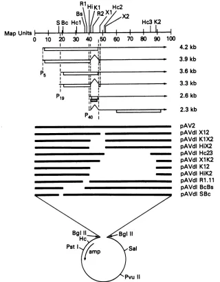
FIG. 1. Structure of the AAV2 DNA andmutantAAVgenomes.Theupperportionshows theAAV2 DNAon ascale of 100mapunits.
Re-striction endonuclease sitesaredesignatedasfollows:S, Sst; Bc, BclI; Hc, HincII; R, EcoRI; Hi,HindIII; K,KpnI;X,XhoI.ForHc, R, K, andX, the sites in AAVaredesignated numerically from the leftas indicatedbythe numberfollowingthe abbreviatedname,e.g., Hc2.
Horizontalarrowsshow thestructuresof the six mRNAshavingthe sizesasindicated(in kilobases)at theright.The caret indicates theAAV
intron. The arrowhead indicates thepolyadenylationsiteat96mapunits.P5,P19,andp40indicatethe threepromotersites.Basedonsequence analysis (45),thepotentialopenreadingframes(orf)accessible from each RNAareindicatedbythe boxes below individual RNAsasfollows:
orf-1,openbox; orf-2, stippled box; orf-3, diagonallyshaded box. The lowerportionshows the structure of the AAV mutants. Theparental AAV plasmid recombinant pAV2 contains the entire AAV2 genome (solid horizontal line) inserted into a pBR322-derived plasmid,
pA11P.Xba (open circle),viaBglIIlinkers. The otherplasmidswerederivedfrompAV2 by deleting regionsof AAVsequenceasindicatedby thegapsinthe horizontal line. Thefigureis drawn approximatelytoscale and several referencerestriction sites areshown in theplasmid
sequenceaswellasthelocation of theampicillinresistancegene(amp).The constructionof individualplasmidsis described in thetext.
within the intron which may be accessible for translation
from the 2.6-kilobase (kb) unspliced P40 transcript. One of
these small reading frames is the same as the carboxyl
terminus of orf-1 expected from translation of the spliced 4.2- and 3.6-kb mRNAs: the other, orf-3, is differentfrom butoverlaps orf-1 and is contained entirely within the intron. Thereisanadditionalopenreading frame, different from
orf-2, between map positions 85 and 97 (45), but this is not shown inFig. 1 because this reading frame isnotapparently accessiblefrom anycurrently known AAV mRNA.
Structure ofindividual mutant AAV genomes. The
struc-tures ofindividual AAV plasmid recombinants are detailed
in Fig. 1. For these constructions we describe the entire
recombinantplasmid, usingtheprefix pAV,e.g.,pAVcdl23. When referring to the AAV genome contained within, or
rescued from, theplasmid, we will notusetheprefix, e.g., dlHc23. The AAV mutants fall into several overlapping classes. Restriction site Xl is 6bpdownstreamof theintron, andmutantdlX12deletes 186bp immediatelydownstream of thiswhich includes the last six codons oftheorf-1 expected from the spliced transcripts. Two mutants, dlHiX2 and dlK1X2, delete the intron as well as the region deleted in dlX12. Mutants dlHc23 and dlX1K2 also overlap mutant
dlX12but extend the deletionto theright to removenearly
all of orf-2. Another pair ofmutants, dlK12 and dlHiK2, have deleted both the intron and mostof orf-2. All ofthese Map Units #
0
r19
In
on November 10, 2019 by guest
http://jvi.asm.org/
[image:3.612.145.465.73.482.2]mutants contain deletions which are to the right of the HindIll site at map position 40. A second class of mutants contained deletions which mapped only to the left ofthis site. These mutants, dlSBc, dlBcBs, and dlR1.11, have deletions of internal portions of orf-1. The mutation in dlR1.11alsodeletes the promoterand the first23nucleotides at the 5' terminus of
P40
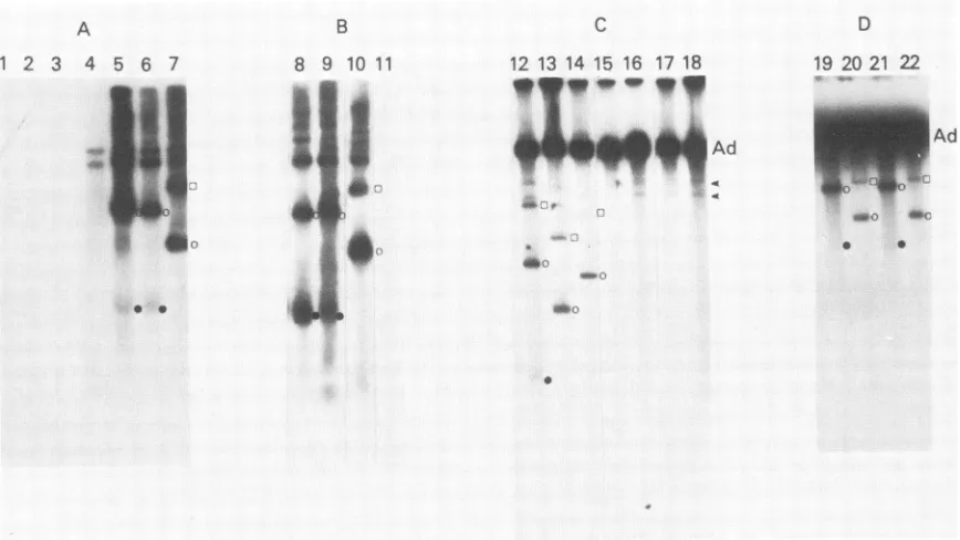
transcripts.Replication of AAV mutants and accumulationof duplexRF DNA. The replication of the AAV mutants was tested by transfection of 293 cells with recombinant plasmids in the presence ofadenovirus. Viral DNA was selectively extract-ed and analyzextract-ed by agarose gel electrophoresis. Several
examples ofexperiments with individual mutants are sum-marized in Fig. 2. In some experiments, AAV DNA was detected byblottingthegels andhybridizing with an in vitro
labeled AAV 32P-DNA probe (Fig. 2A and B). In other
experiments when in vivo
32P
labeling was used, the DNA was detected by direct autoradiography of the gels (Fig. 2C and D).Thetransfections with the wild-type AAV-plasmid pAV2
(Fig. 2, tracks 5, 9, 12) clearly show the three AAV DNA species, including the monomeric and dimeric duplex RF DNAsand the progeny ssDNA. These AAV DNA species, which arise by rescueofthe AAVgenomefrom theplasmid
followedbyreplication,have beencharacterized before (26). In the experiments using in vivo
32P
labeling, adenovirusDNA was also labeled, whereas the residual unreplicated
A
1 2 3 4 5 6 7
.- .
* 0
B 8 9 10 11
_ _
4k.
A& =|
inputrecombinant AAV plasmid DNA was not (Fig. 2C and D). When the experiment was performed by blotting and
hybridizing with an AAV
32P-labeled
DNA probe (Fig. 2A and B), the adenovirus DNA was not observed but the unreplicated input plasmid DNA was detected (Fig. 2A, track 4). The input plasmid DNA is generally observed as two species comprising the linear and open circular forms. However, the adenovirus DNA and both input plasmid DNA species migrated more slowly than the major AAV DNA species.Each of the three mutants containing deletions to the left of the HindlIl site (dlSBc, dlBcBs, dlR1.11) failed to accu-mulate either RF DNA or AAV ssDNA (Fig. 2C, tracks 15 to 17). These three DNA-negative mutants are designated rep-. Allof the other AAV mutants having deletions only to the right of the
Hindlll
site were rescued and replicated intransfectedcells as judged by theaccumulation of duplex RF DNA. Further, each of these mutantsaccumulated RF DNA about as efficiently as wild-type AAV. Based on the
produc-tion ofRF DNA wedescribe these mutants asrep'. That the mutantRF genomes observed in the transfected cells indeed
correspond tothose expected to be rescued from the
trans-fecting
plasmnid
was verified by their size and restriction cleavage (not shown).Accumulation of progeny ssDNA. Although the rep' AAV mutantsefficientlyaccumulated RF DNA, they varied great-ly in their ability to accumulate progeny ssDNA (Fig. 2). For
C
12 13 14 15 16 17 18
*y""A~mpyd
-9
D 19 20 21 22
W"oUW-LcAW:O ox().,(;....
a 0
FIG. 2. Effectof deletionsonreplicationof AAVgenomesintransfected 293 cells. Cultures of 293 cellsweretransfected with adenovirus particlesandsupercoiledAAVplasmidDNA(10,ug).Viral DNAwasisolated andelectrophoresed inagarose gels. (A), (B), (C), and (D) show separateexperiments.In(A)and(B)the DNAwasdetected afterelectrophoresisby blotting andhybridizing with anick-translatedAAV32p_ labeledDNAprobe.In(C)and(D)theDNAwaslabeled in vivo and the agarosegelwasdried down andautoradiographed directly. Tracks 1 and 11showpAV2DNAcleavedwithBglIl to serve as amarker. Tr4ck 2, DNA frommock-transfected cells inwhich bothadenovirus and DNA were omitted. Track 3, Adenovirus-infected cells. Track 4, Transfected with pAV2 DNA in the absence of adenovirus. All other culturesweretransfectedwithadenovirusandsupercoiled plasmidDNAasfollows: track 5,pAV2; track 6, pAVdlX12; track 7, pAVdlHc23; track8,pAVdIX12;track9,pAV2;track10,pAVdIXIK2;track12,pAV2;track 13,pAVdlHc23;track 14,pAVdlKlX2;track 15,pAVdlSBc; track16,pAVdlBcBs;track17,pAVdlR1.11;track18,pA11P.Xba;track 19,pAVdIHiX2;track 20,pAVdlHiK2;track 21,pAVdlKlX2;track 22,pAVdIK12.DNAspeciesforeachmutant aredenotedbythesymbolsattherightof each trackasfollows:(O)duplex dimer RF DNA;(0) duplexmonomerRFDNA; (-)ssDNA. Arrows(C)indicateone ortwocell DNAspeciesusually seen in in vivo labelingexperiments (26). In (C)and (D)the heavy, slowly migrating band is adenovirus DNA. In (A) and (B) the slowly migrating doublet (e.g., track 4) shows input plasmidDNA.
on November 10, 2019 by guest
http://jvi.asm.org/
[image:4.612.98.532.360.604.2]wild-type
AAV DNA(i.e.,
pAV2), the ratio of ssDNA tomonomeric RF DNAshowed somevariation between
differ-ent
experiments
(Fig.
2, tracks 5, 9, 12) but was generally similar in asingle experiment (Fig.
3, tracks 2, 5, 8, 10). Mutant dlX12 accumulated ssDNA about as efficiently aswild-type
AAV(Fig.
2. tracks 6, 8). In contrast, mutantdlHc23did notaccumulate detectable ssDNA (Fig. 2, tracks 7. 13).
Similarly,
the three mutants containing even larger deletions, dlXlK2(Fig.
2. track 10), dlHiK2 (Fig. 2, track20).
and dlK12(Fig.
2. track 22), also did not accumulate detectable ssDNA. It isimportant
to note that it may bedifficulttodetect very low levels of AAV ssDNA because of
a variable
background
ofheterogeneous
material whichmigrated
faster than theduplex
RF DNA and probablyarises frompartially
replicated
molecules. Thisprecludesdetection of low levels of ssDNAsimply by
increased autoradiograph-ic exposure. Nevertheless, with the four mutants dlHc23,dlHiK2, dlK12,
anddlX1K2,
we have reproducibly failed to observe accumulation of ssDNA. We designate the pheno-type of these mutants as cap-.Two mutants, dlK1X2 and
dlHiX2,
showed a very low levelofssDNA in someexperiments
(e.g., Fig. 2, tracks 19,21),
whereas in otherexperiments
(Fig. 2, track 14) no ssDNA was observed. As shown below, these two mutants have aphenotype
designated iif-
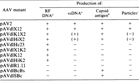
which is different from that of the cap- mutants.Production ofcapsid antigen and infectious virus. Table 1 summarizes for each of the AAV mutants productionof RF and ssDNAas well as accumulation of AAV capsid antigen and infectious
particles.
Production of capsid antigen was measuredby fixing
cells 24 to 30 h after transfection in the presence of adenovirus and staining for nuclearfluores-cence,
using
the indirect immunofluorescence assay withantibody
directedagainst
theAAV capsid protein. In theseexperiments
5 to10% of the cellstransfectedwithwild-typepAV2
showedpositive
staining.
This indicates that the transfectionwasreasonably
efficient(20) since cells infected with AAVparticles
normally do not show more than 50 to 60% positivestaining
forcapsid protein
(7). Mutant dlX12produced
capsid antigen
as efficiently as pAV2. The fourcap- mutants,
dlHc23,
dlX1K2,
dlK12, and dlHiK2, havingdeletions in orf-2 failedto
synthesize
detectable AAVcapsidantigen.
In contrast, for the twoijuf-
mutants, dlHiX2 anddlKlX2,
the number ofcellspositive
for AAV antigen was 25 to 100% of that withpAV2.
The two rep- mutants, dlBcBS and dlSBc, also showed
positive
AAVantigen
staining
at alevel of about 10to 30%that of
wild-type
AAV. The reduced level probably reflects decreased genome copy number in the absence ofreplica-tion.The
lrep-
mutantdIR1.11
retains all of orf-2 but failed tosynthesize
capsid antigen
presumably because the deletionremoves both
P4(
and the 5' terminus of the 2.3- and 2.6-kbmRNAs. These results are consistent with previous evi-dence that theAAV
capsid protein
iscodedbytheright half of the genome(22).
None of theplasmids,
includingthewild-type
AAV,
synthesized
detectable AAV antigen in the absence of adenovirus. This is consistent with previous evidencethat anadenovirusfunctionis requiredfortransla-tion of AAV
capsid
antigen (6, 22) since transcription ofAAVDNA occursin293cellseven in theabsence of helper
(J. Tratschin,
M.West,
T. Sandbank,andB. Carter, submit-ted forpublication).
Production of infectious particles in transfected cells is also summarized
qualitatively
in Table 1. The only mutant thatproduced
infectious virus at significant levels wasdlX12,
which wasabout50% asefficientaswild-type AAV.For most of the mutants, the failure to
produce
infectious virus can beexplained by either failuretoreplicate(rep-)
orfailure to produce
capsid
antigen
(cap-).
The two inf mutants, dlHiX2 andd1K1X2, whichreadily synthesized
RFDNA andcapsid antigen, produced infectious particles
only
very inefficiently ifatall, as determined by the immunofluo-rescenceassay (Table 1). This correlates with poor
accumu-lation of ssDNA by these inf mutants. The failure of mutants to make infectious virus is also shown
by
another assay described below.Complementation ofAAVmutants. Other studies
(P.
Sena-pathy, J.
Tratschin,
and B.Carter,
unpublished data)
showed that naturally occurring mutants of AAV
having
much largerdeletions removingmost oforf-1andalarge part of orf-2 exhibit a rep- phenotype. These mutants could be
complementedfor RF DNA
replication by
awild-type
AAV or arep+
mutant. Recombination was not observedby
analyzing viral DNAfromlysates of the transfected cells and did not interfere with the
complementation
assay for RFDNA synthesis. Recombination did occur at a very much lower level than complementation butwas detected
only
by
successive passaging of cell lysates from the transfected cells (43a).
We examined the
*ep-
mutantsdescribed hereforcomple-mentation for RF DNA replication by cap- mutant dlHc23. The three rep- mutants dlBcBs, dlSBc, and
dlR1.11
all failed to replicate when transfected only with adenovirus (Fig. 3, tracks 1,4, 7). Inamixed transfection with pAV2 ordlHc23, dlBcBs RF DNA genomes were observed (Fig.
3,
tracks 2, 3) which shows that the
rep-
mutant wascomple-mented. For the other two mutants, dlSBc and
dlR1.11,
an RF molecule of the expected size was observedin the mixed transfections with dlHc23 (Fig. 3, tracks 6, 9). Because the deletion in these two mutants is only 150 and 225bp,
respectively, complementation would be obscured in the mixed transfections with wild-type AAV(Fig. 3, tracks 5,8).
However, at least for dlR1.11, the monomeric R1.11 RF
DNA in the complementation with dlHc23 (Fig. 3, track 9)
migrated slightly faster than the wild-type RF DNA in the parallel tracks.
Encapsidation of mutant AAV genomes. The
inft
mutants such asdlK1X2readily synthesized DNA and capsid antigen but yielded very little infectivity asjudged by immunofluo-rescent staining of the P1 cultures for capsid antigen (Table 1). Immunofluorescence may be relatively insensitive for measuring low levels of AAV. Therefore, we used an alternative assay to analyze further the inf- phenotype. These experiments were performed in a similar way, but production of infectious particles in theP0cells was detected by analyzing viral DNA replication in theP1 cells. Deletion mutantdlX12 readily yielded RF and ssDNA in bothP0
and P1 cells (Fig. 4A), whereas cap- mutant dlHc23 yieldedRF
DNA in theP0cells but no DNA replication in the
P1
cells. In the same experiment, theinf mutantdlK1X2
yieldedRF and ssDNA in theP0 cells, but in theP1cells only a very low level of DNA replication was observed upon prolonged autoradiographic exposure (Fig. 4A, track 8). This again suggested that the inf mutant produced only very few infectious particles. Further, in the experiment of Fig. 4A, the P0 culture contained appreciable amounts of dlK1X2 ssDNA but this was still relatively much less than fordlX12.
In this experiment there is also evident some heterogeneous AAV DNA as noted above.
The cap- mutants did not synthesize any capsid antigen or ssDNA which is consistent with other evidence (see Discus-sion) that capsids may be required to allow accumulation of
on November 10, 2019 by guest
http://jvi.asm.org/
1
2
3
4
5
6
7
8
9
10 11
Ad
FIG. 3. Complementation of AAV mutants. Adenovirus-infected 293 cells were transfected with supercoiled plasmid DNA by using the DEAE-dextranprocedure, and in vivo labeled, viral32P-DNAwasanalyzed as in the legend to Fig. 2. As for Fig. 2, DNA species are indicated atthe rightof each track: (O) AAV duplex dimer RF DNA;(0)AAV duplex monomerRF DNA; (0) AAV progeny ssDNA. Ad, Adenovirus DNA;arrow, cell DNA. For tracksIto 9,only mutant AAV DNA species are indicated. Those of AAVwildtype or AVdlHc23 areindicated in tracks 10and 11, respectively. Cells weretransfected with 10 ,ug ofpAVdlBcBs (tracks 1to3),pAVdlSBc (tracks 4 to 6), pAVdIR1.11 (tracks 7to 9), pAV2 (track 10), or pAVdlHc23 (track 11). Cells were also complemented with 5 ,ug of either pAV2 (tracks 2, 5, 8),or pAVdlHc23 (tracks3, 6, 9).
TABLE 1. Properties ofAAV mutants in transfected cells Production of:
AAV mutant DNA ssDNA Capsid Particles' DNAV2
+sDNA"
antigen'
Particlnie
pAV2 + + + +
pAVdlXl2 + + + +
pAVdlKHX2 + (+) +
(-pAVdlHiX2 + + -
-pAVdlHc23 + - -
-pAVdIXlK2 + - -
-pAVdliK12 + - -
-pAVdlHiK2 + - -
-pAVdIR1.11 - -
-pAVdlBcBs - - +
-pAVdISBc - - +
'RF orssDNAsynthesiswasassayedasshown inFig.2or3.(+)indicates that ssDNA wasdetected at low levelsornotatall (see text).
6Capsid antigenwas scoredbyfixingcells 24 h after transfection inthe
presenceof adenovirus andstainingfor AAVcapsidantigen immunofluores-cence.
`Production of AAVparticleswasscoredby transfection ofcells with the
indicatedplasmidinthe presence ofadenovirus.At48hafter transfectionthe cells (P0culture) were lysed,heated at60°C toinactivateadenovirus, and treated with DNase. The lysates were assayed for AAV infectivity by
infection of 293 or KB cells(P1culture)togetherwithadenovirus. followed
eitherbystaining forcapsid antigenoranalysisofAAV DNAreplication.(-)
indicatesnodetection with thecapsid antigenassayand verylow levelsor not
atallwith the DNAreplicationassay (see textfordetails).
single strands. However, it was important to determine whether these mutant genomes could be packaged into virus
particles. This was tested for dlHc23 by the same assay as
described in Fig. 4A. When the dlHc23 mutant was
trans-fected together with wild-type AAV in P0 cells, significant amountsof replicatingdlHc23 DNA, as well as the wild-type DNA, were seen in the second passage (Fig.4B, track 6). In the absence of wild-type AAV in P0, noreplicating dlHc23 DNA was seen inthe second passage(Fig.4B,track 3). We take this to mean that genomes of dlHc23 were packaged
intoAAVparticles when replicated in the presence of wild-typevirus. AgaindlX12did notrequirecomplementation for
encapsidation (Fig. 4B, track 2). Similar experiments (data notshown) with the inf mutants dlHiX2 and dlKlX2,which produced infectious particles at only very low efficiency, showed that the presence of wild-type AAV increased the
efficiency ofencapsidation of themutant genomes. DISCUSSION
The experiments describedhere
provide
an initialgenetic
analysis of AAV gene expression and show that the two
major open reading frames, orf-1 and orf-2, code for a
replication function and acapsid protein,
respectively.
Thedeletions in the rep- mutants dlSBc, dlBcBs, and dlR1.11 were located entirely to the left of map
position
40 and prevented duplexRFreplication. This indicates that there ison November 10, 2019 by guest
http://jvi.asm.org/
[image:6.612.63.301.484.623.2]A
1
2
3
45
6
78
6 78
1
2
3
4 56
pX .
x1"
o0 0
0
i.-..f.
*i:.
0 o _
0
[image:7.612.122.494.70.352.2]0 *
FIG. 4. Encapsidation ofmutant AAV genomes. Cultures (P0) of 293 cells were transfected with AAV plasmids in the presence of adenovirus. At 44 h the cellswereharvestedand part of each culturewasused for isolationandanalysisof viral DNA. The remainder ofeach
P0lysatewastreated with DNase, heatedat60OC. andusedtoinfectfresh 293 cells(P1) togetherwith adenovirus. At 44 h viral DNA was
again isolated from the P1 cultures.DNA from P0 and P1 cultureswasanalyzed byelectrophoresis. In(A)the viral DNAwasdetectedby
blot-ting andhybridization withAAV32P-labeledDNA. In(B)the DNAwasdetectedbylabelinginvivo.(A)TracksIto 4show DNA fromP0
cul-turesand tracks5to8 show DNA from thecorrespondingP1cultures. The P0 culturesweretransfected,inthepresenceofadenovirus, with
noplasmidDNA (track 1)or10jigofpAVdlX12 (track2), pAVdlHc23 (track 3). orpAVdlK1X2 (track 4)DNA. Portionsof thelysatefrom
the P0cultures in tracksIto4wereusedtoinfectthe P1 cultures in tracks 5to8. respectively.Theright-handthreetracksshowalonger
auto-radiographicexposureof tracks6to8.(B) All six tracksshow DNA isolated from P1 cultures. These cultureswereinfectedwithadenovirus
andlysatesfrom P0 cells that had been transfected (in thepresenceofadenovirus)withplasmidDNAasfollows: track1. noDNA;track2,
pAVdlX12 (10 ,ug); track 3,pAVdlHc23 (10 ,ug);track4, pAV2 (10 (Ig);track5, pAVdIX12 (10 p.g) plus pAV2 (5 ,ug);track 6,pAVdlHc23 (10 ,ug) plus pAV2 (5 pg). DNA species aredesignated atthe right of each track: adenovirus (Ad):AAV duplexdimer RF (U); AAVduplex
monomerRF(0);AAVsingle strands (0). In(A)track5,themutantandwild-typegenomesaretoosimilar insizetobereadilyresolved in
thisgel. In (B) tracks 5 and 6, only themutant DNA speciesaredesignated.
an AAV rep function codedfrom orf-1. We had previously
postulatedtheexistenceofsuchanAAVrep function based
uponaconsideration of the propertiesofnaturallyoccurring
AAV varient (defective-interfering) particles which contain internally deletedgenomes(5, 7). Since orf-1canapparently
be entered for translation from either p5- or pl9-promoted
transcripts and mutantsdlSBc anddlBcBs affect bothtypes oftranscript, itisnotknown whetheroneorboth transcripts
specify the rep function. The precise nature of this rep
function alsoisnotknownbutitmight be required foroneor
more of several processes which apparently occur during
AAV replication (2, 9, 16, 46), including cleavage of
cova-lently closed termini in RF molecules, regeneration of the terminal sequence, andconversion ofoligomeric replicating moleculestomonomericunits. Therepfunctionmayalso be
required to rescue AAV genomes which are covalently
integrated into cell chromosomes (10, 15, 19) or from the recombinant plasmid in transfected cells (26, 42, 43). The
transfection experiments do not distinguish between in-volvement ofthe rep function in excision or in subsequent
amplification of duplex RF molecules. Indeed, rep may be
involved in both processes. For instance, the excision of
AAV genomes from plasmids may be analogous to the
conversion ofoligomeric RF DNA to monomeric units.
orf-1 has two possible carboxyl termini depending upon
splicing of the mRNA. Several mutants (dlHiX2,
dlHiK2'
dlK12, and dlKlX2) have deletions extending from map position40or41 through theintron. These deletionsremove
bothtypesofcarboxyl terminusfrom orf-1. Also, in mutant
dlX12, the last six codons from orf-1 downstream of the intron are deleted. Yet all of these mutants have a
rep'
phenotype. Apparently the putative carboxyl termini of the
rep gene products are not required for any rep functions
involved in RF accumulation or excisionfrom the plasmid.
However, we note that for another class of AAV mutants having insertions of foreign DNAat theHindIII site, some,
but not all, of these mutants replicate poorly (Tratschin et al., submitted forpublication).
AAV particles(41) contain threecapsidpolypeptides: the
major component (by mass) is VP3 (61,000 molecular weight) and there are two minor components, VP1 (85,000
molecular weight) and VP2 (72,000 molecular weight). These polypeptides share extensive sequence homology (28, 34).
The cap- mutants(dlHc23, dlX1K2, dlK12, and dlHiK2) all
B
Ad
on November 10, 2019 by guest
http://jvi.asm.org/
synthesizednormal amounts of RF DNA but did not produce
capsidantigen. In addition, the rep- mutant
dIRI.11,
whichis deleted for the promoter
P4o,
also failed to synthesize capsid antigen. In contrast, deletion of the intron and theimmediately following 186 nucleotides (dlHiX2, dlKlX2,
and dlX12) did not prevent abundant synthesis of capsid antigen. These findings suggest that the capsid antigen is coded by the righthalf of the genome, which is consistent withprevious evidence that, in an in vitro translation assay, proteins VP1, VP2, and VP3 were coded by mRNA that cosedimented with the major AAV 2.3-kb mRNA (22).
For cap- mutants such as dlHc23, dlK12, and dlHiK2 which deleted the AAV capsid coding region, RF DNA synthesis was normal but ssDNA was not detected. Mixed transfection with wild-type AAV showed that the cap mutant DNA could be encapsidated. These observations are consistent with a previous hypothesis that AAV capsid production and at least the major capsid protein,
VP3,
is required for accumulation of ssDNA but not for RF DNA synthesis (37, 38). This was interpreted as indicating either arequirement forcapsid to displace ssDNA from replicating DNA or, more likely, to sequester ssDNA from further replication (38).
The two inf mutants dlHiX2 and dlK1X2 synthesized abundant amounts ofcapsid protein butaccumulated ssDNA at a variable, but generally very low, efficiency and pro-duced very fewinfectious particles. This unexpected pheno-type suggests that some component, in addition to that
coded by orf-2, may be required for particle assembly. This component could be part of VP1 or VP2 as discussed below oranother product such as from the small reading frames in the intron. That this function may involve the intron is
supported by the evidence that dlX12 does not exhibit the inf phenotype,although this mutant has a deletion, immedi-atelyfollowing the intron, of 186 bp which corresponds to the 3' region of the deletionsin the two inf mutants. The inf phenotype may reflecta lack of anyparticlescontaining DNA, synthesis of particles which are stable but
noninfec-tious,orassembly of unstable particles. This last possibility is the most attractive because we previously reported evi-dence indicating that, after association of an AAV DNA strand with a preformed empty capsid, maturation to yield infectious particles may occur via an unstable intermediate (37).Such unstableintermediatesmight still allow sequester-ing andaccumulation ofprogeny ssDNA at low levels as was
observedfor the infr mutants.
It has been noted that orf-2, which begins about 640 nucleotides downstream from the 5' terminus of the 2.3-kb mRNA, may be too small to codeentirely for VP1 or perhaps VP2 (45). It wasfurther suggestedthat these minorproteins couldhaveadditionalamino-terminal sequencescodedfrom upstream of the putative initiation codon of orf-2 (34). Recent experiments (J. Janik, M. Houston, and J. Rose, unpublished data) show that the VP1 protein apparently is coded from AAVsequences beginning within the intron and that the 186-nucleotide deletion in dlX12 is an in-frame deletion resulting in the synthesis of a truncated VP1 pro-tein. This might require an unexpected translation initiation onthe major2.3-kb mRNA or another minor mRNA with a different structure. These observations are not inconsistent
with our results because the immunofluorescence assay probably scores preferentially forVP3.
In summary, this paper describes a genetic analysis of AAV and shows at least two complementation groups for a rep function and capsid production, respectively. A third mutant phenotype, inf, was unexpected but suggests that
AAV particle assembly may require some additional func-tion. Other studies (43; P. Senapathy, J. Tratschin, and B.
Carter, J. Mol. Biol., in press) have revealed a fourth mutant phenotype,
ori-,
in which both terminal palindromes aredeleted. This double mutation results in a cis--dominantreplication defect and reflects deletion of at least part of the AAV replication origin. Finally, the mutants described here donotreveal any information about the part of orf-1 which is unique to the transcripts initiated from
p5.
However, these preliminary genetic studies provide a basis for detailed analysisof AAVreplication, and further biochemical experi-ments are required to define the molecular basis of the mutant phenotypes. This work is also important for other studies aimed at developing AAV as a eucaryotic vector.ACKNOWLEDGMENTS
We thank C. Laughlin, P.Senapathy, and M. West for providing some plasmids and N. Nossal and M. West forcritcal review. We thank also J. Rose forcommunicating unpublished data.
J.-D.T. was partially funded by Schweizerischer Nationalfonds zurFoerderung derwissenschaftlichen Forschung.
LITERATURE CITED
1. Atchison, R. W., B. C. Casto, and W. M. Hammon. 1965. Adenovirus-associated defective virus particles. Science 149:754-756.
2. Berns, K. I., W. W.Hauswirth, K. H. Fife, and E. Lusby. 1978. Adeno-associated virus DNA replication. Cold Spring Harbor Symp. Quant. Biol. 43:781-787.
3. Buller, R. M. L., J. E. Janik, E. D. Sebring, and J. A. Rose. 1981. Herpes simplex virus types 1 and 2 completely help adenovirus-associated virus replication. J. Virol. 40:241-247. 4. Buller, R. M. L., and J. A. Rose. 1978. Characterization of
adenovirus-associated virus-induced polypeptides in KB cell. J. Virol. 25:331-338.
5. Carter, B. J. 1983. Variant and defective interfering viruses, p. 205-257. In K. Berns (ed.), Theparvoviruses. Plenum Publish-ingCorp., New York.
6. Carter, B. J., and C. A.Laughlin. 1983. Adeno-associated virus defectiveness and the nature of the helper function, p. 67-127. It K. Berns (ed.), The parvoviruses. PlenumPublishing Corp., NewYork.
7. Carter, B. J., C. A. Laughlin, L. M. de la Maza, and M. W. Myers. 1979. Adeno-associated virus auto-interference. Virolo-gy92:449-462.
8. Carter, B. J., C. A. Laughlin, and C. J. Marcus-Sekura. 1983. Parvovirus transcription, p. 153-207. In K. Berns (ed.), The parvoviruses. Plenum Publishing Corp., New York.
9. Carter, B. J., C. J. Marcus-Sekura, C. A. Laughlin, and G. Ketner. 1983. Properties of an adenovirus mutant, Ad2dl807, having adeletion near the right hand genome terminus: failure to help AAV DNA replication. Virology 126:505-512.
10. Cheung, A. M. K., M. D. Hoggan, W. W. Hauswirth, and K. I. Berns. 1980. Integration ofthe adeno-associated virus genome intocellular DNA in latently infectedhuman Detroit 6 cells. J. Virol. 33:739-748.
11. Garret, J., and R. Young. 1982. Lethal actionofbacteriophage lambda S gene. J. Virol. 44:886-892.
12. Graham, F. L., J. Smiley, W. C. Russell, and R. Naiva. 1977. Characteristics of ahuman cell line transformed by DNA from adenovirustype 5. J. Gen. Virol. 36:59-72.
13. Green, M., and R. G. Roeder. 1980. Definition of a novel promoter for the major adeno-associated virus mRNA. Cell 22:231-242.
14. Gruss, P., N.Rosenthal,M.Konig, R. W. Ellis, T. Y. Shih, E. M. Scolnik, and G. Khoury. 1982. The expression ofviral and cellular p21 rasgenes using SV40 as avector, p. 13-17. InY. Gluzman (ed.), Eukaryotic viral vectors. Cold Spring Harbor Laboratory, Cold Spring Harbor, N.Y.
15. Handa, H., K. Shiroki, and H.Shimojo.1977. Establishment and characteristics of KB cell lines latently infected with adeno-associatedvirus type 1. Virology 82:84-92.
on November 10, 2019 by guest
http://jvi.asm.org/
16. Hauswirth, W. W., and K. I. Berns. 1977. Originand termina-tion of adeno-associated virus DNA replication. Virology 78:488-499.
17. Hirt, B. 1967. Selective extraction of polyoma DNA from infected mouse cell cultures. J. Mol. Biol. 26:365-369. 18. Hoggan, M. I., N. R. Blacklow,andW. P. Rowe. 1966. Studies
ofsmall DNA viruses found in adenovirus preparations: physi-cal, biological and immunological characteristics. Proc. Natl. Acad. Sci. U.S.A. 55:1467-1474.
19. Hoggan, M. D., G. F. Thomas, and F. B. Johnson. 1973. Continuous carriage ofadeno-associated virus genome in cell culture in the absenceofadenovirus,p. 243-249. InProceedings of the Fourth Lepetit Colloquium, Cocoyac, Mexico. North-Holland Publishing Co.,Amsterdam.
20. Holmes, D. S., and M. Quigley. 1981. Arapidboilingmethod for thepreparation ofbacterialplasmids.Anal.Biochem. 114:193-198.
21. Humphreys, G. O., G. A.Willshaw,and E. S. Andersen. 1975. Asimple methodforthepreparationoflargequantitiesof pure plasmid DNA. Biochim. Biophys. Acta383:457-463.
22. Jay,F. T., C. A. Laughlin, and B. J. Carter. 1981. Eukaryotic translational control:adeno-associated virusproteinsynthesisis affected byamutation intheadenovirusDNA-bindingprotein. Proc. Natl. Acad. Sci. U.S.A. 78:2927-2931.
23. Kushner,S. R.1978. Animproved methodfortransformationof Escherichiac(oliwith ColEl-derived plasmids, p. 17. In H. B. Boyer and S. Nicosia (ed.), Genetic engineering. Elsevier/ North-Holland, Amsterdam.
24. Langridge, J., P. Langridge, and P. L. Berquist. 1980. Extrac-tion of nucleic acids from agarose gels. Anal. Biochem. 103:264-271.
25. Laughlin, C. A., M. W. Myers, D. L. Risin, and B. J. Carter. 1979. Defective-interfering particles ofthe human parvovirus. adeno-associated virus. Virology94:162-174.
26. Laughlin, C. A., J.-D. Tratschin, H. Coon, and B. J. Carter. 1983. Cloning ofinfectiousadeno-associated virus genomes in bacterial plasmids. Gene23:65-73.
27. Laughlin, C. A., H. Westphal, and B. J. Carter. 1979. Spliced adeno-associated virus RNA. Proc. Natl. Acad. Sci. U.S.A. 76:5567-5571.
28. Lubeck, M. D.,H. M. Lee, M. D.Hoggan, and F. B. Johnson. 1979. Adenovirus-associated virus structural protein sequence homology. J. Gen. Virol. 45:209-216.
29. Lusby, E. W., andK. I.Berns. 1982. Mappingof 5'-termini of twoadeno-associatedvirus RNAs in the left half of the genome. J. Virol. 41:518-526.
30. Marcus, C. J., C. A. Laughlin,and B. J.Carter. 1981. Adeno-associated virustranscription. Eur. J. Biochem. 121:147-154. 31. Mayor, H. D., K.Torikai,J.L.Melnick,andM.Mandel. 1969.
Plusand minussingle-strandedDNAseparately encapsidatedin adeno-associatedsatellite virions. Science 166:1280-1282. 32. Maxam, A. M., and W. Gilbert. 1980. Sequencingend-labeled
DNAwithbase-specificchemicalcleavages. MethodsEnzymol. 65:499-560.
33. McCutchan, J.H.,andJ.S.Pagano.1968. Enhancement of the infectivity ofsimianvirus40deoxyribonucleic acid with diethy-laminoethyl-dextran. J. Natl. CancerInst. 41:351-357.
34. McPherson, R. A., and J. A. Rose. 1983. Structural proteins of adeno-associated virus: subspecies and their relatedness. J. Virol. 46:523-529.
35. Moore, D. D., K. Denniston-Thompson, M. E. Furth, B. G. Williams, and F. R. Blattner. 1977. Construction of chimeric phages and plasmids containing the origin of replication of bacteriophage lambda. Science 198:1041-1046.
36. Muzyczka, N. 1980. Construction of an SV40-derived cloning vector. Gene 11:63-77.
37. Myers, M. W., and B. J. Carter. 1980. Assembly of adeno-associated virus.Virology 102:71-82.
38. Myers, M. W., and B. J. Carter. 1981. Adeno-associated virus replication. Theeffect of L-canavanine or a helper virus muta-tion on accumulamuta-tion of viral capsids and progeny single strand-edDNA. J. Biol. Chem.256:567-570.
39. Rigby, P. W. J., M.Dieckmnann,C. Rhodes, and P. Berg. 1977. Labeling deoxyribonucleic acid to high specific activity in vitro by nick-translation with DNA polymerase. J. Mol. Biol. 113:237-248.
40. Rose, J. A., K. I. Berns, M. D. Hoggan, and F. J. Koczot. 1969. Evidence for a single-stranded adenovirus-associated virus genome:formationof a density hybrid on release of viral DNA. Proc.Natl. Acad. Sci. U.S.A. 64:863-869.
41. Rose, J. A., J. V. Maizel, J. K. Intnan,and A. J.Shatkin. 1971. Structural proteins of adenovirus-associated viruses. J. Virol. 8:766-770.
42. Samulski, R. J., K. I. Berns, M. Tan, and N. Muzyczka. 1982. Cloning of adeno-associated virus into pBR322: rescue of intact virusfrom therecombinantplasmidin human cells. Proc. Natl. Acad. Sci. U.S.A. 79:2077-2081.
43. Samulski, R. J., A. Srivastava, K. I. Berns, and N. Muzczka. 1983. Rescue of adeno-associated virus from recombinant plas-mids: genecorrection within the terminal repeats of AAV. Cell 33:135-143.
43a.Senapathy, P., and B. J. Carter. 1984. Replication of adeno-associated virus DNA: complementation of naturally occurring rep mutants by a wild type genome or an ori mutant and correction of terminal palindrome deletions. J. Biol. Chem. 259:4661-4666.
44. Southern, E. 1975. Detection of specific sequences among DNA fragments separated by gel electrophoresis. J. Mol. Biol. 98:503-521.
45. Srivastava, A., E. W. Lusby, and K. I. Berns. 1983. Nucleotide sequence and organization of the adeno-associated virus 2 genome.J. Virol. 45:555-564.
46. Straus, S. E., E.D.Sebring,andJ. A. Rose. 1976. Concatemers ofalternating plus and minus strands are intermediates in adeno-associated virus DNAsynthesis. Proc.Natl.Acad. Sci.U.S.A. 73:742-746.
47. Weislander, L. 1979. A simple method to recover intact high molecularweight RNA and DNA afterelectrophoretic separa-tion in low gelling temperature agarose gels. Anal. Biochem. 98:305-310.
48. Wigler,M., A.Pellicer, S. Silverstein, R. Axel, G.Urlaub,andL. Chasin. 1979. DNA mediated transfer of the adenine phosphori-bosyl transferase gene locus into mammalian cells. Proc. Natl. Acad. Sci. U.S.A. 76:1373-1376.