0022-538X/86/031023-14$02.00/0
Copyright
© 1986, American Society forMicrobiologyCharacterization
of the Genes
Encoding Herpes Simplex Virus Type
1
and
Type 2 Alkaline Exonucleases and
Overlapping Proteins
KENNETH G. DRAPER,GAYATHRI DEVI-RAO, ROBERT H. COSTA,t EDWARD D. BLAIR, RICHARD L.
THOMPSON,AND EDWARD K. WAGNER*
DepartmentofMolecularBiology andBiochemistry, University of California, Irvine, California92717 Received 16September 1985/Accepted 19November 1985
A detailed sequence analysis of the herpes simplex virus type 1 (HSV-1) and HSV-2 DNA encoding the
alkaline exonuclease mRNA clusters has been completed. Three partially colinear mRNAs (2.3, 1.9, and 0.9 kilobases) are completly encoded within the DNAsequencepresented. The putative promoter regions of the transcripts were inserted upstream ofaplasmid-borne chloramphenicol acetyl transferase (CAT) gene and assayed for theirability toinducetranscription of theCATgene uponlowmultiplicityofinfection withHSV intransientexpressionassays. We conclude that theexpression of all three transcriptsappeartobecontrolled by individual promoters. The 2.3-kilobase mRNA containsanopen translationalreading frame sufficient to encode 626 amino acids for the HSV-1 alkaline exonucleaseenzyme; thisvalue is 620 aminoacids forHSV-2. Acomparison ofthepredicted amino acid sequencesoftheHSV-1 and HSV-2 alkaline exonucleaseenzymes
revealedsignificant amino acid differencesin theN-terminalportionsof thetwoproteins; however, computer analyses suggest that the three-dimensional structures of the HSV-1 and HSV-2 nuclease enzymesare very
similar. The 0.9-kilobasemRNAcontainsanopenreading frame which sharesasmall amountofout-of-phase overlap with the C-terminal portionofthe alkaline nucleaseopenreading frame. Thisopenreading frame has thecapacity toencodea96-amino-acidpolypeptide(10,500 daltons).
The transcripts of herpes simplex virus type 1 (HSV-1)
mapinarathersimplemanner, aspointedoutintwo recent reviews(49, 50). Inmostcases,individualtranscriptsappear
tobecontrolled by individualpromoters. Further, inspite of
some individual variations, HSV transcripts encode
unspliced open protein translational reading frames. This
one-to-one relationship betweena viral mRNA and its
pro-teinproduct has allowed the useoftranscriptional mapping as atool forhigh-resolution localization ofgenetic markers.
The alkaline exonuclease function of HSV-1 has been localizedtoaregion between 0.16 and 0.18mapunits(m.u.),
which encodes a set ofapparently unspliced, 3'-coterminal mRNAs. We have identified a 2.3-kilobase (kb) mRNA in
thissetwhichcanbe translated in vitrotoproduceapeptide
immunoprecipitable with a monoclonal antibody to the HSV-2 alkalineexonuclease protein (13). Theenzyme
puri-fied fromboth virustypesmigrates witharatecorresponding to 80,000 to 85,000 daltons (Da) in denaturing polyacryl-amide gel electrophoresis. Available immunological data suggestthattheenzymesencoded by HSV-1 and HSV-2are
clearly related (5, 6). The HSV alkaline nuclease enzyme appears tobe important in the replication process of HSV
DNA and canbeidentified within partially purified
replica-tioncomplexes of viral DNA by using monoclonal antibodies (48).
In thiscommunication, we presentacomplete analysis of
thetranscripts and DNA sequenceencoding the HSV-1 and
HSV-2 alkaline nuclease mRNA "families." All transcripts
appeartobe controlled by specificpromoters. This conclu-sion is basedon thefact that chloramphenicol acetyl
trans-ferase (CAT) activity can be inducedupon low-multiplicity
superinfection with HSV in cells transfected withconstructs
ofputative promoter regions linked to the CAT gene. We
*Corresponding author.
tPresent address: Department of Molecular and Cell Biology, RockefellerUniversity, NewYork, NY 10021.
find a translational open reading frame (ORF) sufficientto encode 626 amino acids (620 for HSV-2) for the HSV-1 nucleaseenzyme. We haveidentifiedafifthtranscript in this
nested set which encodes a small translational ORF that
shares a small amount of out-of-phase overlap with the C-terminal portion of the alkaline exonuclease ORF. Com-parisons of the predictedstruturesof the HSV-1and HSV-2
enzymesandpotential relationships between these and other herpesvirus enzymes arediscussed.
MATERIALS ANDMETHODS
Cells and virus. Monolayer cultures of HeLa cells were grownat37°C in Eagle minimal essential medium containing 10% calf serum, penicillin, and streptomycin. For HSV-1
RNApreparation, plaque-purified isolates of the KOS strain ofHSV-1wereused. HSV-2RNAwasprepared from HeLa
cells that had been infected with the HG-52 strainof HSV-2. Enzymes. Restriction enzymes were obtained from
Bethesda Research Laboratories, Inc., and New England Biolabs, Inc. Digestions werecarriedout in buffers
recom-mendedby the suppliers. Bacterial alkaline phosphatase and phage T4 polynucleotide kinase (Bethesda Research Labo-ratories) were used for 5' end labeling as described by
MaxamandGilbert (37). Escherichia coli DNA polymerase I (Klenow fragment; Boehringer-Mannheim Biochemicals)
wasused for 3' end labeling by the method of Maniatisetal. (36). HindIII linkers wereobtained from Collaborative
Re-search, Inc. S1 nuclease (Boehringer-Mannheim) was used
tolocalizemRNA termini as describedpreviously (1, 7).
Isolationandsizefractionation ofpolyribosomal RNA. For HSV-1 RNA, monolayer cultures of HeLa cells (1.2 x 10⁷
cellsperflask)wereinfected for30minatamultiplicity of 10
PFUof viruspercell inphosphate-buffered saline containing 0.1%glucose and 1.0% fetal calfserum. For HSV-2 RNA,
monolayer cultures of HeLa cellswereinfected for60minat
amultiplicity of 2 PFU of viruspercell. RNA wasisolated
fromHSV-infected cells by the guanidinium
isothiocyanate-1023
on November 10, 2019 by guest
http://jvi.asm.org/
1024 DRAPER ET AL.
hot phenol method of Feramisco et al. (23). Poly(A)-containingmRNAwasisolatedfrom total RNA by
oligo(dt)-cellulose (Collaborative Research, Inc.) chromatography. This is referred to as HSV poly(A) mRNA. Details ofthis
procedurewere presented elsewhere (1,2,19-21,24-26, 30,
41). RNAwasisolated 6 hpostinfection forHSV-1 infection
and 20 h postinfection for HSV-2infection. Forisolation of early mRNA, viral DNA synthesis was inhibited by the
additionof1-f3-D-arabinofuranosylthymine(SigmaChemical Co.)toaconcentration of50,ug/ml. RNAwas size
fraction-ated by electrophoresis on 1.4% agarosegels containing 10
mM methylmercury hydroxide (4) as previously described
(1, 2, 24, 30).
Recombinant DNA. All recombinant DNA clones used in the experiments described here were derived from either
BamHI-HindIII fragmentA-I0 (0.151 to 0.182 m.u.) of the KOSstrain ofHSV-1cloned in pBR322orHindlIl fragment
B (0.065to0.260m.u.)ortheHG-52 strainsof HSV-2cloned
in pHC-79. The following subclones were used:
BamHI-BglII fragment A-K (0.151 to 0.163 m.u.), BamHI-XhoI fragment A-G (0.151 to 0.171 m.u.), BglII-XhoI fragment O-G (0.164 to 0.171 m.u.), BgIII-HindIII fragment 0-10 (0.164 to 0.182 m.u.), and XhoI-HindIII fragment C'-IO (0.171to0.182 m.u.)of theKOS strain of HSV-1 orBamHl
fragment Q(0.150to0.174m.u.), BglII fragment P (0.183to 0.221 m.u.),andBamHI-EcoRIfragment D-J (0.174to0.222 m.u.)of theHG-52strain ofHSV-2. Procedures for cloning HSV DNA fragments into pBR322 and pHC-49 were de-scribed previously (1, 14, 35). BglII-XhoI fragment O-G (0.164to0.171 m.u.) was cloned into the m13 phagevector according to published procedures (31, 40, 44). DNA
frag-ments cloned were named as described previously and
locatedby theirmapcoordinatesonthe Parrangementof the HSV-1and HSV-2genomes(14).
Construction of the pSVODori-CAT expression marker
vector used in these studies hasbeen described previously (15). The expression marker vectors containing the HSV-1 alkaline exonuclease (AE-CAT) and the HSV-1 VP5(168) [VP5(168-CAT]promotersweredescribedandcharacterized
elsewhere (15).
The expression marker vectors containing the HSV-1 1.9-kb mRNA promoter region [1.9-CAT and 1.9(R)-CAT]
were constructed as follows: the DNA fragment (HSV-1
nucleotides 366through 617; seeFig. 2),which extends from
anHinfl site 211bases 5' of the primary 1.9-kb capsitetoa
SmaIsite40bases 3' of thecapsite,wasisolated. Flushends
weregeneratedattheHinfl site by usingDNApolymerase I (Klenow fragment) as described by Maniatis et al. (36). HindIlI linkers were inserted on both ends of the DNA fragment, and thispromoterwasligatedinto the HindIll site of the pSVODori-CAT vector. The orientation of the pro-moter fragment with respect to the CAT gene was deter-mined bydiagnostic restriction endonuclease analysis.
The expression marker vectors containing the HSV-1 0.9-kb mRNA promoter region [0.9-CAT and 0.9(R)-CAT]
wereconstructedbyusing the241-nucleotide DNAfragment
thatextends fromanRsaI site 230 bases 5'of the 0.9-kbcap
siteto an Narl site 11 bases 3' of this capsite. Flush ends
were generated at the Narl site with DNA polymerase I (Klenow fragment). HindlIl linkers were inserted on both endsof the DNAfragment, and the promoterfragment was
ligatedinto the CAT vector.
In situ Northern RNA blots. Samples (10 ,ug) of HSV poly(A) RNA werefractionated onmethylmercury gelsand
dried onto Whatman 3MM paper under vacuum as previ-ouslydescribed (19, 24, 30).Theagarose filmwasfloatedoff
the paper in water and hybridized with
appropriate
32P_ labeled DNA probes in 50% formamidecontaining
0.4 MNa+, 0.1 M HEPES
(N-2-hydroxyethylpiperazine-N'-2-ethanesulfonicacid) (pH
8.0),
0.005 MEDTA,andDenhardt solution(17)at50°C
for36 h. Blotswererinsedat50°C.
The firsttworinseswerein50% formamide-2x SSC(lx SSC is 0.15 M NaClplus
0.015 M sodiumcitrate)-0.1%
sodium dodecyl sulfate. The last rinse was in 0.1x SSC-0.1% sodiumdodecyl
sulfate.Autoradiography
was on Kodak XRPfilm with orwithoutintensifying
screens asneeded.Invitro 35P-labeled DNAwas made either
by
nicktrans-lating
appropriate
DNA clones with DNApolymerase I,
DNase I
(Boehringer-Mannheim),
and 50,uCi
of[ot-32P]dCTP
(3,000
Ci/mmol;
AmershamCorp.)
orby primer
extension of the M13hybridization
probe
primer (New England
Biolabs)
into
BglII-XhoI
fragment
O-G DNA cloned in the M131ac phage vector with DNApolymerase
I(Klenow
fragment)
and 15
pLCi
of[a-32P]dCTP
by published procedures (40, 43).
The
32P-labeled single-stranded
M13probe
was nicked with DNase I(200
ng/ml; 4°C,
30min)
beforehybridization
to in situ Northernblots.Nuclease mappingof HSV-1 mRNA. Si nuclease
analysis
ofRNAwas carriedout
essentially
asdescribedpreviously
(1, 12-16,
19-21,
24-26, 30).
Appropriate
HSV-1 and HSV-2 DNAclones(10
,ug)
werecleavedatthedesired sitewith theappropriate
restriction enzyme. The DNA was then 5' end labeled with[y-32P]ATP
(3,000
Ci/mmol;
ICNBiochemicals)
with T4
polyneucleotide
kinase to aspecific
activity
of 100,000 cpm/,Lg ofDNA.Alternatively,
the DNAwas3' end labeledtothe samespecific
activity
withDNApolymerase
I (Klenowfragment).
The DNA
fragments
were then denatured and strandseparated
on5%acrylamide gels
asdescribedby
Maxamand Gilbert(37).
Thestrand-separated
DNAwashybridized
with 10 FgofHSVpoly(A)
mRNAin 0.1 MNa+-0.1
MHEPES (pH 8.0)-0.01 M EDTA at65°C
for 6 to 16 h in a30-,ul
volume, and the
hybrids
weresubjected
toSi
nucleasedigestion. Si-resistant
material was fractionated on 6%acrylamide-8
Mureasequencing gels.
Nucleotide
sequencing.
Asdescribedpreviously (12, 13, 15,
19-21,
24, 26,
30),
nucleotidesequenceanalysis
wasdoneby
the method ofMaxamand Gilbert(37).
Transfection and CAT assay. HeLa cells
(2
x 106cells)
were
passaged
24 h before DNA transfection into75-cm2
tissueculture flasks
(Corning
GlassWorks)
containing Eagle
minimal essential medium
supplemented
with 10% calfse-rum.
Alternatively,
rabbit skin fibroblasts(4
x 105cells)
were
passaged
12 h before transfection into 60-mm tissue cultureplates
containing
Eagle
minimal essential mediumsupplemented
with 5% fetal calf serum. The cells wereplated
sothatthey
would reach 50to70%confluency
before DNAtransfection.The
procedure
for the calciumphosphate
coprecipitation
of DNAontocellswas
essentially
asdescribedby
Busslinger
etal.
(8)
andpreviously (15).
Cells that had beentransfected with HSV-1 promoter CATplasmids
were either infected withHSV-1at amultiplicity
of2PFU/cellormock infected 12 to24h after removal of themediumcontaining
the DNAprecipitant.
Protein extracts from transfected cells were made and
assayed
for CATactivity
asdescribedby
Gormanetal.(29).
The standard CAT assay contained 50 [L of
supernatant
(equivalent
to2 x106 cells),
40LI
of 0.25MTris(pH
7.6),
20 [L of4mMacetyl
coenzymeA(P-L Biochemicals, Inc.),
and 0.25RCi
of['4C]chloramphenicol
(58
mCi/mmol;
NewEn-gland
NuclearCorp.).
The reactionproducts
weresus-J.VIROL.
on November 10, 2019 by guest
http://jvi.asm.org/
2000 0.164
C~
l
*99*p;
1500
Jt*f t *t
' ~ , ~,
1000 500 Approx. Base number 0.171 Mapunits
M,.V°R c)Q,2° x4'f
4eso
Rii i i
i
I
HSV-1$4 HSV-2
-, .. . . ... ,
(St%RSoN* -14P
A}l
3.9 AE
1.9
0.9 ___
(626) (460) (300) (130) _ _ _ (1)
(NuJjase
a.a number)3rir
-o-rT--Il-r
ivir- lrii
r --1nITItW
significdnt6
5n
-1- -fl -T
-111
0--l"
')' amino aciddifferences
It
I
t
I tt
tt
-HSV-1
|Increased
i
I
II
..HSV-2]J Hydrophobicity
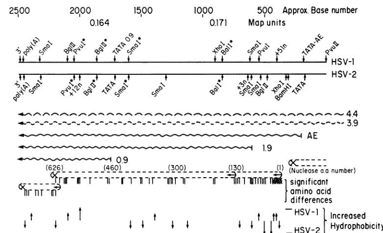
[image:3.612.97.488.71.309.2]FIG. 1. HSV alkaline exonuclease gene. This is aschematic summary ofthegenetic expression of theregion of HSV-1 and HSV-2
encoding the alkaline exonuclease function(s). Map units localizing the region of the P arrangement of the genomes are given. Some characteristic restriction endonuclease sitesareshown. Those whichmapatidenticalpositionsonbothgenomesareindicatedwithasterisks. Sites of nucleotide additionsaremarked(+51in the HSV-1genomes, +3 and +12 in the HSV-2genome). The locations of the 50' and 3'
terminiof mRNAsareshown,whereapplicable.Theoverlapping3.9- and 4.4-kb mRNAsarerepresented bywavybroken lines. Predicted
open translational readingframes are illustrated (+-),and stop (x) signals of the threereading frames are derived from DNA sequence
analysis. Significant amino acid differences between theputative HSV-1 and HSV-2polypeptidesbased onthe simplification protocolsof
Devereuxetal.(18)discussed in thetextarerepresented byvertical lines.Regionalincreasesin thepredictedhydrophobicityof the HSV-1
and HSV-2polypeptidesareshowngraphically byarrows: (t)smalldifferences, (t)largedifferences.
pended in 20
RIl
ofethyl acetate and spotted on silica gel thin-layer plates (J. T. Baker Chemical Co.). The productswere separated by ascending chromatography with a 95%
chloroform-5% methanol solvent mixture. After autoradiog-raphy, spotsof the different forms ofchloramphenicol were
localized, cut out, and counted in PCS scintillation fluid (Amersham Corp.).
RESULTS
Comparativenucleotidesequenceof theregionof theHSV-1 and HSV-2genomesencoding alkaline exonuclease. Apartial restriction mapoftheportionof the HSV-1 and -2genomes
encoding alkaline endonuclease is shown in Fig. 1. This figure also summarizes transcriptional and sequence data reported here as well as previously (13). Five transcripts sharea common3'terminusatca. 0.160m.u. Alltranscript
sizes reported here correspond to the actual number of nucleotides encodedin the DNA; actual migrationon
dena-turing gels is slower because of thepresenceof the200-base poly(A) tail (45). The four largest transcripts have been characterized previously (13). In addition, the figure indi-cates the location of a 0.9-kb transcript detected with a
sensitive single-stranded M13 probe.
The nucleotide sequence of the HSV-1 and -2 DNAs encoding these transcripts wasdetermined by use of
over-lapping cloned DNA fragments. Both strands of both virus DNAtypeswerecompletely sequenced. Ashas beenseenin
previous work in other regions of the genome, there were
occasional regions in both HSV-1 and -2 where
compres-sions of bases made sequencing difficult. Itwasfound that
increasing the percentage of acrylamide and gel running temperaturewassufficienttoresolve such uncertainties. The
sequencesof the HSV-1 and HSV-2 DNAwerecompared by useofan improved dot matrix programdeveloped byJohn Coffin of the Tufts UniversityMedical School Cancer Cen-ter. The comparative analysis is shown in Fig. 2; the data begin at a point 200 bases 5' of the cap site of the HSV-1 alkaline exonucleasetranscriptandcontinuesto ca.50bases 3' of thepolyadenylation sitefor the transcriptcluster.
Thecomparative sequencedata showed that four restric-tion sites occurred in identical locations on the two viral
genomes:theBalI siteatHSV-1base 845 (HSV-2base797), the SmaI site at HSV-1 base 1606(HSV-2 base 1558), the BglII site atHSV-1 base 1866 (HSV-2 base 1818), and the PvuIsiteatHSV-1 base 2061(HSV-2base2025).These sites
were used to align Fig. 1 and are indicated by asterisks.
There is one unusual site in the HSV-2 sequence at base number 1256 where the sequence was determined to be GGGACCTC by sequencing both DNA strands, yet some
lots of the restrictionendonucleaseSau3AI, whose nominal recognitionsite isGATC,consistentlycleavedatthispoint, whereas othersdidnot. Carefulanalysis of thesequencein thisregion showed noevidence fora "swallowed" orextra base.
Theoverall homologybetween theHSV-1 and -2 DNA in thisregionisapproximately81%. This is similartolevelsof homology seenin otherregions of thegenome(see
Discus-sion). Very high homology was seenbetween base numbers
1 and 301, which corresponds to the putative translation termination signal of the 50,000-Da putative capsid protein encodedbytheoverlapping3.9-kbtranscript (13; Draperand Wagner, work in progress). Homology abruptly breaks at this point, and there are a total of 51 fewer bases in the
HSV-2 DNA than in the HSV-1 DNA. Computer analysis 2500
F°FT
I0
e
on November 10, 2019 by guest
http://jvi.asm.org/
HSV-1
HSV-2
HSV-1
HSV-2
HSV
-1
HSV-2
10 20 30 40 50 60 70 80 90 100 110 120 130 140
COCCTCBCScT6CcTIC6CCCT66CC6CTC6CCTAJC6C6CU6TCcACCGCCCTcTTrMTTA
ACTI66CC1MACCCC66CCT6GC6TATQXC6JTCCCGC6CCAMTCSCAA
m i
fttfit§§tttnuf muntt innuum§§§ tIm imun um*#Mu§§4§ itutmftif*tftntit*ttmi,ttt*t*tutm§*§t
rA6CTCTC6CC6AC6CCG66C TCBCCTATC=6C6CW6CATCTcccKcc7cm6TrAAT66Am c=TPATwmcmAoCcocctM
10 20 30 40 50 60 78 8 90 100 110 120 130 140
'TATA'
AE
-AE-
CAP
150 160 170 160 I90 200 210 220 230 240 250 260 270 20
TCCC6MTCGB AC6AC66TTATACACA 6GCWA CTGTC6TC66At66C96TCMA6CAA6CfTlCCf66f6C 6r1TCCCOCACA64C4MCA
unfluftitffltuftt mu§ *filmillunuff§vif*iffit* i mut fiff iffit* **##"ttttt §tAtf* mun§§§tifi umin§§*##ttt##§**§t
TCCCmTTUC66C6A66AcKAC6CTA6CAAAAc 6AC6TCCT6TC6 TCW6C6CnM6caCn6T C66CT66TCCrCCC1CT6TCACMCCC
150 160 170 10 190 200 210 220 230 240 250 260 270 280
Term
cpsd
AE-ORF
2m 200 310 320 330 340 350 360 370 380 30 400 410 42U
6C9C6C66CAC6C6CT8C6TMWTr46CnCACCC66C66C8C6CTCACCACC6CTCCCCAC6 TCOTCTC6MT66TCCA BTC C6ACTM CT BAA CCT
111111 III uuuI itit f1,*tiiMn* * *tt t i t
CM6C6K6CCARCOC9ETGCA6MTGCCIT1CCGCCCTCMCC6CTGCTCGT
AARTG6C66CCCC0CAMC66CGcCCMA6C8M
290 300 310 320 320 340 350 360 370
BamHI
(*1.9)
430 440 450 460 470 400 490 500 510 520 530 540 550 50
HSV-166c2 CCC6mCTCA6CC6CCCCCMTCCCCCCAA6C6CCCCCCTCCTMCC6CAt 6CCCACMCC86CCCC0CCCCCCCCCCC666CCCC66nCCCA1
if 11H it t ft f*141if it If*** Mt *if*ft UM§ ft If# t off fit * ft** ##tf4fittiff §%mi§t H A tt§
380 390 400 410 420 430 440 450 460 470 48 490 500
CAP
1.9
Kb
570 50 590 600 610 620 630 640 650 60 670 600 690
HSV-1 cccCCET1CcCCCACTmm6crACCCACC6
A6CCCC66CMCC6TC6
CCGCTCCCC CCCTn CCCC6M1TCCCCT1CCATA111 *#141111 1 *I* II It * *111 i 11111 111111 if iIIII 11111 1ffI #ff1 ofi111 HIIfff1It11111 1111111
HSV-2 66cCFAwcccmcrAcCcac mc6c6666cc mc6cc66ccc6cCCcc66CTclCA1Cc8cCCMcACCCCCWA1TCCCC1C6CC "C
510 52
50
540 50 56 570 590 60610
620 60 6401.9
Kb
ORF
700 710 720 720 740 75 760 77 m 79 800 810 020 820
HSV-1 C6C80IAB0C6MCCCTCCCM=6CCCWCMAGT6TC0SC66CTGCCMCC CSCTB=CWrAJA1ACTA8>^CH ACCrACCT0C1SC8C8CTBCCCBC
IlIllIllI111 1111111 II 111I1IIl1 ilIIffilfi 1##111 Hff1111 IfH If, fIffiffmi,fIIIlf fflIlIfIl
HSV-2
A7cmmcCwSrCcCC
cCSMCcCM
cccmTSIflCOCMICCCMCG
6C CCCC1ATMr8CVCCM0C8ACNcTCc1snC18C8c86NCC6Tc
650 4" 67 a0 690 0 710 720
720
740
7 760 770 mBall*
XhoI
840 8S0 860 8?0 8S m" 900 910 920 930 90 9 9U1 978
H SV -1 CC C"A6CCC CTCT66CCC6CT00TATCTT6Tr66nCCE6a6TCCTCMc6866C66A
64
C TA6GOOCCCCC6CC86C6
HS V- 2 CCCTr6Cu6CCCr6c6cCTC6CCTs0CAAT1r6TC6lC6TCS6CT
6C 6T6C6C6WCBC6CC6
6CCT6rC
;m 000 10 02 820 840 50 060 00 880 89 90 910 92
Ball*
900 990 1000 1f10 1020 1020 1040 1050 1060 1070 1080 1090 1100 1110 HSV-1 C66ACCC6T66C6CCCC66CCCCTCATe66ST AC0SCssCCA AAACCA66CcCT6CCrA6CTA7666CCCT6CT
666CTCI4CC6
CTCCA-CCc
CCCTIit111111if 1UM §§ #Ill*1111 iilfilIll ufi1IIIIIII1IIlIIlI II IffIlIlI 111fffH#*mIum1111 1111 ftfI 11fiff 1ilIIfl
HS V- 2 CC6XC67C6CCCCCC6CM9TTA0^6ATCWGC66CC66CC 6CWA 6=CCHCSCTS66CCJ0C
6C6CCTCC
C6CCISC P C CSC6C 930 940 950 960 970 90 9 1000 1010 1020 1030 i14 1050 10601120 1130 1140 1150 1160 1170 1108 1190 1200 1210 1220 1230 120 1250
H
SV- 1
TrCoCrcc6T6r6w"CACa6C0CccwI6TACA6TTTC6wCS6ATw6T
6WTCCOGOCS6ST9cmcs
c6W
_
HUH§*1414MU§ I *HHHHHHUM§§§§ * M§ * Hlif §11HUffi§§5§§§§§§§lif"§§§§§§ ff,§§§§t*§§"U§§§§§
H
SV-
2
nRCUCFTCCMWCrACCCA SGCC GM6C66S66ACCCA6TC6TC66COGfT6SA6TTC6
SC666CC 66CAMC6TTCBCACTC61070 1000 100 1100 1110 1120 1130 114 1150 1160 1170 1180 1190 1200
1260 1270 120 1290 130 1310 1320 1320 1340 135 1360 1370 130 1390
HSV-1
C6A66CS6C6CACC6CmAmCrAC
IIIi111111I11111 iii. n 1111 1 *nniiiiwCCA6CCCT6C6CCWMMC6TSCAT
Innn iiilff, HHIIlIIIlIlI,IIACFT60SSCCATMCS6A6rC
1111TCACff 111f6SCE7
lIHIIlIffIlICCCT0M1ATTCTC6TCT6TCC
I t111i IH
SV- 2
OCMcCACACC6SCSCC66SC6CTTCOTCM
CACOMGSCCC66MGCC
GTS CTSCC6TCCTCA7
CIOXSTS66C6ECCCTSG.CAICCTCOITSCCCTA
1210 1220 1230 1240 1250 1260 1270 1280 1290 1300 1310 1320 133 13401400 1410 1420 1430 1440 1450 1460 1470 140 1490 1500 1510 1520 15 HSV-1 66AC6wA- CCTPCC TCCCCCCC0A6CclTMm TKC06cC CACACTCSACCCCAI CCCCBTMCCUCC6MACTTA16A76AC
1111IlIIIIIIIII ffIffllff111111 1111 HU1111 If111 ffIIIlIlIf4l iff11111 fflff mI 11ff, Ifi11iIII11m1I1I,11111111v
HSV- 2 mfcmClC66c8cCCACMTS6CMS CCCCCCOCCCACC
1350 1368 1370 1300 130 1400 1410 1420 1430 1440 145 1460 1470 148
SmaI*
1540 1550 1560 1570 1800 15" i60e 1610 1620 1630 164 1650 1660 1670
HS V-1 C6E9CCCC
c6ASc0CCS66C6TICC6SCSCA6CCTA
CCC SUCC CTCSC C CCCIt111 IfffIfifilifIl tii 1111111I11111H1111n1111 1111ffIIIlffIlIlffIIfIIf IfI I fI f 1fIf
HSV- 2 crTCuC6CcnCCO TCCS6cATCCACCCCrCCBK6C9AC,C6CK SCCO!CC
149 1500 1511 1520 1530 1540 1550 1560 1570 15 15"
160d
1610 1620SmaI*
on November 10, 2019 by guest
http://jvi.asm.org/
166 16" 1766 1711
'TATA'
-0.9Kb-1726 1736
0.9 Kb
176 176 176 1776 1766 1M 1NS 116 H SV- 1 e-6cUTF6Crcc6EcC6U6Tc66644Crrr6TT6rCgmhgr6cr--6-- FC6WCCI CC 16WX6
ninnnnft iiinnaannn*nnNnnnnonennnnennnnnn nnntn *nNnnnnnntn tan*innnn nnn HSV- 2 6MT6CrCC6C6TC 6TT T (nmCNcccMcnM n"cgT66cninc66
1J3 164 15 166 1676 16 16" 1766 1716 1726 173 174 1756 1746
BglU*
1626 136 1 1656 I6N91676 16 15 16 11 im 1936 1946 I
H S
V-i
"Z - r" -AHSV-1 TTc6CC6CGACCM6CBTCTC6CMCTCcCcATCCM% SWCTCTA4Tr669T6CA66MTAcrCTC1VCMMCC1TCCCWT6CACCC6CA7CT6PT6CAfMA7AMCD HSV-2 TT CU TCCCC6=TMMCG^^TMWAMK CCCM.W1;rrrVACWWWMUCrc-A
1776 176 1756 1680 1811 1626 1o 1646 15 16 1867 16 156 1966
BglI[*
Pvu
I*
1966 197 1966 199 266 216 2 N2 264 2656 2646 2670 2666HS V-2 66c@_rrmcu;crams6c%%=ca:omwnwmcCCC c=6Mcc A ccMlIcC"TMlCAlCMCCCC6c
1916 1926 1936 1946 195 19 1976
19C
199 26662016
2620
2636 2640.9Kb
Pvu
I*
ORF
26" 216 2116 2120 2136 214 2156 2146 2176 216 21" 2ni 2216 222 HS V-1 rATcmJcc6 TctTATm66CAT 6c6CT6c1m1AC c6ccm6 nTAISGcCTIcTcc KrcrgcccaCrI
I iii! tiN{111111 H§11111 11111IIIIIIHNII ilIlili Nitilt ii iinttn§tintNtttin nnttnnmNNNn ItNtNititie n innn
HSV- 2 cUcUc6TCTCMCATCC666Cxc^A66 cUCMTAC6ccPA16CCnc6TTCClAAMca7 2656 2064 2076 2006 26" 2166 2110 2126
2136
2146
215 214 2176 216AE
TERM
2236 224 2256 2266 2n27 2266 22m 2366 2316 236 2336 2346 25 2346
HSV-
I1
,6cCTCSRCCWCMCCC_A6m6C60CcttAt§N§tttttttt HUMtMtttttftfunufutttiIftomtAfftiftnfftttt utiftwofNt#ttwmtA tt *t* tfffN*ttZ
H
SV-2
6M66TC VCC6MMMMCCPWATm*CM6
M6&6U67
TC-
PANCIAATAWCM6C6M c6rCOSA=21" 2266 2216 2226 2238 2246 2256 2246 2276 2266 225 2366 2316 2326
poly( A)
TERM
0.9
3'
(3')
23 236 25 26 2416 24 243 2446 2456 246 2476 24 24" 2ml
H
SV-1
cOUacrc N=OC- CC#U=76TcCITC
I 1 1 1t in ftN u*N. inn t ntnt4 t o £0 * ttN tt HHHn mi is fnm ttiten *tinI a m .ini
HSV-2
MCM ~ ~ ~~~6cc6CCTCCT6DC(EcCTrccccCTC70TA6CWTMMrA0JmTT6T7CCfATrTW6cffAr676TTTr1nAA2336 2346 2 23 2376 236 235 2466 2416 2426 2436 244 2456 244
3'
FIG. 2. Nucleotide sequence and homology between HSV-1 KOS DNA and HSV-2 HG-52 DNA encoding the alkaline exonuclease function. HSV-1 and HSV-2 DNA sequences were compared using a dot matrix program as described in the text. Matched bases are designated by asterisks. Nucleotidenurmbersare listed above andbelow the HSV-1 and HSV-2 DNAs, respectively. Positions of the mRNA capsitesand3' terminiof the transcriptsweredeterminedbySinucleaseanalysisasdiscussed in the text. A possible secondary cap site for the1.9-kb transcriptis shown(*1.9). Selectedrestrictionendonucleasesites arelisted, including the uniqueBamHIandXhoI sites, as well astheBalI (BalI*), SmaI (SmaI*),BglII (BglII*),andPvuI(PvuI*)sites, which map at identical positions in the twogenomes. Putative TATA boxes of thepromoterregions forthealkalineexonuclease (AE) and 0.9-kb transcripts and the polyadenylation signal for the transcript cluster areshown. Theputative initiation(ORF)andtermination(term) sites for the open translational reading framesencoded by thisregion are predicted fromcomputeranalysis ofthe DNAsequence.
suggests that residual homology is maximized by the ar-rangementof gaps shown in Fig. 2. Here, it is suggested that there are 33 fewer nucleotides in the HSV-2 sequence just after the translation initiator for the alkaline exonuclease protein (HSV-1 base 362, HSV-2 base 344).
Twootherregions show net differences in the number of bases between HSV-1 and HSV-2. In both cases, the HSV-2 DNA contains additions; there are three extra bases at HSV-2 base 598 (HSV-1 base 648) and 12 extra bases at HSV-2 base 1957 (HSV-1 base 2005). In all cases of nucle-otide number differences between the two sequences, the ORF for the alkaline exonuclease and colinear 1.9-kb tran-scripts is conserved.
As has been reported previously (13), the nucleotide sequence 5' of the alkaline exonuclease transcript has the nominal features of a eucaryotic promoter, whereas the sequence immediately 5' of the 1.9-kb transcript cap site does notcontain suchfeatures. The sequence 5' of the cap
site of the0.9-kb transcript contains an obvious TATA box homology (HSV-1 base 1722, HSV-2 base 1674); however, CAAT box homology is not particularly evident in the region of 60 to 80 bases 5' of the cap site.
The ORF for the alkaline exonuclease protein terminates at HSV-1 base 2242(HSV-2 base 2206), providing capacity to encode 626 amino acids (620 for HSV-2). Some compar-ative properties of the HSV-1 and HSV-2 proteins are summarized in Fig. 1 and discussed below. The 1.9-kb transcript contains a unique translation initiation codon at base 740 (HSV-2 base 692); this ATG codonis in phase with the largeralkalineexonuclease ORF, as was suggested inour earlier report (13). The only ATG triplet encoded by the 0.9-kbtranscript occurs ca. 400 bases 3' of thetranscript cap site (HSV-1 base 2158, HSV-2 base 2122) and initiates an ORF which is out of phase with the ORF of alkaline exonuclease. The ORF of the 0.9-kb transcript is sufficient to encode a protein of 96 amino acids.
on November 10, 2019 by guest
http://jvi.asm.org/
[image:5.612.103.502.67.445.2]1028 DRAPER ET AL.
A
mF. .. - r(r *
HSV-1t
(M13-Bg1-Xho
OG rrprobe
0164-0171
rpl: rB HSV-1
RNA
AroT Late
mRNIA
s ze(Kb)*
44
.- 3 9 doub!et
-2
3(A
E)
-
1.9
-0.9
FIG. 3. In situ RNA (Northern) blot analysis of the mRNA encoded between 0.159 and 0.176 m.u. of the HSV-1 and HSV-2
genomes. Samples (10,ug)of the HSV-1orHSV-2 poly(A)mRNA
were frationated on methylmercury-containing agarose gels and
immobilized by drying in vacuo.The mRNAspecies weredetected
by hybridization with 32P-labeled DNA probes as indicated. Sizes
shown were determinedby positions of HeLa cell rRNA markers (not shown)asdescribed previously(1,2, 32,33, 45).(A)Leftpanel,
KOS strain HSV-1poly(A) mRNA hybridizingtoasingle-stranded
probe made from BglII-XhoI fragment O-G (0.164 to 0.171 m.u.) DNAcloned in the M13 vector. Right panel, HG-52 strain HSV-2
poly(A) mRNAhybridizing toa nick-translated probe made from
BamHI fragment Q (0.154 to 0.174 m.u.) DNA. (B) KOS strain HSV-1 poly(A) mRNA species isolated in the presence (late) or
absence
(1-p-D-arabinofuranosylthymine;
AraT) ofviral DNArep-lication. Hybridizationwas tothe samesingle-stranded M13
BglII-XhoI fragment O-G DNA probe usedabove.
There is a polyadenylation signal (AATAAA) at HSV-1 base 2444 (HSV-2 base 2408). As shownbelow the position of this signalcorresponds wellwiththedetermined3'ends of the transcripts in both HSV-1 and HSV-2. Interestingly, there is a variant of this signal 12 bases 3' ofthis site in
HSV-1 (AATAA) but not inHSV-2.
Transcription patterns in the HSV alkaline exonuclease
gene. Identification ofthe 0.9-kb transcript colinear within
the extreme 3' end of the alkaline exonuclease transcript cluster was facilitated by the use of single-stranded M13
probes. The 970-base HSV-1 BgIII-XhoI fragment O-G (0.164 to 0.171 m.u.) was cloned in M13 to accurately quantitate therelativelevels of thealkalineexonuclease and colinear 1.9-kb transcripts during infection. This probe clearly revealed the presence of the 0.9-kb transcript (Fig.
3A);anidenticaltranscriptwas seeninHSV-2 infection with
nick-translated HSV-2 BamHI fragment Q (0.15 to 0.17
m.u.) as a probe. The 0.9-kb transcript was not readily detectable with nick-translated double-stranded BglII-XhoI fragment O-G as aprobe, presumably due tothe transcript
only extendingca. 120 basesbeyond the BglII site at0.164 m.u. The M13 clone is superior fordetecting this transcript
because the method used for labeling the single-stranded probeyields the highest specific radioactivity near the BglII site.
Wedetermined the kinetic relationship between the alka-line exonucleasetranscript andthe1.9-kbcolineartranscript byusing the single-stranded BglII-XhoI fragment OG probe to measure the relative amounts of the two transcripts detectable in the presence or absence of viral DNA replica-tion. A typicalexperiment isshownin Fig. 3B. Here, based ondensitometry, the ratio of alkalineexonuclease mRNA to the1.9-kb transcript is 2.5 to 3.0 for RNAisolatedfrom cells 6 h postinfection either with or without
1-p-D-ara-binofuranosylthymine to inhibitviral DNA replication. The measure of therelative ratios between the two transcripts overcomes anydifferentialefficiencyof RNA isolation under thedifferent conditions. Because the M13 probe covers the sameregions in both transcripts, the ratiois avalidmeasure of the comparative steady-state levels of these two RNA species.Thesedata indicate that both thealkaline exonucle-ase and the 1.9-kb transcripts belong to the same kinetic class, which we have previously classed as early (beta), based on the ease ofdetection ofthe alkaline exonuclease transcript in theabsenceofDNA replication.We cannotbe as precise about the relative abundance of the0.9-kb transcriptcompared wihthe alkalineexonuclease and 1.9-kb transcripts, since our probe does not cover an equivalentamount of this mRNA. Still, using the M13 probe with HSV-1 RNA, the ratio of the 0.9-kb transcript to alkaline exonuclease transcript increases by a factor of 3 to 5 when DNA replication is allowed to proceed (Fig. 3A). Further, the nick-translated HSV-2 probe indicates that the 0.9-kbmRNA is also arelativelyabundant HSV-2 transcript lateafterinfection. Wecan,therefore, tentatively classify its kinetic class asbeta-gamma.
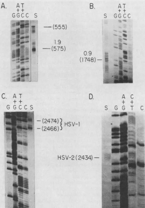
Precise location of thetranscripts. The 5' end of the 1.9-kb mRNA in HSV-1 was previously shown to lie 270 to 280 bases 5' ofthe XhoI site (base 901,Fig.2), (13). Here,itwas preciselylocalizedby Sinuclease analysisofHSV-1poly(A) mRNA hybridized with strand-separated PvuII-Hinfl frag-ment DNA (HSV-1 nucleotides540to 722) 5' end-labeledat the Hinfl site at HSV-1 base 722. The Sl-protected DNA fragment was sizefractionated against a sequence ladder of DNA 5' labeled at the sameHinfl site (Fig. 4A). Note that the sequence ladderis of the DNA strand complementaryto the mRNA. Thus, the cap site of thistranscript is locatedat or near HSV-1 base 575 of Fig. 2. This is approximately 30 bases 5' of our original estimate. A secondary cap site appeared tolie 20 bases 5' of this position at base 555. The DNA sequence within 50 bases 5' ofthe major cap site has no convincingTATAbox homolog.
We precisely located the 5' end of the HSV-1 0.9-kb transcript by carrying out S1 nuclease
analysis
of HSV-1 poly(A) mRNA hybridized withstrand-separatedBg1II-XhoI fragment O-G DNA 5' end labeled at theBglIIsite atHSV-1 base 1870. These data are shown in Fig. 4B, thetranscript
has a cap site at HSV-1 base 1748. This siteis 27 bases 3'of the sequence TAAATA, which is an excellent TATA box homolog.
Thehighhomology of HSV-1 and HSV-2 DNA sequences aroundthe HSV-1 cap sites and the fact that the HSV-1 and HSV-2 transcripts encoded in this
region
areessentially
identical (Fig. 3A) indicate that thecap sites for the HSV-2 transcripts can be expected to lie in the same locations as seen for the HSV-1 mRNA species.
The 3' end of the HSV-1 transcript cluster was
precisely
located by using S1 nuclease analysis ofhybrids between J.VIROL.
on November 10, 2019 by guest
http://jvi.asm.org/
[image:6.612.71.308.67.330.2]HSV-1 poly(A) mRNA and aDNA fragm
at the SmaI site at HSV-1 base 2332 an(
AvaI site 162 baes 3' of nucleotide 2505. fragment was sizefractionated against a s
DNA 5' labeled at thesame SmaI site. TI
inFig.(4C); therearetwomajor 3' end site apart. We suggest that, inHSV-1, the seq
base2456serves as a secondpolyadenyla
tothis 3' "stutter."
Theprecise location ofthe3' ends of the
A.
AT
GGCC
S'
;iJ
gs
11
AT
G GOCCS
B.
-(555)
1.9
-(575)
0.9
(1748)
D.
-(2474)
HSV-1HSV-2
(2434)-FIG. 4. Preciselocalizationof the5'endsof
0.9-kbmRNAsand of the3'termini ofbothth
transcriptclusters. HSVpoly(A)mRNAwash
3' end-labeledsingle-strandedDNA andfollow
Si nuclease. The Si nuclease-resistant mater
againstasequenceladderof the DNAfragment
same restriction site. Nucleotide numbers ar
HSV-1 and -2 DNA sequencesof Fig. 2. (A)
HSV-1 PvuII-Hinfl fragment DNA located in
mentC'-10 (0.171 to0.182m.u.)5'end labeled
localizethemRNAcapsiteof theHSV-11.9-kb sequence. A secondary cap site at base 555 asterisk (B) The 970-nucleotide HSV-1
BglII-DNA(0.164to0.171m.u.)5'endlabeldattheB
locate the 5' terminus of the HSV-1 0.9-kl
335-nucleotideHSV-1 AvaIfragmentDNAloc; fragmentA-K(0.151to0.163m.u.)3'end labe termini of the transcripts in this cluster. (D) fragmentHSV-2 XmaIDNAlocated inBamHI
0.174m.u.)3' endlabeled tolocalize the3'term
ofthisnested set.
ent 3' end labeled cluster was determined by Si nuclease
analysis
ofhybrids
d extending to an between HSV-2poly(A) mRNAandaDNA
fragment
3' end The Si-protected labeled at the SmaI site at HSV-2 nucleotide 2332 and equence ladder of extending to anSmaI site 283 bases 3'ofthenucleotide2469. he data are shown The Si-protected DNAfragment
was size fractionated sthat are 12bases againsta sequenceladderof DNA 5' end labeled atthe sameuence AATAA at SmaI site. The dataare shown in
Fig.
4D;
there isonly
onetion signal leading 3' end site that occurs 22 bases 3' of the consensus
polyadenylation
signal
of AATAAA at HSV-2 base 2408. HSV-2 transcript Thislocation is in close agreement with the positions ofthetermini observed in HSV-1.
Determination ofpromoteractivities in transient
expression
A T assays. An in vitro assay was used for CAT
activity
to+ + quantitate the
ability
ofthe0.9-, 1.9-,
and 2.3-kbpromoters
S GGCC to
expression
control the transcription of CAT mRNA in transientsystems.
Fragments
of HSV-1 DNAcontaining
the promoter regions of the 2.3-, 1.9- and 0.9-kb transcripts with a limited amount of leader sequence (nucleotides 1 to 230,366 to617, and 1518 to 1758, respectively) were inserted immediately 5' of the bacterial CAT gene of the pSVOD (ori-)CAT vector, which has been described previously (15). These constructs are called AE-CAT (bases 1 to 230), 1.9-CAT (bases 366 to 617), and 0.9-CAT (bases 1518 to 1758).Recombinant clones were selected thatcontained the desired promoter region in both the forward (HSV-1 sense) and the reverse(HSV-1 antisense) orientations with respect to the CAT gene.DNA preparations from recombinant clones of AE-CAT A C and 1.9-CAT were transfected into HeLa cells, which were + + then mock infected or superinfected at low multiplicities of S G G T C infection (2 PFU/cell) with HSV-1 KOS strain and assayed
ma& for CAT
activity
asdescribed in Materials and Methods. As*=shown in Fig. 5A and reported elsewhere (15), the alkaline
_-exonucleasepromoter was quite active upon superinfection. We have previously shown that this activity is dependent upon the expression of viral alpha gene products and is independent of viral DNA synthesis.
The atypical promoter for the 1.9-kb tanscript induced significant CAT activity in transfected HeLa cells when superinfected with HSV. The level of CAT activity seen with this construct never reached the levels observed with the alkalineexonuclease promoter constructs. In the experiment shown in Fig. SA, extracts from HeLa cells transfected with the AE-CAT construct converted 97% of chloramphenicol to theacetylated forms in the standard assay, whereas extracts from cells containing the 1.9-CAT construct converted only about 27%. We have described such activity measurements fthe HSV-1 1.9- and previously (15). Interestingly, the 4:1 ratio of CAT activity
eHSV-1 and HSV-2 between these promoters
(AE-CAT/1.9-CAT)
was roughly iybridized with 5' or equivalent to the relative mRNA abundancies of the 2.3- and ted by digestion with 1.9-kb transcripts late in viral infection. In both cases, the ial was fractionated reversed promoter sequences induced no detectable CAT re according to the activity upon superinfection with HSV.The 182-nucleotide The ability ofthe 0.9-kb transcript promoter to induce XhoI-HindIII frag- CATactivity upon superinfection with HSV in rabbit skin Iat the Hinfl site to cells was tested. Although these cells did not support the mRNA on the DNA extremely high levels of CAT activity reported with HeLa is designated by an cells,they have been more reproducible in their response to XhoI fragment O-G superinfection, and smaller amounts of transfecting DNA IglIl site to precisely can be used. A setof transient expression experiments with b mRNA. (C) The
AE-CAT, 1.9-CAT, 0.9-CAT,
and theVP5(168)-CAT
con-ated inBamHi-Bglll struct containing the VP-5 promoter described earlier (15) The 418-nucleotide are shown in Fig. SB. The dataof Fig. SB indicate that the fragment Q (0.14 to properorientation of the0.9-kb promoter induced levels of ini of the transcripts CAT activity that are nearly equivalent to those seen with the AE-CAT construct. Again, the reversed orientation of
on November 10, 2019 by guest
http://jvi.asm.org/
[image:7.612.48.287.195.537.2]1030 DRAPER ET AL.
I
Acetylated CAMr
.,
HSV - +
H-'E eC;\
H5V - + - *.
H-Li-",.".)f.-)
*lf-j , QD)
}V
Acetylated CAMCAM
HSV + + + + -+
o
_0
FIG. 5. CAT expression under the control of various HSV-1
promoters. HSV promoter CAT plasmids were transfected into
either HeLa cellsorrabbit skinfibroblasts, andthetransfected cells wereeithermockinfected (-)orsuperinfected (+)withHSV-1ata
multiplicityof2PFU/cell. Cellextracts werethen assayedfor CAT
activity 22 h postinfection as described previously (15).
Unacetyl-ated (CAM) and acetylated (acetylated CAM) forms of
chlor-amphenicol are shown. Nucleotide numbers ofthe DNA fragment
usedforthepromoterconstructsaregiven in parentheses (see Fig.
2). (A) CAT expression in HeLa cells transfected with AE-CAT,
1.9-CAT, or 1.9(R)-CAT promoter CAT plasmid constructs. (B) CAT expression inrabbit skinfibroblaststransfectedwithAE-CAT, 0.9-CAT, 0.9(R)-CAT, or VP5(168)-CAT promoter CAT plasmid
constructs.
the 0.9-kb transcript promoter [0.9(R)-CAT] showed little activity. Further, itis clear that the 1.9-CAT-directed activ-ity is less than that ofeither AE-CAT or 0.9-CAT in these
cells, as was thecase in HeLa cells.
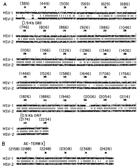
Comparison of the polypeptides encoded by the alkaline
exonuclease transcript cluster. Each of the three transcripts completelycontained in theregionof HSVDNAsequenced in this report contains a unique translational ORF as is
shown in Fig. 1. In HSV-1, the ORF for the alkaline exonucleaseenzymeyieldsapolypeptide of626amino acids
with aresiduemolecularweight of 67,000. TheHSV-2 ORF
isslightly smaller, yieldingapolypeptide of 620amino acids.
The predicted comparative sequences of the HSV-1 and
HSV-2enzymes areshowninFig. 6A. Thepredicted residue
molecularweight issignificantly less thanthevalue of82,000 measured by gel electrophoresis of the HSV-1 and HSV-2 purified enzymes and for the in vitro translation product of
the HSV-1 alkaline exonuclease transcript (13). We suggest
that the deviation in measured and predicted molecular
weight of the alkaline exonuclease protein is a result of the veryhighprolinecontent of the uniqueN-terminal 126 (117 for HSV-2)aminoacidsencoded by the 2.3-kb mRNA. This N-terminal segment of the protein has a predicted proline content of 24% in HSV-1 and 27% in HSV-2. We have previoulsy suggested that thedeviationinthepredicted and measured molecular weights of the HSV-1 glycoprotein C (50,000-Da residue compared with a migration value of 69,000 to 72,000Da[19,24]) isdue tothe high proline content in thatprotein, especially in its N-terminal region. Further support forthissuggestion isthat theresiduemolecular mass (54,000 Da) of the protein encoded by the 1.9-kb colinear transcript, whichcontains9%proline, is much closer to the 57,000- to 60,000-Da value derived from migration rates in denaturing gels.
The codonusefrequencies of the DNAencodingboth the alkaline exonuclease polypeptide and the polypeptide en-coded by the 0.9-kb transcript were very similar to those seenfor DNA encoding other HSV polypeptides (reviewed in references 49 and 50). The 84-nucleotide region of the C-terminal portion of the alkaline exonucleasetranslational ORF, which is out of phase with the ORF of the small protein, deviated from the norm; here, the favored codon use frequency was contained in the latter ORF; the base composition of the DNA in this region tends to blur the deviation, however.
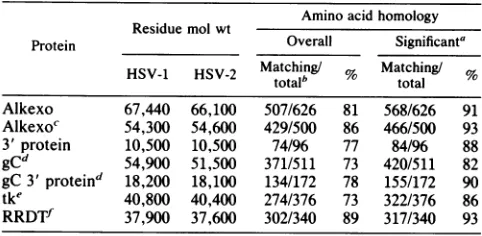
Ignoringgaps, the overall amino acid homologybetween the HSV-1 and HSV-2 alkaline exonuclease proteins was 84%;however, asignificantamount of thedeviation between the two types wasin theN-terminal20%oftheproteins. The overall homology there was only about 50%, whereas the aminoacid homology in the portion ofthe protein overlap-ping the polypeptide encoded by the 1.9-kb transcript was 88%. This value approaches the amino acid homology seen betweenHSV-1 and HSV-2 small subunitsofribonucleotide reductase (21, 27). A summary oftheavailable comparative amino acid homology datafor anumber ofHSV proteins is shown in Table 1.
Functional divergence between the HSV-1 and HSV-2 alkaline exonuclease polypeptides isprobably less thanthat suggested by a simple comparison of the predicted amino acid sequences. We used three different computeranalyses to assess the potential functional significance ofthe amino acid variation. The first isa simplification of the amino acid composition described by Devereux et al. (18). Here, the amino acids were broken into six functional types:
neutral,
weak hydrophobic (A-Pro, Ala, Gly, Ser, Thr), small hydrophilic (D-Gln, Glu, Asn, Asp), large
hydrophilic
(H-His, Arg, Lys), small hydrophobic (I-Leu, Ilu, Val, Met), large hydrophobic (F-Phe, Tyr, Trp), andcross-linking
(C-Cys). A sumamry of thedivergence between theHSV-1 and -2proteins seen withthis simplification
protocol
is shownin Fig. 7 and summarized in Figure 1. Asdiscussedbelow,
the analysis suggested that overall functional amino acid homol-ogy between the portion of the alkaline exonuclease en-zymes encodedby the HSV-1 and HSV-2 1.9-kbtranscripts
was on the order of93%. Such simplication values for the amino acid differences for other HSV-1 and HSV-2
proteins
are also included in Table 1.
The close functional relationship between the HSV-1 and HSV-2 alkaline exonuclease enzymes was also seen with a computer analysis of the average
degree
ofhydrophobicity
throughoutthepredicted amino acid sequences of the HSV-1 and HSV-2 enzymes. The computer program used calculates the averagehydrophobicity overgroups of nine amino acids (42); the results (not shown) are summarized in
Fig.
1.Here,
A.
B.
J.VIROL.
on November 10, 2019 by guest
http://jvi.asm.org/
[image:8.612.73.311.73.387.2]A
HSV-1
HSV-2
HSV-1
HSV-2
(389)
1I
(449)
36(509)
SO
(569)
76
(629)
'6
(686)
110 4ESTGGMEPPR1TIMWLAE07JB MK aPLT1JFU'LPPPMiiS NMP#l 77DPAPLM .PILNWF
I I 11
I1-
11111111|11111 I I I1I1 11 11 I 111 1111 111I 111111 11 I 1111111111 11 III I[1.9 Kb ORF
(746)
(806)
(866)
(926)
(986)
(1046)
136 15 170 I" 211 230
D60LDIMU WPSNIIEFLRHLGLLRPlWA16PLL.DYLcSLCNLEEA DRHWRLLT 6PPWDIaILflIlNPH LAETFELR6LL4R1SPLA16PLWLDYLMCSL LM 6GRHLTRIdRLJ
HSV-1
HSV-2
HSV-1
HSV-2
HSV-1
HSV-2
HSV-1
HSV-2
(1106)
(1166)
(1226)
(1286)
(1346)
(1406)
256 270 290 316 336 350
TASTLGP qILK LRSSARMEPrMSLFRYcW6 EAGTRRFIFHEPSDLAEBJKCUJD6UWDILVCPDIHLMP 11111111 Mll1 III 111111 I IIIIIIIIIIIIIII IlIllIllIll I II 1111IIIIIlIIIIIIIIIIIIIIIII1111111111 111111I 11
LUNDMSRF7FH?GDLAEBMAKAIV6W1NI6fAELD LIORPI6LTL
(1466)
(1526)
(1586)
(1646)
(1706)
(1766)
376 3N 416 436 450 471
LAYEAKCMKAF W0TMDUWUSMWRFWIRS]nIPWW6WtPSRPEMLLVbTSEKRC MCSVMMUP4SEVSLLFMPOL6RKISVA
LAfYP DllSF IRS1PA6ZRJlOODSMA6EIODRCA
(1826)
(1886)
(1946)
(2006)
(2054)
(2114)
4" 510 53 55 57 50
WSSGDLREF IRlKGILVVLD DSHIFP6PtVTF1GRHRTWEEWVTFRLED6....L WVASILW IALIITTFR1DIEIYKAIQRSSRI.NDO 1111 111111 IIIIIIIIIIIIIIIIIIIIIIIIIII 111111 liii111111111111'I 11111111l IIIIIIIIIIIII I II I I 11111111 WSS6ELJMREPIFffHFNFKQILY fLDSHFP0CPL0PLVTFL6MEErVFRLEfR L E ILP MIAI17P 61YR I WI
[0.9
Kb ORF
(2174)
(2234)
611 6
LAELISRSP66AEMSSSPTT6RSSR' 11 11111111 11111 1111 11 111111
_KRns sPAGR*
AE-TERM
X]
(2158) (2188)
(2248)
16 36
(2308)
se
n
(2368)
70
(2428)
n
HSV-1 ?ULsFcc176EYWSLANADFPJIESEEE rW6 UUPGMCMAT6FPPFI5E
III1111111111 1111111111111IIIIIIIIIIIIIIIIIII 11111111 I 111111111111 II 1111111
[image:9.612.81.526.68.601.2]HSV-2
1mmS
R1O6AEFESEEEW
"NOW4
M161111R'SPr
RROPUMILTPPLSSE
FIG. 6. Comparative amino acidsequenceanalysis betweentheputative proteinsencodedby alkaline exonuclease (2.3-kb), 1.9-kb, and
0.9-kbtranscripts of HSV-1 and -2. HSV-1 nucleotide numbersareshowninparentheses (see Fig. 2). Matchedamino acidsaredesignated
byverticallines; regionswheregapswereintroducedbythe computerhomologyprogramtomaximize overallsequencefitareindicated(0). (A) Predicted amino acidsequencesof the HSV-1 and HSV-2 alkaline exonucleaseproteins.Also shownarethepredicted initiationsites for
thein-phase ORF of the 1.9-kbmRNA andfor theout-of-phase ORFofthe 0.9-kbmRNA.(B)Theputativeamino acidsequenceofthe HSV-1 and HSV-210.5-kDaproteinsencodedbythe 0.9-kb mRNA. The site of the termination codon for theout-of-phasealkaline exonuclease ORF
is shown.
regions of minor increases ofhydrophobicity in either the HSV-1 or HSV-2 enzyme were found throughout their
predicted lengths (small arrows). Large differences (large arrows) were only seen in two regions of the proteins,
however. One of these wasin the N-terminalregion of the alkaline exonuclease enzyme. The other major difference
was seeninthe C-terminal addition of four amino acids in the HSV-2 enzyme. This resultedinasignificant loss of
hydro-B
on November 10, 2019 by guest
http://jvi.asm.org/
1032 DRAPER ET AL.
TABLE 1. Summary of homology between predicted polypeptides encoded by HSV-1 and HSV-2
Amino acidhomology Residue mol wt
Protein Overall Significanta
HSV-1 HSV-2
Matching!/
Matching!/
HSV-1 HSV-2 Mtotalb 9c total %
Alkexo 67,440 66,100 507/626 81 568/626 91 Alkexoc 54,300 54,600 429/500 86 466/500 93 3'protein 10,500 10,500 74/96 77 84/96 88 gCd 54,900 51,500 371/511 73 420/511 82 gC 3' proteind 18,200 18,100 134/172 78 155/172 90 tke 40,800 40,400 274/376 73 322/376 86 RRDTf 37,900 37,600 302/340 89 317/340 93
aSimplified as described in the text and in reference18.
bNumberofmatching amino acids/total,ignoring gaps. cProtein of protein encoded by1.9-kbtranscript.
dDatafrom references 19, 24, and 47.
eDatafromreferences 39, 46, and 51. fDatafromreferences 21 and 27.
phobicity of the HSV-2enzymein this regioncomparedwith
its HSV-1 counterpart.
The third analysis performed was a comparison of the
simplified computer predictions of secondary structure and hydrophobicity ofthe proteins. This analysis, whichutilized the parameters of Chou and Fasman (9, 10), has been described by G. Cohen, R. Eisenberg, and E. Golub ofthe UniversityofPennsylvania andtheircolleagues (11, 22). The graphic comparisons were supplied to us by R. Eisenberg
and are shown in Fig. 8. Specific features are described in
the figure legend, and the general properties of the proteins
are similar. Variations in the overall hydrophobicity
(indi-cated by the scaled hexagons) in the N-terminal region and around amino acids 550 to 600 as described above are also
shown in this analysis. Other differences in the "structure" of thetwoproteins with this analysiswere thedifferencesin
beta-turns around amino acids 50, 130, 325, and 425. This analysis does suggest, however, that there are some
signifi-cant differencesinthe overall shapesof the proteinsdespite their general amino acid homology.
A functional analysis of the 96-amino-acid polypeptide encoded by the 0.9-kb transcript and sharing the out-of-phase translational readingframe with the larger transcripts indicated that this protein has 82% homology in predicted
sequence(Fig. 6B). The simplificationcomparison shown in
Fig. 7Band summarizedin Table 1indicatedthatthisprotein is more divergentthan the portion of the alkaline exonucle-ase enzymesencodedby the1.9-kbtranscriptsof HSV-1and
HSV-2. A hydrophobicity plot summarized in Fig. 1,
how-ever, suggested that the overall conformation of the HSV-1
and HSV-2 polypeptides is rather similar. DISCUSSION
Complete analysis ofthe transcript family encoding the HSV-1 alkaline exonuclease and neighboring proteins
re-veals some variant aspects of herpesvirus gene packaging
and expression, althoughthe overalltranscriptional patterns
areconsistent with HSV dataoutlinedin several reviews(49,
50). Variants include the extreme density of packaging of
protein translational reading frames within the transcripts, the atypicalpromotersequencefor the 1.9-kbtranscript, and
the very long (ca. 400-base) leader length for the
transla-tional reading frame unique to the0.9-kb transcript.
Dense packing of protein translational reading frames
withinthe genomeis a general feature of viruses. However, based on available transcription mapping data (49, 50), the presentinstance israther extremefor the long unique region of HSV. Such
density
is approached inthe genes partially encoded in theDNAcontainedinXhoI fragment W (0.690 to 0.703 m.u. [30]), and in the short unique region (38). The translational ORFforthe 50,000-Da putative capsidprotein occupies two-thirds of the unique leader for the alkaline exonuclease polypeptide for both HSV-1 and HSV-2. Theout-of-phase
overlap of translational reading frames between the C-terminal region of alkaline exonuclease and the N-terminalregionof the10,000-Da polypeptide encoded bythe 0.9-kb transcript is another example of this compression of translational information. This is the first reported instance of such an occurrence in the long unique region of HSV, although it does occurin the short unique region (38).Sequence analysis ofthe DNA encoding the overlapping 3.9- and 4.4-kb transcripts ofFig. 1 indicates that there is another out-of-phase overlap between the N-terminal por-tion of the ORF of the 50,000-Da protein encoded by the 3.9-kb mRNA and an ORF unique to the 4.4-kb mRNA (Draper and Wagner, workinprogress). These dataindicate that there is some constraint on the amount of untranslated information in this partofthe long unique region. Whatever the reason for the slight increase in information density, thereis no obvious compression ofnontranscribed DNA in this region of the genome. This conclusion is based on the observation that the extent of untranscribed DNA between the 3'terminusof the transcripts discussed in this report and the 3' endofapresently uncharacterized transcript encoded fromtheoppositeDNAstrand, immediately to theleft of this region, is only about 170 bases in HSV-1 and 140 bases in HSV-2 (data not shown). There is essentially the same amount of DNA between the 3' ends of the transcripts terminating at 0.6 and at 0.645 m.u. (19, 20, 24, 47). Inter-estingly, inboth cases, the HSV-2 genome contains signifi-cantly less bases than is found in HSV-1.
The dataofFig. 3demonstrate that both the 2.3-kb andthe colinear1.9-kb transcripts are expressed with verysimilar,if not identical, kinetics. It is therefore interesting that the "promoter" for the 1.9-kbtranscript is so atypicalfor HSV promoters. There is no compelling TATA box homology in the promoter region, and the characteristics of some early promoters, such as seen in the promoter for the alkaline exonuclease mRNA, are missing. Thefact that the promoter of the 1.9-kb transcript can be activated in transfection assays is evidence that this region of DNA acts as an independent promoter, especially since we have found no evidence of a splice between the cap site of the 1.9-kb transcript and the cap site for the 2.3-kb alkaline exonucle-ase mRNA (13). Indeed, no convincing candidate sequence for a splice acceptor is seen in the HSV-1 and HSV-2 comparative data.
The 400-base leader in the0.9-kbtranscriptis significantly longer than the average value of 150 bases seen with most well-characterized HSV transcription units (49, 50). How-ever, the length is not at great variance with the extreme values seen in other regions ofthe HSV genome. Several leaders in excess of250 bases have been seen in the short region of the HSV-1 genome (38). In addition, the spliced transcript, which we characterized previously and mapped at 0.19 m.u., appears to have a translational leader of approximately 330 bases (16; Draper and Wagner, unpub-lished data). Finally, the6-kbtranscript encoding the major capsid protein VP-5 appears to have a leader sequence at least 240 bases in length (Draper, Costa, and Wagner, J. VIROL.
on November 10, 2019 by guest
http://jvi.asm.org/
[image:10.612.59.300.94.212.2]A.
HSV-1
HSV-2
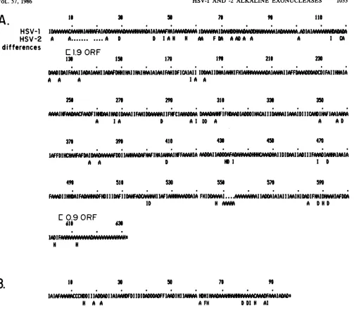
differences
1s
A
3.
A . A D
C
1.9 ORF
13.
50 7.
D IA H A F DA A AD A A
156 170 I"
a a a a a. . * a. a*
DADIM4IFAMIHIADAIAH $DHIHIIHmIW.41AMIMAIFAHIDFICAIIII 11 1Z1z1 F H I I IAFF IFAIIHHIA
A A A IA A
256 276 311 33. 350
mm] la'N I HUAIMIlAI I0FAHII IFHFC ID IFAAIIII DIIAIDI I MHFIMIAHD
A I1A D AlI DD A A AD
37. 410 43. 496 470
IAFFDIHcIMDAI
IwFDDlAHI HFwIHIAHMIHFFAAHIA
MOMII IIDl
D II I IIIFIAHMIAIA
A A D HD I I D
510 556 57.
FAAI
IIFAIH
FHDII IDAFIIINAW
IIAF M1A FHI I....AID H AMI
590
A
DHD
C
0.9 ORF
61' 6.
H H
1i 30 50 7, 90
IAIIIllAMDi IIMHDFDIDID0MDFFIMDIHII
HI.,IlA- IADAD [image:11.612.62.565.55.498.2]H a A A FH D DIH Al
FIG. 7. Significantamino acid differences between thepredictedHSV-1 and HSV-2polypeptidesencodedbythe alkaline exonuclease and 0.9-kbtranscripts. Asimplifiedamino acidsequencewasobtainedby assigningeach amino acid residuetooneof sixcategories, according
tothe chargeof the aminoacid, thestrengthof that charge,ortheabilityof that residue toformcross-linkagesintheproteinstructure.
Symbolsof thecharge assignments given each amino acidgroupare explainedin thetext(andreference 18). The simplified amino acid
sequencesof thepredictedHSV-1polypeptidesaregiven.Differences in the HSV-2polypeptidesareshown below the HSV-1sequences.(A) Amino acidcharge profilesof the HSV-1 and HSV-2 alkaline exonucleaseproteins. Gaps generated bythecomputerprogramtomaintain the best overallsequencefitare shown(d). (B)Theputative amino acidcharge profilesof the HSV-1 and HSV-2polypeptidesencodedbythe 0.9-kbtranscripts.
unpublished data). Such long leader sequences may reflect codingconstraintsimposed by overlapping transcripts;
how-ever,they mayalsobeimportantinthe post-transcriptional regulation ofexpression of certain herpesvirus proteins.
Thefact that theC-terminal 80% of the alkaline
exonucle-asepolypeptide is expressedasanindependent polypeptide viaitsowntranscriptsuggeststhat conservation of functions contained inthis region of the proteinareespecially impor-tantforreplication of the virus. The homology between the HSV-1 and HSV-2 DNA within the region of the genome
encoding this portion of the alkaline exonucleasegene(87%)
is essentially as great as that seen between the 38,000-Da
ribonucleotide reductasegenesof HSV-1 and HSV-2 (88%). This is the highest reported among the HSV-1 and HSV-2
polypeptides (21, 27;Table 1). The analysis summarized in Fig. 8 demonstrates that thisconservation ofsequencedoes
not, however, preclude the possibility that the HSV-1 and HSV-2 proteins will have some rather different structural features.
Davison has recentlyannounced the complete nucleotide
sequence of the DNA ofherpes zoster virus, which has a
46% G+C content compared with a 67% G+C content in HSV (A. Davison, unpublished). Interestingly, there is sig-nificant homology between the predicted amino acid
se-quencesof theproteins describedinthisreportandproteins encodedby herpeszostervirus. Indeed,thearrangementof ORFs in the homologous regions of the two genomes are
quite similar.
n6 110
I CA A
218 230
B.
6 a a a 0 8 a 0 a a 0 a
1- IAII I ASSAA AAMMILAIA& IALWMIAAA ..IMAAWAI I O' ClipqjJA IAVAW.ADIAI-AAAARIAM
on November 10, 2019 by guest
http://jvi.asm.org/
1034 DRAPER ET AL.
A
NH2
58
BNH2
5.~~~~~~~~~~5
Cs3
1~~~~~5
2_e°~~~~~2
300 350 §~~~~~~~~~5
458_
6s 5 5
HOOC
FIG. 8. Predictedsecondary structure andhydrophilicitymaps ofHSV-1 (A)and HSV-2(B)alkaline exonucleaseproteins. Secondary structureswerepredicted
by
acomputerprogram,accordingtothe rules of Chou and Fasman(9, 10).Probabilities for theoccurrenceof helix(Pa,),
pleatedsheet(Pb),and betaturns(P,)wereevaluatedusingmodified coditions: P,>7.5 x i0-5orP,
>5 xiO-';
P,
>Pa andP,>Pb.
Shadedhexagonsrepresenthydrophobic regions; scaledopenhexagonsindicate hydrophilicareas. The dimension of thehexagonsover a
residueisproportio'naltothemeanhydrophilicitycalculated for thatresidue and thenextfive residues(34). The value is therefore distorted at the C-termnal end. Sinewaves are indicative ofalpha helixes, andpleatedsheet regionsareindicated byzig-zagliens.
Despite this conservation of sequence between some herpesviruses, the DNA sequence encoding the alkaline exonuclease gene is not strongly conserved between HSV and Epstein-Barrvirus. Homology searches of the Epstein-Barrvirus genome(3)with the sequence data presented here do not show the length of homology seen either with the ribonucleotide reductase gene or with the spliced HSV-1 transcript (16, 28). Some limited amino acid homology has been seen between the HSV nuclease and two predicted proteins encoded by Epstein-Barr virus (E. Littler, K. G. Draper, E. Wagner, A. McBride, K. L. Powell, and J. R. Arrand, manuscript in preparation). Possible reasons for the duplication in theEpstein-Barr virus genome arediscussed
in that report. All of these data, when taken together, stronglysuggestthat theenzymatic activity encodedbythe herpesvirus alkaline exonuclease genes is importantto the biology oftheviruses as agroup.
The divergenceof theamino acid sequence in the unique 120 N-tertninal atino acids of the alkaline exonuclease protein suggests that a lengthy contiguous amino acid se-quence in this region is not directly involved in the enzy-maticactivityof theprotein. The veryhigh proline content (ca.
25%)
for both HSV-1 and HSV-2 and thecomparative analysisof Fig. 8indicate, however, that the three-dimen-sional structure of the enzyme is conserved in this region. This may be involved in its enzymaticactivity.Inthis light, J. VIROL.on November 10, 2019 by guest
http://jvi.asm.org/
[image:12.612.136.494.58.523.2]itmaybe significant thatalarge changein the
hydrophobic-ityof the protein inthis region is only seen at onesite
(Fig.
6 and 7; summarized in Fig. 1).
We have no idea concerning the possible function of the 96-amino-acidpolypeptide encoded bythe0.9-kbtranscript. The out-of-phase overlap of the first 18amino acids with the C-terminal region of the alkaline exonuclease polypeptide would tend to put some severe constraints ondivergence in this region of the protein. Despite this, there is one signifi-cant amino acid change in the predicted HSV-1 and HSV-2 sequences.
ACKNOWLEDGMENTS
This workwassupported by Public Health ServicegrantCA11861 from the National CancerInstituteand bygrant MV159Afrom the American Cancer Society to E.K.W. R.L.T. is the recipient of Public Health Service FellowshipgrantNS07589from the National Institutes of Health. K.G.D. is a postdoctoral traineesupportedby Public Health Service traininggrantCA-09054-11from theNational Institutes of Health. R.H.C.was atrainee supported by Public Health Service Molecular and Cellular Biology training grant GM-07311 from the National Institutes ofHealth. E.D.B. was therecipient of aWellcome Foundation Travel Award.
We thank M. Rice, M. Young, and C. Blair for their help. We thankR. Eisenberg of the Department of Pathobiology, University of Pennsylvania, School of Veterinary Medicine, for carrying out thecomputeranalysisofFig. 8.
LITERATURECITED
1. Anderson, K. P., R.Frink,G.Devi,B.Gaylord, R. Costa, and E. Wagner.1981.Detailed characterization of the mRNAmapping in theHindIlI fragment K region of the herpes simplex virus type 1 genome. J. Virol. 37:1011-1027.
2. Anderson, K. P., J. Stringer, L. Holland, and E. Wagner. 1979. Isolationandlocalizationofherpes simplex virus type 1mRNA. J. Virol. 30:805-820.
3. Baer, R., A. Bankier, M. Biggin, P. Deininger, P. Farell, T. Gibson, G. Hatfull, G. Hudson, S. Satchwell, C. Seguin, P. Tuffnell, and B. Barrell. 1984. DNAsequence andexpression of the B95-8 Epstein-Barr virus genome. Nature (London) 310:207-211.
4. Bailey, J. M., and N. Davidson. 1976. Methylmercury as a reversible denaturing agent for agarose gel electrophoresis. Anal. Biochem. 70:75-85.
5. Banks, L., D. J. M. Purifoy, P-F. Hurst, R. A. Killington, and K. L. Powell. 1983. Herpes simplex virus nonstructural pro-teins. IV. Purification of the virus induced exonuclease and characterization of theenzyme usingmonoclonal antibodies. J. Gen.Virol. 64:2249-2260.
6. Banks, L. M., I. W. Halliburton, D. J. M. Purifoy, R. A. Killington, and K. L. Powell. 1985. Studies on the herpes simplex virus alkaline nuclease: detection of type-common and type-specific epitopeson theenzyme. J. Gen. Virol. 66:1-14. 7. Berk, A. J., and P. A. Sharp. 1977. Sizing and mapping of early
adenovirusmRNAsby gelelectrophoresisofS1 endonuclease-digestedhybrids. Cell 12:721-732.
8. Busslinger, M., N. Moschonas, and R. Flavell. 1981. Beta' thalassemia: aberrantsplicing results from a single point muta-tion inanintron. Cell27:289-298.
9. Chou, P. Y., and G. D. Fasman. 1974.Conformational parame-ters for amino acids in helical beta sheet and random coil regions. Biochemistry13:211-222.
10. Chou, P. Y., and G. D. Fasman. 1974. Prediction of protein conformation. Biochemistry 13:222-245.
11. Cohen, G. H., B. Dietzschold, M. PonceDeLeon, D. Long, E. Golub, A. Varrichio, L. Pereira, and R. J. Eisenberg. 1984. Localization andsynthesis of an antigenic determinant of herpes simplex virusglycoproteinD that stimulates theproductionof neutralizingantibody. J. Virol.49:102-108.
12. Costa, R., G. Cohen, R. Eisenberg, D. Long, and E. Wagner.
1984. A direct demonstration that the abundant 6-kilobase herpes simplex virus type 1 mRNAmapping between 0.23 and 0.27 encodes the major capsid protein VP-5. J. Virol. 49:287-292.
13. Costa, R., K. Draper, L. Banks, K. Powell, G. Cohen, R. Eisenberg, and E. Wagner. 1983. High resolution characteriza-tion of herpes simplex virus type 1transcriptsencoding alkaline exonuclease and a 50,000-daltonproteintentatively identifiedas
a capsidprotein. J. Virol. 48:591-603.
14. Costa, R. H.,B. G. Devi, K. P. Anderson, B. H. Gaylord,and E. K. Wagner. 1981. Characterization of a major late herpes simplex virus type 1 mRNA. J. Virol. 38:483-496.
15. Costa, R. H., K. G. Draper, G. Devi-Rao, R. L. Thompson, and E. K. Wagner. 1985. Virus-induced modification of the hostcell is required for expression of the bacterial chloramphenicol acetyl transferase gene controlled by a late herpes simplexvirus promoter (VP5). J. Virol. 65:19-30.
16. Costa, R. H., K. Draper, T. Kelly, and E. Wagner. 1985. An unusual spliced herpes simplex virus type 1 transcript with sequence homology to Epstein-Barr virus DNA. J. Virol. 54:317-328.
17. Denhardt, D. T. 1966. A membrane-filter technique for the detection of complementary DNA. Biochem. Biophys. Res. Commun. 23:641-646.
18. Devereux, J., P. Haeberli, and0. Smithies. 1984. A comprehen-sive set of sequence analysis programs for the VAX. Nucleic Acids Res. 12:387-395.
19. Draper, K., R. Costa, G. T.-Y. Lee, P. G. Spear, and E. K. Wagner. 1984. Molecular basis of the glycoprotein C-negative phenotypeof herpes simplex virus type 1 macroplaque strain. J. Virol. 51:578-585.
20. Draper, K., R. Frink, G. Devi, M. Swain, D. Galloway, and E. Wagner. 1984. Herpes simplex virus type 1 and 2 homology in the region between 0.58 and 0.68 map units. J. Virol. 52:615-623.
21. Draper, K., R.Frink, and E.Wagner.1982. Detailed character-ization of an unspliced beta herpes simpelx virus type 1 gene mapping in the interior of another. J. Virol. 43:1123-1128. 22. Eisenberg, R., D. Long, M. Ponce De Leon, J. T. Matthews,
P. G. Spear, M. G. Gibson, L. A. Lasky, P. Berman, E. Golub, and G. H. Cohen. 1985. Localization of epiitopes of herpes simplex virus type 1 glycoprotein D. J. Virol. 53:634 644. 23. Feramisco, J., J. Smart, K. Burridge, D. Helfman, and G. P.
Thomas. 1982. Coexistence of vinculin-like protein of higher molecular weight in smooth muscle. J. Biol. Chem. 257: 11024-11031.
24. Frink, R., R. Eisenberg, G. Cohen, and E. Wagner. 1983. Detailed analysis of the portion of the HSV-1 genome encoding gC. J. Virol. 45:634 647.
25. Frink, R. J., K. P. Anderson, and E. K. Wagner. 1981a. Herpes simplex virus type 1 HindIllfragment L encodes spliced and complementary mRNA species. J. Virol. 39:559-572.
26. Frink, R. J., K. G. Draper, and E. K. Wagner. 1981b. Uninfected cell polymerase efficiently transcribed early but not late herpessimplex virus type 1 mRNA. Proc. Natl. Acad. Sci. USA78:6139-6143.
27. Galloway, D. A., and M. A. Swain. 1984. Organization of the left-hand end of the herpes simplex virus type 2 BglII N fragment. J. Virol.49:724-730.
28. Gibson, T., P. Stockwell, M. Ginsberg, and B. Barrell. 1984. Homology between two EBV early genes and HSV ribonucle-otide reductase and 38K genes. Nucleic Asids Res. 12:5087-5099.
29. Gorman, C., L. Moffat, and B. Howard.
1982.
Recombinant genomes which express chloramphenicol acetyltransferase in mammalian cells. Mol. Cell. Biol. 2:1044 1051.30. HaUl, L., K. Draper, R. Frink, R. Costa, and E. Wagner. 1982. Herpes simplex virus mRNA species mapping in EcoRI frag-mentI.J. Virol.43:594-607.
31. Hines, J. C.,and D. S. Ray. 1980. Construction and character-ization of new coliphageM13cloning vectors. Gene 11:207-218. 32. HoUland, L., K. Anderson, C. Shipman, Jr., and E. Wagner. 1980. Viral DNAsynthesis is required for the efficient