City, University of London Institutional Repository
Citation
:
Kosilo, M. (2017). The contribution of the luminance and opponent chromatic post-receptoral mechanisms to visual working memory. (Unpublished Doctoral thesis, City, Universtiy of London)This is the accepted version of the paper.
This version of the publication may differ from the final published
version.
Permanent repository link:
http://openaccess.city.ac.uk/19357/Link to published version
:
Copyright and reuse:
City Research Online aims to make research
outputs of City, University of London available to a wider audience.
Copyright and Moral Rights remain with the author(s) and/or copyright
holders. URLs from City Research Online may be freely distributed and
linked to.
The contribution of the luminance and
opponent chromatic post-receptoral
mechanisms to visual working memory
Maciej Kosiło
School of Arts and Social Sciences Department of Psychology City, University of London
3
Table of Contents
Table of Contents ... 3
List of Figures ... 9
List of Tables ... 13
Acknowledgments ... 15
Abstract ... 19
Chapter 1: General Introduction ... 21
1. Working memory and the prefrontal cortex (PFC) ... 26
1.1. Summary: working memory and the prefrontal cortex (PFC) ... 31
2. The role of top-down signals in working memory ... 32
2.1. Summary: The role of top-down signals in working memory ... 34
3. Beyond the prefrontal cortex ... 36
3.1. Sensory areas and perceptual processing contributing to working memory. ... 36
3.2. Interactions between perception and visual working memory: impact on theoretical approaches and significance. ... 41
3.3. Summary: common mechanisms of perception and visual working memory ... 45
4. Chromatic and achromatic shape processing: from the retina to the cortex ... 46
4.1. The retina – anatomy and functional organisation ... 47
4.2. Lateral Geniculate Nucleus (LGN) – anatomy and functional organisation ... 55
4.3. Cortex – anatomy and functional organisation ... 58
4.4. Separation of visual channels ... 60
5. Chromatic and achromatic visual channels in perception ... 63
5.1. The role of luminance in facilitating perception via top-down signalling ... 67
5.2. Summary: differential contribution of luminance and chromatic signals to perception ... 70
5.3. The potential role of luminance signals in working memory: top-down benefits, noise reduction... 70
6. Conclusion and the main hypothesis ... 72
Chapter 2: General methods and colourimetry ... 75
7. Colour spaces ... 76
7.1. The CIE 1931 colour space ... 76
7.2. Physiologically meaningful colour spaces ... 79
7.2.1. LMS-cone excitation space ... 79
7.2.2. Cone-opponent colour space: the Derrington-Krauskopf-Lennie (DKL) space ... 80
7.2.3. Use of DKL colour space to stimulate cone-opponent and luminance mechanisms ... 82
7.2.4. Summary ... 85
7.3. Observer’s isoluminance – heterochromatic flicker photometry (HCFP). ... 85
4
7.4.1. Calibration ... 86
7.4.2. Verification procedures ... 89
7.5. Re-calibration ... 91
7.6. Stimuli & on-line DKL conversions ... 92
8. Psychophysics – overview of methods used to estimate threshold. ... 93
8.1.1. Detection and discrimination thresholds ... 93
8.1.2. Adjustment methods ... 95
8.1.3. Method of constant stimuli ... 96
8.1.4. Adaptive procedures ... 96
Chapter 3: The contribution of luminance signals to visual working memory performance – a study using EEG ... 101
9. Investigating visual working memory using EEG ... 104
9.1. Use of ERP components to study visual working memory ... 106
9.1.1. Visual component P1 ... 106
9.1.2. N1 ... 110
9.1.3. P1 and N1 as gain control mechanism... 113
9.1.4. P3 component ... 113
9.1.5. Slow wave ... 116
9.2. Predicted ERP modulations ... 117
9.3. Studies on perception/working memory interaction using the ERP technique ... 118
10. General summary and hypothesis ... 121
11. Methods ... 122
11.1. Participants ... 122
11.2. Procedure ... 122
11.2.1. Working memory measurements: digit span and letter-number test. ... 124
11.2.2. Observer’s isoluminance: Heterochromatic Flicker Photometry test ... 125
11.2.3. Same/different shape discrimination threshold ... 125
11.3. Main WM experiment ... 127
11.3.1. Delayed match-to-sample task ... 127
11.4. EEG data acquisition... 130
11.5. Pre-processing and analysis ... 130
11.6. Note on ERP amplitude and latency extraction ... 134
12. Behavioural results ... 136
12.1. Vision tests ... 136
12.2. Working memory tests ... 137
12.3. Threshold measurements ... 137
12.4. Accuracy ... 139
12.4.1. Match ... 140
5
12.5. Reaction times... 143
12.6. Comparison with mixed signals stimuli ... 144
13. Event-related potential (ERP) results ... 146
13.1. Encoding stage ... 146
13.1.1. P1 Amplitude (Oz, O1, O2; 80-160 ms) ... 146
13.2. Low contrast ... 147
13.3. High contrast ... 148
13.3.1. Latency... 150
13.4. N1 Amplitude (Oz, O1, O2; 130-300 ms) ... 150
13.5. Latency ... 151
13.6. P1 and N1 interactions with behavioural performance ... 152
13.7. P3a Amplitude (C1, C2, Cz; 200-400 ms) ... 154
13.8. Low contrast ... 155
13.9. High contrast ... 155
13.10. Latency ... 156
13.10.1. P3b Amplitude (P3, P4, Pz; 200-500ms) ... 157
13.10.2. Latency... 159
14. ERPs – maintenance stage ... 161
14.1. Stimuli offset peak (700-900 ms) ... 161
14.2. Slow wave (1000-1600 ms) – occipital (electrodes O1, O2, and Oz). ... 161
14.3. Slow wave (1000-1600ms) – frontal (electrodes F3, F4, and Fz). ... 163
15. ERPs – retrieval stage ... 164
15.1. P1 Amplitude (Oz, O1, O2; 80 ms-160 ms) ... 164
15.1.1. Latency... 169
15.2. N1 Amplitude (Oz O1 O2; 130 ms – 300 ms) ... 169
15.2.1. Low contrast ... 171
15.2.2. High contrast ... 171
15.2.3. Latency... 172
15.3. P3a Amplitude (C1, C2, Cz; 200 – 400 ms) ... 172
15.3.1. Latency... 173
15.4. P3b Amplitude (P3, P4, Pz; 200 – 500 ms) ... 173
15.4.1. Latency... 175
15.4.2. Low contrast ... 175
15.4.3. High contrast ... 175
16. Discussion ... 176
16.1. Overall behaviour findings ... 176
16.2. Match/mismatch effects ... 177
6
16.2.2. Lack of correlation between digit span and letter-number tests and delayed match-to-sample task 178
16.3. ERP effects... 179
16.3.1. Perceptual effects ... 179
16.3.2. ERP modulations related to working memory load ... 182
16.4. What mechanism underlies luminance advantage? ... 185
16.5. Further directions and general shortcomings ... 186
16.6. Summary ... 187
Chapter 4: Psychophysical experiments on visual working memory ... 191
17. Psychophysical measurements and sensory perception. ... 193
17.1. Insights into visual perception ... 193
17.2. Studying memory and cognition with psychophysics ... 194
17.3. Summary: studying memory and cognition with psychophysics ... 197
18. Current experiment: visual working memory thresholds ... 199
18.1. Summary – visual working memory threshold experiment ... 202
19. Experiment 1: working memory thresholds ... 203
19.1. Methods ... 203
19.1.1. Participants ... 203
19.1.2. DKL colour space & stimuli ... 203
19.1.3. Threshold normalisation ... 206
19.1.4. Procedure ... 206
19.1.5. Analysis ... 210
20. Results ... 211
21. Control experiments ... 213
22. Experiment 2 – shape detection thresholds ... 214
22.1. Methods ... 214
22.2. Results ... 215
23. Experiment 3: Same/different shape discrimination thresholds ... 217
23.1. Methods ... 217
23.2. Results ... 217
24. Discussion ... 219
Chapter 5: A pilot study of luminance processing and visual working memory in schizophrenia ... 227
25. Cognitive deficits in schizophrenia... 230
25.1. Perceptual and sensory deficits in schizophrenia ... 231
25.2. Interaction between visual perception and working memory in schizophrenia... 234
25.3. Luminance processing in schizophrenia: relevance to working memory performance ... 236
25.4. Investigating luminance – processing in schizophrenia in the context of magnocellular theory and its criticism ... 240
7
25.6. Summary and aims ... 243
26. Methods ... 246
26.1. Participants ... 246
26.2. Procedure ... 248
26.3. Averaged thresholds and Weber contrasts ... 249
26.4. Task design ... 250
26.5. EEG data acquisition, pre-processing and analysis ... 251
26.6. Vision tests and behavioural data analysis ... 254
27. Behavioural results ... 255
27.1. Vision tests ... 255
27.2. Accuracy ... 257
27.2.1. Between-subject effects ... 257
27.2.2. Within-subject effects ... 257
27.3. Reaction times... 260
27.3.1. Between-subject effects ... 260
27.3.2. Within-subject effects ... 261
28. Event-related potential (ERP) results ... 262
28.1. Encoding stage ... 263
28.1.1. P1 Amplitude (Oz, O1, O2; 80-160 ms) ... 263
28.1.2. Low contrast ... 263
28.1.3. High contrast ... 264
28.1.4. Latency... 265
28.2. N1 Amplitude (Oz, O1, O2; 130-300 ms) ... 265
28.2.1. Low contrast ... 267
28.2.2. High contrast ... 268
28.2.3. Latency... 268
28.3. P3a Amplitude (C1, C2, Cz; 200-400 ms) ... 269
28.3.1. Latency... 269
28.4. P3b Amplitude (P3, P4, Pz; 200-500 ms) ... 270
28.4.1. Control group... 271
28.4.2. Patient group ... 272
28.4.3. Latency... 272
29. Maintenance stage ... 273
29.1. Slow wave (1000-1600 ms) – occipital (O1, O2, Oz). ... 273
30. Retrieval stage ... 275
30.1. P1 Amplitude (Oz O1 O2; 80-160 ms) ... 275
30.2. Latency ... 275
30.3. N1 Amplitude (Oz O1 O2; 130 ms – 300 ms) ... 276
8
30.4. P3aAmplitude (C1, C2, Cz; 200-400 ms) ... 277
30.4.1. Latency... 278
30.4.2. Control group... 278
30.4.3. Patient group ... 278
30.5. P3b Amplitude (P3, P4, Pz; 200-500 ms) ... 279
30.5.1. Latency... 280
31. Discussion ... 282
Chapter 6: General discussion ... 292
32. The underlying mechanism(s) behind the luminance advantage in visual working memory .... 296
32.1. Is luminance advantage mediated by a top-down or bottom-up mechanism? ... 296
32.2. Contribution of luminance signals to memory-probe comparison ... 301
32.3. Comparison between WM representation and currently-perceived stimuli – insights from WM literature. ... 301
32.4. Insights from perceptual comparison studies ... 303
33. Summary and implications for future research ... 309
Conclusion... 311
9
List of Figures
FIGURE 1EXAMPLE OF MEMORY FIELDS IN ACTION. ... 27
FIGURE 2FOCUS OF ACTIVATION DURING A WM TASK LOCATED IN THE PFC.. ... 29
FIGURE 3STRUCTURE OF THE MAMMALIAN RETINA BY SANTIAGO RAMON Y CAJAL (1900).. ... 47
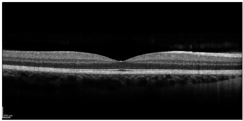
FIGURE 4SCAN OF THE RETINA BELONGING TO THE AUTHOR OF THIS THESIS, SHOWCASING THE LAYERED STRUCTURE.IMAGE: DR IRENE CTORI. ... 48
FIGURE 5A) NORMALISED RESPONSITIVITY SPECTRA OF HUMAN CONE CELLS: SHORT, MEDIUM AND LONG CONES. B) APPROXIMATION OF COLOUR SPECTRUM VISIBLE TO THE HUMAN EYE. C) ISAAC NEWTON’S (1642– 1726/27) DESCRIPTION OF THE COLOUR SPECTRUM, BASED ON OBSERVATION OF WHITE LIGHT SHINED THROUGH A PRISM AND DECOMPOSED AS AN EFFECT.. ... 49
FIGURE 6CONNECTIVITY IN THE PRIMATE RETINA.. ... 54
FIGURE 7LAYERING OF THE LATERAL GENICULATE NUCLEUS (LGN) ... 56
FIGURE 8VISUAL PATHWAYS FROM THE RETINA THROUGH THE LGN TO THE V1. ... 59
FIGURE 9THE CIE1931RGB COLOUR MATCHING FUNCTIONS... 77
FIGURE 10CIE CHROMATICITY DIAGRAM ... 78
FIGURE 11THE DKL COLOUR SPACE. ... 82
FIGURE 12A GENERAL OUTLINE OF CALIBRATION AND VERIFICATION PROCEDURE STEPS.. ... 88
FIGURE 13AN EXAMPLE OF THE SPECTRAL POWER DISTRIBUTION (SPD) OF THE DISPLAY USED IN THE EXPERIMENTS. ... 89
FIGURE 14TRIAL OUTLINE OF THE MATCH-TO-SAMPLE TASK USED TO MEASURE DISCRIMINATION THRESHOLD.. ... 126
FIGURE 15DELAYED MATCH-TO-SAMPLE TASK. ... 128
FIGURE 16RESULTS FROM ACUITYPLUS TESTS AND THE CAD TEST. ... 136
FIGURE 17RESULTS OF THE WM TESTS.. ... 137
FIGURE 18MEAN PROPORTION OF CORRECT RESPONSES FOR S-CONE,L–M AND LUMINANCE STIMULI AT THREE LEVELS OF WM LOAD ... 139
FIGURE 19MEAN PROPORTION OF CORRECT RESPONSES FOR S-CONE,L–M AND LUMINANCE STIMULI AT 3 LEVELS OF WM LOAD. ... 140
FIGURE 20A)PROPORTION OF CORRECT RESPONSES FOR S-CONE,L–M AND LUMINANCE STIMULI AT 3WM LOADS SHOWN SEPARATELY FOR THE MATCH AND MISMATCH PROBES. ... 142
FIGURE 21MEAN OF MEDIAN REACTION TIMES FOR S-CONE,L–M AND LUMINANCE STIMULI AT THREE LEVELS OF WM LOAD ... 143
FIGURE 22LINE PLOT SHOWING ACCURACY IN RESPONSE TO S-CONE,L–M, MIXED SIGNALS AND LUMINANCE-DEFINED SHAPES AT THREE WM LOAD LEVELS, IN RESPONSE TO MATCH OR MISMATCH PROBE.. ... 145
FIGURE 23LOCAL PEAK AMPLITUDES OF COMPONENT P1 FOR S-CONE L–M AND LUMINANCE STIMULI, AT THREE LEVELS OF LOAD FOR LOW AND HIGH CONTRAST. ... 146
FIGURE 24GRAND AVERAGE WAVEFORM AT ELECTRODE O1 DURING ENCODING LOW CONTRAST LUMINANCE-DEFINED SHAPES, AT THREE LEVELS OF WM LOAD. ... 147
FIGURE 25GRAND AVERAGE WAVEFORM AT ELECTRODE O1 DURING ENCODING HIGH CONTRAST LUMINANCE-DEFINED SHAPES, AT THREE LEVELS OF WM LOAD.. ... 148
10
FIGURE 27LOCAL PEAK AMPLITUDES OF COMPONENT N1 FOR EACH DKL DIRECTION, AT THREE LEVELS OF LOAD FOR LOW AND
HIGH CONTRAST, AVERAGED OVER ELECTRODES O1,O2 AND OZ.. ... 151
FIGURE 28GRAND AVERAGE WAVEFORM AT ELECTRODE C1 DURING ENCODING HIGH CONTRAST LUMINANCE-DEFINED,L–M
AND S-CONE SHAPES AT LOAD 3... 156
FIGURE 29P3B AMPLITUDE AT ELECTRODES P3,P4 AND PZ, AT LOW AND HIGH CONTRAST FOR LUMINANCE,L–M AND
S-CONE.. ... 158
FIGURE 30P3B WAVEFORMS AT ELECTRODE P3 AT THREE LEVELS OF WM LOAD, FOR S-CONE,L–M AND LUMINANCE. . 159
FIGURE 31GRAND AVERAGE WAVEFORM AT ELECTRODE O1 DURING MAINTENANCE AT THREE LEVELS OF WM LOAD. ... 162
FIGURE 32GRAND AVERAGE WAVEFORM AT ELECTRODE F3 DURING MAINTENANCE AT THREE LEVELS OF WM LOAD. ... 163
FIGURE 33GRAND AVERAGE WAVEFORM AT ELECTRODE O1 DURING RETRIEVAL OF LOW CONTRAST LUMINANCE-DEFINED
SHAPES, AT THREE LEVELS OF WM LOAD. ... 165
FIGURE 34GRAND AVERAGE WAVEFORM AT ELECTRODE O1 DURING RETRIEVAL OF HIGH CONTRAST LUMINANCE-DEFINED
SHAPES, AT THREE LEVELS OF WM LOAD. ... 166
FIGURE 35CORRELATION BETWEEN THE P1 COMPONENT AT ELECTRODE O1(FOR LOAD 3) AND OVERALL TASK ACCURACY. ... 168
FIGURE 36GRAND AVERAGE WAVEFORM AT ELECTRODE O1 DURING RETRIEVAL OF HIGH CONTRAST LUMINANCE-DEFINED
SHAPES, AT THREE LEVELS OF WM LOAD. ... 170
FIGURE 37 GRAND AVERAGE WAVEFORM AT ELECTRODE CZ DURING RETRIEVAL AT THREE LEVELS OF WM LOAD. DKL
CONDITIONS AND CONTRAST LEVELS WERE COLLAPSED TO HIGHLIGHT THE MAIN EFFECT OF WM LOAD. ... 173
FIGURE 38 GRAND AVERAGE WAVEFORM AT ELECTRODE PZ DURING RETRIEVAL AT THREE LEVELS OF WM LOAD. DKL
CONDITIONS AND CONTRAST LEVELS WERE COLLAPSED TO HIGHLIGHT THE MAIN EFFECT OF WM LOAD.. ... 174
FIGURE 39TWO INTERVAL FORCED-CHOICE PROCEDURE (2IFC) USED TO MEASURE STIMULUS DETECTION THRESHOLDS. ... 208
FIGURE 40MULTIPLES OF DETECTION THRESHOLDS PLOTTED FOR 4DKL CONDITIONS, AT 3 LEVELS OF WM LOAD.. ... 211
FIGURE 41WM THRESHOLDS AT LOAD 3, FOR EACH DKL CONDITION.RED LINES ARE MEAN WM THRESHOLDS ACROSS
OBSERVERS,. ... 212
FIGURE 42 RESULTS OF MODEL FITS FOR THE 2IFC DETECTION THRESHOLD TASK. ... 216
FIGURE 43COMPARISON OF SLOPES OF THE PSYCHOMETRIC FUNCTION FOR DIFFERENT POST-RECEPTORAL MECHANISMS FITTED
TO DATA DERIVED FROM SHAPE DISCRIMINATION TASK ... 218
FIGURE 44 RESULTS FROM A DELAYED MATCH-TO-SAMPLE TASK WITH FIXED STIMULUS CONTRAST FROM THE PREVIOUS
EXPERIMENT ... 220
FIGURE 45TOP: TITLE PAGE OF BLEULER’S 1911 ARTICLE ON DEMENTIA PRAECOX, WHERE HE REFERRED TO THE DISORDER AS
“GROUP OF SCHIZOPHRENIAS”. AND A TITLE PAGE OF THE 1912’S TRANSLATION OF KRAEPELIN'S “LEHRBUCH DER
PSYCHIATRIE”, ORIGINALLY PUBLISHED IN 1899 ... 228
FIGURE 46RESULTS OF THE VISION TESTS FOR PATIENTS AND CONTROLS SHOWN IN BOX PLOTS.. ... 256
FIGURE 47OVERALL MEAN ACCURACY FOR CONTROL AND PATIENT GROUP AND CONTROL/PATIENT GROUP MEAN ACCURACY
SHOWN AT WM LOAD 1 AND LOAD 3, SEPARATELY FOR DKL DIRECTIONS ... 257
FIGURE 48ACCURACY AT LOAD 1 AND LOAD 3 FOR MATCHING AND MISMATCHING MEMORY PROBE, SHOWN FOR EACH DKL
DIRECTION AND CONTROL/PATIENT GROUP SEPARATELY.. ... 259
FIGURE 49OVERALL MATCH & MISMATCH ACCURACY (PATIENTS + CONTROLS) FOR THE DIFFERENT DKL DIRECTIONS AT LOW
AND HIGH WM LOAD. ... 260
FIGURE 50OVERALL REACTION TIMES FOR CONTROL AND PATIENT GROUP AND CONTROL/PATIENT GROUP REACTION TIMES ARE
SHOWN AT WM LOAD 1 AND LOAD 3, SEPARATELY FOR DKL DIRECTIONS. ... 260
FIGURE 51REACTION TIMES FOR S-CONE,L–M AND LUMINANCE CONDITIONS. ... 261
FIGURE 52OVERALL MATCH & MISMATCH REACTION TIMES (PATIENTS + CONTROLS) AND RTS FOR DIFFERENT DKL DIRECTIONS
11
FIGURE 53COMPARISON BETWEEN THE NUMBER OF TRIALS FOR PATIENTS AND CONTROLS. ... 262
FIGURE 54ERP WAVEFORMS DURING THE ENCODING OF LOW CONTRAST LUMINANCE SHAPES FOR CONTROL GROUP AND SCHIZOPHRENIA GROUP ... 263
FIGURE 55ERP WAVEFORMS DURING THE ENCODING OF HIGH CONTRAST LUMINANCE SHAPES ARE SHOWN FOR CONTROL GROUP AND SCHIZOPHRENIA GROUP ... 264
FIGURE 56ERP WAVEFORMS DURING ENCODING OF S-CONE,L–M AND LUMINANCE SHAPES ... 266
FIGURE 57 N1 AMPLITUDE IN RESPONSE TO DIFFERENT DKL DIRECTIONS AT LOW CONTRAST LEVEL FOR CONTROL AND SCHIZOPHRENIA GROUP.. ... 267
FIGURE 58N1 COMPONENT LATENCIES FOR CONTROL AND SCHIZOPHRENIA GROUP, FOR THE THREE DKL DIRECTIONS. ... 268
FIGURE 59N1 COMPONENT LATENCIES FOR CONTROL AND SCHIZOPHRENIA GROUP DURING ENCODING LUMINANCE-DEFINED SHAPES.. ... 269
FIGURE 60P3B AMPLITUDE IN RESPONSE TO S-CONE,L–M AND LUMINANCE SHAPES. ... 270
FIGURE 61ERP WAVEFORMS DURING ENCODING OF S-CONE,L–M AND LUMINANCE SHAPES.. ... 270
FIGURE 62P3B AMPLITUDES AT ELECTRODE O1,O2 AND OZ, AT TWO LEVELS OF LOAD, FOR EACH DKL DIRECTION.. ... 272
FIGURE 63LATE, SLOW WAVE FOR CONTROLS AND PATIENTS PRESENTED AT LOW CONTRAST, AT ELECTRODE OZ ... 273
FIGURE 64SLOW WAVE FOR THE PATIENT GROUP, PRESENTED AT LOW AND HIGH CONTRAST, FOR SEPARATE DKL DIRECTIONS AND LOAD LEVELS.. ... 274
FIGURE 65ERP WAVEFORMS COLLAPSED ACROSS LOAD CONTRASTING PATIENT AND CONTROL LUMINANCE DATA AT RETRIEVAL AT LOW AND HIGH LEVELS OF CONTRAST. ... 275
FIGURE 66WAVEFORMS COLLAPSED ACROSS WM LOAD, ELECTRODE LOCATION & CONTRAST TO SHOW DIFFERENCES BETWEEN DKL DIRECTIONS, FOR CONTROLS AND PATIENTS SEPARATELY. ... 276
FIGURE 67ERP WAVEFORMS COLLAPSED ACROSS CONTRAS LEVELS,WM LOAD AND ELECTRODE LOCATIONS (C1,C2&CZ), PRESENTED AT EACH DKL DIRECTION SEPARATELY ... 277
FIGURE 68WAVEFORMS COLLAPSED ACROSS DKL DIRECTIONS, LOAD AND CONTRAST LEVELS AT ELECTRODE PZ FOR CONTROLS AND PATIENTS ... 279
FIGURE 69P3B AMPLITUDE DURING RETRIEVAL FOR 3DKL DIRECTIONS, AT LOAD 1 AND LOAD 3. ... 280
13
List of Tables
TABLE 1THE THREE CARDINAL AXES IN DKL COLOUR SPACE. ... 81
TABLE 2PSYCHOPHYSICAL MECHANISMS OF INTEREST AND THEIR CORRESPONDING DKL COORDINATES. ... 84
TABLE 3SUMMARY OF TESTS COMPLETED BY ALL PARTICIPANTS. ... 123
TABLE 4EXPERIMENTAL CONDITIONS. ... 130
TABLE 5P1 AMPLITUDE AT ENCODING STAGE FOR LUMINANCE CONDITION AND N1 AMPLITUDE AT ENCODING FOR TWO ISOLUMINANT CONDITIONS CORRELATED WITH OVERALL TASK ACCURACY ACCURACY. ... 153
TABLE 6P1 AMPLITUDE AT ENCODING STAGE FOR LUMINANCE CONDITION AND N1 AMPLITUDE AT ENCODING FOR TWO ISOLUMINANT CONDITIONS CORRELATED WITH TASK ACCURACY AT HIGH CONTRAST ... 154
TABLE 7PEARSON CORRELATION COEFFICIENTS AND P VALUES FOR P1 AMPLITUDE AT RETRIEVAL STAGE WITH OVERALL ACCURACY. ... 167
TABLE 8PEARSON CORRELATION COEFFICIENTS AND P VALUES FOR A CORRELATION BETWEEN P1 AMPLITUDE AT RETRIEVAL STAGE FOR MISMATCHING PROBES AND ACCURACY FOR MISMATCHING PROBE AT THREE WM LOADS. ... 167
TABLE 9PEARSON CORRELATION COEFFICIENTS AND P VALUES FOR P1 AMPLITUDE AT RETRIEVAL STAGE FOR MATCHING PROBES WITH ACCURACY FOR MATCHING PROBE AT THREE WM LOADS. ... 168
TABLE 10PSYCHOPHYSICAL MECHANISMS OF INTEREST AND THEIR CORRESPONDING DKL COORDINATES. ... 205
TABLE 11SUMMARY OF EXPERIMENTAL CONDITIONS. ... 207
TABLE 12WEBER CONTRASTS FOR ACHROMATIC AND CHROMATIC MECHANISMS FOR EACH CONDITION, DERIVED FROM L,M AND S CONTRASTS. ... 223
TABLE 13CODING OF EDUCATIONAL LEVEL AND A SUMMARY OF AGE/QUALIFICATIONS FOR CONTROL AND SCHIZOPHRENIA GROUP. ... 247
TABLE 14INDIVIDUAL DISCRIMINATION THRESHOLDS FOR ALL DKL DIRECTIONS OBTAINED IN THE PREVIOUS EXPERIMENT .. 250
TABLE 15 WEBER CONTRAST VALUES FOR ALL POST-RECEPTORAL MECHANISMS TESTED IN THE EXPERIMENT, BASED ON AVERAGED THRESHOLDS. ... 250
TABLE 16CONDITIONS IN THE CURRENT STUDY. ... 251
15
Acknowledgments
It is impossible to face all the challenges that the PhD brings on one’s own. Although I have been told that earning a doctorate is a long journey, there were times where I felt like all I was doing was falling – slowly, but inevitably – rather than walking towards the goal. I am indebted to many people who (knowingly or not) have helped me to get where I am now.
I would like to thank Dr Corinna Haenschel, my supervisor, for the opportunity, support, and patience; Dr Jasna Martinovic, for her expertise, and for getting me on this path in the first place; Dr Elliott Freeman, my 2nd supervisor, and Dr Ben
J. Jennings, for all their help; my old University of Aberdeen friends and faculty, for making science fun before I started my PhD; thanks to all the doctorate students and postdocs from our department (and beyond), for keeping it that way, and for creating the community I felt so good to be part of (and for all the Jägerbombs); to the City, University of London faculty and staff, for having me here and helping me out along the way; thanks to Emily and Pete, for all the spare parts and help in the lab; to everyone who I shared the EEG lab with (especially Laila and Matthew – I learned a lot from you); to Jenny, Neelam, Shinal and Saim, for their help in running the patient study; to the Polish science blogging community, for broadening my interests; to the “twitter people”, for company, laughs, and advice; to the Antisocial Grumpy Club, for the friendship and gossip; to Paula, for the email you sent me on the 3/11/2015; to my Princess Bubblegum, for all the spaceship adventures; to Megan, for being a true Walrus to me; to Klaus Schmiegel, for general support; to my coinhabitant friends: Ania and Jadzia (and Kota, too) for all the warmth and non-blended soups; to Michał Wawrzyński, my high school Polish teacher, for cultivating my critical thinking; to Żółwik, Ilona, Kasia, Szymon, Gelu, Asuka, Francuz, Gizmo, Ziół, and other friends from old times, who are (or were) always there; to my brother, Marcin, the best brother I could ever ask for; to my dad, Tadeusz, for believing in me and supporting me (dziękuję, tatuś); and to Aga, for the companionship, patience, love, and the cuddles (when I needed them the most) – dziękuję.
Finally, I want to give my thanks – and dedicate this thesis – to my mom, Krystyna. I know you would be proud, and it hurts me I cannot share this moment with you.
17
19
Abstract
Visual Working Memory (visual WM) is an ability to encode and temporarily maintain visual information. There is some evidence that early perceptual processes make an important contribution to successful WM performance. However, perceptual contributions to WM are not yet fully understood.
In vision, signals originating from three classes of photoreceptors in the retina (L, M and S-cones) are combined into three distinct mechanisms, which form the fundamentals of visual perception. These mechanisms are the two opponent chromatic mechanisms (L – M and S – (L + M) and an achromatic, luminance mechanism (L + M). Vision science has been long concerned with properties of these mechanisms and how they contribute to the perception of the world. Despite this, there was little interest to date in how these mechanisms contribute to creating memory representations, i.e. after the visual stimulus has disappeared from the visual field. In a series of experiments presented in this thesis, a
differential contribution of three post-receptoral mechanisms to visual WM was investigated. It was hypothesised that luminance signals will prove to be more efficient in their contribution to WM encoding, maintenance and retrieval than opponent chromatic signals. This was investigated using a variety of
methodologies, from psychophysical measurements and behavioural responses to recordings of neural activity using electroencephalography (EEG).
Results of the experiments have shown that indeed, remembering abstract shapes designed to excite the luminance mechanism contributed to better WM
21
Chapter 1
General Introduction
The term working memory (WM) became established in the scientific literature in the early 1960s (Miller, Galanter, & Pribram, 1960; Pribram, Ahumada, Hartog, & Roos, 1964). It refers to a short-term storage system that temporarily maintains information for immediate use. The important aspect of the WM definition is its emphasis on goal-directed behaviour. Short-term storage of any information that may be no longer present in the environment is the basis for carrying out
complex tasks, such as learning, comprehension, reasoning or operating in a visual world (Baddeley & Hitch, 1974; Logie, 2011; Phillips, 1974).
A decade later, two independent lines of research delivered ideas that still
influence our current understanding of WM (Postle, 2006). Electrophysiological recordings in primates looked at neural activity in the dorsolateral prefrontal cortex (dorsolateral PFC) while subjects were engaged in memory task (Fuster, 1973; Fuster & Alexander, 1971; Niki, 1974). This required primates to remember presented stimulus and retain it over time. It was demonstrated that neurons in the PFC continued to fire after the stimulus was no longer present in the visual field. This was taken as an evidence for sustained memory-related activity. These early findings helped to direct the focus of electrophysiological WM research on the prefrontal areas in the brain.
Almost in parallel to the animal research, there was another development in a different field that would later prove to be extremely influential across
22
WM as a multi-component system. According to it, WM consists of two separate and largely independent storage systems. The phonological loop is responsible for storing auditory and/or phonological information, while the visuospatial sketchpad deals with visual information. Both storage systems were governed by a central executive system. Its primary role is to control and manage information stored in both buffers.
Another influential WM model was later developed by Cowan (1995, 2001, 2005, 2008). He postulated that WM is a system that forms part of long-term memory and is not, therefore, a separate construct – a view similar to the one proposed by Anders and Kintsch (1995). According to Cowan’s model, what is regarded as WM “representation” is simply information held in long-term memory that has been temporary re-activated. He proposed that this re-activating, cognitive “force” is attention.
Attention can be defined as the ability to select and focus on a piece of
information in the environment1. This definition was extended to highlight that
one can focus attention towards internally (mentally) represented stimuli (Itti, Rees & Tsotsos, 2005). Thus, Cowan proposed that the focus of attention can reactivate representations stored in long-term memory and thus make them available for manipulation and immediate usage. While Cowan postulated that there is theoretically no limit to how many long-term memory representations one can hold, the focus of attention is a capacity-limited process. In other words, an individual can reactivate only a number of such representations at a time, thus explaining the limited nature of working memory. Cowan estimated the capacity of focus of attention to be around four “chunks of information”.
An important, although not always spelt-out an aspect of working memory is that it involves differed processing stages. Definitions of working memory (e.g.
1 The definition provided here is constrained to this circumstance, while purposefully avoiding being too
23
Baddeley & Hitch, 1974; Woodman & Vogel, 2005) implicitly assume that prior to retrieval of information, the information that has been remembered (encoded), needs to be maintained over a period of time. In other words, working memory can be conceptualised as a cognitive ability that is comprised of encoding,
maintenance and retrieval/recall. The relative importance of each stage has been explicitly addressed using task designs that allow to clearly separate them
temporally (Bays, Marshall, & Husain, 2011; Haenschel, Bittner, Haertling,
Rotarska-Jagiela, Maurer, Singer, & Linden, 2007; Woodman & Vogel, 2005). For example, Woodman & Vogel (2005) used a modified change-detection paradigm. In this paradigm, participants are required to encode visual stimuli, followed by a maintenance period, and are subsequently presented with a test array.
Participants retrieve remembered stimuli in order to judge whether the probe is the same as or different than the stimuli presented previously. Using a similar paradigm, Bays et al. (2011) showed that working memory capacity may depend on separate limits imposed during encoding and maintenance. More specifically, the encoding limit constraints transfer of information into memory, while the maintenance limit affects the precision with which the stimuli can be retained over time. Furthermore, another study (Haenschel et al., 2007) showed that different WM stages might be selectively affected in conditions with reported working memory deficits, such as schizophrenia. These examples highlight the importance of defining working memory not as a uniform cognitive ability, but rather a construct that can, and should be, divided into separate processing stages. These three stages will be referred to throughout this thesis as the encoding, maintenance, and retrieval (or recall).
One of the important, however implicit implications of this model was defining WM as an independent cognitive system, separate from other faculties, such as perception. This corresponds to a traditional view that perceptual information is
transferred to memory and is not considered an integrated part of it (Magnussen,
2000; Squire & Kendell, 1999).
24
research. Goldman-Rakic (1989, 1990) proposed that delay-period activity in PFC recorded in behaving animals corresponds to storage buffers described in
Baddeley and Hitch multi-component model. An interesting and highly influential consequence of this idea was that working memory was a general phenomenon that could be demonstrated across species. Postle (2006) sees Goldman-Rakic’s work as an important milestone that brought cognitive
psychology and electrophysiological WM research together. Such an integrated approach proved to be productive: for example, Goldman-Rakic proposed that the division between the object (“what”) and spatial location of the object (“where”) seen in the visual system could also hold for visual working memory (Wilson, O’Scalaidhe, & Goldman-Rakic, 1993). The neuroscientific, as well as cognitive and psychological research that followed, has since supported this notion (see Postle, 2006 for references).
It is interesting that Goldman-Rakic and colleagues’ proposal described above (Wilson et al., 1993) was one of the first indications that the rules governing the visual system (and thus, visual perception) can be also applied to working
memory, a supposedly separate construct. Although visual working memory and perception have continued to be regarded and researched largely separately from visual perception, it seems that such conceptual separation can no longer be sustained (Pasternak & Greenlee, 2005). It is now acknowledged that the role of a perceptual system extends from “mere” encoding of stimuli to memory storage as well (Pasternak & Greenlee, 2005). In neuroscientific terms, this implies that the sensory areas that process a given stimuli will be also responsible for its storage when the stimuli are no longer available to the senses, a notion that was
25
Fast-growing research on interactions between perception and WM is a symptom of this new approach (D’Esposito & Postle, 2015; Gao, Gao, Li, Sun, & Shen, 2011; Harrison & Tong, 2009; Lara & Wallis, 2012; Pasternak & Greenlee, 2005; Postle, 2006; Yin et al., 2012).
When talking about interactions between perception and working memory, the encoding stage should be given a special consideration. It is at this stage that perception and working memory are likely to interact, given that the stimulus is still present in the visual field. The general aim of the research presented in this thesis is thus to establish the importance of the encoding stage to WM
performance (Haenschel et al., 2007). Furthermore, another goal is to specify the relationship of this early WM stage with perceptual processing, as well as to determine whether the later stages of WM can also benefit from these interactions and impact task performance.
In the following sections, I will describe the evidence that established dorsolateral PFC as a crucial anatomical structure responsible for WM
26
1.
Working memory and the prefrontal cortex (PFC)
A good deal of evidence pointing to frontal cortices as the processing and storage site for working memory came from animal studies using delayed-discrimination tasks (Goldman-Rakic, 1992, 1995; Levy & Goldman-Rakic, 2000; Rodriguez & Paule, 2009). In such experiments, animals are instructed to remember a
location of a presented target that is subsequently occluded. After a short delay, primates look for a bait in a number of possible locations. Importantly, this task design ensures that the representation has to be updated on a trial-to-trial basis. Hence, target’s location cannot be predicted from the stimulus itself or from its location on a previous trial but has to depend on the memory representation of the target (Goldman-Rakic, 1995).
One of the earliest studies of this kind using electrophysiological recordings were published in the early 70’s (Fuster & Alexander 1971; Kubota & Niki, 1971). These showed that during a delay period (i.e. a time when the memory representation was stored “in mind” in the absence of the stimulus) a subset of neurons in the PFC sustained their activation.
With time, new paradigms and more accurate techniques became available, which led to the further advancement of the field. For instance, Funahashi et al. (1989) used an oculomotor paradigm, where monkeys were required to maintain fixation on the target during stimulus presentation and retention. This design ensured that animals maintained their fixation on the display, and hence ensured that the performance depended on the stimulus encoding (successful or not), and not on poor fixation or distractibility.
27
the PFC demonstrated a prolonged target-related activity in the absence of the target, but only at locations that were earlier identified as preferred by these neurons (see Figure 1 for an example). Such delay-period activity associated with a target previously presented in a specific location is referred to as the memory
[image:28.595.125.523.181.554.2]field.
Figure 1 Example of memory fields in action. The target was presented in one of the eight
possible locations. In this case, the target appeared at an eccentricity of 270°. On the roster plot in the middle of the bottom row, we can observe an increased spiking of a prefrontal neuron in the absence of the target, during a maintenance stage. Note that the responses of the same neuron do not differ from a baseline if the target was presented at different eccentricities. Figure
from Goldman-Rakic, 1995.
28
spatial working memory tasks. Although generally, all studies reported
activations in the frontal areas, they were not restricted only to these locations. For example, some studies showed activations in Broca’s area (i.e. the brain area classically indicated in language processing), in addition to activity in the PFC (Paulesu et al., 1993). This location did not correspond to areas that were
previously associated with working memory in primates. It was thought that the differences within human studies and between human and animal studies arise because of a verbal component in these tasks. To address that, McCarthy et al. s(1994) attempted to provide a more accurate localisation of working memory processing in humans using a task that would restrict verbal involvement. Their experiments employed magnetic resonance imaging (MRI) rather than PET, which also allowed for a more precise localisation of WM processing. Subjects performed a variation of the delayed – response task: they were presented with an array of stimuli randomly flashed at different locations. Participants had to
29
Figure 2 Focus of activation during a WM task located in the PFC. The figure is taken from
McCarthy et al. (1994).
Nevertheless, although the prefrontal cortex has been regarded as playing a pivotal role in working memory, it was by far not the only area employed during WM. It was apparent that a distributed network is responsible for WM
30
concluded that the activity in the PFC during working memory task is not unitary. During WM, activity in PFC reflects activity associated with different stages of working memory, as well as any non-mnemonic processes that must be employed during these tasks. More specifically, encoding, maintenance and retrieval engage differed portions of the PFC to a different extent. For example, while encoding is associated predominantly with the activity in the dorsolateral PFC, maintenance engages ventrolateral as well as dorsolateral PFC. If a
manipulation of a stimulus was required in addition to simple maintenance, activation in dorsolateral part could be observed on top of the storage-related activations. When the memorised information has to be accessed to complete a task, dorsolateral PFC is again recruited, although this activation might be reflecting preparation of motor response in addition to processes related to “scanning” memorized information.
Another group of researchers (Pessoa, Gutierrez, Bandettini, & Ungerleider, 2002) took a different approach to show the relevance of activity in different parts of the brain to the task. They compared the activity of correct and incorrect trials during encoding, maintenance and working memory retrieval during a delayed discrimination task. Unlike D’Esposito et al. (2000), a visual rather than verbal stimuli were used. Performance-related activity was mostly demonstrated in the frontal and parietal cortex. The results showed that activity in frontal eye fields and intraparietal sulcus as well as in dorsolateral PFC during memory delay strongly predicted behavioural performance on a trial-by-trial basis. However, performance-related activation during encoding and recall also correlated with performance. Pessoa et al. suggested that variability in behavioural performance occurs due to fluctuations in attention, which can occur at any WM stage, thus explaining the evident correlation between behavioural performance and BOLD activity throughout the task, at encoding, maintenance and retrieval.
31
maintain memory representation: activity in these regions was interpreted as related to processing of the stimuli, and not of an active WM storage (Munk et al., 2002; Pessoa et al., 2002). For example, Munk et al. (2002) showed that occipital-temporal areas are recruited predominantly during stimulus encoding. Activity in this areas would return to baseline after the initial stimulus-related activity and could be observed again only during a presentation of a test probe, after the maintenance period. Pessoa et al. (2002) additionally demonstrated that successful behavioural performance in a working memory task could not be predicted from activity in sensory areas during encoding. At the same time, the activation in the frontoparietal network was associated with performance, further supporting the idea that sensory cortices are not involved in the storage of
information per se.
1.1.
Summary: working memory and the
prefrontal cortex (PFC)
To sum up, it was generally agreed that the prefrontal cortex is the main brain structure responsible for short-term memory storage. While the sensory cortices are implicated as well, their role was usually described in the context of
processing the stimuli before it was “transported” to memory storage. At the same time, research had indicated that the PFC and sensory cortices do not work in separation. Indeed, recent research recognises that working memory involves multiple, coordinated brain areas, with the PFC being only one of them
32
2.
The role of top-down signals in working memory
The notion of “top-down” processing stems from Gregory’s (1970) theory of perception. According to his theory, visual perception is an active process which is influenced by prior assumptions and knowledge about the world. This is in contrast with Gibson’s (1966) bottom-up account of perception, which states that what we perceive is predominantly stimulus-driven. Both accounts are therefore attempting to establish to what extent our perception relies on information that is present in the environment, versus the information that we already hold. On the neural level, bottom-up processing would refer to processing that goes from the retina “up” the visual hierarchy, through the LGN to sensory cortex, and from there to other brain areas. On the other hand, top-down processing would refer to signals originating in higher-order cortices, such as frontal areas, which
propagate back “down” to the sensory cortex. The dorsolateral prefrontal cortex is one candidate for such top-down controller, given its extensive network of
reciprocal anatomical connections with other cortical and subcortical regions (Knudsen, 2007; Miller & Cohen, 2001).One of the top-down factors that can influence ongoing perception are predictions, referred to Gregory (1970) as hypotheses. These hypotheses are important to cognition, as they can direct and constrain the incoming information and facilitate its processing (O’Callaghan, Kveraga, Shine, Adams, & Bar, 2017). In terms of perception, it has been shown, for example, that predictions can facilitate recognition of objects (Kveraga, Boshyan, & Bar, 2007; Martinovic, Mordal, & Wuerger, 2011).
Today, the distinction between top-down and bottom-up processing is widely used and is not constrained solely to perception. It can be successfully applied to describe other cognitive functions, such as memory.
One of the top-down factors that have been implicated in a wide range of
33
Zanto et al. asked human subjects to selectively remember the target feature (motion or colour) while ignoring the distractor (irrelevant feature). Using rTMS over a region in PFC – inferior frontal junction, or IFJ – they hoped to disrupt top-down, attentional modulation of visual processing and working memory
encoding. Their results showed that rTMS had an effect on perceptual processing. This was reflected in a decreased amplitude of the EEG signal associated with visual processing (indexed by an event-related component P1) around 100 ms after the stimulus appeared on the screen. Importantly, this decrease in P1 was associated with a decrease in behavioural performance. Zanto et al. suggested that the decrease in both behavioural performance and neural activity was a consequence of disrupted top-down attentional modulation. They hypothesised that top-down signalling (which in their case means selective attention) helps to create a high-fidelity memory representation of the stimulus. Additionally, they speculated that top-down signals from the inferior frontal junction (IFJ) help to update task representations (i.e. maintaining the current task requirement). This agrees with previous studies that also suggested that the activity in the PFC in general, and IFJ in particular, is involved in updating task representations (Brass & Von Cramon, 2004). The finding also agrees with the account that the activity in the PFC interacts with other areas responsible for stimulus or other task-relevant processing (Baddeley 2003, Constantinidis & Wang 2004; Knudsen, 2007). Anatomical connections between the PFC and other brain regions would serve as a neural substrate for such interactions, as mention above (Knudsen, 2007).
34
to contribute to greater efficiency of stimulus encoding (Hawkins et al., 1990; Reinitz, 1990). Greater efficiency might also lead to the greater fidelity of mental representation of the stimulus, which is crucial for working memory performance (Zanto & Gazzaley, 2009) 2. It has also been shown that this top-down control
predicts WM performance (Rutman, Clapp, Chadick, & Gazzaley, 2009), thus highlighting the importance of such signals in working memory processing.
2.1.
Summary: The role of top-down signals in
working memory
The previous section established that multiple brain areas are involved in visual working memory, with the storage located in the prefrontal cortex. Working memory depends on the coordinated activity between these regions, with a special role of top-down inputs from the prefrontal cortex to sensory areas (Knudsen, 2007; Lara & Wallis, 2012). It is interesting to note that the sensory cortex and perception are usually discussed in the context of the PFC and higher cognitive processes (such as attention). Coincidently, focusing on higher-order cortical areas while “downplaying” the importance of sensory cortices (at least in a sense that they cannot sustain memory representations on their own) is in line with the tendency to view memory and perception as separate constructs (Squire & Kandel, 1999).
However, recent years have seen a certain shift in the way the visual cortex (and, more generally, perception) is viewed in the context of visual working memory. The evidence for an active involvement of perceptual systems in WM has been accumulating in recent years and is currently enjoying much attention
(D’Esposito, 2007; D’Esposito & Postle, 2015; E. Ester, Serences, & Awh, 2010; Harrison & Tong, 2009; Pasternak & Greenlee, 2005). The view that similar
2 It is important to note that by fidelity, Gazzeley and others mean how well the target can be distinguished
35
mechanisms (and, consequently, similar brain areas and networks) are involved in both encoding and storage of information over short periods of time became known as the sensory recruitment hypothesis (Harrison & Tong, 2009; Pasternak & Greenlee, 2005). This is a big departure from a traditional view, in which frontal areas are solely responsible for memory storage. More and more studies provide evidence in support for the sensory recruitment hypothesis (D’Esposito & Postle, 2015). The evidence in its favour spans a range of different methodologies, from single-cell recordings, human neuroimaging, electrophysiology, and
36
3.
Beyond the prefrontal cortex
3.1.
Sensory areas and perceptual processing
contributing to working memory.
While prefrontal areas were taking most of the spotlight in WM research, reports of an active involvement of visual areas and perception in WM started to appear relatively early (Fuster & Jervey, 1982; Miyashita and Chang, 1988).
Fuster and Jervey (1982) recorded the single unit activity of neurons in the inferotemporal cortex in monkeys during a visual delayed match-to-sample task. Monkeys were presented with a sample colour and, after the delay, required to choose the previously presented one from two alternatives. The results showed colour – dependent increase in firing rates not only during stimulus encoding but also during the retention interval. Fuster and Jervey interpreted this results as evidence for the involvement of a subset of inferotemporal neurons in memory. Importantly, neurons that exhibited such pattern did not appear to be specialised for memory storage. Rather, their participation in memory functions occurred in addition to their visual functions. Miyashita & Chang (1988) extended these findings by showing that neurons in the temporal lobe of a non-human primate are able to sustain the memory of more complex objects as well. After identifying sets of neurons responsive to particular shapes, they showed that these neurons sustained their firing rates during retention interval after the shapes were removed from the visual field. Interestingly, the activity of those neurons, while related to the content of the stimuli, were independent of their size or
orientation.
Low-level visual areas have also been implicated in working memory using single-cell recordings in the macaque. For example, one study (Super, Spekreijse, & Lamme, 2001) required macaques to remember objects in a
37
both correct and incorrect trials. Super et al. (2001) demonstrated that activity related to this figure-ground separation in the visual cortex was sustained during the maintenance period. Interestingly, this activity was sustained only for correct trials, while it would disappear for incorrect trials.
These findings were further extended by studies showing that activity of populations of neurons, rather than single cells, are also related to working memory (Lee, Simpson, Logothetis, & Rainer, 2005). This can be achieved by recording local field potentials (LFP) and looking at the task-related oscillatory activity. Populations of neurons firing in synchrony create functional networks which can be related to the task at hand (Buzsáki & Draguhn, 2004; Buzsáki & Wang, 2012; Canolty et al., 2006; Whitman, Ward, & Woodward, 2013). For instance, Lee et al. (2005) demonstrated that local field potentials recorded from occipital visual cortex during delayed-discrimination task showed an energy enhancement in the theta band (4 – 8 Hz) during memory maintenance. This pattern was interpreted as an evidence for an active involvement of extrastriate visual cortex in working memory. Another study on non-human primates (Liebe, Hoerzer, Logothetis, & Rainer, 2012) showed evidence for inter-areal
communication between prefrontal and sensory areas during a visual short-term memory task. This was evidenced by phase synchronisation in the theta range (defined in this experiment as 3 – 9 Hz) between the areas, i.e. the oscillatory activity in one area was tightly related to activity in the other area. Additionally, single unit activity in this areas was also shown to be related to the ongoing theta oscillations.
Human EEG recordings also point to the crucial role that oscillations originating in sensory cortices seem to be playing in WM. For example, intracranial
recordings in humans indicated higher activity in theta frequencies during WM over occipital sites, among other regions (Raghavachari et al., 2001). These findings match the pattern of results derived from non-human studies described above (Lee et al., 2005; Liebe et al., 2012). Other studies also demonstrate a recurrent interaction between prefrontal and occipital cortex, driven by
38
– 12 Hz) between these areas (e.g. Klimesch, Freunberger, Sauseng, & Gruber, 2008; Sarnthein, Petsche, Rappelsberger, Shaw, & Von Stein, 1998; Sauseng, Klimesch, Schabus, & Doppelmayr, 2005).
Another line of evidence for the involvement of sensory cortices in working memory comes from studies using fMRI (Harrison & Tong, 2009). Similarly to single-cell recordings from non-human primates or EEG recordings in humans, fMRI experiments aim to determine whether activity in early visual areas during task performance can be related to WM processing. These experiments would usually employ delayed-discrimination tasks (Greenlee et al., 2000, Pessoa et al., 2002). In particular, results showed that apart from patterns of activity in the PFC, memory-related activity was also elevated in the occipital and parietal cortex.
One of the most compelling evidence for the active involvement of sensory areas in working memory came from a study by Harrison and Tong (2009). They demonstrated that it is possible to decode the contents of working memory from the sensory cortices alone using fMRI decoding methods. This notion has been previously challenged by findings indicating that early visual areas are showing only a weak sustained activity during the maintenance period or none at all (Bisley, Zaksas, Droll, & Pasternak, 2003; Offen, Schluppeck, & Heeger, 2009). In their task, Harrison & Tong applied fMRI decoding methods to try to decode remembered orientation during the maintenance period. They demonstrated that it is possible to do so with high accuracy, despite the fact that overall activity in visual areas is low. This has been shown to be true not only for orientation (Ester et al., 2013, Harrison & Tong, 2009), but contrast as well (Xing, Ledgeway, McGraw, & Schluppeck, 2013). This further supports a notion that early visual cortex stores memory of visual features, and that this observation is not limited to only one specific stimulus features, such as orientation.
It is apparent that, although imaging generally supports the idea of the
39
produce weaker results than delayed-discrimination tasks; in the former case, the activity is observed predominantly in the areas outside the sensory cortices (prefrontal or posterior parietal cortex). Pasternak and Greenlee (2005) propose that one possible explanation is that the memory representations of the target and non-target stimuli are interacting in N-back tasks, making the detection of sensory-specific activation more difficult.
A final line of evidence for the involvement of sensory areas in working memory (and thus for an interaction between perception and working memory processes) can be derived from psychophysical studies3. Over the years, researchers have
tested visual memory for single stimulus attributes, such as orientation, contrast, frequency, motion and colour (for a review, see Pasternak & Greenlee, 2005). The rationale behind these studies is that if such features are important for visual perception (being essentially “building blocks of perception”), they should also be important for visual memory (Magnussen, 2000; Magnussen & Greenlee, 1999). To test this idea, a delayed-discrimination task is usually used. Participants are presented with a stimulus and after a delay required to decide whether a
currently presented probe is the same or different to the previously remembered stimulus. Single stimulus dimension (such as orientation) is manipulated to probe the memory for this particular feature. It was demonstrated that the retention of basic stimulus features in visual memory is remarkably stable, even with longer delay periods (Magnussen & Greenlee, 1999).
Interestingly, however, the memory trace for different visual attributes does not decay at the same rate (Pasternak & Greenlee, 2005). For example, attributes such as spatial frequency, size, orientation and speed of motion can be retained for longer periods without a significant decay in precision. This is not the case for luminance contrast, texture or direction of motion, which decay more quickly than other attributes (for a review of studies, see Pasternak & Greenlee, 2005). To account for these findings, Magnussen (2000) proposed that the storage of visual
3 Interestingly, the idea of probing memory using psychophysics is almost as old as the field itself, as
40
attributes is achieved by a system consisting of a series of parallel memory stores which are narrowly tuned to individual stimulus dimensions. A strong support for such parallel, stimulus-selective storage system can be derived from visual masking paradigms. In such tasks, an irrelevant stimulus is presented during working memory encoding (“memory mask”). If the attributes of the mask interfere with the memory of the target, it is concluded that the mask is processed by the same system as the target. On the other hand, if a specific
attribute does not seem to interfere with successful encoding, it is concluded that the mask is stored by a different mechanism (Magnussen, Greenlee, Asplund, & Dyrnes, 1991). In one such experiment, Magnussen et al. (1991) showed that delayed discrimination of spatial frequency can be impaired by introducing a mask during a delay period. Importantly, delayed discrimination is most affected if the mask has a different spatial frequency than the memory probe. At the same time, the orientation of the mask proved to be irrelevant to performance. These results suggest that spatial frequency is maintained during the delay period by a similar mechanism that was involved in its encoding.
In summary, it appears that the fidelity of memorised stimulus dimensions closely matches the fidelity achieved during “on-line” perception (i.e. when the stimulus is still present in the visual field). Furthermore, the system responsible for storing these features (Magnussen, 2000) exhibits properties of a narrowly-tuned, spatially localised filters (Magnussen et al., 1991). These findings are taken as an evidence for the contribution of sensory cortical areas to memory storage of basic stimulus dimensions (Pasternak & Greenlee, 2005).
Additional support for the overlap between perception and visual working
memory comes from studies demonstrating that vision can be “contaminated” by the contents of memory. In one notable study (Kang, Hong, Blake, & Woodman, 2011) participants viewed patterns of dots moving clockwise or counterclockwise and were required to remember the direction of motion. Subsequently, they were presented with another moving – dot pattern and were required to indicate the direction of the movement. Finally, subjects indicated the direction of the
41
(Hiris & Blake, 1996; Marshak & Sekuler, 1979) which is usually reported only when two motion stimuli are presented simultaneously. In this phenomenon, the direction of motion of one stimulus is distorted by another stimulus. Remarkably, in this study, the memorized motion influenced the appearance of perceived motion during the retention period. Later experiments followed this pattern and showed that indeed perception can be distorted (or biased) by the contents of working memory. For example, it has been shown that working memory can bias orientation processing (Scocchia, Cicchini, & Triesch, 2013) and colour
appearance (Olkkonen, Allred, Shevell, Singer, & Muckli, 2014). Furthermore, saccade target selection seems to be also influenced by memory contents
(Hollingworth et al., 2013). These results suggest that visual features represented in WM can interact with perception, further supporting the notion that these processes might share a common mechanism.
3.2.
Interactions between perception and visual
working memory: impact on theoretical
approaches and significance.
Findings cited in the previous sections opened up a new approach to WM research, one in which working memory and perception share common mechanisms, rather than being two separate systems. This had a considerable impact on theoretical approaches to visual working memory.
For example, Zimmer (2008) argues that the visual WM can be thought of as an emergent property of perceptual processing performed on visual and spatial information. In his view, parts of the environment – such as visual stimuli – are represented by networks which stay activated as long as the stimulus is available. After it is gone (for instance, when the stimulus is removed from the visual field), the activity associated with the stimulus starts to decay, unless it is continuously reactivated by triggers of external or internal origin. While attentional processing could serve as such trigger, it could be also any operation that needs to be
42
system that is being acted upon by any other cognitive process. Naturally, this implies that the neural networks responsible for working memory are the same as the ones used by perception to represent parts of the environment (in addition to any other networks that are needed to perform additional processing on this representation). In a similar manner to Zimmer’s, Postle (2006) also argues for viewing working memory as an emergent property of perceptual processing. According to him, visual working memory functions have evolved from the same systems as those dedicated to sensory and action-related functions and are achieved by coordinated recruitment of those systems.
One of the consequences of localising memory storage to the same networks that are responsible for perceptual encoding is the need to re-evaluate the role of sustained activity in the prefrontal cortex. It is remarkable that the sustained activity has been demonstrated during a variety of working memory tasks, using different methodologies, at different levels (from single-unit recordings to EEG and fMRI) and using human as well as non-human subjects (Lara &Wallis, 2015; Postle, 2006). If it is not related to storage, what is its function?
Perhaps a bit surprisingly, the role of the PFC can be best captured by the classic Baddeley and Hitch’ model (1974). As D’Esposito and Postle (2015) point out, the multicomponent model actually distinguished between the storage buffers and a system dedicated to controlling and manipulation of those representations – the central executive. It can be therefore argued that the sustained activity in the prefrontal cortex is simply a neural correlate of the central executive. In more concrete terms, the interpretation of the sustained activity in the prefrontal cortex should not focus on storage per se (Lara &Wallis, 2015), but be considered instead as a reflection of top-down processing of sustained perceptual
43
modulatory role, i.e. providing other brain areas with top-down signals that shape working memory processing (e.g. Duncan, 2001; Fuster, 2008; Miller & Cohen, 2001; Shallice, 1982). However, new findings suggesting that the same areas that encode stimuli also serve as a memory storage means that to
understand working memory, we need to specify the nature of the interactions between the “central executive” (i.e. frontal areas) and sensory areas responsible for perceptual/memory representations more closely than before (Lara & Wallis, 2015). Indeed, current research supports the view that the recurrent interactions between frontal and visual areas are crucial for visual working memory
performance (Liebe et al., 2012).
However, not everyone agrees with the view that the PFC (or frontoparietal network in general) is not involved in memory storage at all (Ester, Sprague, & Serences, 2015). This view has been challenged by studies showing that it is possible to decode stimulus-specific activity from the frontoparietal network, suggesting that these areas are indeed involved in working memory storage after all (Ester et al., 2015). While the debate is still ongoing, it is nevertheless evident that the role of sensory cortices should be viewed as an active player in working memory network. Thus, apart from looking at top-down interactions between frontal areas and sensory cortex, there is also a need to focus on the activity in the sensory cortices itself, especially during perceptual encoding (Lara & Wallis, 2015).
One line of research downplays the role of perception-related activity during encoding in working memory. For example, an already mentioned study by Pessoa et al. (2002) showed that frontoparietal activity during encoding predicted correct performance on a working memory task. At the same time, activity in the visual cortex was not crucial to working memory processing. This is not to say that encoding activity in the sensory areas is not important: after all, one would expect that weakly encoded stimuli will lead to worse task
44
prefrontal cortex did not predict successful performance. Furthermore, delay activity could not be distinguished between correct and incorrect trials. On the other hand, for well-encoded trials, activity in the dorsolateral prefrontal cortex during maintenance did predict successful performance (Pessoa et al., 2002). At the same time, activity in the visual cortex for well-encoded stimuli during encoding did not differ between correct and incorrect trials and was not predictive of successful performance. This implies that, although accurate
encoding plays role in task performance in the sense that stimuli need to be well perceived to be remembered, it is the frontal (and frontoparietal) activity that can be directly related to working memory processing and task performance.
45
visual working memory is crucial for working memory and that 2) activity in the extrastriate visual areas during this stage contributes to successful performance.
3.3.
Summary: common mechanisms of
perception and visual working memory
To summarise, the converging suggests that working memory and perception share common mechanisms. This opens up a new approach in WM research, one in which perception, subserved by activity in the sensory areas, is crucial to working memory performance.
There are at least two research directions that can be taken from here. Firstly, one can investigate in detail the dynamic interactions between frontal and sensory (visual) areas to see how the two give rise to working memory (Lara and Wallis, 2015). On the other hand, one can also investigate how the memory representation is created in the first place – and how top-down signals can influence this process. The assumption is that mechanisms involved will most likely be the same as perception, and therefore subserved by the visual system. These approaches hold a good promise, considering that a good deal is known about how perceptual representation is created, from the retina to visual cortex and beyond. Therefore, it is sensible to take advantage of the tools used in visual psychophysics and visual neuroscience to investigate whether the rules governing perception are applicable to visual working memory.
46
4.
Chromatic and achromatic shape processing:
from the retina to the cortex
One way to approach visual processing is to model it as a series of progressing stages along which the visual information is carried over from the retina to the cortex. In this chapter, the processing that is carried at each such stage will be described. Computations integrating visual information will be provided, along with the anatomy and physiology that underlie them. This will be done at a single-cell and network level. More specifically, it will be considered how the light reaching the retina is carried and processed in the form of neural signals, from photoreceptors through various retinal cell types, through the optic nerve, lateral geniculate nucleus (LGN) and finally, the cortex. In primates, most of the visual information reaches the cortex via this route (Lennie & Movshon, 2005). This is certainly not the only route; some visual information reaches the striate cortex without the involvement of the LGN, for example via the superior
colliculus (Adams, Gattas, Webster & Ungerleider, 2000). Additionally, LGN also projects to extrastriate cortex (Fries, 1981; Bullier & Kennedy, 1983; Lysakowski, Standage & Benevento, 1988). However, such connections are sparse compared to the LGN-cortex (geniculo-extrastriate) route; it is also apparent that these
connections alone are not sufficient to activate the cortex appropriately (Lennie & Movshon, 2005; Schiller & Malpeli, 1977; Rodman, Gross & Albright, 1990; Collins, Lyon & Kaas, 2003). Therefore, in this thesis, we will focus solely on the retinogeniculate-striate pathway.
While discussing the flow of visual information, special attention will be given to the processing of luminance and chromatic signals that arise from combinations of cone inputs. The extent to which luminance and chromatic signals are
separated (or not) will be discussed. This is a relevant issue as the main question of this thesis is whether luminance and chromatic signals contribute to working memory differently. In the last section of this chapter, I will describe how
luminance and chromatic signals contribute to building perceptual representation. The aim of doing so is to show that their contribution is