R E S E A R C H A R T I C L E
Open Access
Human cytomegalovirus and Epstein-Barr
virus infections, risk factors, and their
influence on the liver function of patients
with acute-on-chronic liver failure
Jianhua Hu
1†, Hong Zhao
1†, Danfeng Lou
2†, Hainv Gao
2, Meifang Yang
1, Xuan Zhang
1, Hongyu Jia
1and Lanjuan Li
1*Abstract
Background:Studies on human cytomegalovirus (HCMV) and Epstein-Barr virus (EBV) have focused primarily
on the immunosuppressed population. Few studies have considered immunocompetent and not severely immunocompromised patients. We determined the infection rates of HCMV and EBV, their risk factors and their influence on liver function in patients with HBV-related acute-on-chronic liver failure (ACLF).
Methods: Patients infected with ACLF-based hepatitis B virus (HBV) from 1 December 2016 to 31 May 2018
were enrolled in our study and were divided into infected and uninfected groups. The risk factors for HCMV and EBV infection and their influence on liver function were analysed.
Results: A total of 100 hospitalized patients with ACLF due to HBV infection were enrolled in this study. Of
these patients, 5% presented HCMV deoxyribonucleic acid (DNA) and 23.0% presented EBV DNA. An HBV DNA count of < 1000 IU/mL increased the occurrence of HCMV infection (P= 0.003). Age, especially older than 60 years, was a risk factor for EBV infection (P= 0.034, P= 0.033). HCMV-infected patients had lower alanine aminotransferase (ALT) levels; albumin levels and Child–Pugh scores in EBV-infected patients were higher than those in uninfected patients.
Conclusions: HCMV and EBV were detected in patients with ACLF caused by HBV infection. Lower replication
of HBV (HBV DNA < 1000 IU/mL) may increase the probability of HCMV infection; age, especially older than 60 years of age, was a risk factor for EBV infection. HCMV infection may inhibit HBV proliferation and did not increase liver injury, while co-infection with EBV may influence liver function and may result in a poor prognosis.
Keywords:Acute-on-chronic liver failure, Human cytomegalovirus, Epstein-Barr virus, Hepatitis B virus
Background
Human cytomegalovirus (HCMV) is a βhuman herpes-virus with positive rates of antibodies of 50–100% [1, 2]. Primary HCMV infection is often acquired during early childhood, and in later development stages, HCMV es-tablishes lifelong latency or persistence within a person
through cells of myeloid lineage [1] or granulocyte-monocyte lineage [3]. However, reactivated or exogenous reinfection with HCMV may occur and may become a major viral cause of morbidity and mortality in immuno-compromised patients, such as organ or haematopoietic stem cell transplant (HSCT) recipients [4–6] and pa-tients with acquired immune deficiency syndrome (AIDS) [1,2].
Epstein-Barr virus (EBV) is a γ human herpesvirus with positive rates of antibodies of up to 90% [7, 8]. Pri-mary EBV infection mostly manifests as infectious mononucleosis, with fever, angina, lymphadenopathy,
* Correspondence:ljli@zju.edu.cn
†Jianhua Hu, Hong Zhao and Danfeng Lou contributed equally to this work.
1
State Key Laboratory for Diagnosis and Treatment of Infectious Diseases, Collaborative Innovation Center for Diagnosis and Treatment of Infectious Diseases, The First Affiliated Hospital, College of Medicine, Zhejiang University, 79 QingChun Road, Hangzhou 310003, Zhejiang, China Full list of author information is available at the end of the article
and liver and spleen enlargement. After a primary infec-tion occurs, EBV also develops a lifelong latency in B cells [9]. Latent EBV may be reactivated under certain conditions, thereby inducing abnormal B cell prolifera-tion and leading to neoplastic disease.
Studies on HCMV have focused mostly on immuno-suppressed individuals [2, 4, 5]. Studies on EBV have mostly examined patients with neoplastic disease [10] and post-transplant lymphoproliferative disorder follow-ing HSCT [5, 11, 12]. Limited studies have considered immunocompetent and not severely immunocomprom-ised patients.
Acute liver failure (ALF) is a well-established medical emergency defined as a severe liver injury. However, a proportion of patients who present features mimicking ALF suffer from an underlying chronic liver disease or liver cirrhosis. These patients, who have been defined as patients with acute-on-chronic liver failure (ACLF), dis-play various degrees of immune disorders and short-term mortality rates [13]. HCMV may induce fulminant hepatic failure [14, 15], and EBV may trigger acute liver failure [15–17].
HCMV may be reactivated in patients with cirrhosis [18–20]. Rosi, S. et al. [19] recently revealed that HCMV causes or contributes to hepatic decompensation or ACLF in patients with cirrhosis even if they are not se-verely immunocompromised. However, the patients in the above study [19] had alcohol-related and mixed-aeti-ology cirrhosis (hepatitis C virus and alcohol-related cir-rhosis). To date, studies have yet to describe HCMV or EBV infections in patients with ACLF due to hepatitis B virus (HBV)-related chronic liver disease or liver cirrhosis.
Therefore, this study aims to identify HCMV and EBV infection in patients with ACLF-based HBV in our hos-pital. The specific objective of this study was to explore the HCMV and EBV infection rate, the risk factors of these infections, and their influence on the liver function of patients with ACLF-based HBV, which will improve the survival of these patients through an effective intervention.
Methods Patients
The databases of patients with liver disease were reviewed in our hospital, and patients diagnosed with ACLF due to HBV-related chronic liver disease or liver cirrhosis were identified. HCMV/EBV-infected and –un-infected patients were included in this retrospective study. Eligible patients must have been hospitalized at First Affiliated Hospital, College of Medicine, Zhejiang University from 1 January 2016 to 31 May 2018. The demographics, clinical characteristics, and experimental results, such as liver function and HBV DNA level, were
reviewed by a trained team of physicians and entered in duplicate into a computerized system.
Enrolment criteria
ACLF was defined in accordance with the following cri-teria specified by the Asian Pacific Association for the Study of the Liver (2014) [13] and the Guideline for Diagnosis and Treatment of Liver Failure in China [21]: (1) acute deterioration of pre-existing HBV-related chronic liver disease or liver cirrhosis; (2) extreme fa-tigue with severe digestive symptoms, such as observable anorexia, abdominal distension, or nausea and vomiting; (3) progressively worsening jaundice within a short period (serum total bilirubin of≥10 mg/dL or daily ele-vation of ≥1 mg/dL); (4) a haemorrhagic tendency, with a prothrombin activity of≤40% (or international normal-ized ratio (INR)≥1.5); (5) decompensation ascites; and (6) with or without hepatic encephalopathy. The absence of any of these six criteria precluded a diagnosis of ACLF.
Exclusion criteria
Patients meeting the following criteria were excluded: (1) ACLF due to HBV-unrelated chronic liver disease or liver cirrhosis, such as alcoholism, autoimmunity, hepa-tolenticular degeneration, non-alcoholic fatty liver dis-ease, etc.; (2) patients with hepatocellular carcinoma complications; (3) patients aged below 18 years old; (4) women undergoing pregnancy and lactation; (5) patients with AIDS; (6) patients with underlying diseases requir-ing long-time corticosteroids or immunosuppressive treatments. Patients who met any of these six criteria were excluded from this study.
Quantitative polymerase chain reaction assay for HCMV and EBV deoxyribonucleic acid (DNA)
Risk factors of HCMV and EBV infection
Previous studies revealed that among organ transplant and HSCT recipients, the risk factors of HCMV and EBV infection were age, male gender, T-cell depletion, immunosuppressive agents, serostatus matching, acute and chronic graft-versus-host disease, rejection and use of human leukocyte antigen (HLA)-mismatched or unre-lated donors [3,5,6,11, 12, 22]. However, the potential risk factors of HCMV and EBV infection in immuno-competent and not severely immunocompromised pa-tients, such as those with ACLF due to HBV infection, remain unclear. Previous reports have described the in-teractions between HBV and HCMV [23, 24], and be-tween HBV and EBV [25]. Thus, we attempted to explore whether in addition to gender and age, HBV-re-lated indexes, including the proliferation state (HBV DNA levels and HBV DNA < 1000 IU/mL), serological status of HBV (HBsAg levels, HBcAb levels, HBeAg positivity (>0.18 PEIU/ml)) and liver cirrhosis are risk factors for HCMV and EBV infection in patients with ACLF due to HBV infection.
Liver function, child-Pugh score, and MELD score
Liver function, including albumin, alanine amino-transferase (ALT), aspartate aminoamino-transferase (AST), albumin/globulin (A/G), total bilirubin (TB); direct bilirubin (DB), and γ-glutamyl transpeptidase (GGT), was reviewed by Student’s t tests or Mann-Whitney tests. The indexes above that were significantly different were further analysed by binary logistic regression.
The Child-Pugh [26] and model for end-stage liver disease (MELD) [27] scores can be used to accurately as-sess the liver function and prognosis of patients with end-stage liver disease and liver failure. In addition to the aforementioned functional indexes, we also calcu-lated the Child-Pugh and MELD scores of the patients. The calculation for the MELD score is as follows: MELD score = 3.8 × loge (bilirubin[mg/dL]) + 11.2 × loge(INR) + 9.6 × loge (creatinine [mg/dL]) + 6.4 (aetiology: 0 if cho-lestatic or alcoholic; otherwise, 1) [27].
Statistical analysis
Statistical analyses were performed using SPSS software. The results were expressed as the means ± standard de-viations, median (quartile) and percentages. The means for continuous variables were compared by using independent-group Student’s t tests for normally distrib-uted data (age, albumin, TB, DB, MELD score) and Mann-Whitney tests for non-normally distributed data (ALT, AST, GGT, Child-Pugh score). The categorical variables were analysed by performing chi-square or Fisher’s exact tests. The risk factors of HCMV and EBV infection as well as the relationship between liver func-tion and HCMV/EBV infecfunc-tion were analysed using
binary logistic regression. All p-values were based on a two-tailed test of significance.
Results
HCMV and EBV infection
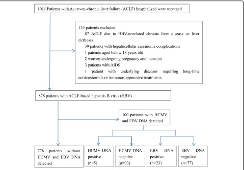
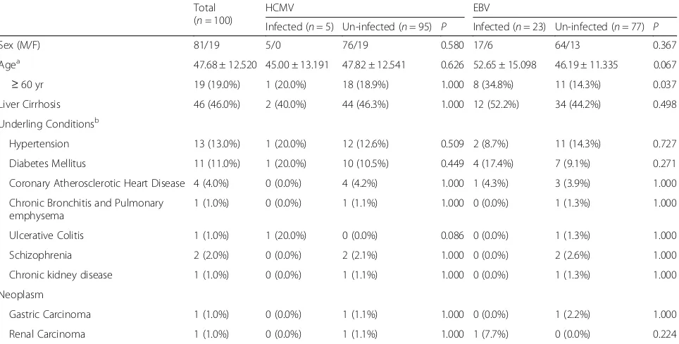
As described in Fig. 1 and Table 1, a total of 1011 pa-tients hospitalized in our hospital with ACLF were screened between 1 January 2016 and 1 September 2017. A total of 878 subjects were hospitalized due to ACLF due to HBV, and the others were excluded. A total of 100 patients were assessed for HCMV and EBV, of which five (5.0%) patients were HCMV DNA-positive, with a mean of 1.39 × 10⁴ copies/mL, and 23 (23.0%) patients were EBV DNA-positive, with a mean of 2.7 × 10³ copies /mL. In addition, one patient was both HCMV DNA-and EBV DNA-positive. The subjects consisted of 81 men and 19 women, with a mean age of 47.7 years old. A total of 19.0% of the patients were at least 60 years old. The demographic and clinical characteristics of the participants are presented in Table1.
Risk factors for HCMV and EBV infection
Among the aforementioned possible risk factors, we found that HBV DNA < 1000 IU/mL was a risk factor for HCMV infection (P= 0.003), with a dramatically in-creased risk of 34.00-fold (95% CI: 3.453–334.800). The ages of the EBV-infected patients were older than the uninfected patients. A greater number of EBV-infected patients were > 60 years of age. Age, especially older than 60 years (P= 0.034,P= 0.033) was a risk factors for EBV infection by 1.042-fold and 3.200-fold, respectively (95% CI: 1.003–1.082, 1.098–9.324). (Table1, Table2). Liver function, child-Pugh score, and MELD score
Discussion
Human herpes viruses include herpes simplex virus type 1 (HSV-1), herpes simplex virus type 2 (HSV-2), varicella zoster virus (VZV), HCMV, EBV, human herpesvirus 6 (HHV-6), human herpesvirus 7 (HHV-7), and human herpesvirus 8 (HHV-8). Although HCMV and EBV are more studied in this respect, after initial infections with no or mild symptoms, all human herpes viruses may be-come latent under certain conditions. However, most studies of latent HCMV and EBV infection have focused on immunosuppressed individuals. In this study, we aimed to focus our investigation on HCMV and EBV in immunocompetent individuals.
Our study evaluates a cohort of 100 hospitalized tients with ACLF due to HBV infection, of which 5.0% pa-tients presented with HCMV DNA, 23.0% papa-tients presented with EBV DNA, and one patient was positive for both HCMV DNA and EBV DNA. HBV DNA < 1000 IU/mL was a risk factor for HCMV infection, and age, especially older than 60 years, were risk factors for EBV infection. HCMV-infected patients had lower ALT levels, indicating that HCMV infection may not increase liver injury. In contrast, EBV infection possibly influenced
liver function, as EBV-infected patients had lower albumin levels and higher Child-Pugh scores.
By reviewing the medical records and laboratory data, we found that of the 69 patients in this study in whom HCMV IgG and EB viral capsid antigen (EB-VCA) IgG were measured, all were HCMV IgG-and EB-VCA IgG-positive. HCMV IgG-and EBV primary infection mostly occur in childhood. Thus, in this study, it is likely that the observed HCMV and EBV infections were due to reactivation rather than pri-mary infection.
[image:4.595.60.539.87.422.2]reported infection rate because chronic HBV infection and liver cirrhosis were both included in our study.
Immunosuppressive drugs, high-dose corticosteroids, T-cell depletion, acute and chronic GVHD, rejection, and virus coinfection were identified as the most com-mon risk factors for HCMV or EBV infection [3, 5, 6,
11, 12, 22]. These risk factors were determined mainly based on immunodeficient or immunosuppressed pa-tients, such as organ transplant recipients, HSCT recipi-ents, and patients with AIDS. Few studies have considered immunocompetent and not severely immunocomprom-ised patients. In the present study, we focused on patients with ACLF due to HBV infection.
ACLF is defined as an acute hepatic insult manifesting as jaundice and coagulopathy that is complicated within 4 weeks by ascites and/or encephalopathy in patients with previously diagnosed or undiagnosed chronic liver disease [13, 21] with various degrees of immune disor-ders. Considering the relationship among HCMV, EBV, and HBV [23, 25, 28–30], we attempted to explore whether HBV-related indexes and liver cirrhosis are po-tential risk factors in addition to age and gender.
[image:5.595.58.540.100.344.2]We found that HCMV-infected patients had lower HBV DNA levels than HCMV-uninfected patients (P= 0.001). Eighty percent of the HCMV-infected patients had HBV DNA < 1000 IU/mL, which was greater than Table 1Characteristics of patients with acute-on-chronic liver caused by HBV infection
Total (n= 100)
HCMV EBV
Infected (n= 5) Un-infected (n= 95) P Infected (n= 23) Un-infected (n= 77) P
Sex (M/F) 81/19 5/0 76/19 0.580 17/6 64/13 0.367
Agea 47.68 ± 12.520 45.00 ± 13.191 47.82 ± 12.541 0.626 52.65 ± 15.098 46.19 ± 11.335 0.067 ≥60 yr 19 (19.0%) 1 (20.0%) 18 (18.9%) 1.000 8 (34.8%) 11 (14.3%) 0.037 Liver Cirrhosis 46 (46.0%) 2 (40.0%) 44 (46.3%) 1.000 12 (52.2%) 34 (44.2%) 0.498
Underling Conditionsb
Hypertension 13 (13.0%) 1 (20.0%) 12 (12.6%) 0.509 2 (8.7%) 11 (14.3%) 0.727
Diabetes Mellitus 11 (11.0%) 1 (20.0%) 10 (10.5%) 0.449 4 (17.4%) 7 (9.1%) 0.271
Coronary Atherosclerotic Heart Disease 4 (4.0%) 0 (0.0%) 4 (4.2%) 1.000 1 (4.3%) 3 (3.9%) 1.000
Chronic Bronchitis and Pulmonary emphysema
1 (1.0%) 0 (0.0%) 1 (1.1%) 1.000 0 (0.0%) 1 (1.3%) 1.000
Ulcerative Colitis 1 (1.0%) 1 (20.0%) 0 (0.0%) 0.086 0 (0.0%) 1 (1.3%) 1.000
Schizophrenia 2 (2.0%) 0 (0.0%) 2 (2.1%) 1.000 0 (0.0%) 2 (2.6%) 1.000
Chronic kidney disease 1 (1.0%) 0 (0.0%) 1 (1.1%) 1.000 0 (0.0%) 1 (1.3%) 1.000
Neoplasm
Gastric Carcinoma 1 (1.0%) 0 (0.0%) 1 (1.1%) 1.000 0 (0.0%) 1 (2.2%) 1.000
Renal Carcinoma 1 (1.0%) 0 (0.0%) 1 (1.1%) 1.000 1 (7.7%) 0 (0.0%) 0.224
Note:amean ± standard deviation, years;bone patient underling conditions included coronary atherosclerotic heart disease, hypertension, chronic bronchitis and emphysema pulmonum; one patient coexisting conditions included diabetes Mellitus, hypertension; and one patient coexisting conditions included coronary atherosclerotic heart Disease, diabetes mellitus, hypertension
Table 2Binary logistic analysis for HCMV, EBV risk factors
Variable HCMV EBV
OR 95% CI P OR 95% CI P
Age 0.981 0.909–1.059 0.623 1.042 1.003–1.082 0.034
Age≥60 yr 1.069 0.113–10.153 0.953 3.200 1.098–9.324 0.033
Male 0.000 0.000–0.000 0.998 1.738 0.575–5.248 0.327
Liver Cirrhosis 0.773 0.123–4.837 0.783 1.380 0.542–3.510 0.499
HBV DNA levels 1.000 0.999–1.000 0.241 1.000 1.000–1.000 0.890
HBV DNA (<1000 IU/ml) 34.000 3.453–334.800 0.003 0.900 0.228–3.546 0.880
HBsAg titer 1.000 1.000–1.000 0.607 1.000 1.000–1.000 0.317
HBcAb titer 1.108 0.859–1.430 0.429 0.956 0.769–1.198 0.687
HBeAg positive (>0.18PEIU/ml) 0.773 0.123–4.837 0.783 2.187 0.844–5.669 0.107
[image:5.595.55.543.570.724.2]the number of HCMV-uninfected patients with similar levels (9.5%). In addition, we found that HBV DNA < 1000 IU/mL was a risk factor for HCMV infection. HBV was strongly correlated with HCMV. Our findings were similar to those of previous reports [23,24]. Cavanaugh, V. J. et al. [24] discovered that cytokines produced by murine cytomegalovirus inhibited HBV replication and gene expression. Bayram, A. et al. [23] revealed that the inflammation caused by HCMV can contribute to viral clearance during chronic HBV infection. Therefore, we suggest that HBV and HCMV replication may mutually inhibit each other. Thus, when HBV replication is in-active, as indicated by a lower titre of HBV DNA, espe-cially HBV DNA < 1000 IU/mL, HCMV replication activity is facilitated. However, the specific mechanism underlying this phenomenon must be further studied.
Our previous study posited that male donors, condi-tioning regimens including ATG and GVHD prophy-laxis, and prednisone increase the risk of EBV infection in patients receiving HSCT [5]. However, EBV infection and its risk factors were rare in patients who are not
severely immunocompromised, such as those with ACLF. In the present study, HBV-related indexes, in-cluding proliferation state, serological status of HBV and liver cirrhosis, were not risk factors of EBV infection in HBV-related ACLF. However, age was a risk factor for EBV infection, especially in patients > 60 years of age. XM. Zhang [31] previously reported that the rate of EBV-1 infection in this age group was significantly higher than that in the < 40 age group. Age was related to EBV-1 infection in patients with chronic periodontitis. It was suggested that the immune system and the envir-onment of the host change with the age, and thus, latent EBV was released and propagated in the host cell [31].
Bayram, A. et al. [23] indicated that the mean ALT and intrahepatic HBV DNA levels in patients with HCMV co-infected with chronic viral hepatitis B were lower than those in patients without HCMV co-infection. Similarly, we found that the levels of ALT, AST, and HBV DNA were lower in patients with ACLF due to HBV co-infection with HCMV. However, further analysis using binary logistic regression indicated that only the ALT level was related to HCMV infection. Therefore, we suggest that HCMV infection may inhibit HBV prolifera-tion, decrease inflammatory activity, reduce liver cell damage, and reduce ALT levels. However, the specific mechanism underlying this phenomenon must be fur-ther studied.
Previous studies have reported that EBV infection causes acute liver failure [15–17]. In the present study, EBV-infected patients had lower albumin levels than un-infected patients. In addition, EBV-un-infected patients also had higher Child-Pugh scores. Thus, EBV infection may be related to lower albumin and higher Child-Pugh score. EBV infection may increase liver injury and de-crease the liver synthesis ability in ACLF patients. Fur-thermore, Child-Pugh score is a suitable prognostic evaluation indictor for patients with end-stage liver dis-ease; a higher score corresponds to higher mortality in the short term [26]. Thus, the influence of EBV infection on liver function may result in poor prognosis.
[image:6.595.304.540.108.180.2]This work is limited by a number of factors. First, a slight bias is present in any retrospective study, which will result in a certain statistical bias. Second, we did not conduct follow-up for the included patients and did not Table 3Liver function between HCMV infected and un-infected
patients
HCMV infected (n= 5)
HCMV un-infected (n= 95)
P
ALT (U/L) 34.0 (30.0, 48.0) 166.0 (87.0, 446.0) 0.001
AST (U/L) 45.0 (43.5, 52.0) 134.0 (78.0, 254.0) 0.002
Albumin (g/L) 31.86 ± 3.002 32.42 ± 4.397 0.779
TB (U/L) 358.20 ± 191.525 328.91 ± 119.722 0.606
DB (U/L) 241.80 ± 125.398 228.74 ± 85.521 0.746
GGT (U/L) 46.0 (38.5, 142.5) 75.0 (55.0, 125.0) 0.300
Child-pugh Score 12.0 (9.0, 12.0) 11.0 (10.0, 12.0) 0.994
Meld Score 20.43 ± 2.328 23.54 ± 5.759 0.235
Note:ALTalanine aminotransferase,ASTaspartate aminotransferase,A/G
Albumin/Globulin,TBtotal bilirubin,DBdirect bilirubin,GGT
γ-glutamyl transpeptadase
Table 4Liver function between EBV infected and un-infected patients
EBV infected (n= 23) EBV un-infected (n= 77) P
ALT (U/L) 162.0 (76.0, 504.0) 153.0 (74.5.0, 408.0) 0.734
AST (U/L) 133.0 (50.0, 332.0) 130.0 (69.0, 241.0) 0.889
Albumin (g/L) 29.72 ± 3.626 33.19 ± 4.217 0.001
TB (U/L) 291.52 ± 109.996 341.98 ± 124.966 0.084
DB (U/L) 202.10 ± 74.653 237.55 ± 89.320 0.087
GGT (U/L) 70.0 (46.0, 92.0) 78.0 (54.5, 128.0) 0.206
Child-pugh Score 12.0 (11.0, 13.0) 10.0 (9.0, 12.0) 0.008
Meld Score 23.48 ± 4.551 23.35 ± 5.992 0.925
Note:ALTalanine aminotransferase,ASTaspartate aminotransferase,A/G
Albumin/Globulin,TBtotal bilirubin,DBdirect bilirubin,GGT
[image:6.595.56.291.109.245.2]γ-glutamyl transpeptadase
Table 5Binary logistic analysis for liver function and HCMV, EBV infection
Virus variable OR (95% CI) P
HCMV ALT 1.067 (1.003~ 1.135) 0.039
AST 1.078 (0.994~ 1.169) 0.071
EBV Albumin 1.242 (1.088~ 1.416) 0.001
Child-pugh Score 1.493 (1.106~ 2.015) 0.009
[image:6.595.56.291.580.707.2]analyse the confirmed influence of prognosis on HCMV and EBV infection in the short and long terms. Third, we unfortunately did not include all human herpes vi-ruses in this study. Fourth, we found that HCMV-in-fected patients had lower HBV DNA levels, and thus HCMV viral replication is likely to be detrimental to HBV replication in these 5 HCMV-infected cases. This may be due to the inflammatory cytokines caused by HCMV contributing to viral clearance during chronic HBV infection. However, we unfortunately did not meas-ure the proinflammatory cytokines released by HCMV that prevent HBV replication. Fifth, we cannot distinguish between primary infection and reactivation of HCMV and EBV infection for all patients, although there all 69 pa-tients in our study who were evaluated were found to be HCMV IgG- and EBV IgG-positive. We will address these drawbacks in our future studies. Finally, although we found that EBV-infected patients had lower albumin levels and higher Child-Pugh scores, as indicated by binary lo-gistic regression, this may be associated with overall physiological decline in these patients. Although this study does have limitations, our evaluation of patients with HBV-related ACLF may begin to fill in gaps of knowledge on HCMV and EBV infection in immunocompetent patients.
Conclusions
This study revealed that HCMV and, more notably, Epstein-Barr virus (EBV) were detected in patients with ACLF due to HBV infection. Lower replication levels of HBV (HBV DNA < 1000 IU/mL) was associated with an increased probability of HCMV infection; age, especially older than 60 years, were associated with EBV infection. HCMV infection may inhibit HBV proliferation and re-duce liver injury, while EBV infection possibly influenced liver function, as EBV-infected patients had lower albu-min levels and higher Child-Pugh scores. HCMV in-fection did not increase liver injury. However, co-infection with EBV may influence liver function and may induce poor prognosis in HBV-related ACLF. Thus, at-tention should be paid to the possibility that older pa-tients, especially those older than 60 years with ACLF due to HBV, may be infected with EBV. In this case, ef-fective antiviral therapy may be needed; otherwise, EBV infection may influence the patients,prognosis.
Abbreviations
A/G:Albumin/Globulin; ACLF: Acute-on-chronic liver failure; AIDS: immune deficiency syndrome; ALF: Acute liver failure; ALT: Alanine aminotransferase; AST: Aspartate aminotransferase; DB: Direct bilirubin; DNA: Deoxyribonucleic acid; EBV: Epstein-Barr virus; GGT:γ-glutamyl transpeptadase; HBeAg: Hepatitis B e antigen; HBsAg: Hepatitis B surface antigen; HBV: Hepatitis B virus; HCMV: Human cytomegalovirus; HLA: Human leukocyte antigen; HSCT: Hematopoietic stem cell transplant recipients; INR: International normalized ratio; MELD: Model for end-stage liver disease scores; PCR: Polymerase chain reaction; TB: Total bilirubin
Acknowledgments Not applicable.
Funding
This study was supported by the national science and technology major project (2017ZX10202202–003-004), which was funded by ministry of science and technology.
Availability of data and materials
The data that support the findings of this study are available from the First Affiliated Hospital, College of Medicine, Zhejiang University upon ethics committee approval and approval by the research ethics committee of the First Affiliated Hospital, College of Medicine, Zhejiang University, which conformed to the ethical guidelines of the Helsinki Declaration.
Authors’contributions
Study design: LJL, JHH; Data collection: DFL, XZ; Data analysis: HZ, HNG, MFY, HYJ; Paper writing: JHH, HZ, DFL. JHH, HZ, DFL contributed equally to this work. All authors read and approved the final manuscript.
Ethics approval and consent to participate
This study was approved by the research ethics committee of the First Affiliated Hospital, College of Medicine, Zhejiang University, which conformed to the ethical guidelines of the Helsinki Declaration. Written informed consent was obtained from the subjects.
Consent for publication
Provided consent also included consent to potentially-identifying information of patients.
Competing interests
The authors declare that they have no competing interests.
Publisher’s Note
Springer Nature remains neutral with regard to jurisdictional claims in published maps and institutional affiliations.
Author details 1
State Key Laboratory for Diagnosis and Treatment of Infectious Diseases, Collaborative Innovation Center for Diagnosis and Treatment of Infectious Diseases, The First Affiliated Hospital, College of Medicine, Zhejiang University, 79 QingChun Road, Hangzhou 310003, Zhejiang, China.2Shulan (Hangzhou) Hospital, 848 Dongxin Road, Hangzhou 310004, Zhejiang, China.
Received: 10 July 2018 Accepted: 31 October 2018
References
1. Gandhi MK, Khanna R. Human cytomegalovirus: clinical aspects, immune regulation, and emerging treatments. Lancet Infect Dis. 2004;4:725–38. https://doi.org/10.1016/S1473-3099(04)01202-2.
2. Jiang XJ, Zhang J, Xiong Y, Jahn G, Xiong HR, Yang ZQ, Liu YY. Human cytomegalovirus glycoprotein polymorphisms and increasing viral load in AIDS patients. PLoS One. 2017;12:e0176160.https://doi.org/10.1371/journal. pone.0176160.
3. Ljungman P, Hakki M, Boeckh M. Cytomegalovirus in hematopoietic stem cell transplant recipients. Infect Dis Clin North Am. 2010;24:319–37.https:// doi.org/10.1016/j.idc.2010.01.008.
4. Vietzen H, Gorzer I, Honsig C, Jaksch P, Puchhammer-Stockl E. Association between antibody functions and human cytomegalovirus (HCMV) replication after lung transplantation in HCMV-seropositive patients. J Heart Lung Transplant. 2017.https://doi.org/10.1016/j.healun.2017.07.010.
5. Fan J, Jing M, Yang M, Xu L, Liang H, Huang Y, Yang R, Gui G, Wang H, Gong S, et al. Herpesvirus infections in hematopoietic stem cell transplant recipients seropositive for human cytomegalovirus before transplantation. Int J Infect Dis. 2016;46:89–93.https://doi.org/10.1016/j.ijid.2016.03.025. 6. Preiksaitis JK, Brennan DC, Fishman J, Allen U. Canadian society of
7. Taylor GS, Long HM, Brooks JM, Rickinson AB, Hislop AD. The immunology of Epstein-Barr virus-induced disease. Annu Rev Immunol. 2015;33:787–821. https://doi.org/10.1146/annurev-immunol-032414-112326.
8. Maeda A, Sato T, Wakiguchi H. Epidemiology of Epstein-Barr virus (EBV) infection and EBV-associated diseases. Nihon Rinsho. 2006;64(Suppl 3): 609–12.
9. Vouloumanou EK, Rafailidis PI, Falagas ME. Current diagnosis and management of infectious mononucleosis. Curr Opin Hematol. 2012;19: 14–20.https://doi.org/10.1097/MOH.0b013e32834daa08.
10. Ma J, Li J, Hao Y, Nie Y, Li Z, Qian M, Liang Q, Yu J, Zeng M, Wu K. Differentiated tumor immune microenvironment of Epstein-Barr virus-associated and negative gastric cancer: implication in prognosis and immunotherapy. Oncotarget. 2017;8:67094–103.https://doi.org/10.18632/ oncotarget.17945.
11. Peric Z, Cahu X, Chevallier P, Brissot E, Malard F, Guillaume T, Delaunay J, Ayari S, Dubruille V, Le Gouill S, et al. Features of Epstein-Barr Virus (EBV) reactivation after reduced intensity conditioning allogeneic hematopoietic stem cell transplantation. Leukemia. 2011;25:932–8.https://doi.org/10.1038/ leu.2011.26.
12. Kullberg-Lindh C, Mellgren K, Friman V, Fasth A, Ascher H, Nilsson S, Lindh M. Opportunistic virus DNA levels after pediatric stem cell transplantation: serostatus matching, anti-thymocyte globulin, and total body irradiation are additive risk factors. Transpl Infect Dis. 2011;13:122–30.https://doi.org/10. 1111/j.1399-3062.2010.00564.x.
13. Sarin SK, Kedarisetty CK, Abbas Z, Amarapurkar D, Bihari C, Chan AC, Chawla YK, Dokmeci AK, Garg H, Ghazinyan H, et al. Acute-on-chronic liver failure: consensus recommendations of the Asian Pacific Association for the Study of the liver (APASL) 2014. Hepatol Int. 2014;8:453–71.https://doi.org/10. 1007/s12072-014-9580-2.
14. Hsu JY, Tsai CC, Tseng KC. Fulminant hepatic failure and acute renal failure as manifestations of concurrent Q fever and cytomegalovirus infection: a case report. BMC Infect Dis. 2014;14:651.https://doi.org/10.1186/ s12879-014-0651-8.
15. Gupta E, Ballani N, Kumar M, Sarin SK. Role of non-hepatotropic viruses in acute sporadic viral hepatitis and acute-on-chronic liver failure in adults. Indian J Gastroenterol. 2015;34:448–52.https://doi.org/10.1007/s12664-015-0613-0. 16. Zhang W, Chen B, Chen Y, Chamberland R, Fider-Whyte A, Craig J, Varma C, Befeler AS, Bisceglie AM, Horton P, et al. Epstein-Barr Virus-Associated Acute Liver Failure Present in a 67-Year-Old Immunocompetent Female. Gastroenterology Res. 2016;9:74–8.https://doi.org/10.14740/gr718e. 17. Mellinger JL, Rossaro L, Naugler WE, Nadig SN, Appelman H, Lee WM,
Fontana RJ. Epstein-Barr virus (EBV) related acute liver failure: a case series from the US acute liver failure study group. Dig Dis Sci. 2014;59:1630–7. https://doi.org/10.1007/s10620-014-3029-2.
18. Tanaka S, Toh Y, Minagawa H, Mori R, Sugimachi K, Minamishima Y. Reactivation of cytomegalovirus in patients with cirrhosis: analysis of 122 cases. Hepatology. 1992;16:1409–14.
19. Rosi S, Poretto V, Cavallin M, Angeli P, Amodio P, Sattin A, Montagnese S. Hepatic decompensation in the absence of obvious precipitants: the potential role of cytomegalovirus infection/reactivation. BMJ Open Gastroenterol. 2015;2:e000050.https://doi.org/10.1136/bmjgast-2015-000050. 20. Varani S, Lazzarotto T, Margotti M, Masi L, Gramantieri L, Bolondi L, Landini
MP. Laboratory signs of acute or recent cytomegalovirus infection are common in cirrhosis of the liver. J Med Virol. 2000;62:25–8.
21. Failure L, Artificial Liver Group CSoID. Chinese Medical Association, severe liver diseases and artificial liver group, Chinese Society of Hepatology, Chinese Medical Association. Guideline for diagnosis and treatment of liver failure. Chin J Clin Infect Dis. 2012;5:321–7.
22. Hanley PJ, Bollard CM. Controlling cytomegalovirus: helping the immune system take the lead. Viruses. 2014;6:2242–58.https://doi.org/ 10.3390/v6062242.
23. Bayram A, Ozkur A, Erkilic S. Prevalence of human cytomegalovirus co-infection in patients with chronic viral hepatitis B and C: a comparison of clinical and histological aspects. J Clin Virol. 2009;45:212–7.https://doi.org/ 10.1016/j.jcv.2009.05.009.
24. Cavanaugh VJ, Guidotti LG, Chisari FV. Inhibition of hepatitis B virus replication during adenovirus and cytomegalovirus infections in transgenic mice. J Virol. 1998;72:2630–7.
25. An H, Zhou XJ, Miu CM. Clinical study on the relationship between EBV infection and liver diseases infected by HBV. Zhong XI Yi Jie He Gan Bing Za Zhi. 2005;15:144–5.
26. Pugh RN, Murray-Lyon IM, Dawson JL, Pietroni MC, Williams R. Transection of the oesophagus for bleeding oesophageal varices. Br J Surg. 1973;60: 646–9.
27. Kamath PS, Wiesner RH, Malinchoc M, Kremers W, Therneau TM, Kosberg CL, D'Amico G, Dickson ER, Kim WR. A model to predict survival in patients with end-stage liver disease. Hepatology. 2001;33:464–70.https://doi.org/10. 1053/jhep.2001.22172.
28. Aldona K, Jacek W, Maciej Z, Wieslawa B, Joanna SZ. Cellular expression of human cytomegalovirus (HCMV) in children with chronic hepatitis B. Przegl Epidemiol. 2001;55(Suppl 3):146–52.
29. Zuschke CA, Herrera JL, Pettyjohn FS. Cytomegalovirus hepatitis mimicking an acute exacerbation of chronic hepatitis B. South Med J. 1996;89:1213–6. 30. Lian YL, Wu WF, Shi YM, Liu QC, Tang XP. Preliminary study on relationship between different viral pathogenesis and disease prognosis in patients with severe viral hepatitis. Zhonghua Shi Yan He Lin Chuang Bing Du Xue Za Zhi. 1999;13:355–7.