R E S E A R C H
Open Access
A potent anti-inflammatory peptide from
the salivary glands of horsefly
Lin Wei
1†, Chunjing Huang
1†, Hailong Yang
2, Min Li
1, Juanjuan Yang
3, Xue Qiao
4, Lixian Mu
2, Fei Xiong
1,
Jing Wu
2*and Wei Xu
1*Abstract
Background:A diverse group of physiologically active peptides/proteins are present in the salivary glands of horseflyTabanus yao(Diptera, Tabanidae) that facilitate acquisition of blood meal. However, their roles in the regulation of local inflammation remains poorly understood.
Methods:Induction expression profiles of immune-related molecules in the salivary glands ofT. yaowas analyzed
by quantitative PCR (qPCR) after bacterial feeding. A significantly up-regulated molecule (cecropin-TY1) was selected for anti-inflammatory assay in lipopolysaccharide (LPS)-stimulated mouse peritoneal macrophages. The transcription levels of inducible NO synthase (iNOS) and pro-inflammatory cytokines were quantified by qPCR. Nitric oxide (NO) production was determined by Griess reagent. Pro-inflammatory cytokine production was determined by an enzyme-linked immunosorbent assay (ELISA). The inflammatory signals were assayed by Western blotting analysis. The secondary structure of cecropin-TY1 was measured by Circular dichroism (CD) spectroscopy. Interaction of cecropin-TY1 with LPS was evaluated by the dissociation of fluorescein isothiocyanate (FITC)-conjugated LPS aggregates and neutralization of LPS determined by a quantitative Chromogenic End-point Tachypleus amebocyte lysate (TAL) assay kit. Homology modeled structure analysis and mutation of key residues/structures were
performed to understand its structure-activity relationship.
Results:Cecropin-TY1 was demonstrated to possess high anti-inflammatory activity and low cytotoxicity toward
mouse macrophages. In LPS-stimulated mouse peritoneal macrophage, addition of cecropin-TY1 significantly inhibited the production of nitric oxide (NO) and pro-inflammatory cytokines. Further study revealed that cecropin-TY1 inhibited inflammatory cytokine production by blocking activation of mitogen-activated protein kinases (MAPKs) and transcriptional nuclear factor-κB (NF-κB) signals. Cecropin-TY1 even interacted with LPS and neutralized LPS. The secondary structure analysis revealed that cecropin-TY1 adopted unordered structures in hydrophobic environment but converted toα-helical confirmation in membrane mimetic environments. Homology modeled structure analysis demonstrated that cecropin-TY1 adopted twoα-helices (Leu3-Thr24, Ile27-Leu38) linked by a hinge (Leu25-Pro26) and the structure surface was partly positively charged. Structure-activity relationship analysis indicated that several key residues/structures are crucial for its anti-inflammatory activity includingα-helices, aromatic residue Trp2, positively charged residues Lys and Arg, hinge residue Pro26 and N-terminal amidation.
(Continued on next page)
* Correspondence:wujing_205@163.com;xuweifd828@126.com †Equal contributors
2
School of Basic Medical Sciences, Kunming Medical University, 1168 West Chunrong Road, Yuhua Avenue, Chenggong District, Kunming 650500, Yunnan Province, China
1Jiangsu Key Laboratory of Infection and Immunity, Institutes of Biology and Medical Sciences, Soochow University, 199 Ren-Ai Road, Suzhou Industrial Park, Suzhou 215123, Jiangsu Province, China
Full list of author information is available at the end of the article
(Continued from previous page)
Conclusions:We found a novel anti-inflammatory function of horsefly-derived cecropin-TY1 peptide, laying groundwork for better understanding the ectoparasite-host interaction of horsefly with host and highlighting its potency in anti-inflammatory therapy for sepsis and endotoxin shock caused by Gram-negative bacterial infections.
Keywords:Horsefly, Salivary gland, Hematophagous arthropod, Cecropin, Anti-inflammation
Background
Hematophagous arthropods have evolved effective mecha-nisms to suppress their host’s hemostatic system and im-mune response to get a blood meal successfully. Their salivary glands can produce a wide array of active com-pounds including antihemostatic and immunoregulatory substances [1–9]. A majority of antihemostatic com-pounds have been identified and can be divided into several categories including inhibitors of coagulation factors (Factors VII, V, IIa, and Xa) and platelet functions, fibrin(ogen)olytic enzymes, and vasoactive peptides [3–5]. Immunoregulatory factors from arthropods especially from ticks have been extensively investigated [10–15]. These immunoregulatory factors such as immunosuppres-sive peptides and antimicrobial peptides (AMPs) are crucial for blood-sucking arthropods to suppress the immune response including innate immunity, adaptive immunity and inflammation [5, 14–17]. Horseflies are economically important blood-feeding insects and vectors for pathogens such as filariasis [5]. There have been re-ported a diverse group of active compounds in the salivary glands of horseflies like other hematophagous ar-thropods including mosquitoes [1], flies [2], and ticks [10, 12]. Although the antihemostaic substances in horsefly have been extensively exploited in previ-ous work [5, 18, 19], comparatively few investiga-tions on the anti-inflammatory effects of horsefly-derived AMPs were conducted.
AMPs are small gene-encoded defensive effectors and play key roles in the innate immunity in all living organisms. Insects are an important source of AMPs [20]. Since the first observation of antimicrobial activ-ity in the hemolymph of bacteria-challenged pupae of the giant silk moths (Samia Cynthia and Hyalophora cecropia) in 1974 and the first purification of AMP from the hemolymph of H. cecropia in 1980, over 200 AMPs have been identified or purified from insects [20]. Generally, insect AMPs are comprised of four groups based on their structural motifs and unique sequences. They are (i) α-helical peptides (cecropin and moricin), (ii) cysteine-rich peptides with intramo-lecular cysteine disulfide bonds forming hairpin-like
β-sheets or α-helical/β-sheet mixed structures (insect defensin and drosomycin), (iii) proline-rich peptides (apidaecin, drosocin, and lebocin), and (iv) glycine-rich peptides/proteins (attacin and gloverin) [20].
Among these insect-derived AMPs, cecropins consti-tute a large family of cationic α-helical peptides com-posed of 35–39 amino acids, and most of them are amidated at the C-terminus [20]. Cecropin was the first insect AMP purified from the hemolymph ofH. cecropia
in 1980 [21]. Since then, a variety of insect-derived cecropin AMPs were identified in lepidopteran, dipteran, and coleopteran [20]. Cecropins usually have broad anti-microbial spectrum against various microorganisms includ-ing Gram-positive and Gram-negative bacteria [22, 23], fungi [24, 25], parasites [26, 27] and HIV-1 virus [28]. In addition to antimicrobial activity, several cecropins showed strong anti-inflammatory activity in LPS-stimulated macro-phages through interaction with LPS on the basis of their
α-helical structures [29, 30]. Cecropins usually adopted ran-dom coil conformations in aqueous solutions. While these unordered structures converted to amphipathic α-helical structures in the membrane-like environments to exert its biological activity [20, 29, 30].
Methods
Induction expression analysis
Horseflies were collected from Shanxi province as previ-ously described [3, 5]. The collectedT. yao (~800 flies) were randomly grouped in two cages (100 × 80 × 60 cm) covered with grenadine and kept at temperature of 25 ± 2 °C, humidity of 80–90 % and a 12 h/12 h photoperiod. After a 12-h starvation, horseflies were fed with fresh chicken blood containing 1 % sodium citrate (w/v) sup-plemented with Gram-negative bacteria Escherichia coli
ATCC 8739 (2 × 10⁶/mL) [36]. The control group was fed with the same chicken blood without bacteria. The salivary glands of horseflies were dissected under a microscope at 0, 6, 12, 24, 36, 48 and 72 h after blood meal. The salivary gland of each horsefly was excised in phosphate buffer solution (100 mM, pH 6.0) on ice im-mediately [3, 5]. Total RNA extraction was performed by Trizol reagent (Life Tech, USA) according to the kit instruction. cDNA was synthesized with PrimeScript® Reverse Transcriptase Kit (Takara, Japan). The induction expression of immune-related genes including tabim-muregulins and AMPs was analyzed by qPCR to screen anti-inflammatory agents in the salivary glands of horse-flies as described in qPCR section.
Ethical approval
The study was approved by the Animal Care and Use Ethics Committee of Kunming Medical University.
Quantitative PCR
qPCR was performed using SYBR green master mix (Takara, Japan) on a Realplex Mastercycler real-time PCR system (Eppendorf, Germany) according to the manufacturer’s instruction. The gene transcription levels were normalized to GAPDH or β-actinas as illustrated in figure legends and calculated by ΔΔCt method. The accuracy of qPCR results were checked by melting curve analysis. qPCR primers were listed in Additional file 1: Table S1.
Peptide synthesis
Cecropin-TY1 and its derivatives (disruption of α -helices, mutation of aromatic residue Trp2, mutation of positively charged residues, deletion of hinge residue
Pro26 and mutation of N-terminal amidation) were syn-thesized for anti-inflammatory effects investigation as listed in Table 1. The peptides were synthesized by solid phase synthesis on an Applied Biosystems model 433A peptide synthesizer by GL Biochem (Shanghai) Ltd. (Shanghai, China). The synthetic peptides were purified by high-performance liquid chromatography. The purified peptides were subjected to an automated Edman degrad-ation protein sequencer to confirm the accuracy of amino acid sequence, and MALDI-TOF mass spectrometry to confirm the purity > 98 %.
Cytotoxicity assay
Mouse peritoneal macrophages were prepared according to previous method [37]. Brewer thioglycollate medium (3 %, w/v, Sigma-Aldrich, USA) was intraperitoneally injected to C57BL/6 mouse. After 3 days, the mouse was euthanized and intraperitoneally injected with 20 mL RPMI-1640 medium to collect peritoneal macrophages. RAW264.7 cells were cultured in RPMI 1640 medium (10 % FBS, 100 U/mL penicillin and 100μg/mL streptomycin, Gibco, USA) at 37 °C humidified with 5 % CO2. Mouse macrophages,
peritoneal macrophages and RAW264.7 cells, were seeded in a 96-well (2 × 10⁴cells/well) plate, and cultured in RPMI 1640 medium (100μL) supplemented with 2 % FBS, 100 U/mL penicillin and 100 μg/mL streptomycin (Gibco, USA) at 37 °C in a humidified 5 % CO2incubator. The
cytotoxicity of cecropin-TY1 against mouse macrophages was determined by Cell Counting Kit-8 assay (CCK-8) following the kit instruction. In brief, serial 2-fold peptide dilutions were added to each well, and con-trol wells received the same volume of phosphate-buffered solution (PBS, 8 g/L NaCl, 0.2 g/L KCl, 0.2 g/L KH2PO4, 2.89 g/L Na2HPO4· 12H2O, pH 7.4).
After 24-h incubation, CCK-8 solution (10 μL/well) was added and incubated for an additional 4 h. The absorbance at 450 nm was monitored on a microplate reader (Epoch Etock, BioTek, USA).
Detection of NO production in macrophages
[image:3.595.55.545.640.725.2]Peritoneal macrophages were seeded in two 24-well plates (2.5 × 10⁵cells/well) and cultured in RPMI-1640 contain-ing 2 % FBS, 100 U/mL penicillin, and 100μg/mL strepto-mycin (Gibco, USA). Cells were incubated with peptides
Table 1The amino acid sequence of the derivatives of cecropin-TY1
Mutant name Amino acid sequence Mutation sitea
N39 mutant GWLKKIGKKIERVGQNVRNAAISTLPIAQGAAGVAGALN-COOH N39CONH2→N
39 COOH
W2 mutant GALKKIGKKIERVGQNVRNAAISTLPIAQGAAGVAGALN-NH2 W
2 →A2
P26 mutant GWLKKIGKKIERVGQNVRNAAISTL_IAQGAAGVAGALN-NH2 lacked the hinge residue P 26
KR mutant GWLAAIGAAIEAVGQNVANAAISTLPIAQGAAGVAGALN-NH2 K
4
→A4, K5→A5, K8→A8, K9→A9, R12→A12, R18→A18 scr-cec-TY1 RGQANILAGKNIKIRSGAAAGVGKTPQKANVEVLALGIW-NH2 Scrambled cecropin-TY1, disrupted the helices
a
(0, 5, 10, and 20μg/mL) in the presence or absence of LPS (100 ng/mL, from Escherichia coli 0111:B4, Sigma-Aldrich, USA). After 6-h incubation, cells in one plate were washed twice with ice-cold PBS and lysed by Trizol reagent (Life Tech, USA) for total RNA extraction. Prime-Script® Reverse Transcriptase Kit (Takara, Japan) was used to synthesize cDNA for qPCR to examine the transcrip-tion levels of inducible nitric oxide synthase (iNOS), which is necessary for NO production [38]. After 24-h in-cubation, culture medium of each wells of another plate was harvested for nitrite detection, which indirectly reflected the NO production [39, 40]. Nitrite accumulation levels were determined by NO detection kit (Beyotime, China) following the kit instruction.
Cytokine production analysis by qPCR and ELISA
Peritoneal macrophages were cultured in a 24-well (2.5 × 10⁵/well) plate in RPMI-1640 containing 2 % FBS, 100 U/mL penicillin, and 100 μg/mL streptomycin (Gibco, USA). Cells were incubated with peptides (0, 5, 10, and 20 μg/mL) with or without 100 ng/mL LPS (fromE. coli0111:B4, Sigma-Aldrich, USA) for 6 h. After treatment, culture medium was collected for TNF-α, IL-1β and IL-6 quantification using enzyme-linked im-munosorbent assay (ELISA) kits (Dakewei, China). Cells were washed with ice-cold PBS and lysed by Trizol re-agent (Life Tech, USA). Total RNA was extracted for cDNA synthesis to quantify TNF-α, IL-1βand IL-6 tran-scription levels by qPCR as described in qPCR section.
Western blot analysis
Peritoneal macrophages were seeded in a 6-well (2 × 10⁶/ well) plate and cultured in RPMI-1640 supplemented with 2 % FBS, 100 U/mL penicillin, and 100μg/mL streptomycin (Gibco, USA). Cells were treated with different concentra-tions of cecropin-TY1 (0, 5, 10, and 20μg/mL) in the pres-ence or abspres-ence of 100 ng/mL LPS (from E. coli0111:B4, Sigma-Aldrich, USA). After 30-min incubation, cells were washed twice with ice-cold PBS and lysed with RIPA lysis buffer (Beyotime, China) on ice for 30 min according to our previous method [14]. Protein concentration was quan-tified by BCA Protein Assay Kit (Thermo, Germany). About 40μg protein was separated on a 10 % SDS-PAGE gel and transferred to a polyvinylidene difluoride mem-brane. The membrane was blocked by incubating with 5 % BSA (BD, USA) dissolved in Tris-buffered solution Tween-20 (TBST, 2.42 g/L Trisbase, 8 g/L NaCl, 0.1 % Tween Tween-20, pH 7.6) for 2 h at room temperature. The membrane was then incubated with primary antibodies against ERK, phospho-ERK, p38, phospho-p38, JNK, phospho-JNK, phospho-IκBα, NF-κB p65 and phospho-NF-κB p65 (1:2000, Cell Signaling Technology, USA) and GAPDH/β -actin (1:5000, Santa Cruz Biotechnology, USA) overnight at 4 °C, respectively. After incubation, the immunoblot was
washed three times with TBST for 5 min each time, and in-cubated with secondary antibody (1:5000, Cell Signaling Technology, USA) at room temperature for 1 h. Signals were measured by enhanced chemiluminescence kit in a dark room after washing three times with TBST for 10 min each time (TIANGEN, China).
Interaction between cecropin-TY1 and LPS
Fluorescein isothiocyanate (FITC)-conjugated LPS (1 μg/ mL, Sigma-Aldrich, USA) was excited at 480 nm and monitored the changes in the emission of FITC-LPS at 515 nm in the incubation of different concentrations of peptides (0, 12.5, 25, 50, 100μg/mL). Peptides were dis-solved in 10 mM phosphate buffer at pH 6.0. The interac-tions between peptides and LPS were further assessed by a quantitative Chromogenic End-point Tachypleus amebo-cyte lysate (TAL) assay kit (Xiamen Houshiji, China) fol-lowing the kit instruction. Briefly, different concentrations of peptides (0, 12.5, 25, 50, 100 μg/mL) were incubated with LPS (1 μg/mL, Sigma-Aldrich, USA) at 37 °C for 30 min. After incubation, 100μL TAL solution was added to 100 μL LPS-peptide mixtures in a pyrogen-free tube and incubated at 37 °C for 10 min. Then, LPS-peptide mixtures were added with pre-warmed substrate solution for additional 6-min incubation at 37 °C. Lastly, the ab-sorbance at 545 nm was measured on a microplate reader (Epoch Etock, BioTek, USA). The percentages of LPS-neutralizing activity were calculated.
Circular dichroism analysis
CD spectra were collected on a Jasco-810 spectropolar-imeter (Jasco, Tokyo, Japan) with a 1-mm path-length cell at 25 °C and 0.2-nm intervals from 190 to 260 nm. Cecropin-TY1 was dissolved in H2O, TFE/water
solu-tion, SDS/water solution and LPS/water solution at the concentration of 0.2 mg/mL. The data from three scans were averaged and smoothed using the Jasco-810 soft-ware for each spectrum. CD data were expressed as the mean residue ellipticity (θ) in deg.cm2.dmol−1.
Structure modeling analysis
Three-dimensional (3D) structure of cecropin-TY1 was modeled by Easymodeller version 2.0. The solution NMR structures of papiliocin (59 % identity, PDB entry code 2LA2) from swallowtail butterfly ofPapilio xuthus
was selected as the template for homology modeling. The comparative 3D structure model of cecropin-TY1 was optimized using MODELLER and visualized using PYMOL software (http://www.pymol.org/) [7].
Statistical analysis
TX, USA). Data were presented as mean ± SEM, and compared using two-tailed equal variance Student’s t -test. *P< 0.05 and **P< 0.01 were considered as statis-tical significance.
Results
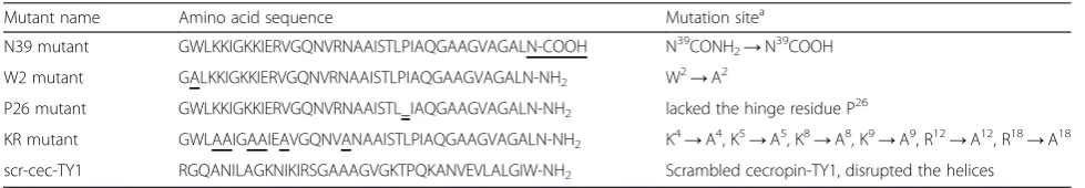
Induction expression of cecropin-TY1 in the salivary glands ofT. yao
Release of LPS during Gram-negative bacteria infection can induce strong inflammatory response [41]. To screen anti-inflammatory molecules, the induction ex-pression of immune-related molecules were analyzed in the salivary glands of T. yao after bacterial feeding. By analysis of the transcription of tabimmuregulins and AMPs in the salivary glands, most of them were up-regulated (data not shown). Among these up-up-regulated innate immunity molecules, cecropin-TY1 was dramatic-ally up-regulated as shown in Fig. 1. At 6, 12, 24, 36, 48 and 72 h post bacterial feeding, the transcription levels of cecproin-TY1 increased by 2.0, 2.4, 4.9, 5.3, 6.4, and 6.3 fold compared to the control group, respectively (Fig. 1). Cecropin-TY1 (GenBank accession No. ABX80069.1) was previously identified as a member of cecropin AMPs with known mature peptide amino acid sequence, molecular
weight, net charges and theoretical pI as listed in Additional file 1: Table S2 [5].
Cecropin-TY1 is non-toxic to mouse macrophages
In order to evaluate the effects of cecropin-TY1 on mouse macrophages, the cell viability was determined in the presence of serial concentrations of peptides incu-bated with peritoneal macrophages and RAW264.7 cells. At the concentration as high as 200 μg/mL, cecropin-TY1 didn’t show any cytotoxicity against mouse periton-eal macrophages and RAW264.7 cells.
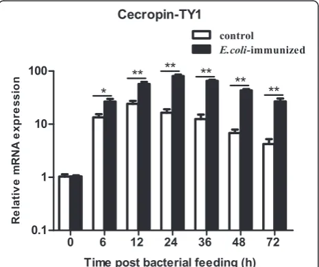
Inhibition of LPS-induced NO production
To determine the effect of cecropin-TY1 on LPS-induced NO production, iNOS transcription and nitrite production upon LPS stimulation in the presence or ab-sence of cecropin-TY1 were detected. 100 ng/mL LPS significantly induced iNOS transcription, and cecropin-TY1 inhibited LPS-activated iNOS transcription in a dose-dependent manner (Fig. 2a). At the concentration of 5, 10, and 20 μg/mL, cecropin-TY1 attenuated 58.8, 68.7 and 84.2 % iNOS transcription, respectively. iNOS is the synthase required for NO production [38]. So we next detected the nitrite accumulation in culture medium. As shown in Fig. 2b, LPS (100 ng/mL) induced about 20.4 μM nitrite production, and cecropin-TY1 at-tenuated nitrite production in a dose-dependent manner. At the concentration of 5, 10, and 20μg/mL, cecropin-TY1 attenuated about 40.0, 63.7 and 75.0 % LPS-induced nitrite production, respectively. The derivatives of cecropin-TY1 showed reduced inhibitory effects on LPS-induced nitrite production as shown in Fig. 2c.
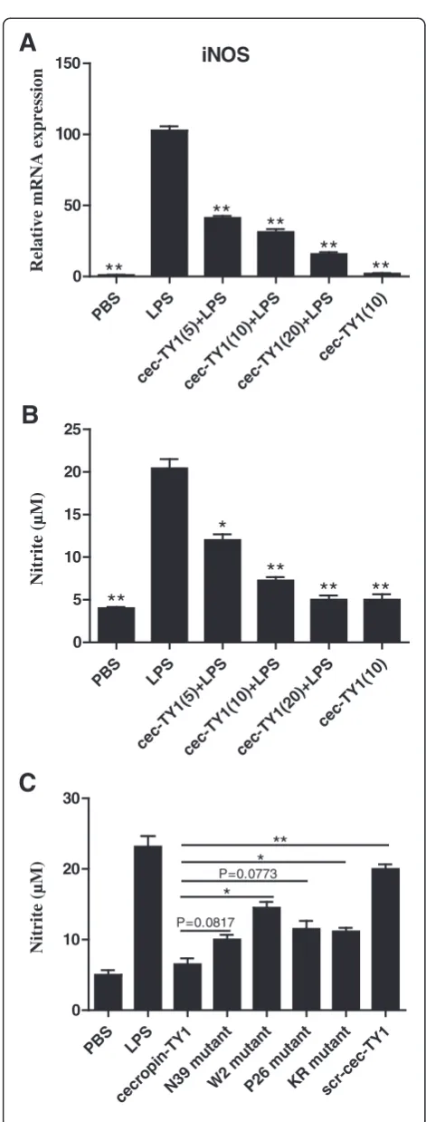
Inhibition of LPS-induced pro-inflammatory cytokine production
To determine the effects of cecropin-TY1 on the produc-tion of pro-inflammatory cytokines, we tested the transcrip-tion and productranscrip-tion of LPS-induced pro-inflammatory cytokines including TNF-α, IL-1β and IL-6 in peritoneal macrophages in the presence or absence of cecropin-TY1 as illustrated in Fig. 3. LPS (100 ng/mL) alone significantly activated the transcription and production of TNF-α, IL-1β and IL-6, and cecropin-TY1 significantly reduced the tran-scription and production of these three pro-inflammatory cytokines in a dose-dependent manner. At the concentra-tion of 20μg/mL, cecropin-TY1 attenuated LPS-stimulated TNF-α, IL-1β and IL-6 transcription by 60.3, 67.5 and 94.1 %, respectively (Fig. 3a–c). As a result, 20μg/mL of cecropin-TY1 subsequently reduced TNF-α, IL-1βand IL-6 production by 72.1, 63.4 and 76.2 % induced by LPS, re-spectively (Fig. 3d–e). The derivatives were less active than cecropin-TY1 on LPS-induced inflammatory cytokine pro-duction (Fig. 3g–i).
[image:5.595.58.291.408.603.2]Inhibition of LPS-induced inflammatory signal pathways
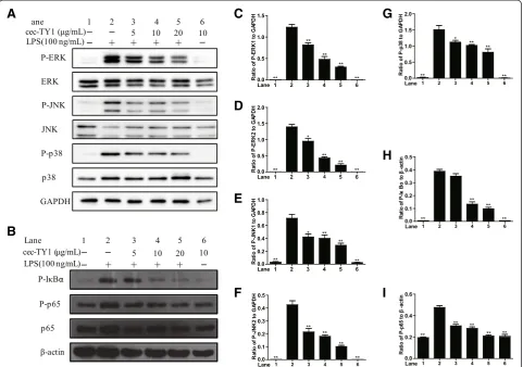
To address the anti-inflammatory mechanisms of cecropin-TY1 on LPS-stimulated mouse peritoneal macrophages, the effects of cecropin-TY1 on LPS-induced inflammatory signal pathways were investigated. As illustrated in Fig. 4, LPS (100 ng/mL) significantly activated MAPKs and NF-κB signal pathways. Cecropin-TY1 significantly blocked the ac-tivation of MAPKs and NF-κB signal pathways in a dose-dependent manner. At the concentration of 20 μg/mL, cecropin-TY1 inhibited 75.5 % P-ERK1, 84.9 % P-ERK2, 61.9 % JNK1, 66.1 % JNK2, 46.4 % p38, 74.8 % P-IκBα and 55.9 % P-p65 expression induced by LPS, respectively.
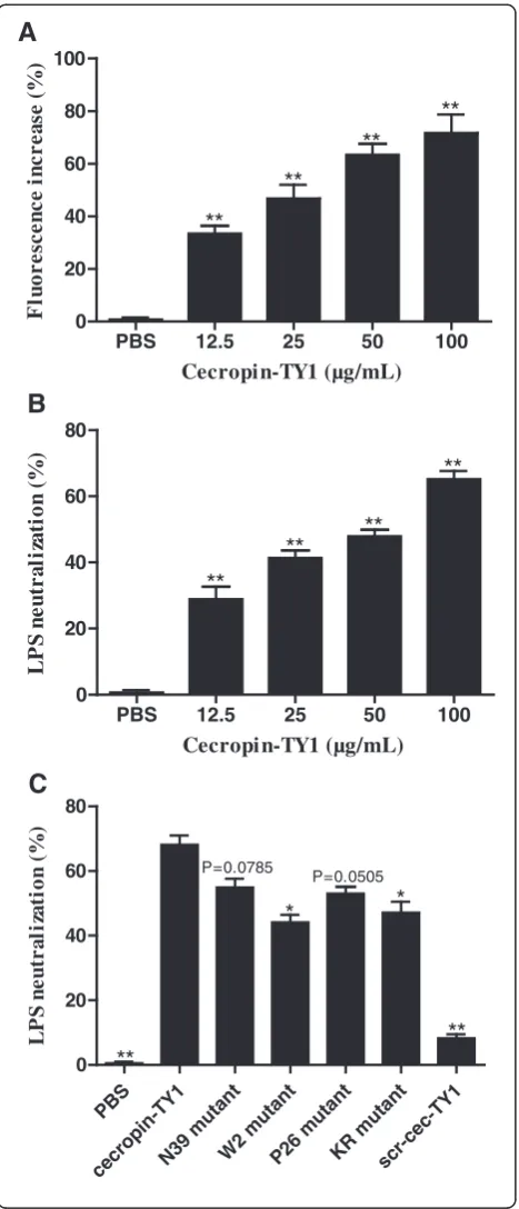
Cecropin-TY1 interacts with LPS and neutralizes its activity
To address the underlying anti-inflammatory mechanism of cecropin-TY1 in LPS-stimulated peritoneal macro-phages, the interaction between cecropin-TY1 and LPS was investigated. Interaction of AMPs with LPS can be measured as an increase in fluorescence of FITC-conjugated LPS, which indirectly reflects the dissociation of large LPS aggregates into smaller sizes [30, 42, 43]. As shown in Fig. 5a, the addition of cecropin-TY1 caused a dose-dependent increase of FITC-LPS fluorescence, indi-cating that the interaction of cecropin-TY1 with LPS re-sulted in the dissociation of LPS aggregates. At the concentration of 12.5, 25, 50 and 100μg/mL, cecropin-TY1 increased 33.4, 46.7, 63.3, and 71.6 % fluorescence, re-spectively. The interaction of cecropin-TY1 with LPS was further evaluated by TAL assay for LPS-neutralization ac-tivity. Cecropin-TY1 neutralized LPS in a dose-dependent
PBS LPS
cec-TY1( 5)+LPS
cec-TY1( 10)+
LPS
cec-TY1( 20)+
LPS
cec-T Y1(1
0)
0 50
100 150
A
iNOS
**
**
**
**
**
Relative mRNA expression
PBS LPS
cec-T Y1(5)+LPS
cec-TY1( 10)+
LPS
cec-TY1( 20)+
LPS
cec -TY
1(1 0)
0 5 10 15 20 25
*
**
**
**
**
B
Nitrite (µM)
PBS LPS
cecropin -TY
1
N39 muta
nt
W2 mutant
P26 m utan
t
KR muta
nt
scr-ce c-TY1
0 10 20 30
*
P=0.0817*
**
C
P=0.0773
Nitrite (µM)
[image:6.595.55.291.80.700.2]manner (Fig. 5b). At the concentration of 12.5, 25, 50 and 100 μg/mL, cecropin-TY1 neutralized 28.8, 41.3, 47.9 and 65.1 % LPS, respectively. The LPS-neutralizing activity of cecropin-TY1 is more potent than that of derivatives (Fig. 5c).
Secondary structures of cecropin-TY1
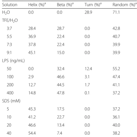
To assess the secondary structures of cecropin-TY1 in membrane mimetic environments, the CD spectra of cecropin-TY1 in different membrane-like solutions were recorded and analyzed as shown in Fig. 6 and Table 2.
A
B
C
D
E
F
G
H
I
[image:7.595.58.540.87.535.2]The CD spectra of cecropin-TY1 dissolved in H2O showed
a strong negative absorption at 199 nm, demonstrating that cecropin-TY1 mainly adopted random coil structures in aqueous solution. The conformations exhibited changes in TFE/H2O solutions, LPS micelles and SDS micelles. In
these membrane-like environments, the CD spectra exhibited double negative absorptions at 208 and 222 nm, indicating that cecropin-TY1 adopted a sig-nificant degree ofα-helical conformations in membrane-like solutions (Fig. 6). In aqueous solution, the sec-ondary structures of cecropin-TY1 are composed of 71.1 % random coil conformation without α-helical conformation. While in TFE/H2O (9:1), LPS/H2O
(400 ng/mL) and SDS/H2O (40 mM) solutions, the α
-helical structures increased to 45.1, 14.8 and 54.4 %, respectively (Table 2).
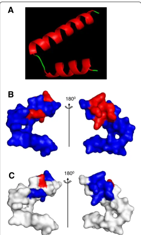
Homology modeled structure analysis of cecropin-TY1
The amphiphilic structure of helix and electrostatic sur-face of such host defensive peptides are important for their interactions with LPS [30, 40]. To investigate the structural basis of cecropin-TY1, the homology modeled structures were analyzed as shown in Fig. 7. It exhibited a helix-hinge-helix confirmation, suggesting the presence of an α-helix (in red) from residues Leu3 to Thr24 linked by a hinge region to anotherα-helix (in red) from Ile27 to Leu38 (Fig. 7a). The amphiphilic α-helix is the predominant structural feature for cecropin-TY1. It also showed that the 6 basic residues (4 Lys and 2 Arg), which have positive charges, were partly distributed in the surface of 3D structure (Fig. 7b, in red). Electrostatic surface analysis revealed that most regions of the elec-trostatic surface are positively charged (Fig. 7c in blue).
A
C
D
E
F
G
H
I
B
[image:8.595.58.540.90.428.2]Discussion
Horseflies have been used as anti-thrombosis materials for hundreds of years in China and some other eastern countries [3]. Recently, a variety of physiologically active compounds have been identified in the salivary glands of horsefly,T. yao. They are (i) fibrin(ogen)olytic enzymes, (ii) Arg-Gly-Asp-motif containing proteins, (iii) a serine protease inhibitor, (iv) a serine protease, (v) a protease, (vi) a apyrase, (vii) vasodilator peptides, (viii) a peroxid-ase, (ix) two metallothioneins, (x) a hyaluronidperoxid-ase, (xi) immunosuppressive peptides and (xii) three family of AMPs [3, 5, 31–35]. All these active compounds can be generally classified into several groups such as antihe-mostatic factors, immunosuppressive factors, antimicro-bial factors and allergens. But anti-inflammatory agents were poorly understood in the salivary glands ofT. yao.
In the present work, a novel anti-inflammatory molecule (cecropin-TY1) was identified in the salivary glands of
T. yao. The mRNA transcription levels of cecropin-TY1 were dramatically up-regulated upon Gram-negative bac-teria challenge at different time course (Fig. 1), suggesting that cecropin-TY1 might involve in the inflammatory response induced by the release of LPS during Gram-negative bacteria infection. In LPS-stimulated mouse peri-toneal macrophages, cecropin-TY1 showed strong anti-inflammatory effects by inhibiting the transcription of iNOS and pro-inflammatory cytokines including TNF-α, IL-1βand IL-6, as well as the production of NO and these pro-inflammatory cytokines (Fig. 2 & Fig. 3). Cecropin-TY1 showed no cytotoxicity toward mouse macrophages at the concentration as high as 200μg/mL, suggesting that its anti-inflammatory effects in LPS-stimulated macro-phages were not dependent on cytotoxicity. LPS, also called endotoxin, is the characteristic components of the cell wall of Gram-negative bacteria. LPS acts as a strong stimulator to activate the innate immune system of cells, which are components of the innate immunity of diverse organisms [41, 44, 45]. Release of LPS during Gram-negative bacteria infections triggers the production of higher concentration of systemic pro-inflammatory cyto-kines and NO, and results in sepsis. The excessive produc-tion of systemic pro-inflammatory cytokines and NO
PBS 12.5 25 50 100
0 20 40 60 80 100
A
**
**
**
**
Cecropin-TY1 (µg/mL) F luo r e scen c e in crea se ()PBS 12.5 25 50 100
0 20 40 60 80
**
**
**
**
B
Cecropi n-TY1 (µg/mL)
L P S n e u tr al iz at io n ( ) PBS cecr opi n-TY1 N39 muta nt W2 mut ant P26 mut ant KR mut ant sc r-ce c-TY1 0 20 40 60 80
C
**
*
*
P=0.0785 P=0.0505 L P Sn e u tr a li za ti o n( )**
[image:9.595.58.292.88.633.2]called cytokines storm, which are responsible for the pathophysiology of septic shock and other immune dis-eases [46]. To address the anti-inflammatory mecha-nisms, the effects of cecropin-TY1 on LPS-triggered
inflammatory pathways were investigated as illustrated in Fig. 4. Cecropin-TY1 significantly inhibited the ac-tivation (phosphorylation) of MAPKs and NF-κB sig-nals induced by LPS in mouse peritoneal macrophage. These results suggested that cecropin-TY1 exerted its anti-inflammatory effects via blockade the activation (phosphorylation) of MAPKs and NF-κB signal pathways, which in turn suppressed iNOS and pro-inflammatory cytokines mRNA transcription, finally reduced NO and pro-inflammatory cytokines production.
A majority of anti-inflammatory peptides are also known to have high binding affinity and LPS-neutralizing activity such as papiliocin, cecropin A, LL-37, SMAP-29, and fowlicidin-1. These anti-inflammatory peptides can interact with LPS through binding to LPS and neutralizing LPS [29, 30, 43, 47, 48]. To clarify whether cecropin-TY1 could interact with LPS, we mon-itored the fluorescence change after the incubation of FITC-conjugated LPS with cecropin-TY1. As indicated in Fig. 5a, the addition of cecropin-TY1 caused a dose-dependent increase in FITC-conjugated LPS fluorescence, implying that the interaction between cecropin-TY1 and LPS resulted in the dissociation of large LPS aggregates into smaller sizes. The interaction of cecropin-TY1 with LPS was further evaluated by TAL assay, and cecropin-TY1 showed a reduced activation of LPS-induced TAL en-zyme in a dose-dependent manner, suggesting that the addition of cecropin-TY1 caused the neutralization of LPS (Fig. 6b). The CD spectra indicated that cecropin-TY1 adopted a random coil structure in aqueous solution
[image:10.595.57.291.86.615.2]Fig. 6CD spectra of cecropin-TY1 in different solutions.aTFE/H2O solution.bLPS/H2O solution.cSDS/H2O solution. TFE, LPS and SDS solutions were prepared as indicated concentrations. Cecropin-TY1 was dissolved in H2O, TFE/water solution, LPS/water solution and SDS/water solution at the concentration of 0.2 mg/mL
Table 2Secondary structural components of cecropin-TY1 in different solutions
Solution Helix (%)a Beta (%)a Turn (%)a Random (%)a
H2O 0.0 0.0 28.9 71.1
TFE/H2O
3:7 28.4 28.7 0.0 42.8
5:5 36.9 22.4 0.0 40.7
7:3 37.8 22.4 0.0 39.9
9:1 45.1 15.0 0.0 39.9
LPS (ng/mL)
50 0.0 32.4 12.4 55.2
100 2.9 46.6 3.1 47.4
200 12.7 44.5 1.7 41.1
400 14.8 47.8 0.1 37.2
SDS (mM)
5 45.3 17.5 0.0 37.2
10 41.2 22.7 0.0 36.1
20 46.6 13.4 0.0 40.0
40 54.4 7.4 0.0 38.2
a
[image:10.595.301.539.110.348.2](H2O), but it converted to α-helical structures in the
hydrophobic and/or negatively charged environments in-cluding TFE/water solution, LPS/water solution, and SDS/ water solution (Fig. 6 and Table 2). The structural charac-terizations were further investigated by 3D structure hom-ology modeling. It revealed that cecropin-TY1 adopted two α-helical regions in residues Leu3-Thr24 and Ile27-Leu38 linked by a hinge region (Leu25-Pro26) (Fig. 7). Like papiliocin (37 residues, helical regions in residues 3– 21 and 25–36) from P. xuthus, cecropin A (37 residues, helical regions in residues 5–21 and 24–37) from H.
cecropia and sarcotoxin-IA (39 residues, helical region in residues 3–23), α-helical confirmations are amphipathic structures which are responsible for the interaction of cecropins with LPS [30, 49, 50]. The same situation as cecropin-TY1, papiliocin also adopted a random coil structure in aqueous solution but converted to α -helical structure in the presence of LPS micelles, and the N-terminal amphiphilic α-helix (residues 3–18) of sarcotoxin-IA was formed upon interaction with mi-celles [30, 50]. In addition to amphiphilic interaction, electrostatic interaction also plays key roles in the interaction between such anti-inflammatory peptides and LPS micelles [30, 40]. There are 4 Lys residues and 2 Arg residues in the amphipathic helix in the N-terminus of cecropin-TY1, which may involve in the electrostatic interaction of cecropin-TY1 with anionic LPS (Table S2 and Fig. 7a). The electrostatic surface an-alyzed by 3D structure homology modeling revealed that its surface was partly positively charged (Fig. 7c). The C-terminal amidation of cecropin is important for its interaction with liposome [51]. Cecropin-TY1 is also amidated at C-terminus, which may contribute to its interaction with LPS. Besides, aromatic residues like Trp2 in N-terminus are required for the interaction of cecropin peptides with LPS [52, 53]. The N-terminus of cecropin-TY1 localized Trp2 residue, which is possibly important for its interaction with LPS (Table S2). The presence of Gly and Pro residues in the hinge region is important for the flexibility of the hinge [54]. There-after, the presence of Pro26 in the hinge between two hel-ical regions may contribute to the flexibility and bending potential in the central part of cecropin-TY1 that allows the hydrophobic α-helix of the N-terminus and C-terminus to interact deeply with LPS. The mutation of these predicted structures/residues indicated that the de-rivatives were less active than cecropin-TY1 (Fig 3c, Fig 4g–i, Fig 6c), suggesting that these key structures/ residues are crucial for the anti-inflammatory activity of cecropin-TY1.
Cecropin-TY1 was demonstrated to be a potent cecropin antimicrobial peptide in previous report [5]. In vertebrates, insects, and plants, antimicrobial peptides play pivotal roles in contribution to host defense against infections by patho-genic microorganisms. In insects, the cecropin AMPs con-stitute a large family of cationic α-helical peptides with antimicrobial activities against Gram-positive bacteria, Gram-negative bacteria, fungi, parasites and HIV-1 virus [20]. However, anti-inflammatory effects of cecropin family AMPs and their associated mechanisms are comparatively less investigated. As far as we know, the anti-inflammatory activities and the underlying mechanisms of insect-derived cecropin peptides have not been extensively investigated except papiliocin and cecropin A. Papiliocin and cecropin A are naturally occurred in either fat bodies or
A
1800 1800
B
C
Fig. 7Homology structure analysis of cecropin-TY1.aRepresentative solution structure of cecropin-TY1 derived from homology modeling. It mainly consists of twoα-helical regions (in red) in residues Leu3-Thr24 and Ile27-Leu38 linked by a hinge (in green) in residues Leu25-Pro26.
[image:11.595.57.290.87.475.2]hemocytes and then release into the hemolymph [21, 30]. But no cecropins with anti-inflammatory effects were identified in salivary glands of any insects. The present work provide the first investigation of anti-inflammatory effects of cecropin antimicrobial peptide (cecropin-TY1) and its associated mechanisms of action from the salivary glands ofT. yao.
Due to the special feeding pattern, horseflies have many chances to be infected with various microorgan-isms. Such defensive peptides in the salivary glands of horseflies can facilitate them to kill the microorganisms in blood meal, and protect them from pathogenic infec-tion during feeding [5]. It has been reported that female horseflies required large amounts of blood (up to 0.5 mL) for egg production. Approximately ten feeding episodes on a host are essential for horseflies to complete a blood meal, and one landing point lasts for 3– 5 min [55, 56]. The multiple landings points, continued landing time and substantial amounts of blood meal of horseflies on a host suggest that they must possess diverse potent strategies to overcome the immune re-sponse of host [55]. Not surprisingly, horseflies have evolved various countermeasures to affect the host immune response. So far, a total of 17 immunosup-pressive peptides belonging to immunoregulin family have been identified and characterized from salivary glands of the horsefliesT. yao, Hybomitra atriperoides
and Tabanus pleskei, respectively [5, 18, 19]. These immunosuppressive peptides are highly conserved and composed of 30 or 35 amino acid residues. All the immuregulins exerted a decreased effect on IFN-γand/or MCP-1 production but an increased effect on IL-10 pro-duction in LPS-stimulated mouse splenocytes. Among these immunosuppressive peptides, up to 12 members (tabimmuregulins 1–12) were identified from the salivary ofT. yao[5]. In addition to the immunosuppressive effects of immuregulins, perhaps the anti-inflammatory effect of cecropin-TY1 is another strategy of horsefly to affect the immune response of host. As a result of co-evolution, cecropin-TY1 is possibly a potent molecule that the horse-fly of T. yao has developed to protect their host from pathogen infection and pathogen-induced inflammatory response during blood sucking.
Conclusion
Taken together, a potent anti-inflammatory peptide, cecropin-TY1, was identified from the horsefly salivary glands ofT. yao. Cecropin-TY1 was demonstrated to inter-act with LPS and neutralize LPS, which in turn endowed cecropin-TY1 with strong anti-inflammatory effects in LPS-induced mouse macrophages without cytotoxicity. These properties make cecropin-TY1 a potential peptide candidate for the future treatment of sepsis and endotoxin shock caused by Gram-negative bacterial infections. The
results also reveal a novel hint for understanding the ectoparasite-host interaction between horsefly and their host, and more work does need to be eventually done to show the true role of this peptide in the saliva in future.
Additional file
Additional file 1: Table S1.Primers for Q-PCR.Table S2.Primary structural and biochemical characteristics of cecropin-TY1. (DOCX 14 kb)
Competing interests
The authors declare that they have no competing interests.
Authors’contributions
These authors contributed equally to this work. The manuscript was written through contributions of all authors. All authors have given approval to the final version of the manuscript.
Acknowledgments
This work was supported by Chinese National Natural Science Foundation (81402830, 81373380, 81360253, 81260258, 81560581), Jiangsu Province Natural Science Foundation (BK20140362, 14KJA310005, 14KJD350003), and Chinese Postdoctor Science Foundation (2015 M571815).
Author details
1
Jiangsu Key Laboratory of Infection and Immunity, Institutes of Biology and Medical Sciences, Soochow University, 199 Ren-Ai Road, Suzhou Industrial Park, Suzhou 215123, Jiangsu Province, China.2School of Basic Medical Sciences, Kunming Medical University, 1168 West Chunrong Road, Yuhua Avenue, Chenggong District, Kunming 650500, Yunnan Province, China. 3College of Biological Science and Technology, Fuzhou University, Fuzhou 350108, Fujian, China.4Institute of Marine Biological Technology, School of Life Science and Biotechnology, Dalian University of Technology, Dalian 116024, Liaoning, China.
Received: 7 August 2015 Accepted: 7 October 2015
References
1. Arca B, Lombardo F, de Lara Capurro M, della Torre A, Dimopoulos G, James AA, et al. Trapping cDNAs encoding secreted proteins from the salivary glands of the malaria vector Anopheles gambiae. Proc Natl Acad Sci U S A. 1999;96:1516–21.
2. Charlab R, Valenzuela JG, Rowton ED, Ribeiro JM. Toward an understanding of the biochemical and pharmacological complexity of the saliva of a hematophagous sand fly Lutzomyia longipalpis. Proc Natl Acad Sci U S A. 1999;96:15155–60.
3. Ma D, Wang Y, Yang H, Wu J, An S, Gao L, et al. Anti-thrombosis repertoire of blood-feeding horsefly salivary glands. Mol Cell Proteomics. 2009;8:2071–9. 4. Ribeiro JM. Characterization of a vasodilator from the salivary glands of the
yellow fever mosquito Aedes aegypti. J Exp Biol. 1992;165:61–71. 5. Xu X, Yang H, Ma D, Wu J, Wang Y, Song Y, et al. Toward an understanding
of the molecular mechanism for successful blood feeding by coupling proteomics analysis with pharmacological testing of horsefly salivary glands. Mol Cell Proteomics. 2008;7:582–90.
6. Wang J, Bian G, Pan W, Feng T, Dai J. Molecular characterization of a defensin gene from a hard tick, Dermacentor silvarum. Parasit Vectors. 2015;8:25.
7. Wei L, Mu L, Wang Y, Bian H, Li J, Lu Y, et al. Purification and
characterization of a novel defensin from the salivary glands of the black fly, Simulium bannaense. Parasit Vectors. 2015;8:71.
8. Nayduch D, Lee MB, Saski CA. Gene discovery and differential expression analysis of humoral immune response elements in female Culicoides sonorensis (Diptera: Ceratopogonidae). Parasit Vectors. 2014;7:388. 9. Tonk M, Cabezas-Cruz A, Valdes JJ, Rego RO, Chrudimska T, Strnad M, et al.
10. Ferreira BR, Silva JS. Successive tick infestations selectively promote a T-helper 2 cytokine profile in mice. Immunology. 1999;96:434–9.
11. Ribeiro JM, Weis JJ, Telford 3rd SR. Saliva of the tick Ixodes dammini inhibits neutrophil function. Exp Parasitol. 1990;70:382–8.
12. Urioste S, Hall LR, Telford 3rd SR, Titus RG. Saliva of the Lyme disease vector, Ixodes dammini, blocks cell activation by a nonprostaglandin E2-dependent mechanism. J Exp Med. 1994;180:1077–85.
13. Kopecky J, Kuthejlova M, Pechova J. Salivary gland extract from Ixodes ricinus ticks inhibits production of interferon-gamma by the upregulation of interleukin-10. Parasite Immunol. 1999;21:351–6.
14. Wu J, Wang Y, Liu H, Yang H, Ma D, Li J, et al. Two immunoregulatory peptides with antioxidant activity from tick salivary glands. J Biol Chem. 2010;285:16606–13.
15. Carvalho-Costa TM, Mendes MT, da Silva MV, da Costa TA, Tiburcio MG, Anhe AC, et al. Immunosuppressive effects of Amblyomma cajennense tick saliva on murine bone marrow-derived dendritic cells. Parasit Vectors. 2015;8:22.
16. Kern A, Collin E, Barthel C, Michel C, Jaulhac B, Boulanger N. Tick saliva represses innate immunity and cutaneous inflammation in a murine model of Lyme disease. Vector Borne Zoonotic Dis. 2011;11:1343–50.
17. Liu L, Dai J, Zhao YO, Narasimhan S, Yang Y, Zhang L, et al. Ixodes scapularis JAK-STAT pathway regulates tick antimicrobial peptides, thereby controlling the agent of human granulocytic anaplasmosis. J Infect Dis. 2012;206:1233–41. 18. Yan X, Feng H, Yu H, Yang X, Liu J, Lai R. An immunoregulatory peptide
from salivary glands of the horsefly, Hybomitra atriperoides. Dev Comp Immunol. 2008;32:1242–7.
19. Zhao R, Yu X, Yu H, Han W, Zhai L, Han J, et al. Immunoregulatory peptides from salivary glands of the horsefly, Tabanus pleskei. Comp Biochem Physiol B: Biochem Mol Biol. 2009;154:1–5.
20. Yi HY, Chowdhury M, Huang YD, Yu XQ. Insect antimicrobial peptides and their applications. Appl Microbiol Biotechnol. 2014;98:5807–22.
21. Hultmark D, Steiner H, Rasmuson T, Boman HG. Insect immunity. Purification and properties of three inducible bactericidal proteins from hemolymph of immunized pupae of Hyalophora cecropia. Eur J Biochem. 1980;106:7–16. 22. Hultmark D, Engstrom A, Bennich H, Kapur R, Boman HG. Insect immunity:
isolation and structure of cecropin D and four minor antibacterial components from Cecropia pupae. Eur J Biochem. 1982;127:207–17. 23. Moore AJ, Beazley WD, Bibby MC, Devine DA. Antimicrobial activity of
cecropins. J Antimicrob Chemother. 1996;37:1077–89.
24. Cavallarin L, Andreu D, San Segundo B. Cecropin A-derived peptides are potent inhibitors of fungal plant pathogens. Mol Plant Microbe Interact. 1998;11:218–27.
25. DeLucca AJ, Bland JM, Jacks TJ, Grimm C, Cleveland TE, Walsh TJ. Fungicidal activity of cecropin A. Antimicrob Agents Chemother. 1997;41:481–3. 26. Arrowood MJ, Jaynes JM, Healey MC. In vitro activities of lytic peptides
against the sporozoites of Cryptosporidium parvum. Antimicrob Agents Chemother. 1991;35:224–7.
27. Barr SC, Rose D, Jaynes JM. Activity of lytic peptides against intracellular Trypanosoma cruzi amastigotes in vitro and parasitemias in mice. J Parasitol. 1995;81:974–8.
28. Wachinger M, Kleinschmidt A, Winder D, von Pechmann N, Ludvigsen A, Neumann M, et al. Antimicrobial peptides melittin and cecropin inhibit replication of human immunodeficiency virus 1 by suppressing viral gene expression. J Gen Virol. 1998;79(Pt 4):731–40.
29. Lee E, Shin A, Kim Y. Anti-inflammatory activities of cecropin A and its mechanism of action. Arch Insect Biochem Physiol. 2015;88:31–44. 30. Kim JK, Lee E, Shin S, Jeong KW, Lee JY, Bae SY, et al. Structure and function
of papiliocin with antimicrobial and anti-inflammatory activities isolated from the swallowtail butterfly. Papilio xuthus J Biol Chem. 2011;286:41296–311. 31. Ma D, Gao L, An S, Song Y, Wu J, Xu X, et al. A horsefly saliva antigen 5-like
protein containing RTS motif is an angiogenesis inhibitor. Toxicon. 2010;55:45–51. 32. An S, Ma D, Wei JF, Yang X, Yang HW, Yang H, et al. A novel allergen Tab y
1 with inhibitory activity of platelet aggregation from salivary glands of horseflies. Allergy. 2011;66:1420–7.
33. Ma D, Xu X, An S, Liu H, Yang X, Andersen JF, et al. A novel family of RGD-containing disintegrins (Tablysin-15) from the salivary gland of the horsefly Tabanus yao targets alphaIIbbeta3 or alphaVbeta3 and inhibits platelet aggregation and angiogenesis. Thromb Haemost. 2011;105:1032–45. 34. Zhang Z, Gao L, Shen C, Rong M, Yan X, Lai R. A potent anti-thrombosis
peptide (vasotab TY) from horsefly salivary glands. Int J Biochem Cell Biol. 2014;54:83–8.
35. Ma D, Li Y, Dong J, An S, Wang Y, Liu C, et al. Purification and characterization of two new allergens from the salivary glands of the horsefly, Tabanus yao. Allergy. 2011;66:101–9.
36. Telleria EL, Sant'Anna MR, Alkurbi MO, Pitaluga AN, Dillon RJ, Traub-Cseko YM. Bacterial feeding, Leishmania infection and distinct infection routes induce differential defensin expression in Lutzomyia longipalpis. Parasit Vectors. 2013;6:12.
37. Zhang X, Goncalves R, Mosser DM. The isolation and characterization of murine macrophages. Curr Protoc Immunol. 2008;Chapter 14:Unit 14.1. doi:10.1002/0471142735.im1401s83.
38. Rochette L, Lorin J, Zeller M, Guilland JC, Lorgis L, Cottin Y, et al. Nitric oxide synthase inhibition and oxidative stress in cardiovascular diseases: possible therapeutic targets? Pharmacol Ther. 2013;140:239–57.
39. Green LC, Wagner DA, Glogowski J, Skipper PL, Wishnok JS, Tannenbaum SR. Analysis of nitrate, nitrite, and [15N]nitrate in biological fluids. Anal Biochem. 1982;126:131–8.
40. Wei L, Yang J, He X, Mo G, Hong J, Yan X, et al. Structure and function of a potent lipopolysaccharide-binding antimicrobial and anti-inflammatory peptide. J Med Chem. 2013;56:3546–56.
41. Alexander C, Rietschel ET. Bacterial lipopolysaccharides and innate immunity. J Endotoxin Res. 2001;7:167–202.
42. Mueller M, Lindner B, Kusumoto S, Fukase K, Schromm AB, Seydel U. Aggregates are the biologically active units of endotoxin. J Biol Chem. 2004;279:26307–13.
43. Rosenfeld Y, Papo N, Shai Y. Endotoxin (lipopolysaccharide) neutralization by innate immunity host-defense peptides. Peptide properties and plausible modes of action. J Biol Chem. 2006;281:1636–43.
44. Cohen J. The immunopathogenesis of sepsis. Nature. 2002;420:885–91. 45. De Castro C, Parrilli M, Holst O, Molinaro A. Microbe-associated molecular
patterns in innate immunity: Extraction and chemical analysis of gram-negative bacterial lipopolysaccharides. Methods Enzymol. 2010;480:89–115. 46. Papo N, Shai Y. A molecular mechanism for lipopolysaccharide protection of
Gram-negative bacteria from antimicrobial peptides. J Biol Chem. 2005;280:10378–87.
47. Jerala R, Porro M. Endotoxin neutralizing peptides. Curr Top Med Chem. 2004;4:1173–84.
48. Rosenfeld Y, Shai Y. Lipopolysaccharide (Endotoxin)-host defense antibacterial peptides interactions: role in bacterial resistance and prevention of sepsis. Biochim Biophys Acta. 2006;1758:1513–22. 49. Holak TA, Engstrom A, Kraulis PJ, Lindeberg G, Bennich H, Jones TA, et al.
The solution conformation of the antibacterial peptide cecropin A: a nuclear magnetic resonance and dynamical simulated annealing study.
Biochemistry. 1988;27:7620–9.
50. Iwai H, Nakajima Y, Natori S, Arata Y, Shimada I. Solution conformation of an antibacterial peptide, sarcotoxin IA, as determined by 1H-NMR. Eur J Biochem. 1993;217:639–44.
51. Nakajima Y, Qu XM, Natori S. Interaction between liposomes and sarcotoxin IA, a potent antibacterial protein of Sarcophaga peregrina (flesh fly). J Biol Chem. 1987;262:1665–9.
52. Okemoto K, Nakajima Y, Fujioka T, Natori S. Participation of two N-terminal residues in LPS-neutralizing activity of sarcotoxin IA. J Biochem. 2002;131:277–81.
53. Lee E, Kim JK, Jeon D, Jeong KW, Shin A, Kim Y. Functional roles of aromatic residues and helices of papiliocin in its antimicrobial and anti-inflammatory activies. Sci Rep. 2015;5:12048.
54. Oh D, Shin SY, Lee S, Kang JH, Kim SD, Ryu PD, et al. Role of the hinge region and the tryptophan residue in the synthetic antimicrobial peptides, cecropin A(1–8)-magainin 2(1–12) and its analogues, on their antibiotic activities and structures. Biochemistry. 2000;39:11855–64.
55. Hollander AL, Wright RE. Impact of tabanids on cattle: blood meal size and preferred feeding sites. J Econ Entomol. 1980;73:431–3.
![1 [2,6 Dichloro 4 (trifluoromethyl)phenyl] 5 [(4 methoxyphenyl)methyleneimino] 1H pyrazole 3 carbonitrile](data:image/gif;base64,R0lGODlhAQABAIAAAP///wAAACH5BAEAAAAALAAAAAABAAEAAAICRAEAOw==)