R E S E A R C H A R T I C L E
Open Access
World-wide variation in incidence of
Acinetobacter
associated ventilator
associated pneumonia: a meta-regression
James C. Hurley
1,2,3Abstract
Background:
Acinetobacter
species such as
Acinetobacter baumanii
are of increasing concern in association with
ventilator associated pneumonia (VAP). In the ICU,
Acinetobacter
infections are known to be subject to seasonal
variation but the extent of geographic variation is unclear. The objective here is to define the extent and possible
reasons for geographic variation for
Acinetobacter
associated VAP whether or not these isolates are reported as
Acinetobacter baumanii
.
Methods:
A meta-regression model of VAP associated
Acinetobacter
incidence within the published literature was
undertaken using random effects methods. This model incorporated group level factors such as proportion of
trauma admissions, year of publication and reporting practices for Acinetobacter infection.
Results:
The search identified 117 studies from seven worldwide regions over 29 years. There is significant variation
in
Acinetobacter
species associated VAP incidence among seven world-wide regions. The highest incidence is
amongst reports from the Middle East (mean; 95 % confidence interval; 8.8; 6 · 2
–
12 · 7 per 1000 mechanical
ventilation days) versus that from North American ICU
’
s (1 · 2; 0 · 8
–
2 · 1). There is a similar geographic related
disparity in incidence among studies reporting specifically as
Acinetobacter baumanii
. The incidence in ICU
’
s with a
majority of admission being for trauma is >2.5 times that of other ICU
’
s.
Conclusion:
There is greater than fivefold variation in
Acinetobacter
associated VAP among reports from various
geographic regions worldwide. This variation is not explainable by variations in rates of VAP overall, admissions for
trauma, publication year or Acinetobacter reporting practices as group level variables.
Keywords:
Intensive care unit, Geographic variation, Ventilator associated pneumonia,
Acinetobacter
Background
Acinetobacter
species are opportunistic gram negative
bacteria which are of emerging concern in intensive care
units worldwide [1
–
102].
Acinetobacter
species accounted
for 7.9 % of bronchoscopically documented ventilator
associated pneumonia in a series drawn predominantly
from centres in Europe and The United States of America
[103]. In contrast to other VAP pathogens,
Acinetobacter
species varies in incidence worldwide for reasons that
remain to be fully defined.
Acinetobacter baumannii
has the greatest clinical
importance amongst
Acinetobacter
species as it is
typic-ally associated with outbreaks in the hospital setting and
it has major antimicrobial resistance issues. There is
evidence for [104] and against [105] an increase in
at-tributable mortality in association with
Acinetobacter
baumannii
infections in the ICU.
Any survey of the worldwide epidemiology of
Acineto-bacter associated with VAP would be challenging for several
reasons. First is that Acinetobacter infections in ICU are
subject to seasonal influences [106
–
108]. For example, this
is most apparent for
Acinetobacter
species blood stream
in-fections versus other bacterial isolates such as
Pseudomonas
aeruginosa
and appears to correspond with the higher
out-door air temperature [107]. For example, for each 10 °F
Correspondence:jamesh@bhs.org.au;hurleyjc@unimelb.edu.au1
Department of Rural Health, Melbourne Medical School, University of Melbourne, Ballarat 3353, Australia
2Internal Medicine Service, Ballarat Health Services, PO Box 577, Ballarat 3353,
Australia
Full list of author information is available at the end of the article
© 2016 The Author(s).Open AccessThis article is distributed under the terms of the Creative Commons Attribution 4.0 International License (http://creativecommons.org/licenses/by/4.0/), which permits unrestricted use, distribution, and reproduction in any medium, provided you give appropriate credit to the original author(s) and the source, provide a link to the Creative Commons license, and indicate if changes were made. The Creative Commons Public Domain Dedication waiver (http://creativecommons.org/publicdomain/zero/1.0/) applies to the data made available in this article, unless otherwise stated.
HurleyBMC Infectious Diseases (2016) 16:577
increase, Perencevich et al. observed a 17 % increase in the
monthly rates of infection from multiple body sites caused
by
A. baumanii
at the University of Maryland Medical
Center over a seven year period [108].
The second challenge is its potentially complex
epidemi-ology.
A. baumanii
infection has the potential for the
simultaneous occurrence of endogenous outbreak and
non-outbreak strains in an ICU together with, in tropical areas,
natural disasters and military deployments, occasional
com-munity acquired infections [70, 109–111].
However, the greatest difficulty is in the vagaries and
vari-ations in the reporting practices of Acinetobacter species.
The differentiation among Acinetobacter species may be
challenging for a busy clinical microbiology laboratory as
the microbiological identification on the basis of phenotypic
characteristics is difficult [101, 102]. As a consequence, the
reporting and documentation to Acinetobacter species level
may not be uniform across laboratories around the world
and may be a high priority only in low incidence countries.
While
Acinetobacter baumannii
is reported as the most
fre-quently isolated species (>90 percent of Acinetobacter
spe-cies isolates) [101], this predominance may reflect the
reporting practices of clinical microbiology laboratories.
Regional variation in the worldwide incidence has
pre-viously been described [102]. However, the
quantifica-tion of this variaquantifica-tion and moreover, the degree to which
it may be explainable, are uncertain. The objective here
is to define the extent of geographic variation within the
published literature using meta-regression methods.
Methods
The literature search and analytic approach used here is as
described previously [112]. In brief, an electronic search of
PubMed, The Cochrane database and Google Scholar for
systematic reviews containing potentially eligible studies
was undertaken using the following search terms;
“ventila-tor associated pneumonia”,
“mechanical ventilation”,
“in-tensive care unit”, up to June 2016. This search was
expanded to include reports that used the number of
mechanical ventilation days as the denominator in addition
to those reports that used the number of patients receiving
prolonged mechanical ventilation as the denominator.
These publications were reviewed for listing of VAP isolates
including
Acinetobacter.
Because this analysis was based on
a literature survey, institutional review board approval was
not required.
The VAP associated
Acinetobacter
is the number of
patients with VAP having an
Acinetobacter
species
iso-lated from respiratory sampling. Where necessary, this
number was derived as the number of patients with VAP
multiplied by the proportion of VAP isolates that were
Acinetobacter
species. In addition, the following were
also extracted where available; the number of ICU
pa-tients surveyed, the overall incidence of VAP per 1000
mechanically ventilated day (MVD), whether the mode
of diagnosis of VAP required bronchoscopic sampling
and whether the ICU was a trauma ICU (defined as
more than 50 % of admissions for trauma). Also,
whether the mode of reporting of Acinetobacter
infec-tion was as
Acinetobacter baumanii
versus other modes
such as
Acinetobacter species
was used as an indicator
variable.
The assignment of countries to near neighbour
group-ings was solely determined in relation to geographic
proximity without regard to political, economic or other
considerations. For the purpose of generating a world
map of Acinetobacter VAP incidence by country,
sum-mary rates by country were estimated were at least two
study reports were available for that country.
A meta-regression model of VAP associated
Acineto-bacter
was undertaken. The weight in this model is the
inverse of the study variance. Because heterogeneity both
within and between regions is to be expected, a random
effects method was used for these estimates. The
pre-dictor variables in the regression model were the region
from where the study originated, the mode of diagnosis
of VAP, mode of reporting of Acinetobacter infection,
trauma ICU and year of publication.
Results
The search identified 117 studies contained in 100
publica-tions (Additional file 1) published over a period spanning
29 years [1–100]. The studies are detailed in the Additional
file 1: Table S1-S6. The studies were classified by
geo-graphic region as detailed in Table 1. There were 13
multi-national ICU surveys from four publications which were
classified separately (Additional file 1: Tables S1) as the
in-cidence data in each of these ICU’
s were anonymized by
originating country in these publications. The majority of
the ICU’s in these multinational studies were from outside
of Europe and North America.
While none of the studies were undertaken in the context
of an outbreak, six studies were undertaken in the context
of an infection control intervention targeting overall ICU
infection rates generally [4, 5, 72, 75, 92] or VAP infections
specifically [70]. The period of study ranged from 1 to
150 months. There were 11 studies [1, 44, 54, 60, 69, 74,
83, 86, 91, 92, 97] that could have been subject to seasonal
variation in Acinetobacter incidence as the period of study
in each was less than 12 months. These were excluded from
the meta-regression model. There were 18 studies that
re-ported for trauma ICU populations [2, 22, 32, 40, 43, 47,
50, 53, 55, 66, 67, 70, 82,-85, 94, 95].
Table 1
Characteristics of studies
aMultinational Europeb Mediterraneanb Asiab Middle Eastb Central & South
Americab USA/Canada
b
Ungroupedb
Sources [ref] Additional file1:
Table S1 [S1-S4]
Additional file1: Table S2 [S5-S37]
Additional file1: Table S3 [S38-S57]
Additional file1: Table S4 [S58-S64]
Additional file1: Table S4 [S65-S73]
Additional file1: Table S5 [S74-S81]
Additional file1: Table S5 [S82-S96]
Additional file1: Table S6 [S97-S100]
Number of groups 13 35 20 7 11 9 18 4
MV for >48 h for <75 %c 0 1 1 1 0 1 2 0
Trauma ICUsd 1 2 6 0 4 0 6 0
Bronchoscopic samplinge 3 18 13 1 1 1 7 1
Intervention period 1 2 0 0 1 1 1 0
Study publication year (range) 1993-2012 1988-2016 1987-2012 2001-2016 2000-2013 1995-2013 1987-2014 1987-2015
Numbers of patients per study group; median (IQR)
2082; 1029-3413 385; 145-764 194; 101-318 952; 301-16426 260; 100-724 270; 233-427 340; 277-678 174; 65-331
Duration of MV (days); median (IQR)
6; 5-7 11; 7.4-14 8; 6.5-10.2 7.5; 6-9 8.9; 7.1-10 9.6; 7.6-10 5.5; 4–10.5 9.2; 4–10.6
VAP incidence;
•per 1000 ventilator days; omean;f
o95 % CI
30 · 6; 20 · 4–40 · 7
24 · 3; 18 · 1–30 · 4
29.8 % 21 · 4–38 · 2
29 · 7; 15.9–43 · 5
34 · 0; 22.9–44 · 9
31 · 5; 19 · 3–43 · 6
26 · 7; 17 · 9–35 · 5
33 · 7; 15 · 4–51 · 9
Acinetobacter(all) VAP incidenceg
•per 1000 ventilator days; omean;h
o95 % CI
4 · 2; 2 · 8–6 · 2
1 · 3; 0.7–2 · 5
3 · 5; 2.0–6 · 1
5.5; 2 · 4–12 · 8
8.8; 6 · 2–12 · 7
3 · 3; 1 · 8–6 · 2
1 · 2; 0 · 8–2 · 1
3 · 1; 1 · 9–5 · 2
Acinetobacter baumaniiVAP incidencei
•per 1000 ventilator days; omean;j
o95 % CI
8 · 2; 0 · 7–3 · 5
0 · 51; 0.1–0 · 9
2 · 8; 1 · 4–5 · 4
6.6; 2 · 1–20 · 1
18.0; 9 · 8–33 · 1
4 · 4; 2 · 0–10 · 0
1 · 2; 0 · 6–2 · 6
a
Abbreviations; ICU, Intensive care unit; MV; Mechanical ventilation; NA not applicable; VAP ventilator associated pneumonia; IQR, interquartile range b
Europe includes France, Germany, United Kingdom, Switzerland, Sweden, Iceland, and Poland; Mediterranean includes Spain, Italy, Greece and Tunisia; Asia includes China, India, Pakistan and Bangladesh; Middle East includes Turkey, Iraq, Lebanon and Saudi Arabia; Central & South America includes Argentina, Brazil, Chile, Colombia, Cuba and Guatemala; Northern America includes USA and Canada; Ungrouped includes Australia and South Africa
c
Studies for which less than 75 % of patients were reported to receive more than 48 h of mechanical ventilation d
Trauma ICU defined as an ICU with >50 % of patient admissions for trauma e
Bronchoscopic versus tracheal sampling toward the diagnosis of VAP f
Mean VAP incidence (per 1000 MV days) was not significantly different between the six geographic regions; p = 0.74 g
Acinetobacter (all) refers to Acinetobacter regardless of whether listing in the original study was as Acinetobacter species,Acinetobacter baumanii, or other speciation h
MeanAcinetobacterVAP incidence (per 1000 MV days) was significantly different between the six geographic regions; p = 0.003 i
Only from those studies that specifiedAcinetobacter baumanii j
MeanAcinetobacterVAP incidence from studies reporting asAcinetobacter baumanii(per 1000 MV days) was significantly different between the six geographic regions; p = 0.014
(four studies);
and
Acinetobacter anitratus
(two studies).
There was no instance of any study reporting more than
one Acinetobacter species type.
There was no significant difference in the overall VAP
incidence across the region categories (p
= 0.36; Table 1;
Additional file 1: Table S7). There was a significant
vari-ation in mean VAP associated
Acinetobacter
across the
region categories (Fig. 6;
p
= 0.003) with the
Acinetobac-ter
species associated VAP incidence being highest
amongst reports from ICU’s in the Middle East (mean;
95 % confidence interval; 8.8; 6 · 2–12 · 7 per 1000
mech-anical ventilation days) versus reports from Northern
Europe (1.3; 0.7–2 · 5) and North American ICU’s (1 · 2;
0 · 8–2.1) (Table 1).
A meta-regression of Log Acinetobacter VAP
inci-dence per thousand MV days revealed no significant
association with use of bronchoscopy for VAP
diagno-sis, or year of publication for
Acinetobacter
species
as-sociated VAP incidence (Table 2). For the purpose of
the meta-regression and also for the caterpillar plots
(Figs. 1, 2, 3, 4 and 5), the incidence in French studies
was used as the benchmark incidence given that the
largest number of studies originated from French
ICU’s. Both origin of study from the Middle East and
also origin from a trauma ICU were each significant
factors for a positive association for
Acinetobacter
species associated VAP incidence. Surprisingly, the
mode of reporting of Acinetobacter VAP infection,
whether as
Acinetobacter baumanii
versus other
modes such as
Acinetobacter species, was not a
signifi-cant factor in these models. This is apparent in a
sum-mary figure for all studies (Fig. 6). The results of a
meta-regression model limited to those studies that
specifically reported as
Acinetobacter baumanii
gave
similar findings (Table 2).
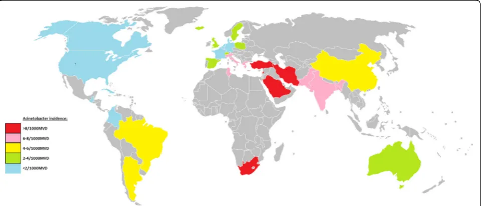
On the basis of the results of studies reporting
inci-dence for single countries, a world map of Acinetobacter
VAP infection incidence can be produced (Fig. 7).
Discussion
This is a survey of the incidence of
Acinetobacter
species
associated VAP among published studies using
meta-analysis to characterize the variation in incidence
world-wide. It reinforces and further characterizes previous
observations [101, 102]. It reveals a more than fivefold
variation in incidence among seven broad world-wide
and multinational regions that is not explainable by a
limited number of group level factors.
[image:4.595.57.543.424.664.2]There are major logistical challenges in undertaking
any international survey and there are few
prospect-ive multinational comparisons of hospital acquired
infections published. A worldwide prevalence survey
Table 2
Log
Acinetobacter
VAP incidence per thousand MV days; meta-regression models
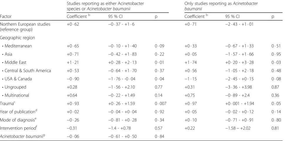
aStudies reporting as either Acinetobacter species orAcinetobacter baumanii
Only studies reporting asAcinetobacter baumanii
Factor Coefficientb 95 % CI p Coefficientb 95 % CI p
Northern European studies (reference group)
+0 · 62 −0 · 37 - +1 · 6 +0 · 71 −2 · 43 - +1 · 01
Geographic region
•Mediterranean +0 · 65 −0 · 10 - +1 · 40 0 · 09 +0 · 33 −0 · 67 - +1 · 33 0 · 51
•Asia +0 · 71 −0 · 42 - +1 · 83 0 · 22 +0 · 05 −1 · 57 - +1 · 66 0 · 95
•Middle East +1 · 21 +0 · 28 - +2 · 13 0 · 01 +1 · 74 +0 · 20 - +3 · 28 0 · 03
•Central & South America +0 · 53 −0 · 64 - +1 · 70 0 · 37 +0 · 56 −1 · 05 - +2 · 18 0 · 48
•USA & Canada −0 · 90 −1 · 76 - -0 · 04 0 · 04 −1 · 15 −2 · 45 - +0 · 15 0 · 08
•Ungrouped +0.28 −1 · 56 - +2.10 0.77 +0.31 −3 · 36 - +3.98 0.87
•Multinational +0.64 −0 · 22 - +1.49 0.14 +0.75 −0 · 89 - +2.4 0.36
Traumac +0 · 93 +0 · 26 - +1.59 0 · 007 +0 · 97 +0 · 001 - +1.94 0 · 05
Year of publicationd +0 · 02 −0 · 04 - +0 · 04 0 · 92 +0 · 05 −0 · 02 - +0 · 12 0 · 14
Mode of diagnosise −0 · 26 −0 · 81 - +0 · 28 0 · 34 +0 · 10 −0 · 71 - +0 · 91 0 · 80
Intervention periodf
−0.31 −1.4 - +0.78 0.57 +0.22 −1.58−+2.02 0.81
Acinetobacter baumaniig
−0 · 06 −0 · 61 - +0 · 50 0 · 84
a
This table displays the results of a meta-regression analysis for logAcinetobacterVAP incidence per thousand MV days
b
Interpretation. The reference group is the Northern European studies and this coefficient equals the difference in log from 0 (a log equal to 0 equates to a rate of 1. The other coefficients represent the difference in log for groups positive for that factor versus the reference group
c
The co-efficient for trauma represents the increment in log for an ICU having a majority of admissions for trauma
d
The co-efficient for year of publication represents the linear increment in log for each year after 1980
e
For sampling using bronchoscopic versus tracheal sampling
f
Studies undertaken during an infection control intervention
g
of
Pseudomonas aeruginosa
associated VAP across 11
countries during 2011–2012 revealed an insignificant
variation
in
prevalence
of
both
P.
aeruginosa
ventilator-associated pneumonia and also VAP overall
across four regions; the United States, Europe, Latin
America, and Asia Pacific [113]. By contrast, an
anonymized survey of 55 ICUs of 46 hospitals in
Argentina, Brazil, Colombia, India, Mexico, Morocco,
Peru, and Turkey revealed an overall rate of VAP of
24.1 per 1000 MV days with
Acinetobacter
species
accounting for between 3 and 46 % of VAP isolates
amongst the eight non-identified countries [3].
How-ever, the extent to which any possible association
with admission for trauma account for differences in
VAP microbiology is difficult to establish in short
term single center studies [114, 115].
Seasonal variation is another challenge to attempts at
sur-veillance [106–108]. The seasonal variation amongst
hos-pital acquired pneumonia and bloodstream
Acinetobacter
species infections was first documented in National
Noso-comial Infections Surveillance System (NNIS) data and
more recently within The Surveillance Network-USA
data-base [106].
Acinetobacter
species infections in these surveys
were ~50 % more common in summer than winter months.
The variation seen here in this worldwide survey exceeds
that explainable by seasonal variation. A possible
mechan-ism to account for this seasonal and possibly geographic
variation, and by contrast to species that do not exhibit the
same variation, is that Acinetobacter and particularly
A.
baumanii
have an exceptional ability to survive desiccation.
It remains to speculate how this property of Acinetobacter
could account for the variation found here. Of interest in
this regard however, amongst a panel of Acinetobacter
iso-lates, this ability to survive desiccation was notable for
A.
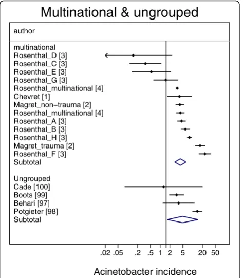
Fig. 1Caterpillar plots of the group specific (small diamonds) and
summary (large open diamond, broken vertical line) Acinetobacter VAP incidence per 1000 mechanical ventilation days and 95 % CI for groups from the multinational (top) and ungrouped (bottom) studies. For comparison, the summary Acinetobacter VAP incidence (vertical line) derived from the studies from French groups is shown for reference. Studies are listed in Additional file 1: Tables S1 & S6. Note that the x axis is a logarithmic scale
Fig. 2Caterpillar plots of the group specific (small diamonds) and
summary (large open diamond) Acinetobacter VAP incidence per 1000 mechanical ventilation days and 95 % CI for groups from the French (top) studies, and studies from other European countries. For comparison, the summary Acinetobacter VAP incidence (vertical line) derived from the studies from French groups is shown for reference. Studies are listed in Additional file 1: Tables S2. Note that the x axis is a logarithmic scale
[image:5.595.304.538.86.489.2] [image:5.595.57.294.88.359.2]baumanii
that had caused an outbreak of hospital acquired
respiratory tract infections [116].
The advantage of a literature survey is that
pub-lished data is readily available and the meta-regression
methods for analysing these types of data are
estab-lished. In contrast to multi-country incidence studies,
which tend to be a snap shot over typically less than
six months, most of the studies here extended over
more than twelve months. Here a random effects
methods is used. By using this method, the precision
associated with each individual study estimate is
in-corporated in the derivation of both the overall
sum-mary estimate and in the derivation of the
meta-regression model. Moreover, in contrast to a fixed
ef-fects model, a random effect meta-regression model
will generate more conservative summary estimates
(i.e. wider 95 % confidence limits) as the method
in-corporates both within and between study variability.
In this way, comparisons to address questions of study
specific [115] and contextual [117] influences that
would not be apparent within a single center study are
enabled. As an example, the use of meta-regression
can be used to benchmark control group pneumonia
[112] and bacteremia [118] incidences in published
prevention studies of VAP. The finding here of
vari-ability in incidence by region raises the possibility of
contextual factors behind the variation.
There are several limitations to this literature
based study. This is an analysis at the group level
and is unable to take account of patient specific risk
factors for
Acinetobacter
species associated VAP. For
example, the usage of empiric antibiotic therapy in
each study is an important unknown as use or
non-use may account for vulnerability to
Acinetobacter
Fig. 3Caterpillar plots of the group specific (small diamonds) and
summary (large open diamond) Acinetobacter VAP incidence per 1000 mechanical ventilation days and 95 % CI for groups from the Mediterranean studies. For comparison, the summary Acinetobacter VAP incidence (vertical line) derived from the studies from French groups is shown for reference. Studies are listed in Additional file 1: Tables S3. Note that the x axis is a logarithmic scale
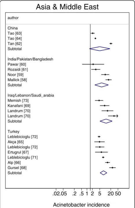
Fig. 4Caterpillar plots of the group specific (small diamonds) and
summary (large open diamond) Acinetobacter VAP incidence per 1000 mechanical ventilation days and 95 % CI for groups from the studies from Asia (top) and the middle East (bottom). For
[image:6.595.305.539.88.447.2] [image:6.595.56.294.88.408.2]species associated VAP at the level of the individual
patient [119, 120].
The grouping of countries into near neighbour
group-ings is somewhat arbitrary. Country and even regional
groupings could be confounded by other variables such as
infection control practices, prevalence of antibiotic use
and standards of care for patients receiving mechanical
ventilation that have not been able to be considered in the
analysis here. Another limitation and difficult to exclude
bias is the possible influence of publication bias.
The main limitation of a literature based survey is the
lack of standardization across jurisdictions. It could be
anticipated that there might be a range of clinical
defi-nitions used in the diagnosis of VAP at the level of the
individual patient. That the mode of VAP diagnosis was
not a significant factor in the regression model (Table 2)
implies that this bias is likely to be minimal within a
group level analysis as here. Likewise, the possibility of
a linear time trend has been considered within the
meta-regression model but this does not exclude the
impact of trends more complex than linear.
An additional limitation is that for some reports,
the VAP associated
Acinetobacter, being the number
of patients with VAP having an
Acinetobacter
species
isolated from respiratory sampling, was not available.
For these reports this number was derived as the
number of patients with VAP multiplied by the
pro-portion of VAP isolates that were
Acinetobacter
spe-cies. This is likely a reasonable approximation for a
relatively rare outcome as found here. This
approxi-mation allows for VAP patients with multiple isolates.
A more difficult issue is that of laboratory
documenta-tion and reporting of Acinetobacter species type across
jurisdictions. The striking observation among this survey
was that all studies reported only one classification type of
Acinetobacter. This was apparent in even the surveys with
the most number of isolates [4, 27, 63]. The most
com-mon mode of reporting was as Acinetobacter species.
Aci-netobacter infections were less commonly reported as
Acinetobacter baumanii
from studies outside of Northern
European and North American centers. It is possible that
second line Acinetobacter species had been identified and
listed within the category of
‘other’
gram negative
infec-tions. However, it remains a plausible explanation that the
common practice in the literature reported here of the
listing of a single Acinetobacter species generally reflects
the mode of local reporting practices. In this regard and of
pertinence to this survey of
Acinetobacter baumanii, the
reports of Acinetobacter species cannot be easily
dismissed.
Of particular note, the rates of Acinetobacter VAP
re-ported from studies reporting as Acinetobacter species
ver-sus studies reporting as
Acinetobacter baumanii
showed
similar patterns of regional variation despite similar rates of
overall VAP infection. Moreover, this regional variation in
rates of Acinetobacter VAP were not explainable in a
meta-regression model by trauma ICU, year of publication and
Acinetobacter reporting practice as group level variables
whereas a comparable meta-regression model of VAP
showed no major regional variation in overall VAP rates
(Additional file 1: Table S7).
The methods in use in clinical microbiology
labora-tories likely varied not only geographically but also
likely temporally over the three decades encompassed
in this survey [121–125]. Because of the limitations of
the traditional phenotypic testing methods [121, 122]
for identification, a broad category of
Acinetobacter
calcoaceticus-A. baumanii
complex was suggested at
one point [121]. These phenotypic methods are being
superseded by newer and more specific molecular
methods [123–125]. Moreover, these and even newer
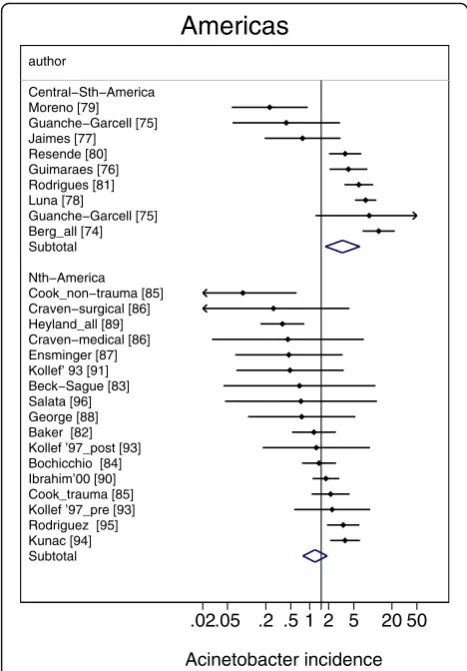
Fig. 5Caterpillar plots of the group specific (small diamonds) and
summary (large open diamond) Acinetobacter VAP incidence per 1000 mechanical ventilation days and 95 % CI for groups from the North (bottom) and central and outh (top) American studies. For comparison, the summary Acinetobacter VAP incidence (vertical line) derived from the studies from French groups is shown for reference. Studies are listed in Additional file 1: Tables S5. Note that the x axis is a logarithmic scale
[image:7.595.56.290.87.423.2]methods such as MALDI-TOF for microbial detection
and identification will likely further refine the
identifi-cation and reporting of Acinetobacter species clinical
isolates going forward.
The lower prevalence of
Acinetobacter
in cooler
sea-sons [106–108] is consistent with the finding here that
the prevalence is lower in reports from countries further
away from the equator. The biological mechanism for
this difference remains speculative. However,
Acineto-bacter
species are gram negative bacteria which have
im-portant additional international public health issue for
two further reasons. The transportation of patients
around the world create the potential for infection
con-trol failures [111]. Moreover,
Acinetobacter
species are
commonly multi-drug resistant although rates of
resist-ance vary from region to region [126].
Fig. 6A scatter plot of worldwideAcinetobacterVAP incidence (per 1000 MV days) among published studies in seven geographic regions with rates
from studies reporting Acinetobacter infections asAcinetobacter baumanii(open symbols) versus reporting as Acinetobacter species or otherwise (closed symbols; Note logarithmic scale of incidence). The vertical lines are for reference at incidence rates of 0.1, 1 and 10 per 1000 mvd
Fig. 7A map ofAcinetobacterVAP incidence per 1000 MV days for each country or world sub region for which at least two reports were
[image:8.595.61.539.87.288.2] [image:8.595.59.540.477.683.2]Conclusion
There is a greater than fivefold variation in
Acinetobacter
associated VAP among published reports from various
geographic regions worldwide. This variation is not
ex-plainable by variations in rates of VAP overall,
admis-sions for trauma, publication year or Acinetobacter
reporting practices as group level variables.
Additional file
Additional file 1:Tables of study data, a meta-regression of VAP inci-dence and listing of 100 references. (PDF 740 kb)
Abbreviations
ICU:Intensive Care Unit; MV: Mechanical ventilation; VAP: Ventilator associated pneumonia
Funding
This work was supported by the Australian Government Department of Health and Ageing through the Rural Clinical Training and Support (RCTS) program. The funding agency had no role in the preparation of the manuscript nor its approval for submission.
Availability of data and materials
The datasets supporting the conclusions of this article are included within the article and its additional file 1.
Authors’contributions
The author undertook the literature search, data analysis, manuscript preparation and approved its submission for publication and is the guarantor for this article.
Competing interest
The author declares that he has no competing interest.
Ethics approval and consent to participate
This article does not contain any studies with human participants performed by the author. Consent was not applicable.
Author details
1Department of Rural Health, Melbourne Medical School, University of
Melbourne, Ballarat 3353, Australia.2Internal Medicine Service, Ballarat Health Services, PO Box 577, Ballarat 3353, Australia.3Infection Control Committees,
St John of God Hospital and Ballarat Health Services, Ballarat, Victoria, Australia.
Received: 26 July 2016 Accepted: 12 October 2016
References
1. Chevret S, Hemmer M, Carlet J, et al. Incidence and risk factors of pneumonia acquired in intensive care units. Results from a multicenter prospective study on 996 patients. European Cooperative Group on Nosocomial Pneumonia. Intensive Care Med. 1993;19:256–64.
2. Magret M, Amaya-Villar R, Garnacho J, Lisboa T, Diaz E, DeWaele J, Deja M, Manno E, Rello J, EU-VAP/CAP Study Group. Ventilator-associated pneumonia in trauma patients is associated with lower mortality: results from EU-VAP study. J Trauma Acute Care Surg. 2010;69(4):849–54. 3. Rosenthal VD, Maki DG, Salomao R, Moreno CA, Mehta Y, Higuera F, Cuellar
LE, Arikan OA, Abouqal R, Leblebicioglu H. Device-associated nosocomial infections in 55 intensive care units of 8 developing countries. Ann Intern Med. 2006;145(8):582–91.
4. Rosenthal VD, Rodrigues C, Madani N, Mitrev Z, Ye G, Salomao R, Ulger F, Guanche-Garcell H, Kanj SS, Cuéllar LE, Higuera F. Effectiveness of a multidimensional approach for prevention of ventilator-associated pneumonia in adult intensive care units from 14 developing countries of
four continents: Findings of the International Nosocomial Infection Control Consortium. Crit Care Med. 2012;40(12):3121–8.
5. Bercault N, Wolf M, Runge I, et al. Intrahospital transport of critically ill ventilated patients: a risk factor for ventilator-associated pneumonia–a matched cohort study. Crit Care Med. 2005;33:2471–8.
6. Bornstain C, Azoulay E, De Lassence A, Cohen Y, Costa MA, Mourvillier B, Descorps-Declere A, Garrouste-Orgeas M, Thuong M, Schlemmer B, Timsit JF. Sedation, sucralfate, and antibiotic use are potential means for protection against early-onset ventilator-associated pneumonia. Clin Infect Dis. 2004;38(10):1401–8.
7. Bregeon F, Papazian L, Visconti A, Gregoire R, Thirion X, Gouin F. Relationship of microbiologic diagnostic criteria to morbidity and mortality in patients with ventilator-associated pneumonia. JAMA.
1997;277:655–62.
8. Chastre J, Trouillet JL, Vuagnat A, et al. Nosocomial pneumonia in patients with acute respiratory distress syndrome. Am J Respir Crit Care Med. 1998;157:1165–72.
9. Fagon JY, Chastre J, Domart Y, Trouillet JL, Pierre J, Darne C, Gibert C. Nosocomial pneumonia in patients receiving continuous mechanical ventilation. Prospective analysis of 52 episodes with use of a protected specimen brush and quantitative culture techniques. Am Rev Respir Dis. 1989;139:877–84.
10. Garrouste-Orgeas M, Chevret S, Arlet G, et al. Oropharyngeal or gastric colonization and nosocomial pneumonia in adult intensive care unit patients. A prospective study based on genomic DNA analysis. Am J Respir Crit Care Med. 1997;156:1647–56.
11. Georges H, Leroy O, Guery B, Alfandari S, Beaucaire G. Predisposing factors for nosocomial pneumonia in patients receiving mechanical ventilation and requiring tracheotomy. Chest. 2000;118:767–74.
12. Gruson D, Hilbert G, Vargas F, Valentino R, Bebear C, Allery A, Bebear C, Gbikpi-benissan GE, Cardinaud JP. Rotation and restricted use of antibiotics in a medical intensive care unit: impact on the incidence of ventilator-associated pneumonia caused by antibiotic-resistant gram-negative bacteria. Am J Respir Crit Care Med. 2000;162(3):837–43.
13. Gruson D, Hilbert G, Vargas F, et al. Strategy of antibiotic rotation: long-term effect on incidence and susceptibilities of Gram-negative bacilli responsible for ventilator-associated pneumonia. Crit Care Med. 2003;31:1908–14. 14. Guerin C, Girard R, Chemorin C, De Varax R, Fournier G. Facial mask
noninvasive mechanical ventilation reduces the incidence of nosocomial pneumonia. Intensive Care Med. 1997;23(10):1024–32.
15. Jaillette E, Nseir S. Relationship between inhaledβ2-agonists and ventilator-associated pneumonia: A cohort study. Crit Care Med.
2011;39(4):725–30.
16. Luyt CE, Guerin V, Combes A, et al. Procalcitonin kinetics as a prognostic marker of ventilator-associated pneumonia. Am J Respir Crit Care Med. 2005;171:48–53.
17. Mahul P, Auboyer C, Jospe R, Ros A, Guerin C, el Khouri Z, Galliez M, Dumont A, Gaudin O. Prevention of nosocomial pneumonia in intubated patients respective role of mechanical subglottic secretions drainage and stress ulcer prophylaxis. Intensive Care Med. 1992;18:20–5.
18. Markowicz P, Wolff M, Djedaini K, Cohen Y, Chastre J, Delclaux C. Multicenter prospective study of ventilator-associated pneumonia during acute respiratory distress syndrome. Incidence, prognosis, and risk factors. ARDS Study Group. Am J Respir Crit Care Med. 2000;161:1942–8. 19. Moine P, Timsit JF, De Lassence A, et al. Mortality associated with late-onset
pneumonia in the intensive care unit: results of a multi-center cohort study. Intensive Care Med. 2002;28:154–63.
20. Nseir S, Di Pompeo C, Soubrier S, Cavestri B, Jozefowicz E, Saulnier F, Durocher A. Impact of ventilator-associated pneumonia on outcome in patients with COPD. Chest. 2005;128(3):1650–6.
21. Papazian L, Bregeon F, Thirion X, et al. Effect of ventilator-associated pneumonia on mortality and morbidity. Am J Respir Crit Care Med. 1996;154:91–7. 22. Stéphan F, Mabrouk N, Decailliot F, Delclaux C, Legrand P.
Ventilator-associated pneumonia leading to acute lung injury after trauma: importance of Haemophilus influenzae. Anesthesiol. 2006;104:235–41. 23. Timsit JF, Chevret S, Valcke J, et al. Mortality of nosocomial pneumonia in
ventilated patients: influence of diagnostic tools. Am J Respir Crit Care Med. 1996;154:116–23.
24. Trouillet JL, Chastre J, Vuagnat A, Joly-Guillou ML, Combaux D, Dombret MC, Gibert C. Ventilator-associated pneumonia caused by potentially drug-resistant bacteria. Am J Respir Crit Care Med. 1998;157(2):531–9.
25. Daschner F, Kappstein I, Schuster F, et al. Influence of disposable (‘Conchapak’) and reusable humidifying systems on the incidence of ventilation pneumonia. J Hosp Infect. 1988;11:161–8.
26. Daschner F, Kappstein I, Engels I, Reuschenbach K, Pfisterer J, Krieg N, Vogel W. Stress Ulcer Prophylaxis and Ventilation Pneumonia Prevention by Antibacterial Cytoprotective Agents? Infect Control. 1988;9(02):59–65. 27. Kohlenberg A, Schwab F, Behnke M, Geffers C, Gastmeier P. Pneumonia
associated with invasive and noninvasive ventilation: an analysis of the German nosocomial infection surveillance system database. Intensive Care Med. 2010;36(6):971–8.
28. Myny D, Depuydt P, Colardyn F, Blot S. Ventilator-associated pneumonia in a tertiary care ICU analysis of risk factors for acquisition and mortality. Acta Clin Belg. 2005;60:114–21.
29. Nielsen SL, Roder B, Magnussen P, et al. Nosocomial pneumonia in an intensive care unit in a Danish university hospital: incidence, mortality and etiology. Scand J Infect Dis. 1992;24:65–70.
30. Verhamme KM, De Coster W, De Roo L, De Beenhouwer H, Nollet G, Verbeke J, Demeyer I, Jordens P. Pathogens in early-onset and late-onset intensive care unit–acquired pneumonia. Infect Control Hosp Epidemiol. 2007;28(04):389–97.
31. Woske HJ, Röding T, Schulz I, Lode H. Ventilator-associated pneumonia in a surgical intensive care unit Epidemiology, etiology and comparison of three bronchoscopic methods for microbiological specimen sampling. Crit Care. 2001;5:167–73.
32. A’Court CH, Garrard CS, Crook D, et al. Microbiological lung surveillance in mechanically ventilated patients, using non-directed bronchial lavage and quantitative culture. Q J Med. 1993;86:635–48.
33. Duszyńska W, Rosenthal VD, Dragan B, Węgrzyn P, Mazur A, Wojtyra P, Tomala A, Kübler A. Ventilator-associated pneumonia monitoring according to the INICC project at one centre. Anaesthesiol Intensive Ther. 2015;47(1):34–9. 34. Hugonnet S, Uçkay I, Pittet D. Staffing level: a determinant of late-onset
ventilator-associated pneumonia. Crit Care. 2007;11(4):R80.
35. Hyllienmark P, Gardlund B, Persson JO, Ekdahl K. Nosocomial pneumonia in the ICU: a prospective cohort study. Scand J Infect Dis. 2007;39:676–82. 36. Magnason S, Kristinsson KG, Stefansson T, Erlendsdottir H, Jonsdottir K,
Kristjansson M, Gudmundsson S. Risk factors and outcome in ICU‐acquired infections. Acta Anaesthesiol Scand. 2008;52:1238–45.
37. Reusser P, Zimmerli W, Scheidegger D, Marbet GA, Buser M, Gyr K. Role of gastric colonization in nosocomial infections and endotoxemia: a prospective study in neurosurgical patients on mechanical ventilation. J Infect Dis. 1989;160:414–21.
38. Alvarez-Lerma F. ICU-acquired Pneumonia Study Group. Modification of empiric antibiotic treatment in patients with pneumonia acquired in the intensive care unit. Intensive Care Med. 1996;22(5):387–94.
39. Baraibar J, Correa H, Mariscal D, Gallego M, Valles J, Rello J. Risk factors for infection by Acinetobacter baumannii in intubated patients with nosocomial pneumonia. Chest. 1997;112(4):1050–4.
40. Cavalcanti M, Ferrer M, Ferrer R, et al. Risk and prognostic factors of ventilator-associated pneumonia in trauma patients. Crit Care Med. 2006;34:1067–72. 41. Cardenosa Cendrero JA, Sole-Violan J, Bordes Benitez A, et al. Role of
different routes of tracheal colonization in the development of pneumonia in patients receiving mechanical ventilation. Chest. 1999;116:462–70. 42. de Latorre FJ, Pont T, Ferrer A, et al. Pattern of tracheal colonization during
echanical ventilation. Am J Respir Crit Care Med. 1995;152:1028–33. 43. Ewig S, Torres A, El-Ebiary M, et al. Bacterial colonization patterns in
mechanically ventilated patients with traumatic and medical head injury. Incidence, risk factors, and association with ventilator-associated pneumonia. Am J Respir Crit Care Med. 1999;159:188–98.
44. Jimenez P, Torres A, Rodriguez-Roisin R, et al. Incidence and etiology of pneumonia acquired during mechanical ventilation. Crit Care Med. 1989;17:882–5.
45. Rello J, Quintana E, Ausina V, et al. Incidence, etiology, and outcome of nosocomial pneumonia in mechanically ventilated patients. Chest. 1991; 100:439–44.
46. Rello J, Lorente C, Diaz E, et al. Incidence, etiology, and outcome of nosocomial pneumonia in ICU patients requiring percutaneous tracheotomy for mechanical ventilation. Chest. 2003;124:2239–43. 47. Rincón-Ferrari MD, Flores-Cordero JM, Leal-Noval SR, Murillo-Cabezas F,
Cayuelas A, Muñoz-Sánchez MA, Sánchez-Olmedo JI. Impact of ventilator-associated pneumonia in patients with severe head injury. J Trauma Acute Care Surg. 2004;57(6):1234–40.
48. Ruiz-Santana S, Garcia Jimenez A, Esteban A, et al. ICU pneumonias: a multi-institutional study. Crit Care Med. 1987;15:930–2.
49. Tamayo E, Álvarez FJ, Martínez-Rafael B, Bustamante J, Bermejo-Martin JF, Fierro I, Eiros JM, Castrodeza J, Heredia M, Gómez-Herreras JI, Valladolid Sepsis Study Group. Ventilator-associated pneumonia is an important risk factor for mortality after major cardiac surgery. J Crit Care.
2012;27(1):18–25.
50. Tejada Artigas A, Bello Dronda S, Chacon Valles E, et al. Risk factors for nosocomial pneumonia in critically ill trauma patients. Crit Care Med. 2001;29:304–9.
51. Torres A, Aznar R, Gatell JM, et al. Incidence, risk, and prognosis factors of nosocomial pneumonia in mechanically ventilated patients. Am Rev Respir Dis. 1990;142:523–8.
52. Violan JS, Sanchez-Ramirez C, Mujica AP, Cendrero JC, Fernandez JA, de Castro FR. Impact of nosocomial pneumonia on the outcome of mechanically-ventilated patients. Crit Care (Lond). 1998;2:19–23. 53. Antonelli M, Moro ML, Capelli O, et al. Risk factors for early onset
pneumonia in trauma patients. Chest. 1994;105:224–8.
54. Apostolopoulou E, Bakakos P, Katostaras T, et al. Incidence and risk factors for ventilator-associated pneumonia in 4 multidisciplinary intensive care units in Athens, Greece. Respir Care. 2003;48:681–8.
55. Kallel H, Chelly H, Bahloul M, Ksibi H, Dammak H, Chaari A, Hamida CB, Rekik N, Bouaziz M. The effect of ventilator-associated pneumonia on the prognosis of head trauma patients. J Trauma Acute Care Surg. 2005;59(3):705–10.
56. Piazza O, Iasiello A, PapaIanni C, De Robertis E, Servillo G, Rossano F, Tufano R. Incidence of antimicrobial-resistant ventilator associated pneumonia: an eighteen-month survey. Panminerva Med. 2005;47(4):265–7.
57. Sofianou DC, Constandinidis TC, Yannacou M, et al. Analysis of risk factors for ventilator-associated pneumonia in a multidisciplinary intensive care unit. Eur J Clin Microbiol Infect Dis. 2000;19:460–3.
58. Mallick UK, Faruq MO, Ahsan AA, Fatema K, Ahmed F, Asaduzzaman M, Islam M, Sultana A. Spectrum of Early Onset and Late Onset Ventilator Associated Pneumonia (VAP) in a Tertiary Care Hospital of Bangladesh: A Prospective Cohort Study. Bangladesh Crit Care J. 2015;3(1):9–13. 59. Noor A, Hussain SF. Risk factors associated with development of ventilator
associated pneumonia. J Coll Physicians Surg Pak. 2005;15:92–5.
60. Pawar M, Mehta Y, Khurana P, Chaudhary A, Kulkarni V, Trehan N. Ventilator-associated pneumonia: incidence, risk factors, outcome, and microbiology. J Cardiothorac Vasc Anesth. 2003;17(1):22–8.
61. Rozaidi SW, Sukro J, Dan A. The incidence of nosocomial infection in the Intensive Care Unit, Hospital Universiti Kebangsaan Malaysia: ICU-acquired nosocomial infection surveillance program 1998–1999. Med J Malaysia. 2001;56(2):207–22.
62. Tan X, Zhu S, Yan D, Chen W, Chen R, Zou J, Yan J, Zhang X, Farmakiotis D, Mylonakis E. Candida spp. airway colonization: A potential risk factor for Acinetobacter baumannii ventilator-associated pneumonia. Med Mycol 2016;54(6):557–66.
63. Tao L, Hu B, Rosenthal VD, Gao X, He L. Device-associated infection rates in 398 intensive care units in Shanghai, China: International Nosocomial Infection Control Consortium (INICC) findings. Int J Infect Dis. 2011;15(11):e774–80.
64. Tao L, Hu B, Rosenthal VD, Zhang Y, Gao X, He L. Impact of a
multidimensional approach on ventilator-associated pneumonia rates in a hospital of Shanghai: findings of the International Nosocomial Infection Control Consortium. J Crit Care. 2012;27(5):440–6.
65. Akça O, Koltka K, Uzel S, et al. Risk factors for early-onset, ventilator-associated pneumonia in critical care patients: selected multiresistant versus non-resistant bacteria. Anesthesiol. 2000;93:638–45.
66. Alp E, Güven M, Yıldız O, Aygen B, Voss A, Doganay M. Incidence, risk factors and mortality of nosocomial pneumonia in intensive care units: a prospective study. Ann Clin Microbiol Antimicrob. 2004;3(1):1.
67. Ertugrul BM, Yildirim A, Ay P, Oncu S, Cagatay A, Cakar N, Ertekin C, Ozsut H, Eraksoy H, Calangu S. Ventilator-associated pneumonia in surgical emergency intensive care unit. Saudi Med J. 2006;27(1):52–7.
68. Gursel G, Aydogdu M, Nadir Ozis T, Tasyurek S. Comparison of the value of initial and serial endotracheal aspirate surveillance cultures in predicting the causative pathogen of ventilator-associated pneumonia. Scand J Infect Dis. 2010;42:341–6.
susceptibility patterns of isolated microorganisms. Infect Control Hosp Epidemiol. 2003;24:864–9.
70. Landrum ML, Murray CK. Ventilator associated pneumonia in a military deployed setting: the impact of an aggressive infection control program. J Trauma Acute Care Surg. 2008;64(2):S123–8.
71. Leblebicioglu H, Rosenthal VD, Arıkan ÖA, Özgültekin A, Yalcin AN, Koksal I, Usluer G, Sardan YC, Ulusoy S. Device-associated hospital-acquired infection rates in Turkish intensive care units. Findings of the International Nosocomial Infection Control Consortium (INICC). J Hosp Infect. 2007;65(3):251–7.
72. Leblebicioglu H, Yalcin AN, Rosenthal VD, Koksal I, Sirmatel F, Unal S, Turgut H, Ozdemir D, Ersoz G, Uzun C, Ulusoy S. Effectiveness of a
multidimensional approach for prevention of ventilator-associated pneumonia in 11 adult intensive care units from 10 cities of Turkey: findings of the International Nosocomial Infection Control Consortium (INICC). Infect. 2013;41(2):447–56.
73. Memish ZA, Cunningham G, Oni GA, et al. The incidence and risk factors of ventilator-associated pneumonia in a Riyadh hospital. Infect Control Hosp Epidemiol. 2000;21:271–3.
74. Berg DE, Hershow RC, Ramirez CA, Weinstein RA. Control of nosocomial infections in an intensive care unit in Guatemala City. Clin Infect Dis. 1995;21:588–93.
75. Guanche-Garcell H, Morales-Perez C, Rosenthal VD. Effectiveness of a multidimensional approach for the prevention of ventilator-associated pneumonia in an adult intensive care unit in Cuba: findings of the International Nosocomial Infection Control Consortium (INICC). J Infect Public Health. 2013;6:98–107.
76. Guimaraes MM, Rocco JR. Prevalence of ventilator-associated pneumonia in a university hospital and prognosis for the patients affected. J Bras Pneumol. 2006;32:339–46.
77. Jaimes F, De La Rosa G, Gómez E, Múnera P, Ramírez J, Castrillón S. Incidence and risk factors for ventilator-associated pneumonia in a developing country Where is the difference? Respir Med. 2007;101:762–7.
78. Luna CM, Blanzaco D, Niederman MS, et al. Resolution of ventilator-associated pneumonia: prospective evaluation of the clinical pulmonary infection score as an early clinical predictor of outcome. Crit Care Med. 2003;31:676–82.
79. Moreno CA, Rosenthal VD, Olarte N, Gomez WV, Sussmann O, Agudelo JG, Rojas C, Osorio L, Linares C, Valderrama A, Mercado PG. Device-associated infection rate and mortality in intensive care units of 9 Colombian hospitals: findings of the International Nosocomial Infection Control Consortium. Infect Control. 2006;27(04):349–56.
80. Resende MM, Monteiro SG, Callegari B, Figueiredo PM, Monteiro CR, Monteiro-Neto V. Epidemiology and outcomes of ventilator-associated pneumonia in northern Brazil: an analytical descriptive prospective cohort study. BMC Infect Dis. 2013;13(1):119.
81. Rodrigues PM, Neto C, Santos LR, Knibel MF. Ventilator-associated pneumonia: epidemiology and impact on the clinical evolution of ICU patients. J Bras Pneumol. 2009;35(11):1084–91.
82. Baker AM, Meredith JW, Haponik EF. Pneumonia in intubated trauma patients. Microbiology and outcomes. Am J Respir Crit Care Med. 1996; 153:343–9.
83. Beck-Sague CM, Sinkowitz RL, Chinn RY, et al. Risk factors for ventilator-associated pneumonia in surgical intensive-care-unit patients. Infect Control Hosp Epidemiol. 1996;17:374–6.
84. Bochicchio GV, Joshi M, Bochicchio K, et al. A time-dependent analysis of intensive care unit pneumonia in trauma patients. J Trauma. 2004;56:296–301.
85. Cook A, Norwood S, Berne J. Ventilator-associated pneumonia is more common and of less consequence in trauma patients compared with other critically ill patients. J Trauma Acute Care Surg. 2010;69(5):1083–91. 86. Craven DE, Kunches LM, Lichtenberg DA, et al. Nosocomial infection and
fatality in medical and surgical intensive care unit patients. Arch Intern Med. 1988;148:1161–8.
87. Ensminger SA, Wright RS, Baddour LM, Afess B. Suspected ventilator-associated pneumonia in cardiac patients admitted to the coronary care unit. Mayo Clin Proc. 2006;81:32–5.
88. George DL, Falk PS, Wunderink RG, Leeper Jr KV, Meduri GU, Steere EL, Glen MC. Epidemiology of ventilator-acquired pneumonia based on protected bronchoscopic sampling. Am J Respir Crit Care Med. 1998;158:1839–47.
89. Heyland DK, Cook DJ, Griffith L, Keenan SP, Brun-Buisson C. The attributable morbidity and mortality of ventilator-associated pneumonia in the critically ill patient. The Canadian Critical Trials Group. Am J Respir Crit Care Med. 1999;159:1249–56.
90. Ibrahim EH, Ward S, Sherman G, Kollef MH. A comparative analysis of patients with early-onset vs late-onset nosocomial pneumonia in the ICU setting. Chest. 2000;117:1434–42.
91. Kollef MH. Ventilator-associated pneumonia. A multivariate analysis. JAMA. 1993;270:1965–70.
92. Kollef MH, Silver P, Murphy DM, et al. The effect of late-onset ventilator-associated pneumonia in determining patient mortality. Chest. 1995;108:1655–62.
93. Kollef MH, Vlasnik J, Sharpless L, Pasque C, Murphy D, Fraser V. Scheduled change of antibiotic classes A strategy to decrease the incidence of ventilator-associated pneumonia. Am J Respir Crit Care Med. 1997;156:1040–8.
94. Kunac A, Sifri ZC, Mohr AM, Horng H, Lavery RF, Livingston DH. Bacteremia and Ventilator-Associated Pneumonia: A Marker for Contemporaneous Extra-Pulmonic Infection. Surg Infect. 2014;15:77–83.
95. Rodriguez JL, Gibbons KJ, Bitzer LG, et al. Pneumonia: incidence, risk factors, and outcome in injured patients. J Trauma. 1991;31:907–12.
96. Salata RA, Lederman MM, Shlaes DM, Jacobs MR, Eckstein E, Tweardy D, Toossi Z, Chmielewski R, Marino J, King CH. Diagnosis of nosocomial pneumonia in intubated, intensive care unit patients. Am Rev Respir Dis. 1987;135:426–32.
97. Behari AA, Kalafatis N. Incidence and outcome of ventilator-associated pneumonia in Inkosi Albert Luthuli and King Edward VIII Hospital surgical intensive care units. Southern African J Crit Care (Online).
2015;31(1):16–8.
98. Potgieter PD, Linton DM, Oliver S, Forder AA. Nosocomial infections in a respiratory intensive care unit. Crit Care Med. 1987;15:495–8.
99. Boots RJ, Phillips GE, George N, Faoagali JL. Surveillance culture utility and safety using low‐volume blind bronchoalveolar lavage in the diagnosis of ventilator‐associated pneumonia. Respirology. 2008;13:87–96.
100. Cade JF, McOwat E, Siganporia R, Keighley C, Presneill J, Sinickas V. Uncertain relevance of gastric colonization in the seriously ill. Intensive Care Med. 1992;18:210–7.
101. Munoz-Price LS, Weinstein RA. Acinetobacter infection. New Eng J Med. 2008;358(12):1271–81.
102. Falagas ME, Karveli EA, Siempos II, Vardakas KZ. Acinetobacter infections: a growing threat for critically ill patients. Epidemiol Infect.
2008;136(08):1009–19.
103. Chastre J, Fagon JY. Ventilator-associated pneumonia. Am J Respir Crit Care Med. 2002;165(7):867–903.
104. Falagas ME, Bliziotis IA, Siempos II. Attributable mortality of Acinetobacter baumannii infections in critically ill patients: a systematic review of matched cohort and case–control studies. Crit Care. 2006;10(2):1.
105. Garnacho J, Sole-Violan J, Sa-Borges M, Diaz E, Rello J. Clinical impact of pneumonia caused by Acinetobacter baumannii in intubated patients: a matched cohort study. Crit Care Med. 2003;31(10):2478–82.
106. McDonald LC, Banerjee SN, Jarvis WR. National Nosocomial Infections Surveillance System. Seasonal variation ofAcinetobacterinfections: 1987– 1996. Clin Infect Dis. 1999;29(5):1133–7.
107. Eber MR, Shardell M, Schweizer ML, Laxminarayan R, Perencevich EN. Seasonal and temperature-associated increases in gram-negative bacterial bloodstream infections among hospitalized patients. PLoS ONE. 2011;6(9):e25298. 108. Perencevich EN, McGregor JC, Shardell M, Furuno JP, Harris AD, Morris JG,
Fisman DN, Johnson JA. Summer peaks in the incidences of gram-negative bacterial infection among hospitalized patients. Infect Control Hosp Epidemiol. 2008;29(12):1124–31.
109. Fournier PE, Richet H, Weinstein RA. The epidemiology and control of Acinetobacter baumanniiin health care facilities. Clin Infect Dis. 2006;42(5):692–9. 110. Anstey NM, Currie BJ, Withnall KM. Community-acquired Acinetobacter
pneumonia in the Northern Territory of Australia. Clin Infect Dis. 1992;14:83–91.
111. Uckay I, Sax H, Harbarth S, Bernard L, Pittet D. Multi-resistant infections in repatriated patients after natural disasters: lessons learned from the 2004 tsunami for hospital infection control. J Hosp Infect. 2008;68(1):1–8. 112. Hurley JC. Ventilator Associated Pneumonia prevention methods using
topical antibiotics: herd protection or herd peril? Chest. 2014;146(4):890–8.
113. Kollef MH, Chastre J, Fagon JY, François B, Niederman MS, Rello J, et al. Global prospective epidemiologic and surveillance study of ventilator-associated pneumonia due to Pseudomonas aeruginosa. Crit Care Med. 2014;42(10):2178–87.
114. Agbaht K, Lisboa T, Pobo A, et al. Management of ventilator-associated pneumonia in a multidisciplinary intensive care unit: does trauma make a difference? Intensive Care Med. 2007;33(8):1387–95.
115. Hurley JC. Profound effect of study design factors on ventilator-associated pneumonia incidence of prevention studies: benchmarking the literature experience. J Antimicrob Chemother. 2008;61:1154–61.
116. Wendt C, Dietze B, Dietz E, Rüden H. Survival ofAcinetobacter baumanniion dry surfaces. J Clin Microbiol. 1997;35(6):1394–7.
117. Hurley JC. Inapparent outbreaks of ventilator-associated pneumonia: an ecologic analysis of prevention and cohort studies. Infect Control Hosp Epidemiol. 2005;26:374–90.
118. Hurley JC. Topical antibiotics as a major contextual hazard toward bacteremia within selective digestive decontamination studies: a meta-analysis. BMC Infect Dis. 2014;14:714.
119. Medina J, Formento C, Pontet J, Curbelo A, Bazet C, Gerez J, Larrañaga E. Prospective study of risk factors for ventilator-associated pneumonia caused by Acinetobacter species. J Crit Care. 2007;22(1):8–26.
120. Garnacho-Montero J, Ortiz-Leyba C, Fernández-Hinojosa E, Aldabó-Pallás T, Cayuela A, Marquez-Vácaro JA, Garcia-Curiel A, Jiménez-Jiménez FJ. Acinetobacter baumanniiventilator-associated pneumonia: epidemiological and clinical findings. Intensive Care Med. 2005;31(5):649–55.
121. Turton JF, Shah J, Ozongwu C, Pike R. Incidence of Acinetobacter species other thanA. baumanniiamong clinical isolates of Acinetobacter: evidence for emerging species. J Clin Microbiol. 2010;48(4):1445–9.
122. Gerner-Smidt PE, Tjernberg I, Ursing J. Reliability of phenotypic tests for identification of Acinetobacter species. J Clin Microbiol. 1991;29(2):277–82. 123. Bosshard PP, Zbinden R, Abels S, Böddinghaus B, Altwegg M, Böttger EC.
16S rRNA gene sequencing versus the API 20 NE system and the VITEK 2 ID-GNB card for identification of nonfermenting Gram-negative bacteria in the clinical laboratory. J Clin Microbiol. 2006;44(4):1359–66.
124. Bernards AT, Van der Toorn J, Van Boven CP, Dijkshoorn L. Evaluation of the ability of a commercial system to identifyAcinetobacter genomic species. Eur J Clin Microbiol Infect Dis. 1996;15(4):303–8.
125. Lee MJ, Jang SJ, Li XM, Park G, Kook JK, Kim MJ, Chang YH, Shin JH, Kim SH, Kim DM, Kang SH. Comparison of rpoB gene sequencing, 16S rRNA gene sequencing, gyrB multiplex PCR, and the VITEK2 system for identification of Acinetobacter clinical isolates. Diag Microbiol Infect Dis. 2014;78(1):29–34. 126. Lob SH, Hoban DJ, Sahm DF, Badal RE. Regional differences and trends in antimicrobial susceptibility ofAcinetobacter baumannii. Int J Antimicrob Agent. 2016;47(4):317–23.
• We accept pre-submission inquiries
• Our selector tool helps you to find the most relevant journal
• We provide round the clock customer support
• Convenient online submission
• Thorough peer review
• Inclusion in PubMed and all major indexing services
• Maximum visibility for your research
Submit your manuscript at www.biomedcentral.com/submit