White Rose Research Online [email protected]
Universities of Leeds, Sheffield and York
http://eprints.whiterose.ac.uk/
This is an author produced version of a paper published in Journal of Clinical Endocrinology and Metabolism.
White Rose Research Online URL for this paper:
http://eprints.whiterose.ac.uk/8641 (Figures 1-3 for the paper also available from this URL)
Published paper
Debono M, Ghobadi C, Rostami-Hodjegan A, Ross RJ et al. (2009) Modified-Release Hydrocortisone to Provide Circadian Cortisol Profiles, Journal of Clinical
Endocrinology and Metabolism, 94(5) pp.1548-1554
Modified-release hydrocortisone to provide circadian cortisol profiles
Short title: Circadian hydrocortisone therapy
Authors: Miguel Debono1, Cyrus Ghobadi2, Amin Rostami-Hodjegan2, Hiep Huatan3, Michael J
Campbell4, John Newell-Price1, Ken Darzy5, Deborah P Merke6, Wiebke Arlt7 and Richard J
Ross1.
Affiliations: 1Academic Unit of Diabetes, Endocrinology & Metabolism & 2Academic Unit of
Clinical Pharmacology, School of Medicine, Royal Hallamshire Hospital, University of Sheffield,
Sheffield, UK; 3H2Pharma; 4Health Services Research ScHARR, University of Sheffield, Regent
Court, 30 Regent street, Sheffield, UK; 5Diabetes and Endocrinology, East and North
Hertfordshire NHS Trust, Howlands, Welwyn Garden City, UK; 6National Institutes of Health
Clinical Center and The Eunice Kennedy Shriver National Institute of Child Health and Human
Development, Bethesda, MD, USA; 7Section Endocrinology, Diabetes & Metabolism, School of
Clinical & Experimental Medicine, University of Birmingham, Birmingham, UK
Corresponding author: Professor Richard JM Ross, University of Sheffield, Room 112 Floor
M, Royal Hallamshire Hospital, Glossop Road, Sheffield, S10 2JF, UK.
E-mail: [email protected]
Disclosure statement
MD, CG, MC, JN-P, KD, WA have nothing to declare. DPM received research funds from
Phoqus Plc. RR & AR have equity interests in (Diurnal Ltd). HH is employed as a consultant for
Diurnal Ltd.
This is an un-copyedited author manuscript copyrighted by The Endocrine Society. This may not
be duplicated or reproduced, other that for personal use or within the rule of “Fair Use of
Copyrighted Materials” (section 107, Title 17, U.S. Code) without permission of the copyright
owner, The Endocrine Society. From the time of acceptance following peer review, the full text
of this manuscript is made freely available by The Endocrine Society at
http://www.endojournals.org/. The final copy edited article can be found at
errors or omissions in this version of the manuscript or in any version derived from it by the
National Institutes of Health or other parties. The citation of this article must include the
following information: author(s), article title, journal title, year of publication, and DOI.
Word Count: 2755 (main text), 250 (abstract), 4 Tables; 3 Figures
Key Terms: hydrocortisone, modified-release, adrenal insufficiency, circadian
Precis: A modified release formulation of hydrocortisone that allows for delayed and sustained
release of hydrocortisone can potentially replicate normal un-stressed physiological cortisol
Abstract
Context: Cortisol has a distinct circadian rhythm regulated by the brain’s central pacemaker.
Loss of this rhythm is associated with metabolic abnormalities, fatigue and poor quality of life.
Conventional glucocorticoid replacement cannot replicate this rhythm.
Objectives: To define key variables of physiological cortisol rhythm and by pharmacokinetic
modelling test whether modified-release hydrocortisone (MR-HC) can provide circadian cortisol
profiles.
Setting: Clinical Research Facility.
Design and Methods: Using data from a cross-sectional study in healthy reference subjects
(n=33) we defined parameters for the cortisol rhythm. We then tested MR-HC against
immediate-release (IR-HC) in healthy volunteers (n=28) in an open-label, randomised, single-dose,
crossover study. We compared profiles to physiological cortisol levels, and modelled an optimal
treatment regimen.
Results: The key variables in the physiological cortisol profile included: peak 15.5mcg/dL (95%
reference range 11.7–20.6), acrophase 0832h (95%CI 0759h–0905h), nadir<2mcg/dL (95%
reference range 1.5-2.5), time of nadir 0018h (95%CI 2339h–0058h), and quiescent phase (below
the mesor) 1943h to 0531h. MR-HC 15mg demonstrated delayed and sustained release with a
mean (SEM) Cmax of 16.6 (1.4) mcg/dL at 7·41 (0·57) hrs after drug. Bioavailability of MR-HC 5,
10 & 15mg was 100, 79, & 86% that of IR-HC. Modelling suggested that MR-HC 15 to 20mg at
2300h and 10mg at 0700h could reproduce physiological cortisol levels.
Conclusion: By defining circadian rhythms and using modern formulation technology it is
Introduction
Cortisol secretion follows a distinct circadian rhythm, with circulating levels low at sleep onset,
beginning to rise between 0200h and 0400h, peaking within an hour of waking and then declining
through the day (1). This circadian rhythm is determined by the central endogenous clock
(pacemaker) of the hypothalamic-pituitary-adrenal (HPA) axis, located in the hypothalamic
supra-chiasmatic nucleus (SCN), which drives release of corticotropin releasing hormone (CRH),
in turn leading to secretion of adrenocorticotropin (ACTH) from the pituitary and thus cortisol
from the adrenal.
The central SCN clock has an approximate period length of 24·2 hours, and requires daily
adjustment by photoperiod to synchronize to the 24 hour day/night cycle. In addition to the
central clock, there are molecular oscillators (peripheral clocks) in most mammalian cells (2). The
phase of these peripheral clocks is reset by signals from the central pacemaker. The specific
signals from the central to peripheral clocks have not been fully established. Circadian gene
expression can be induced by serum shock (3), and glucocorticoids are able to phase delay or
advance peripheral oscillators (4). Thus cortisol appears to act as one secondary messenger from
central to peripheral clocks.
The HPA axis plays an important role in maintaining alertness and modulating sleep (5).
Conditions associated with insomnia including depression, sleep apnoea, and chronic fatigue,
disrupt the circadian rhythm of cortisol leading to metabolic abnormalities and increased
cardiovascular risk (6, 7). Patients with adrenal insufficiency have loss of the normal circadian
rhythm of cortisol and excess mortality mainly from cardiovascular events and infections (8-10).
This may be explained in part by the fact that current replacement regimens cannot replace the
majority of patients with adrenal insufficiency report impaired health-related quality of life
(HR-QOL) (12, 13), and early morning fatigue (14, 15) with subsequent socioeconomic health
problems. Hence, physiological cortisol replacement in patients with adrenal insufficiency may
be advantageous when compared to conventional treatments.
Infusions of hydrocortisone (cortisol) can mimic the normal circadian rhythm of cortisol and
improve biochemical control and quality of life in patients with adrenal insufficiency (16, 17).
Since infusions are not a practical solution, we designed a modified release hydrocortisone
(MR-HC). A small proof of concept study confirmed the potential utility of the formulation to
reproduce the normal overnight rise in cortisol levels (18).
We have now analyzed circadian cortisol levels from a group of healthy subjects to define the
parameters of physiological cortisol secretion. These can then be used to judge any new therapy
that aims for physiological circadian replacement of cortisol. We have then performed a detailed
phase 1 study on new modified-release formulations of hydrocortisone (MR-HC) and compared
them to the kinetics of 10mg immediate-release hydrocortisone (IR-HC), the conventional
hydrocortisone preparation currently used in routine clinical care for adrenal insufficiency and
congenital adrenal hyperplasia. These data were then used to define optimal treatment regimens
with MR-HC for mimicking physiological cortisol levels, a novel concept in the delivery of
glucocorticoid replacement.
Subjects and Methods
Healthy reference group
33 normal individuals who had undergone detailed, 24-hour, 20-minute, cortisol profiling
data set was used as all individual measurements were available to us and the data was generated
with the same modern cortisol assay. To further validate that this data set is representative of the
general population all mean (±95% confidence intervals (CI)) cortisol concentrations, AUC(0-24)
and time variables obtained from the healthy reference group were compared to similar variables
derived from the previously published literature (20-29).
Defining physiological ranges
Cortisol levels generally show a skewed distribution. Therefore, in defining physiological ranges
cortisol levels were transformed to the natural logarithm enabling the cortisol geometric mean to
be calculated at each time point. 95% CI were calculated for: the AUC(0-24) (area under the curve
from time 0 to 24 hours), peak cortisol, trough cortisol and 24-hour mean cortisol. In addition we
report 95% reference ranges (±2SD) for the peak and trough cortisol.
Cosinor analysis
For each individual cortisol profile, a cosinor model with a second harmonic was fitted to the data
(30). A group cosinor model was computed by averaging the coefficients from the individual fits.
Circadian timing estimates were obtained for each individual cortisol profile. This allowed us to
calculate the mesor (rhythm adjusted mean), the acrophase (time of peak in rhythm), the nadir
(lowest point of the rhythm), and the quiescent phase (start, taken as the time when the cortisol
level was equal or less to the mesor for more than an hour, and end, when the cortisol level was
equal or more to the mesor for more than an hour). In a previous study reporting the quiescent
phase, the cutoff for the start and end was taken as 5 mcg/dL (138nmol/l) (21), a very similar
value to the mesor 5.2 mcg/dL (143·6nmol/l) that we obtained for use in this study.
Pharmacokinetic Analysis for modified-release hydrocortisone
32 healthy male subjects were recruited. Entry criteria: 18 to 50 years; no illness, operation or
the Plymouth Independent Ethics Committee, UK and all subjects gave informed written consent.
The sampling was performed in the Chiltern Clinical Research Unit, UK.
Dose-response study
20 subjects were randomized to receive three of the following four single-dose regimes: 5mg
MR-HC (1x5mg), 15mg MR-HC (1x15mg), 30mg MR-HC (2x15mg), 10mg immediate release
hydrocortisone (IR-HC) with a one week washout between treatments. 12 other subjects were
randomized to receive either 10mg (2x5mg) of MR-HC or 10mg IR-HC with a one-week washout
period.
The MR-HC or IR-HC dose was taken at 2200h. Participants had their HPA axis suppressed with
1mg oral dexamethasone at 1800h and 2200h on Day 1 and then at 0600h, 1200h and 1800h on
Day 2 during each treatment period. Plasma ACTH levels were taken at 2155h on Day 1 and on
0600h on Day 2 whilst serum cortisol levels were taken prior to ingestion of the drug and were
repeated every 30 minutes for the first four hours, then at 0200h, 0300h, 0400h, 0500h, 0600h,
0800h, 1000h, 1300h, 1600h, 2200h (Table 2).
MR-HC
The tablet has an insoluble barrier coat protecting all but the upper face of the tablet. The
unprotected face exposes a delaying layer that slowly erodes in the small intestine to present the
sustained release drug-containing layer. Two different dose units of MR-HC were available, 5mg
and 15mg. MR-HC was supplied by Phoqus PLC (Kent, UK).
IR-HC
10mg tablets were from MSD (Hertfordshire, UK). A 10mg dose was used to avoid
our view would complicate the analysis when comparing PK values to a modified release
formulation of hydrocortisone (31).
Assays
Serum cortisol was measured using the Bayer Advia Centaur Automated Immunoassay System.
The inter-assay coefficient variation was 7% at 7·2μg/dl (200nmol/l), and 8% at 38μg/dl
(1,050nmol/l). Plasma ACTH was measured using the DPC Immulite 2000 assay. The inter-assay
coefficient variation was 4% at 28ng/l (6·2pmol/l). The same assay methodology was used in the
healthy reference group.
Statistical Analysis
Pharmacokinetic (PK) parameters were computed by non-compartmental analysis. As only a
10mg dose of IR-HC was used, to assess relative bioavailability MR-HC was dose adjusted as
follows: (AUC(MR-HC)×Dose(IR-HC)/(AUC(IR-HC)×Dose(MR-HC). Independent sample T-
test was used to compare PK data of MR-HC and IR-HC. A significant difference was taken as p
<0.05. Dose proportionality was determined by comparing AUC(0-inf), and Cmax of the 5mg, 15mg
and 30mg MR-HC doses using a linear model approach on the log-transformed pharmacokinetic
parameters versus log-transformed doses. The slope of the response against dose estimated, and
dose proportionality accepted if the 90% confidence interval for the slope included unity.
In modeling we simulated giving MR-HC doses at different times and once or twice daily to
establish whether MR-HC could produce a 24-hour physiological profile. To examine the best fit
we calculated the ratio between the MR-HC AUC versus the physiological AUC at each of the
pharmacokinetically relevant time intervals, e.g. the trapezoidal segment between each sampling
interval. A ratio between 0·8 and 1·2 (within 20% of identity) was considered to indicate an
levels after MR-HC that fell between the upper and lower 95% reference ranges for physiological
cortisol levels.
Results
Defining the Physiological Cortisol Circadian Rhythm
A circadian rhythm is clearly demonstrated in the data from the healthy reference group
(Figure1). A cosinor model with second harmonic gave an excellent fit to the mean cortisol data
(r2=0·97, p<0·001). All individuals had a significant (P<0.001) sinusoidal rhythm. Cortisol levels
reached a peak at around 0832h (95%CI 0759h–0905h), then levels gradually decreased until
reaching a nadir at 0018h (95%CI 2339 – 0058h). Two smaller peaks occurred at meal times.
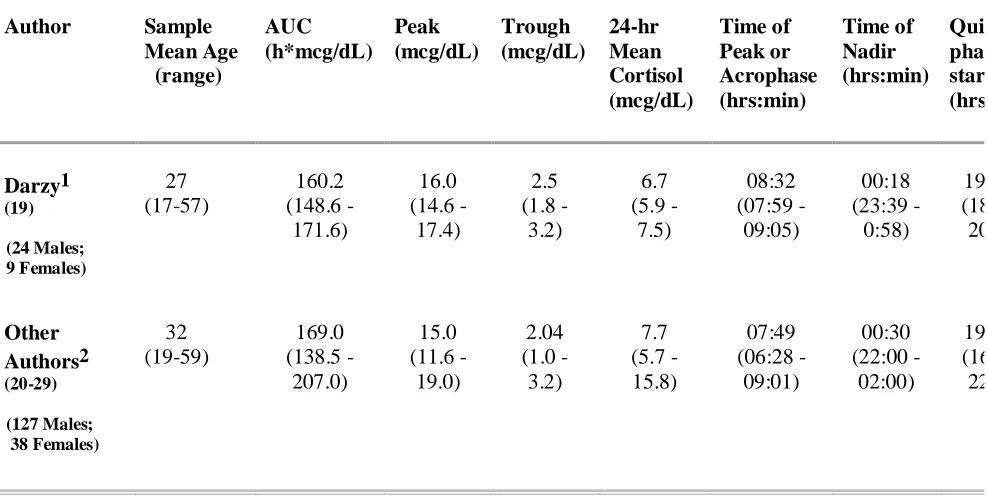
The means for all variables of the healthy reference group were similar to the previously
published literature with overlap of all 95% CIs (Table 1) (20-29). In this cohort there was no
difference in the level or phase of the circadian rhythm between men and women.
Pharmacokinetic Analysis of MR-HC and Comparison to IR-HC
Thirty two subjects were recruited. Four participants were withdrawn; two for violation of the
protocol, one withdrew consent and one had an adverse event whilst on IR-HC (hiccups). The
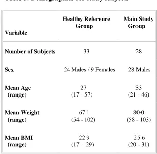
twenty eight other subjects completed the study (Table 3). 98% of all ACTH values were below
the lower limit of the assay confirming dexamethasone-induced suppression of endogenous HPA
axis, a mandatory precondition for correct pharmacokinetic analysis of cortisol bioavailability.
The cortisol concentration-time profiles following administration of MR-HC and IR-HC are
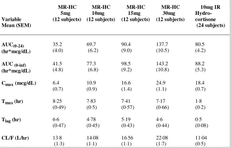
shown in Figure 2. The summary statistics for the pharmacokinetic parameters is represented in
Table 4 for comparison. The meangeo (90%CI) relative bioavailability of MR-HC formulations
compared to that of IR-HC, as measured by dose-normalized AUC values, were: 100%
respectively. The corresponding analysis for the maximum exposure, as measured by Cmax
(without dose–normalization), indicated equivalence of the MR-HC 15mg dose to IR-HC 10mg
[92% (77% - 107%)]. As expected from a delayed and sustained release formulation, the MR-HC
15mg dose showed a marked delay in mean Tmax, prolongation in Tlag, and lower dose-normalized
Cmax compared to IR-HC 10mg dose [Tmax (95% CI): (7.41 (6.16 - 8.66) hrs vs 1.8 (1.4 – 2.2) hrs,
p≤0.001); Tlag (5.19 (4.3 – 6.09) hrs vs 0.5 (0.33 vs 0.67) hrs, p<0.001; dose-adjusted Cmax (11.04
(9.02 – 13.06) mcg/dL vs 18.4 (16.9 – 19.9) mcg/dL, p≤0.001). All other formulations also
showed characteristics of sustained and delayed release (Table 4). The peak concentrations and
cortisol exposure increased predictably with increasing MR-HC dose. Dose proportionality was
seen for MR-HC 5mg and 15mg: slope (90% CIs) were 0·82 (0·62-1·02) and 0·9 (0·69-1·1) for
AUC(0-inf) and Cmax, respectively. Dose proportionality was not shown between 15 mg and 30 mg
MR-HC formulation: slopes (90%CI) for AUC(0-inf), and Cmax were, 0·58 (0·30-0·85) and 0·64
(0·40-0·87), respectively. There was no correlation between weight and any variable of MR-HC
pharmacokinetics.
Comparison of MR-HC to the Physiological Cortisol Profile
Administration of 30mg MR-HC provided the best cortisol exposure over 24 hours based on
AUC compared to the physiological profile: mean (90%CI) = 88% (77–99%). However, the
30mg MR-HC Cmax was above that seen for the physiological peak (mean±SEM = 24.9±1.1 vs
15.5±0.8 mcg/dL) and the peak occurred earlier than the physiological peak (mean: 0600h vs
0832h). The cortisol profile following MR-HC suggests that it only provides approximately 12
hours exposure to hydrocortisone (Figure 2) and when looking at the AUC for 12 hours after
2200h for 15mg MR-HC cortisol exposure was 84% (71–97%) of physiological, the Cmax similar
to physiological peak (16.6±1.4 vs 15.5±0.8 mcg/dL) but occurred earlier (0600h vs 0832h) when
Modeling MR-HC Data to Provide Physiological Dosing Regimen
Based on the phase 1 MR-HC data obtained from the dose-response study using single dose regimes, we simulated giving MR-HC doses at different times and once or twice daily. For MR-HC 30mg given at 2200h, only 2 cortisol AUCs of the 16 (12.5%) fell within the
0.8 to 1.2 ratio. However, 12 (75%) of 16 cortisol AUCs fell within the 0.8 to 1.2 ratio for
MR-HC given as 20mg at 2300h and 10mg at 0700 . Using this analysis we defined the best dose
combination to provide physiological cortisol levels as either 15 or 20mg MR-HC at 2300h and
10mg MR-HC at 0700h. An example profile is shown in Figure 3 against physiological cortisol
levels.
Discussion
Physiological cortisol replacement therapy for adrenal insufficiency is not possible with current
oral hydrocortisone formulations (32). To address this we have developed a novel
modified-release formulation of hydrocortisone (MR-HC). We have undertaken detailed pharmacokinetics
of MR-HC and compared these with the physiological profile of cortisol in normal individuals.
Based on pharmacokinetic modelling we suggest that a twice daily regimen of MR-HC given at
2300 and 0700h can provide levels of cortisol similar to normal physiological cortisol; AUC, Cmax
and Tmax.
Defining the normal physiological profile of cortisol was critical to our study. We had access to a
dataset of 33 healthy individuals who had undergone 20 minute blood sampling over a 24 hour
period (19). The generalisability of these data is reflected by the fact that the peak and nadir
cortisol, the timing of the peak and nadir, the duration of the quiescent phase and the onset of
cortisol secretion and cosinor analysis were all very similar to that in the literature (20-29). The
reference range 11.7 – 20.6), with the acrophase at 0832h (95%CI 0759h – 0905h), a nadir
cortisol of <2 mcg/dL (95% reference range 1.5 – 2.5) occurring at 0018h (95%CI 2339h –
0058h) and the quiescent phase (below the mesor) occurring from 1943h to 0531h.
We found no difference in physiological cortisol profiles according to weight or gender. Gender
has previously been reported to have a minor effect on physiological cortisol levels (21) although
not found in all studies (33). BMI does not have any significant effect on parameters quantifying
the 24-hour cortisol profile in lean or modestly overweight individuals (21). In the literature there
is an age effect on the cortisol profile and older subjects have an earlier onset of the cortisol
rhythm (22). Most of our subjects were young and Caucasian. However, different populations and
ethnic groups may exhibit differences in circadian cortisol rhythm parameters. For example,
trough and peak cortisol levels occur earlier in Chinese men compared to Caucasians, possibly
due to genetic differences and or environmental influences including social activity and everyday
habits on entrainment of circadian rhythms, such as an average earlier bedtime (26).
The pharmacokinetics of MR-HC showed similar bioavailability to IR-HC with
dose-proportionality for MR-HC between 5 and 15mg but not between 15 and 30mg. This is likely to
be explained by the binding characteristics of CBG. Under basal conditions about 5% to 10% of
circulating cortisol is free, about 75% is bound to CBG, and the remainder is bound to albumin.
Increases in total plasma cortisol concentrations above 20 mcg/dL (550nmol/L) exceed the
binding capacity of CBG and result in rapid increases in levels of free cortisol concentration,
which thus exhibits more rapid clearance from the circulation (31). As the peak cortisol following
30mg MR-HC exceeded 20 mcg/dL, the lack of dose-proportionality is probably explained by
The profile following MR-HC was compared to physiological cortisol levels. The
pharmacokinetic studies were all undertaken with subjects taking a single dose at 2200h. Based
on this the onset of cortisol release preceded the end of the physiological quiescent phase and the
peak preceded the timing of physiological peak cortisol. In addition a single dose of MR-HC
could only replace the 24 hour AUC if the peak cortisol exceeded the physiological peak. We
simulated, therefore, giving MR-HC at different time points and once or twice daily. In the
simulations AUC at time intervals were compared with the normal circadian rhythm. This
analysis shows that MR-HC 20mg and 10mg, given at 2300 and 0700h, respectively, could
provide a cortisol profile with the least difference from normal physiological cortisol levels.
However, this conclusion requires validation by an appropriate clinical study and it should be
recognised that all simulations have been done on data derived from adults and a different
regimen both in timing and dose will be required for children
The future of endocrine replacement lies in using modern pharmaceutical formulations to provide
hormone replacement that replicates physiological hormone levels. We recognise that it is
unlikely that any future drug regimen will be able to completely replicate the rapid adaptation of
physiological cortisol secretion to different conditions of stress. However, we have shown that a
modified release formulation of hydrocortisone that allows for delayed and sustained release of
hydrocortisone can potentially replicate normal un-stressed physiological cortisol levels.
Currently, in patients with adrenal insufficiency, health-related quality of life is significantly
compromised, affecting their ability to work and cope with activities of daily life, and even more
significant, mortality is increased. Future studies will determine the beneficial effects of
physiological cortisol replacement but we have demonstrated here that it is possible to generate
Acknowledgements
The study was funded by Diurnal Ltd & Phoqus Plc. WA is an MRC Senior Clinical Fellow
(GA116/172). This research was supported (in part) by the Intramural Research Program of the
National Institutes of Health.
References
1. Krieger DT, Allen W, Rizzo F, Krieger HP 1971 Characterization of the normal
temporal pattern of plasma corticosteroid levels. Journal of Clinical Endocrinology & Metabolism 32:266-284
2. Dunlap JC 1999 Molecular bases for circadian clocks. Cell 96:271-290
3. Balsalobre A, Damiola F, Schibler U 1998 A serum shock induces circadian
gene expression in mammalian tissue culture cells. Cell 93:929-937
4. Balsalobre A, Brown SA, Marcacci L, Tronche F, Kellendonk C, Reichardt
HM, Schutz G, Schibler U 2000 Resetting of circadian time in peripheral tissues
by glucocorticoid signaling. Science 289:2344-2347
5. Buckley TM, Schatzberg AF 2005 On the interactions of the
hypothalamic-pituitary-adrenal (HPA) axis and sleep: normal HPA axis activity and circadian rhythm, exemplary sleep disorders. Journal of Clinical Endocrinology & Metabolism 90:3106-3114
6. Plat L, Leproult R, L'Hermite-Baleriaux M, Fery F, Mockel J, Polonsky KS,
Van Cauter E 1999 Metabolic effects of short-term elevations of plasma cortisol
are more pronounced in the evening than in the morning. Journal of Clinical Endocrinology & Metabolism 84:3082-3092
7. Spiegel K, Leproult R, Van Cauter E 1999 Impact of sleep debt on metabolic
and endocrine function. Lancet 354:1435-1439
8. Bergthorsdottir R, Leonsson-Zachrisson M, Oden A, Johannsson G 2006
Premature mortality in patients with Addison's disease: a population-based study. Journal of Clinical Endocrinology & Metabolism 91:4849-4853
9. Tomlinson JW, Holden N, Hills RK, Wheatley K, Clayton RN, Bates AS,
Sheppard MC, Stewart PM 2001 Association between premature mortality and
hypopituitarism. West Midlands Prospective Hypopituitary Study Group. Lancet 357:425-431
10. Bensing S, Brandt L, Tabaroj F, Sjoberg O, Nilsson B, Ekbom A, Blomqvist
P, Kampe O 2008 Increased death risk and altered cancer incidence pattern in patients with isolated or combined autoimmune primary adrenocortical insufficiency. Clinical Endocrinology 69:697-704
11. Mah PM, Jenkins RC, Rostami-Hodjegan A, Newell-Price J, Doane A,
monitoring hydrocortisone replacement therapy in patients with adrenal insufficiency. Clinical Endocrinology 61:367-375
12. Arlt W, Allolio B 2003 Adrenal insufficiency. Lancet 361:1881-1893
13. Hahner S, Loeffler M, Fassnacht M, Weismann D, Koschker AC, Quinkler
M, Decker O, Arlt W, Allolio B 2007 Impaired subjective health status in 256 patients with adrenal insufficiency on standard therapy based on cross-sectional analysis. Journal of Clinical Endocrinology & Metabolism 92:3912-3922
14. Lovas K, Loge JH, Husebye ES 2002 Subjective health status in Norwegian
patients with Addison's disease. Clinical Endocrinology 56:581-588
15. Lovas K, Husebye ES, Holsten F, Bjorvatn B 2003 Sleep disturbances in
patients with Addison's disease. European Journal of Endocrinology 148:449-456
16. Merza Z, Rostami-Hodjegan A, Memmott A, Doane A, Ibbotson V,
Newell-Price J, Tucker GT, Ross RJ 2006 Circadian hydrocortisone infusions in patients with adrenal insufficiency and congenital adrenal hyperplasia. Clinical Endocrinology (Oxf) 65:45-50
17. Lovas K, Husebye ES 2007 Continuous subcutaneous hydrocortisone infusion in
Addison's disease. European journal of endocrinology / European Federation of Endocrine Societies 157:109-112
18. Newell-Price J, Whiteman M, Rostami-Hodjegan A, Darzy K, Shalet S,
Tucker GT, Ross RJ 2008 Modified-release hydrocortisone for circadian
therapy: a proof-of-principle study in dexamethasone-suppressed normal volunteers. Clinical Endocrinology (Oxf) 68:130-135
19. Darzy KH, Shalet SM 2005 Absence of adrenocorticotropin (ACTH)
neurosecretory dysfunction but increased cortisol concentrations and production rates in ACTH-replete adult cancer survivors after cranial irradiation for nonpituitary brain tumors. Journal of Clinical Endocrinology & Metabolism 90:5217-5225
20. Weitzman ED, Fukushima D, Nogeire C, Roffwarg H, Gallagher TF,
Hellman L 1971 Twenty-four hour pattern of the episodic secretion of cortisol in normal subjects. Journal of Clinical Endocrinology & Metabolism 33:14-22
21. Van Cauter E, Leproult R, Kupfer DJ 1996 Effects of gender and age on the
levels and circadian rhythmicity of plasma cortisol. Journal of Clinical Endocrinology & Metabolism 81:2468-2473
22. Sharma M, Palacios-Bois J, Schwartz G, Iskandar H, Thakur M, Quirion R,
Nair NP 1989 Circadian rhythms of melatonin and cortisol in aging. Biological Psychiatry 25:305-319
23. Hurwitz S, Cohen RJ, Williams GH 2004 Diurnal variation of aldosterone and
plasma renin activity: timing relation to melatonin and cortisol and consistency after prolonged bed rest. J Appl Physiol 96:1406-1414
24. Selmaoui B, Touitou Y 2003 Reproducibility of the circadian rhythms of serum
cortisol and melatonin in healthy subjects: a study of three different 24-h cycles over six weeks. Life Sciences 73:3339-3349
25. Selmaoui B, Lambrozo J, Touitou Y 1997 Endocrine functions in young men
exposed for one night to a 50-Hz magnetic field. A circadian study of pituitary, thyroid and adrenocortical hormones. Life Sciences 61:473-486
26. Zhao ZY, Xie Y, Fu YR, Li YY, Bogdan A, Touitou Y 2003 Circadian rhythm
27. Kanabrocki EL, Sothern RB, Scheving LE, Vesely DL, Tsai TH, Shelstad J,
Cournoyer C, Greco J, Mermall H, Ferlin H, et al. 1990 Reference values for
circadian rhythms of 98 variables in clinically healthy men in the fifth decade of life. Chronobiology International 7:445-461
28. Kronfol Z, Nair M, Zhang Q, Hill EE, Brown MB 1997 Circadian immune
measures in healthy volunteers: relationship to hypothalamic-pituitary-adrenal axis hormones and sympathetic neurotransmitters. Psychosomatic Medicine 59:42-50
29. Reinberg A, Lagoguey M, Cesselin F, Touitou Y, Legrand JC, Delassalle A,
Antreassian J, Lagoguey A 1978 Circadian and circannual rhythms in plasma hormones and other variables of five healthy young human males. Acta endocrinologica 88:417-427
30. Chakraborty A, Krzyzanski W, Jusko WJ 1999 Mathematical modeling of
circadian cortisol concentrations using indirect response models: comparison of several methods. Journal of Pharmacokinetics and Biopharmaceutics 27:23-43
31. Tunn S, Mollmann H, Barth J, Derendorf H, Krieg M 1992 Simultaneous
measurement of cortisol in serum and saliva after different forms of cortisol administration. Clinical Chemistry 38:1491-1494
32. Crown A, Lightman S 2005 Why is the management of glucocorticoid
deficiency still controversial: a review of the literature. Clinical Endocrinology 63:483-492
33. Halbreich U, Asnis GM, Zumoff B, Nathan RS, Shindledecker R 1984 Effect
Table 1: Characteristics of Cortisol Circadian Rhythm – Comparison of
Mean (±95% Confidence Intervals) Cortisol Concentrations, AUC
and Time Variables in our healthy reference group (Darzy 2005)
to similar variables in previously published data.
Author Sample Mean Age (range) AUC (h*mcg/dL) Peak (mcg/dL) Trough (mcg/dL) 24-hr Mean Cortisol (mcg/dL) Time of Peak or Acrophase (hrs:min) Time of Nadir (hrs:min) Quiescent phase start (hrs:min) Darzy1 (19) (24 Males; 9 Females) 27 (17-57) 160.2 (148.6 - 171.6) 16.0 (14.6 - 17.4) 2.5 (1.8 - 3.2) 6.7 (5.9 - 7.5) 08:32 (07:59 - 09:05) 00:18 (23:39 - 0:58) 19:43 (18:36 20:49) Other Authors2 (20-29) (127 Males; 38 Females) 32 (19-59) 169.0 (138.5 -207.0) 15.0 (11.6 -19.0) 2.04 (1.0 - 3.2) 7.7 (5.7 - 15.8) 07:49 (06:28 -09:01) 00:30 (22:00 - 02:00) 19:40 (16:30 22:00) 1
Variable Mean (95% Confidence Intervals) 2
Values have been estimated from published data: Mean (Range) b1
Period starts when Cortisol level ~ 5.2 mcg/dL or less for at least 1h b2
Period starts when Cortisol level ~ 5.0 mcg/dL or less for at least 1h c1
Period starts when Cortisol levels ~ 5.2 mcg/dL or more for at least 1h c2
Period starts when Cortisol levels ~ 5.0 mcg/dL or more for at least 1h
Notes:
- Sample – subgroups used in analysis: (21) 20-29 yr subgroup, (22)19-25 yr subgroup, (24) S1 session, (25) Control group, (26) Chinese, 30 yr sub group
- AUC: Ref (23) - curve represents average of harmonic regressions
Table 2: Study Scheme: Subjects admitted to Clinical Research Facility
on day 1. Subjects tested on up to 3 occasions with a one week washout
between treatments
DAY 1
DAY 2
STUDY ASSESSMENT
1800h 2200h 0600h 1200h 1800h 2200h
ACTH MEASUREMENT
ü ü
DEXAMETHASONE 1MG ORALLY ü
ü ü ü ü
20* SUBJECTS DOSED WITH MR-HC5MG,
15MG,30MG OR IR-HC10MG
12 SUBJECTS DOSED WITH MR-HC10MG
OR IR-HC10MG
ü
CORTISOL MEASUREMENT
Predose, postdose sampling every 30 minutes for the first 4 hours then at 5, 6, 7, 8, 10, 12, 15, 18 and 24 hours post dose
Table 3: Demographics for study subjects
Variable
Healthy Reference Group
Main Study Group
Number of Subjects
33
28
Sex
24 Males / 9 Females
28 Males
Mean Age (range)
27 (17 - 57)
33 (21 - 46)
Mean Weight (range)
67.1 (54 - 102)
80·0 (58 - 103)
Mean BMI (range)
22·9
(17 - 29)
Table 4: Pharmacokinetic Data for MR-HC and IR-HC
Variable Mean (SEM) MR-HC 5mg (12 subjects) MR-HC 10mg (12 subjects) MR-HC 15mg (12 subjects) MR-HC 30mg (12 subjects) 10mg IR Hydro- cortisone (24 subjects) AUC(0-24) (hr*mcg/dL) 35.2 (4.0) 69.7 (6.2) 90.4 (9.0) 137.7 (10.5) 80.5 (4.2)AUC (0-inf)
(hr*mcg/dL) 41.5 (4.8) 77.3 (6.8) 98.5 (9.2) 143.2 (10.8) 88.2 (5.3)
Cmax(mcg/dL) 6.4
(0.7) 10.9 (0.9) 16.6 (1.4) 24.9 (1.1) 18.4 (0.7)
Tmax (hr) 8·25 (0·49)
7·83 (0·5)
7·41 (0·57)
7·17 (0·66)
1·8 (0·2)
Tlag (hr) 6·6 (0·47)
4·78 (0·45)
5·19 (0·43)
4·6 (0·44)
0·5 (0·08)
CL/F (L/hr) 13·8 (1·3)
14·08 (1·1)
16·56 (1·1)
22·08 (1·7)
11·04 (0·5)
AUC(0-24) Area under the plasma concentration-time curve for 24 hours
AUC(0-inf) Area under the plasma concentration-time curve extrapolated to infinity
Cmax Maximum observed concentration
Tmax Time to reach Cmax
Tlag Time delay between drug administration and cortisol concentration > 3.5 mcg/dL
Figure Legends
Figure 1: Physiological Cortisol Circadian Rhythm: The figure shows the geometric mean
(▬) plus/minus 2SD (▬) serum cortisol concentration calculated from 20-minute sampling over
a 24-hour period in 33 healthy subjects. The fitted cosinor (▬) is the average of harmonic
regressions that were a fit for the individual subject data. Cortisol has a distinct circadian rhythm
with a peak of 15.5 mcg/dL (95% reference range 11.7 – 20.6) occurring at 0832h and a nadir <
2.0 mcg/dL (95% reference range 1.5 – 2.5) at 0018h. Mesor = Midline estimating statistic of
rhythm. Acrophase = Time of peak using a 24-hour clock with midnight taken as origin. Nadir =
Time of trough cortisol level. The mean and 95%CI are shown for the mesor, acrophase and
nadir.
Figure 2: Concentration – time profiles for MR-HC and IR-HC: Concentration – time
profiles for different doses of MR-HC given at 2200h compared to 10mg IR-HC using geometric
means (±SEM) of serum cortisol concentrations at 18 different time points over 24 hours.
Profiles of MR-HC show a prolonged Tmax, Tlag, and a lower dose-adjusted Cmax (not shown)
when compared to IR-HC, all typical of a formulation with delayed and sustained release
characteristics.
Figure 3: Simulation of Physiological Cortisol rhythm using MR-HC: Phase 1 PK data were
used to simulate giving MR-HC doses at different times and once or twice daily. PK modelling
included comparison of MR-HC to the physiological cortisol rhythm over 12 and 24 hours, and at
different time intervals (trapezoidal segments), using AUCs. The graph shows modeled
concentration-time profile (▬) obtained when giving 20mg (15mg + 5mg) MR-HC at 2300h and
10mg MR-HC at 0700h superimposed on the physiological cortisol rhythm (geometric mean (▬)
± 2SD (▬) serum cortisol concentration calculated from 20-minute sampling over a 24-hour