IN VITRO CHARACTERISATION OF THE
NEUTROPHIL IN RESPIRATORY
SYNCYTIAL VIRUS BRONCHIOLITIS
Gemma Louise Saint
A thesis submitted in accordance with the requirements of the University of Liverpool for the Degree of Doctor of Philosophy
June 2015
Department of Women’s and Children’s Health
Institute of Translational Medicine
2
Abstract
Background: Respiratory Syncytial Virus (RSV) is a common cause of bronchiolitis during infancy; virtually all children are infected with this virus by the age of 2 years. Although the majority have a self-limiting, mild infection, 1-3% have disease severe enough to warrant hospital admission. These children have an abundance of neutrophils (>80% of bronchoalveolar lavage cells) within their airways. There is however, a lack of detailed investigation into their role in viral infection. Our group has previously shown RSV associated with ex vivo neutrophils from infected infants. To explore this observation further, I have modelled and visualised the RSV-neutrophil interaction, hypothesising that neutrophils play a role in the innate viral response in the airway.
Aims: The aims of this thesis were to (i) establish an in vitro model of RSV-neutrophil interaction, (ii) determine whether RSV is taken up into the neutrophil, (iii) determine if RSV productively replicates inside the neutrophil, (iv) compare neutrophil-RSV interaction in adult and infant neutrophils and (v) investigate the response of the neutrophil to RSV.
Methods: An in vitro model of RSV-neutrophil interaction was established using highly purified adult and cord blood neutrophils. Neutrophils were analysed by quantitative RT-PCR, western blot, and by confocal microscopy using indirect immunocytochemistry. In addition, ex vivo BAL neutrophils from RSV infected infants were examined by confocal microscopy. Following validation of this model, confirming the presence of virus within the neutrophil, a human gene microarray was performed.
Results: Interaction of neutrophils with RSV was modelled in vitro and demonstrated by PCR analysis and western blot. RSV N gene expression revealed maximal neutrophil uptake at 4 hours. A time course over 20 hours showed no increase in RSV expression, implying that active RSV replication was not occurring. Confocal microscopy revealed internalisation of RSV, with virus distributed throughout the cytoplasm. Imaging of ex vivo neutrophils extracted from bronchoalveolar lavage of RSV-infected infants showed a similar distribution. RSV induced significant change in neutrophil gene expression with over 1000 differentially expressed genes being identified. Pathway analysis revealed an RNA virus specific transcriptional response. Results for key molecules were subsequently validated by RT-PCR.
3
Table of contents
Abstract………...2
Table of contents………...3
Table of figures………..9
Table of tables………..12
Acknowledgments………13
Author’s declaration……… 14
Abbreviations………15
Chapter 1 General Introduction……….18
1.1 Respiratory Syncytial Virus ... 18
1.1.1 Classification ... 18
1.1.2 Structure ... 18
1.1.3 Viral replicative cycle ... 20
1.2 Respiratory Syncytial Virus bronchiolitis ... 21
1.2.1 Epidemiology ... 21
1.2.2 Incidence ... 21
1.2.3 Spread of RSV ... 23
1.2.4 Clinical presentation ... 23
1.2.5 Delayed respiratory sequelae of RSV bronchiolitis ... 23
1.2.6 Management ... 24
1.2.7 Prophylaxis ... 24
1.3 Innate immune response in RSV bronchiolitis ... 25
1.3.1 Viral recognition ... 25
1.3.2 Inflammatory response ... 26
1.3.3 Macrophages ... 26
1.3.4 Neutrophils ... 28
1.3.5 Natural Killer Cells ... 28
1.3.6 Dendritic cells ... 28
1.4 Adaptive Response in RSV bronchiolitis ... 28
1.4.1 Humoral response ... 28
1.4.2 Cell-mediated immune response ... 29
1.5 RSV pathogenesis ... 29
4
1.5.2 Immune pathogenesis ... 30
1.6 The Neutrophil ... 33
1.6.1 Structure ... 33
1.6.2 Pathogen destruction ... 35
1.6.3 Resolution of inflammation ... 36
1.6.4 Neutrophil-derived cytokines ... 36
1.6.5 Crosstalk with other immune cells ... 38
1.6.6 Neutrophils in viral infection ... 38
1.7 Neutrophils and RSV ... 39
1.8 Aims of the study ... 40
Chapter 2 Methods and optimisation of an in vitro model of neutrophil-RSV interaction……….41
2.1 Participant recruitment ... 41
2.1.1 Mucin study ... 41
2.1.2 Cord blood study ... 41
2.2 Blood neutrophil collection and processing ... 42
2.2.1 Negative immunoselection methodology ... 44
2.2.2 Neutrophil ultra-purification by magnetic immunoselection ... 44
2.3 Romanowsky staining technique ... 46
2.4 Neutrophil culture ... 46
2.5 RSV preparation ... 46
2.6 Plaque assay ... 47
2.7 Phagocytosis assay ... 48
2.7.1 Isolated neutrophils are capable of phagocytosis ... 48
2.7.2 Autologous serum enhances neutrophil phagocytosis ... 49
2.8 Flow cytometer analysis and set-up ... 52
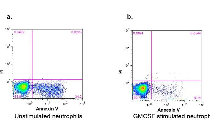
2.9 Annexin V/PI apoptosis assay ... 52
2.10 Neutrophil survival ... 52
2.10.1 Neutrophil survival can be extended in vitro by GM-CSF ... 52
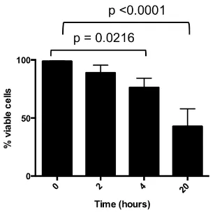
2.10.2 The effect of RSV on neutrophil survival ... 55
2.11 In vitro neutrophil-RSV culture system ... 55
2.12 Western blotting ... 59
2.12.1 Principle of western blotting ... 59
5
2.12.3 Running gel and blotting ... 60
2.12.4 Antibody staining ... 60
2.12.5 RSV proteins measured by Western Blot from infected epithelial cells ... 61
2.12.6 RSV proteins measured by Western blot from neutrophils incubated with RSV ... 61
2.12.7 Principle of Reverse Transcription qPCR ... 64
2.12.8 RNA Extraction ... 66
2.12.9 cDNA synthesis ... 67
2.12.10 Quantitative Polymerase Chain Reaction ... 67
2.12.11 Analysis of qPCR data ... 67
2.13 Imaging ... 68
2.13.1 Sample preparation for imaging ... 68
2.13.2 Indirect immunocytochemistry staining ... 69
2.13.3 Confocal microscopy ... 69
2.13.4 Image Processing ... 70
2.14 Statistical Analyses ... 70
Chapter 3 Characterisation of RSV-neutrophil interactions………..71
3.1 Introduction ... 71
3.2 Hypothesis ... 73
3.3 Aims ... 73
3.4 Specific methods ... 74
3.5 Results ... 74
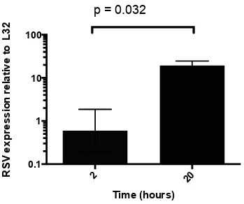
3.5.1 RSV measured by qPCR using a RSV N primer ... 74
3.5.2 No evidence of RSV replication in neutrophils ... 74
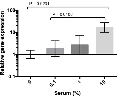
3.5.3 RSV uptake is optimally enhanced by 10% autologous serum .... 75
3.5.4 Western blot analysis confirms qPCR time course results ... 79
3.5.5 Neutrophils can not disseminate virus to epithelial cells ... 79
3.5.6 Cytochalasin D is an effective phagocytosis inhibitor ... 82
3.5.7 RSV uptake is not inhibited by the cytochalasin D ... 82
3.5.8 RSV uptake is not enhanced by a RSV monoclonal antibody ... 82
3.5.9 RSV can be visualised by confocal microscopy ... 85
3.6 Discussion ... 87
6 Chapter 4 Visualisation of RSV within the neutrophil by confocal
microscopy………96
4.1 Introduction ... 96
4.2 Hypothesis ... 99
4.3 Aims ... 99
4.4 Specific methods ... 99
4.4.1 Z stack ... 100
4.5 Results ... 100
4.5.1 Validation of RSV F monoclonal antibody by Western blot ... 100
4.5.2 Optimisation of RSV antibody using epithelial cells as a positive control ... 100
4.5.3 RSV is internalised within the cytoplasm of the neutrophil ... 103
4.5.4 Measurement of discrete RSV labelled areas within neutrophils 109 4.5.5 RSV is not co-localised with dextran in endosomes ... 109
4.5.6 RSV uptake is not prevented by inhibitors ... 109
4.6 Discussion ... 113
4.7 Summary ... 120
Chapter 5 Interaction of cord blood derived neutrophils with RSV…………121
5.1 Introduction ... 121
5.2 Overall aim ... 122
5.3 Specific aims ... 122
5.4 Specific Methods ... 122
5.4.1 Cord blood collection ... 122
5.4.2 Cord blood neutrophil isolation ... 122
5.4.3 BAL sample collection and processing ... 123
5.4.4 BAL neutrophil isolation ... 123
5.5 Results ... 123
5.5.1 Maternal characteristics ... 123
5.5.2 Cord blood neutrophils can be isolated to 98.7% purity as determined by CD66c positivity ... 124
5.5.3 Cord blood neutrophil survival can be extended in vitro by GM-CSF ... 124
7
5.5.6 RSV uptake not inhibited by phagocytosis inhibitor ... 127
5.5.7 Bronchiolitis patients’ characteristics ... 131
5.5.8 RSV identified by confocal in ex vivo BAL neutrophils ... 131
5.6 Discussion ... 134
5.7 Summary ... 136
Chapter 6 Innate antiviral response to RSV………..137
6.1 Introduction ... 137
6.2 Hypothesis ... 139
6.3 Aims ... 139
6.4 Specific methods ... 139
6.4.1 Microarray ... 139
6.4.2 qPCR validation of microarray findings ... 140
6.4.2.1 cDNA synthesis ... 140
6.4.2.2 Quantitative Polymerase Chain Reaction ... 141
6.4.3 Housekeeping gene selection ... 141
6.5 Results ... 145
6.5.1 RNA Pico chip traces ... 145
6.5.2 Data quality assessment ... 145
6.5.3 Differential gene expression analysis ... 150
6.5.4 Ingenuity Pathway Analysis ... 151
6.5.5 IPA statistics ... 151
6.5.6 Canonical pathways ... 151
6.5.7 Disease and function analysis ... 152
6.5.8 Upstream regulator analysis ... 152
6.5.9 Validation of microarray results by qPCR ... 159
6.5.10 RSV uptake with reduction over time confirmed ... 159
6.6 Discussion ... 162
6.6.1 Early response ... 162
6.6.2 Pattern recognition receptors ... 163
6.6.3 The interferon regulatory factor pathway ... 164
6.6.4 Jak-STAT Pathways ... 166
6.6.5 IFN stimulated genes (ISGs) ... 166
6.6.5.1 Ubiquitin Specific Peptidase 18 ... 166
8
6.6.5.3 Tetherin ... 168
6.6.5.4 Double stranded RNA-specific endoribonuclease ... 168
6.6.5.5 APOBEC3 enzymes ... 169
6.6.5.6 TRIM Proteins ... 169
6.6.5.7 IFIT protein encoding genes ... 170
6.6.6 Late response ... 170
6.6.7 Immune crosstalk ... 171
6.7 Summary ... 173
Chapter 7 General discussion……….174
7.1 Future directions ... 179
7.2 Final conclusions ... 180
Appendix 1………..182
Appendix 2………..185
Appendix 3………..187
Appendix 4………..193
Appendix 5………..195
9
Table of figures
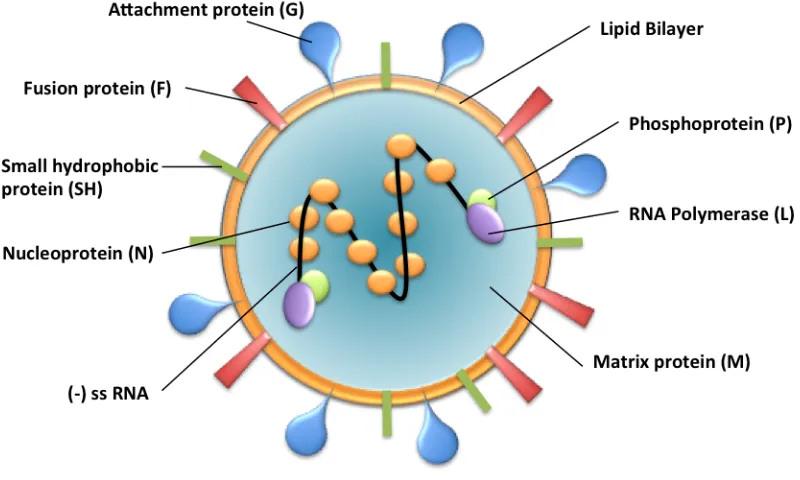
Figure 1-1 Schematic representation of an RSV virion identifying key proteins
... 19
Figure 1-2 Number of RSV/week at Alder Hey Hospital ... 22
Figure 1-3 RSV infection modifies the inflammatory environment in the airways. ... 27
Figure 1-4 Neutrophil granulocyte ... 34
Figure 1-5 Cytokines expressed and/or produced by human neutrophils .... 37
Figure 2-1 Neutrophil purity using polymorph preparation method ... 43
Figure 2-2 Neutrophil purity using negative immunoselection method ... 45
Figure 2-3 Neutrophil phagocytosis using pHrodo E. coli ... 50
Figure 2-4 Autologous serum enhances neutrophil phagocytosis of pHrodo ... 51
Figure 2-5 Illustration of flow cytometric analysis of Annexin V/PI apoptosis assay ... 53
Figure 2-6 Neutrophil survival time course ... 54
Figure 2-7 Neutrophil survival can be extended in vitro by GM-CSF ... 56
Figure 2-8 Neutrophil survival is affected in vitro by RSV ... 57
Figure 2-9 In vitro culture system for neutrophil-RSV infection ... 58
Figure 2-10 RSV proteins identified from infected epithelial cells ... 62
Figure 2-11 RSV proteins identified from neutrophils incubated with RSV .. 64
Figure 2-12 Calculation of PCR efficiency ... 65
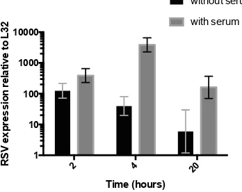
Figure 3-1 RSV N gene expression relative to L32 in the epithelial cell line A549 ... 76
Figure 3-2 RSV N gene expression relative to L32 in neutrophils incubated with RSV... 77
Figure 3-3 RSV uptake is enhanced by the presence of 10% autologous serum ... 78
Figure 3-4 Western blot time course for RSV from neutrophils ... 80
Figure 3-5 Plaque assay of A549s exposed to supernatant from RSV exposed neutrophils and A549s ... 81
10 Figure 3-7 RSV uptake following pre-treatment with cytochalasin D or
palivizumab. ... 84
Figure 3-8 Confocal fluorescence microscopy with superimposed brightfield microscopy ... 86
Figure 3-9 Modes of cellular internalisation by phagocytes ... 91
Figure 3-10 Macropinosome ruffling formation ... 92
Figure 4-1 RSV F antibody validation by Western blot ... 101
Figure 4-2 RSV antibody optimised to show BEAS2B RSV infection by confocal microscopy ... 102
Figure 4-3 Neutrophils visualised by confocal microscopy showing positive staining for RSV F protein ... 104
Figure 4-4 Orthogonal view showing RSV within the neutrophil cytoplasm 105 Figure 4-5 Surface-rendered neutrophil projection ... 106
Figure 4-6 Surface rendered neutrophil ... 107
Figure 4-7 3D reconstruction of neutrophils and internalised RSV ... 108
Figure 4-8 Measurement of internalised RSV ... 110
Figure 4-9 Dextran and RSV visualised by confocal in a neutrophil ... 111
Figure 4-10 Dextran and RSV are not colocalised in neutrophils ... 112
Figure 4-11 RSV visualised in neutrophils with and without inhibition ... 114
Figure 4-12 Endocytic pathways used by viruses ... 116
Figure 5-1 Cord blood neutrophil purity ... 125
Figure 5-2 GM-CSF prevents significant neutrophil apoptosis ... 126
Figure 5-3 Western blot time course of RSV proteins from cord blood neutrophils ... 128
Figure 5-4 Cord blood neutrophils visualised by confocal microscopy showing RSV ... 129
Figure 5-5 Orthogonal view showing RSV within the cord blood neutrophil cytoplasm ... 130
Figure 5-6 Ex vivo neutrophils showing RSV by immunocytochemistry ... 132
Figure 5-7 Orthogonal view of ex vivo neutrophils ... 133
Figure 6-1 Average expression stability of reference targets ... 143
Figure 6-2 Determination of the optimal number of reference targets ... 144
Figure 6-3 Electrophoresis run summary of total RNA on Pico chip ... 146
11 Figure 6-5 Distribution of log expression signal before and after
normalisation ... 148
Figure 6-6 PCA plot of log2 gene expression for all samples ... 149
Figure 6-7 Target genes from microarray validated by qPCR ... 160
Figure 6-8 Relative RSV gene expression ... 161
12
Table of tables
Table 1-1 Neutrophil granule types and contents ... 34
Table 2-1 Antibodies used for neutrophil identification ... 42
Table 2-2 Primary antibodies used for western blot analysis ... 61
Table 2-3 Secondary antibodies used for western blotting ... 61
Table 2-4 Standard curve characteristics of qRT-PCR experiments ... 66
Table 2-5 Pre-designed gene expression assays used for qPCR analysis 68 Table 2-6 RSV Primer and probe for qPCR ... 68
Table 2-7 Primary antibodies used for indirect immunocytochemistry staining ... 69
Table 2-8 Secondary antibodies used for indirect immunocytochemistry staining ... 69
Table 5-1 Table of RSV patient recruit characteristics ... 131
Table 6-1 Reverse transcription mastermix used in cDNA synthesis. ... 140
Table 6-2 Standard curve characteristics of qRT-PCR experiments ... 142
Table 6-3 Pre-designed gene expression assays used for qPCR analysis 142 Table 6-4 Number of differentially expressed probes and genes and regulation direction ... 150
Table 6-5 Top canonical pathways of early response ... 152
Table 6-6 Top canonical pathways of late response ... 152
Table 6-7 Disease and function table of early response ... 154
Table 6-8 Disease and function table of late response ... 156
13
Acknowledgments
I have enjoyed the past 3 years immensely and feel incredibly privileged to have been able to undertake this PhD. I cannot possibly thank everybody fully here, but would like to record those who have been of particular importance. Firstly, thanks to my supervisors who have allowed me three different perspectives, to have their direction and encouragement has been instrumental to this work. Dr Brian Flanagan has always been on hand for impromptu meetings and has coped admirably well with my bad science day grumps! I am very grateful to Dr Paul McNamara for his support, guidance and most excellent tutoring in the art of removing extraneous words! I am most appreciative of Prof Rosalind Smyth who always bought thoughtful critique to the project and pushed me to work to the highest standard.
A number of people from research groups at the University of Liverpool and around the country have given generously of their time, and offered invaluable advice and tutoring. My particular thanks to Ian Sabroe, Lynne Prince, Claire Smith, Rob Hirst, Marco Marcello, Lucille Rainbow, Mark Turner, David Mason and Jonathon David. I thank the research nurses who have supported the studies, both at Alder Hey and Liverpool Women’s Hospital. The kind participation of the children and families involved in this study is so appreciated and I thank them for their contribution. The support of the Alder Hey PICU physiotherapy team and the Liverpool Women’s midwifery team is also appreciated. I give heartfelt thanks to the ICH team, with particular recognition to members of the ABLE group, including Rachel Corkhill, Angela Hackett, Kate Phillips and Katie Rose. Thank you also to Moira Saphier, the true boss! I would like to thank my charitable funder, the Wellcome Trust: their generous Fellowship award has allowed me to carry out this research which has fostered a desire to continue in paediatric medical research.
14
Author’s declaration
I declare that, except where explicit reference is made to the contribution of others, this thesis is the result of my own work. The material contained in this thesis has not been presented, nor is currently being presented, either wholly or in part for any other degree of qualification.
This research was carried out at Alder Hey Children’s Hospital, the Institute of Child Health and the Institute of Integrative Biology, University of Liverpool.
Signature……….
15
Abbreviations
ADE antibody dependant enhancement ALRI acute lower respiratory infection APC antigen presenting cell
AU airy unit
BAL bronchoalveolar lavage
CCA chimpanzee coryzal agent
CGR Centre for Genomic Research
CMV cytomegalovirus
DC dendritic cell
DDX58 DEAD(Asp-Glu-Ala-Asp) box polypeptide 58 DHX58 DEXH (Asp-Glu-X-His) box polypeptide 58 DICER1 double stranded RNA-specific endoribonuclease DMEM Dulbecco’s modified eagle medium
DNA deoxyribonucleic acid
DTT dichlorodiphenyltrichloroethane
EBV Epstein-Barr virus
ERS European Respiratory Society
FCS foetal calf serum
FDR false discovery rate FI-RSV formalin inactivated RSV
FSC forward scatter
GA gestational age
GFP green fluorescent protein
GM-CSF granulocyte macrophage-colony stimulating factor
HMPV human metapneumovirus
IFN interferon
Ig immunoglobulins
IL-1RA IL-1 receptor antagonist IPA Ingenuity Pathway Analysis
IPS-1 interferon promoter-stimulating factor 1 IRF interferon regulatory factor
16 LRTI lower respiratory tract infection
LXA4 lipoxin A4
MAVS mitochondrial antiviral signalling protein MDA-5 melanoma differentiation associated gene 5
MDC monodansylcadaverine
MHC major histocompatability complex MOI multiplicity of infection
MPO myeloperoxidase
MX1 myxovirus resistance 1
NETs neutrophil extracellular traps
NFKB nuclear factor-KB
NK cells natural killer cells
NO nitric oxide
NPA nasophayngeal aspirate
P value probability value
PAMPs pathogen-associated molecular patterns PBS phosphate buffered saline
PCA principal component analysis pDC plasmacytoid dentritic cell PEV primary endocytic vesicle
PFA paraformaldehyde
PFU plaque forming units
PI propidium iodide
PICU paediatric intensive care unit
PMNL polymorphonuclear neutrophil leucocytes PRR pattern recognition receptor
PVDF polyvinylidene difluoride
qPCR quantitive polymerase chain reaction REC research ethics committee
RIG-I retinoic acid-inducible gene I-like receptor
RIN RNA integrity number
RNA ribonucleic acid
17 RSV Respiratory Syncytial Virus
RT real time
SDS sodium dodecyl sulfate
SEM standard of error
SSC side scatter
ssRNA single stranded RNA
STAT signal transducer and activator of transcription TBST Tris-buffered saline and Tween 20
TGF transforming growth factor
Th T helper
TLR toll like receptor
TNF tumour necrosis factor
TRAIL TNF-related apoptosis-inducing ligand TRIM tripartite motif-containing proteins URTI upper respiratory tract infection USP18 ubiquitin specific peptidase 18 WHO World Health Organisation
18
Chapter 1 General Introduction
1.1 Respiratory Syncytial Virus
Since its discovery in 1956, respiratory syncytial virus (RSV) has been recognised as a pathogen causing a significant burden of disease to the paediatric population. It is an important cause of severe respiratory illness in infants and young children and the leading cause of bronchiolitis. Despite increasing understanding of the immunology and pathology of the virus, the number of admissions to hospital continues to rise. In the UK, admissions coded as acute bronchiolitis increased from 21 330 in 2004/5 to 34 668 in 2012/13 (1).
RSV was first identified in chimpanzees with symptoms of an upper respiratory tract infection (2). It was initially named Chimpanzee Coryzal Agent (CCA). In 1957, a virus which was indistinguishable from CCA, was isolated from throat swabs from two separate infants by Chanock and Finberg (3). These three isolates were grouped and named respiratory syncytial virus because of the ability they showed in culture to cause epithelial cells to fuse forming syncytia.
1.1.1 Classification
RSV is an RNA virus of the Paramyxoviridae family. The Paramyxoviridae
family is subdivided into Paramyxovirinae and Pneumovirinae. The Pneumovirinae family is split into the Pneumovirus and Metapneumovirus
genus, with RSV belonging to the Pneumovirus genus. Avian pneumovirus and human metapneumovirus belong to the Metapneumovirus genus. RSV contains more genes/proteins than any other member of the Paramyxoviridae
family.
1.1.2 Structure
19 large glycoprotein G, fusion protein F and small hydrophobic protein SH. These viral glycoproteins form separate homo-oligomers that appear as short surface spikes (Figure 1-1). RSV lacks neuraminidase or haemagglutinin activity.
Figure 1-1 Schematic representation of an RSV virion identifying key proteins
The RSV virion is composed of a lipid bilayer which encapsulates the negative sense single stranded RNA. There are 3 surface glycoproteins, G, F, and SH. Inside the lipid bilayer is the matrix protein (M). The nucleocapsid is made up of 4 proteins, L, M, N and P.
Lipid envelope
20
Ribonucleocapsid
The nucleocapsid, contained inside the lipid bilayer, is 12-15nm in diameter and made of 4 nucleocapsid proteins which replicate and transcribe the RSV genome, these are the nucleocapsid protein N, the phosphoprotein P, the anti-termination factor M2-1 and the large polymerase subunit L. M2 is the second matrix protein encoding both M2-1 (elongation factor) and M2-2 (transcription regulation)
Non-structural proteins
NS1 and NS2 are non-structural proteins that are unique to the Pneumovirus
genus. These proteins act cooperatively to interfere with innate immune responses including interferon induction and signalling. They suppress the activation and nuclear translocation of the IFN-regulatory factor IRF-3 (5). NS1 and NS2 have also been shown to activate antiapoptotic genes of the host cell, thereby prolonging the life of the cell and increasing viral yield (6). 1.1.3 Viral replicative cycle
21
1.2 Respiratory Syncytial Virus bronchiolitis
Viral bronchiolitis occurs predominantly in infants under the age of one year and is the commonest reason for hospitalisation in this age group. Approximately 1 in 5 infants will develop bronchiolitis in their first year of life. RSV is the leading cause of bronchiolitis, resulting in up to 80% of cases. 1.2.1 Epidemiology
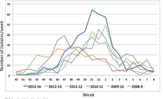
RSV is a common infection during infancy; virtually all children will be infected by the age of 2 years (14). Although the majority have a self-limiting, mild infection, 1-3% of all infants have disease severe enough to warrant admission to hospital (15-18). Most of these hospitalised children are previously healthy, although several groups of infants are predisposed to severe RSV infection; premature or extremely low birth weight infants, and those with chronic lung disease, congenital heart disease or immunodeficiency (19-21). Incomplete development of the airway and airway hyperreactivity may contribute to the increased risk of disease seen in preterm infants. Pulmonary hypertension and cyanosis are associated with worse outcome in those infants with pre-existent heart disease (22). RSV epidemics occur annually. In temperate climes this occurs during the winter season. In Liverpool, at Alder Hey Children’s Hospital, peak RSV bronchiolitis is seen between November and January (Figure 1-2).
1.2.2 Incidence
22
Figure 1-2 Number of RSV/week at Alder Hey Hospital
Line graph showing number of RSV isolates/week from 1st week of October to end of February during 2008-2014 at Alder Hey Children’s Hospital, Liverpool Image generated from data collated by the Microbiology department at Alder Hey Children’s Hospital, Liverpool
N
um
be
r'o
f'Is
ol
ate
s/
w
ee
k'
23 priority for many national health organisations for over 20 years, with renewed attention in 2000 following the establishment of the Millennium Development Goals, which included reduction in child mortality (23). The WHO have also recognised the importance of vaccine development for RSV and their ‘Product Development for Vaccines Advisory Committee’ (PDVAC) have identified it as a priority area (24).
1.2.3 Spread of RSV
RSV is one of the most contagious human pathogens (25). It is spread by droplet and secretion transmission between individuals in close contact. The incubation period is between two to eight days. By two years of age, almost all children will have had an RSV infection at least once (26).
1.2.4 Clinical presentation
Initial symptoms are generally those of an upper respiratory tract infection with coryza, rhinitis and cough (27). A third of children develop otitis media (28). Symptoms can progress over 3-4 days to involvement of the bronchioles leading to dyspnoea, subcostal recession and poor feeding. When examined, chest auscultation may reveal wheeze and/or crackles. Infants younger than 6 weeks of age may present with apnoea and no other clinical signs. Bronchiolitis can lead to acute respiratory failure, characterised by hypoxia and carbon dioxide retention. The acute illness improves over 5-7 days but a persistent cough lasting for more than two weeks occurs in 50% of children (29).
1.2.5 Delayed respiratory sequelae of RSV bronchiolitis
In some children, a ‘post bronchiolitis syndrome’ occurs characterised by months of episodic wheeze, typically with other viral infections. Stein et al
double-24 cohort, follow-up study in 27 centres recruited 421 pre-term infants, who either did or did not received prophylactic palivizumab. The recruits were followed up for two years, and an association between palivizumab prophylaxis and improved respiratory outcomes was statistically significant even after adjustment for confounding factors. The authors concluded that preventing RSV LRTI using palivizumab may reduce subsequent recurrent wheezing in pre-term infants (31). In 2013 Blanken et al showed that
palivizumab treatment resulted in a significant reduction in wheezing during the first year of life, even after the end of treatment, thus implicating RSV as an important cause of recurrent wheeze during the first year of life in such infants.
1.2.6 Management
Supportive care is the mainstay of treatment. Children are admitted to hospital if they have apnoea, take less than 75% of usual feed volume, and/or have severe respiratory distress and/or hypoxia. Care is given in the form of hydration (nasogastric feeds or intravenous fluid), and/or supplemental oxygen for hypoxaemia. To date, no treatments for bronchiolitis have been shown to be efficacious, despite many being trialled, including antibiotics, hypertonic saline, adrenaline, salbutamol, montelukast, ipratropium bromide or systemic inhaled corticosteroids for bronchiolitis (32). However, there are a number of new promising anti-virals coming through (33). In particular, oral GS-5806, an oral RSV-entry inhibitor, has shown promising results in an RSV challenge model using healthy adults; reducing viral load and severity of clinical disease (34).
1.2.7 Prophylaxis
Palivizumab is a humanised monoclonal antibody (IgG11K) that is used to
25 infants for whom RSV infection is likely to cause significant illness or death (37). These are:
• Preterm infants who have chronic lung disease (defined as oxygen
dependency for at least 28 days from birth). Eligibility however, depends on their gestational age at birth and their corrected age at the start of the RSV season.
• Pre-term infants who have haemodynamically significant, acyanotic
congenital heart disease.
• Infants < 24 months with severe combined immunodeficiency disorder.
• Infants on long term ventilation under certain conditions (38).
1.3 Innate immune response in RSV bronchiolitis
Inoculation of RSV into the nose or eyes results in virus entering the nasopharynx. Once within the respiratory tract, it can be trapped by mucus lining the airways. Over several days the virus can spread to the lower airways. The mechanism by which this occurs is not known but may be due to spread along the epithelium or through aspiration of the nasopharyngeal secretions. It is the ciliated cells of the small bronchioles and type 1 pneumocytes in the alveoli that are infected, with the basal cells being spared (39).
1.3.1 Viral recognition
Viral infection of epithelial cells leads to recognition of viral components within these cells by pattern recognition receptors (PRRs). RNA viruses are detected by two kinds of PRR; cytoplasmic recognition receptors such as retinoic acid-inducible gene I-like receptor (RIG-1), and Toll-like receptors (TLRs). RIG-1 is a highly inducible cytoplasmic RNA helicase that signals antiviral responses after binding dsRNA (40, 41). In contrast to TLRs, these receptors are cytoplasmic and can therefore recognise intracellular virus (42). TLRs are membrane bound receptors. In RSV infection, TLR 3 and TLR 4 are thought to mediate RSV signalling (43-45). This signalling takes the form of nuclear factor KB (NFKB) activation and cytokine secretion. TLR2 and
26 migration into the lungs (46). RSV can modulate changes in the expression of these TLRs, enhancing inflammatory responses, for example in epithelial cells an upregulation of TLR4 has been described (47). In addition, neutrophils from BAL of RSV infected pre-term infants express higher levels of TLR4 than healthy infants (48).
1.3.2 Inflammatory response
NFKB is translocated to the nucleus upon activation of PRRs, leading to the
production and secretion of cytokines and chemokines (illustrated below in
Figure 1-3 by airway epithelium infected with RSV). Of particular significance to viral infection are type I interferons (IFNs). Not only produced by epithelial cells, they are also produced by plasmacytoid dendritic cells (pDCs) and macrophages in response to TLR and RIG-1 activation by virus. Type I IFNs bind to cell surface receptors activating signalling pathways, promoting an anti-viral response. Downstream signalling molecules vary depending on the cell type but transcription of over 100 IFN stimulating genes (ISGs) lead to an anti-viral state. STAT1/STAT2 signalling is the best described of these pathways. As well as the production of these specific antiviral factors there is also expression of CXC and CC chemokines that stimulate the recruitment of a number of cells including neutrophils, natural killer (NK) cells, and dendritic cells (49). One of the first cells, alongside epithelial cells, to encounter RSV in the airways, is the macrophage.
1.3.3 Macrophages
27
Figure 1-3 RSV infection modifies the inflammatory environment in the airways.
This image illustrates how RSV infection of the epithelium modifies the inflammatory environment of the airways. RSV infects the ciliated airway epithelial cells, and as a result the infected cells mount a defence mechanism. Activation of NFkB by RSV-derived molecular patterns precedes secretion of type I IFNs. Engagement of surface PRRs, such as TLR4, by RSV induces production of several chemokines and cytokines. These messengers trigger an influx of inflammatory cells, including neutrophils. In the meantime viral replication is ongoing inside the cells, spreading from cell to cell.
28 1.3.4 Neutrophils
Neutrophils intensify the cellular response through the production of further chemokines (53). Although these cells are numerically the major inflammatory cell within the airway in infants with RSV bronchiolitis, their specific role is largely undetermined (55). The function of these important cells is discussed in more detail in Section 1.6.
1.3.5 Natural Killer Cells
Work in mice has revealed that natural killer cells are present in the lung in the initial stages of infection and produce abundant quantities of IFN-γ, which appears to shape the subsequent T cell response (56). Virus infected cells are recognised by decreased MHC class 1 molecule expression.
1.3.6 Dendritic cells
Dendritic cells (DCs) link the innate to the adaptive immune response. From murine studies it has been shown that plasmacytoid DCs, thought to limit viral replication, are recruited early on in infection (57). In contrast myeloid DCs increase in number later in the infection and are matured by RSV, and subsequently activate naïve T cells (58).
1.4 Adaptive Response in RSV bronchiolitis
1.4.1 Humoral response
29 Most term newborn babies have RSV neutralising antibodies due to placental transfer of maternal immunoglobulin. These antibodies appear to confer a degree of protection; correlation has been noted between low cord blood RSV antibody titres and severe RSV disease (27) and early RSV disease (62). In addition, prophylactic administration of RSV-neutralising antibody provides protection to high-risk infants (63, 64).
1.4.2 Cell-mediated immune response
Both CD4+ T helper and CD8 cytotoxic T+ cells are recruited to the lung in
response to RSV infection and are believed to play a critical role in terminating acute RSV infection (65, 66). Humans with compromised T cell immunity shed virus for months, and in mice depletion of CD4+ and CD8 T+ cells also cause prolonged shedding (20). In mice, this T cell depletion despite causing prolonged viral replication, reduces pulmonary disease (65).
1.5 RSV pathogenesis
RSV infection is usually completely resolved by the innate and adaptive immune responses described above (67, 68). However, infection does result in widespread alteration in cellular gene expression, and upregulation of cytokines and chemokines as the body mounts its immune response. The combination of host immune response and viral factors are thought to be responsible for RSV induced pathology although their relative contributions are debated.
1.5.1 Infant and viral factors contributing to pathogenesis
30 infants are vulnerable due to the absence of previous exposure and consequent lack of immune memory. Reinfection is common, even within the same season (26). This is not unique to infants; the same is true for healthy adults and even more commonly in exposed adults such as health-care workers (68, 71). Reinfection of healthy individuals usually results in disease of reduced severity but this ability to re-infect ensures that RSV remains present within the population, at all ages.
Individual proteins within the RSV virion contribute to its pathogenicity. RSV subverts the antiviral IFN response through two IFN antagonists, NS1 and NS2 (72). They inhibit the induction of IFN-alpha/beta by blocking the activation of IFN regulatory factor 3 and by inhibiting type I IFN signalling through the JAK/STAT pathway (5). The JAK/STAT pathway would otherwise amplify the IFN response and upregulate IFN stimulated genes, resulting in the antiviral state described in Section 1.3.
RSV F and G surface proteins induce neutralising antibody production and are the major antigenic determinants of protection. The G protein differs from most other receptor binding proteins of the Paramyxoviridae family with respect to its structure and antigenic properties. For not fully elucidated reasons, the G protein requires multiple antibodies in order to be efficiently neutralised (73). It also influences epithelial and antigen presenting cell responses by down-regulating expression of inflammatory mediators through the inhibition of the NF-kB-pathway (74) (75).
31 1.5.2 Immune pathogenesis
Disease manifestation of RSV infection varies widely from upper respiratory tract infection (URTI), through to lower respiratory tract infection (LRTI), which can be mild or life threatening. Although the direct cytopathic effect of the virus and viral load likely contribute to these different manifestations, several observations lead us to believe that the host’s own immunity plays a role in disease severity.
Histopathology of the lung from an infant with RSV who died in a car accident rather than as a result of infection, revealed significant numbers of immune cells, specifically T lymphocytes, monocytes and neutrophils (39). Inflammatory cells were noted in the submuscularis between the arterioles and bronchioles, but also identified moving through gaps in smooth muscle. This, the author’s conjecture, could impact smooth muscle tone and affect airway hyperreactivity. The small airways were noted to contain inflammatory cells, fibrin and mucus, suggesting that mechanical obstruction of these airways plays a role in disease (39).
32 The failure of the formalin inactivated RSV (FI-RSV) vaccination in the 1960s provides further evidence of the potential for immune mediated pathogenesis. This vaccine, given to RSV naive infants, was not protective and primed for severe disease on infection with naturally occurring RSV infection. When this occurred, 80% required hospitalisation and two infants died. Histopathological analysis, supported by subsequent animal data, revealed that an atypical antibody response resulted in antigen-antibody complexes and complement activation in the lungs (83, 84). In addition, a Th2 biased response contributed to immunopathology with an overly robust response of virus-specific CD4+ T lymphocytes (85). It is not clear however what relevance the pathogenesis of FI-RSV disease has to naturally occurring human disease as similar heightened responses do not occur.
There are an abundance of inflammatory cytokines and granulocytes in the airways of infants with severe RSV, with the neutrophil being the most abundant inflammatory cell, accounting for up to 84% of airway leucocytes (55, 86). This is much greater than that seen in influenza virus infection (87). There is a high airway concentration of predominantly epithelial derived CXCL8, a major neutrophil chemoattractant (49). In a study of the dynamics of neutrophil responses in infants admitted to the intensive care unit, the appearance of neutrophil precursors in the blood, preceding their influx into the lungs, closely followed the peak of virus shedding and was concurrent with clinical symptoms. Whether this represented a protective response or a contributor to the disease severity cannot be determined (81).
33
1.6 The Neutrophil
Paul Ehrlich in 1886 first described as ‘polynuclear cells’, the cells we now know as polymorphonuclear neutrophil leucocytes, PMNL, PMN or just neutrophils (88). When it was recognised that the neutrophil had in fact a single multi-lobed nucleus rather than a number of nuclei they were re-named to the present terminology (89). Initially there was misunderstanding as to the role of the neutrophil in inflammation, with the belief that the neutrophil spontaneously generated until Waller demonstrated that they originated from blood (90). Secondly it was thought that the presence of neutrophils, which were now known to produce pus, assisted the growth of microorganisms. However, Metchnikoff showed that neutrophils were capable of chemotaxis and could phagocytose. He argued that the presence of phagocytic cells were beneficial to mammals as a natural immunity against infection by microorganism (91). Other scientists in the field initially refuted Metchnikoff’s theory on the killing ability of neutrophils but in 1905 he was awarded a Nobel Prize for his findings. Previously considered to be short-lived, phagocytic cells, they are increasingly being found to have more complex and far-reaching functions. Their role as the first immune cell to respond to invasion by bacteria and fungi has been extensively demonstrated and, neutrophil depletion generally results in catastrophic consequences to whichever species is being studied. Neutrophils are recruited in large numbers to the site of many viral infections and although there is evidence that they play a role in anti-viral innate immunity, the evidence is limited and more investigation is warranted.
1.6.1 Structure
34
Figure 1-4 Neutrophil granulocyte
This is a schematic diagram of a neutrophil showing the component parts. The nucleus has a characteristic multi-lobed shaped. Within the cytoplasm is the small golgi apparatus, and multiple granules.
Granule type Content
Azurophilic (primary)
myeloperoxidase, permeability-increasing protein, defensins, neutrophil elastase and cathepsin G
Specific (secondary)
alkaline phosphatase, lysozyme, NADPH oxidase, collagenase, lactoferrin, cathelicidin
Tertiary cathepsin, gelatinase
Table 1-1 Neutrophil granule types and contents
35 extracellularly along with cytotoxic substances when activated through Fcγ receptors.
During the inflammatory response, the neutrophil functions in a variety of ways, with regulatory factors at every step. Their killing capability is three pronged, and comprises engulfment of the organism (phagocytosis), release of secretory granules containing reactive oxygen species (degranulation) and release of neutrophil extracellular traps (NETs) capable of trapping microorganisms and potentially killing them with the aid of attached antimicrobial peptides.
1.6.2 Pathogen destruction
36 principle role of the neutrophil is to destroy invading microorganisms and this it does most effectively as described above. The paradox however is that in order to do so it generates chemicals such as ROS and defensins, which not only destroy the invading organism but potentially also damage host tissue (98, 99). Thus to limit this damage, controlled cell death or apoptosis, is required (100).
1.6.3 Resolution of inflammation
Apoptosis is not the only way that the neutrophil contributes to the resolution of inflammation. Neutrophils also have anti-inflammatory capabilities through their ability to block and scavenge chemokines such as CCL3 and CCL5 (101), and produce anti-inflammatory cytokines such as IL-1 receptor antagonist (IL-1RA) and TGF-β1&2 (102). They can also produce pro-resolving lipid mediators such as lipoxin A4 (LXA4) that inhibit further neutrophil recruitment (103). Furthermore, they contribute to the biosynthesis of resolvins, which contribute to inhibit neutrophil transendothelial migration (103).
1.6.4 Neutrophil-derived cytokines
Chemokines, pro-inflammatory, anti-inflammatory, immunoregulatory, angiogenic and fibrogenic cytokines as well as ligands to the TNF superfamily are all expressed and produced by the neutrophil (Figure 1-5)
37
Figure 1-5 Cytokines expressed and/or produced by human neutrophils
This figure lists the cytokines that have been validated in human neutrophils through gene expression techniques, immunohistochemistry, enzyme-linked immune-adsorbent assays or biological assays. * refers to studies only performed at the mRNA level. ? refers to controversial data.
38 1.6.5 Crosstalk with other immune cells
Through its ability to produce cytokines and chemokines, the neutrophil can recruit discrete cell populations as and when needed. Neutrophils produce chemotactic signals that recruit monocytes, dendritic cells, NK cells, and T-helper type 1 and type 17 cells (106). TNF-alpha is produced by neutrophils, which can drive DC and macrophage differentiation. This crosstalk is bi-directional with products from macrophages and DCs activating neutrophils as well. Cell to cell contact between CD11b on neutrophils and DC-SIGN (DC-specific ICAM3 grabbing non-integrin) on monocyte derived DCs induces maturation (107). Neutrophils are also a source of BAFF, a member of the TNF superfamily, which is needed for B cell function, maturation and survival (108). Neutrophils can also modulate NK cell survival, proliferation and IFNy production through the release of ROI and granule components (109). There is complex interplay between T cells and neutrophils, with effects on recruitment to inflamed sites and cell survival (102).
1.6.6 Neutrophils in viral infection
39
1.7 Neutrophils and RSV
On the basis of the studies presented above, it would appear that neutrophils have a role at the frontline of antiviral immunity. What is known about their contribution in RSV infection? As referenced previously neutrophils are the predominant cell in the airways of infants with RSV bronchiolitis. Epithelial cells drive this migration through the production of neutrophil chemoattractants in response to RSV, particularly CXCL8 (53, 116-119). CXCL8 concentration is elevated in the bronchoalveolar lavage (BAL) fluid of severely infected infants and at levels higher than in other viral pulmonary infections such as adenovirus and rhinovirus (118, 120, 121). In vitro studies show that there is an initial CXCL8 peak two hours after RSV infection suggesting establishment of viral infection is not required for this early inflammatory response. A further peak is seen at 24 hours with a requirement for active viral replication at this later stage (122). There is evidence to suggest that CXCL8 correlates with disease severity, the NPA CXCL8 mRNA showing a strong dose response correlation (123). Neutrophils have been found to represent a median of 93% of cells in the upper airway, collected by nasopharyngeal aspiration, and between 76% and 84% in the lower airway, counted from BAL fluid (55, 86). There is some evidence, from in vitro studies, that RSV infection increases neutrophil adherence to epithelial cells, which is hypothesised to contribute to epithelial damage and detachment (124, 125). Most studies on the role of neutrophils in RSV disease have concentrated on the damage inflicted by the neutrophil (119, 126). There is one report, albeit published 30 years ago, which describes neutrophil mediated cytotoxicity against RSV. Human neutrophils were shown to mediate cytotoxicity, in the form of cell lysis, of RSV infected epithelial cells (HEP-2) measured by a chromium-51 release assay, in the presence of complement. Activation of complement by RSV-infected epithelial cells was observed only in the presence of neutrophils, not lymphocytes or monocytes (127).
40 murine model of RSV, a monoclonal antibody Ly6G was used effectively to deplete neutrophils in blood and lungs. Neutrophil depleted mice lost more weight and had increased lung damage evidenced by greater bronchiolar and alveolar oedema, alveolar haemorrhage, and vasculitis. There was no significant change in viral load but there was a reduction in mucin production and a change in cytokine expression, with a reduction in Th2 cytokines (128). This study, albeit in mice, would suggest that the neutrophil protects against lung damage and disease severity rather than causing it.
One further important study, discussed in more detail in Chapter 3, explored the in vivo interaction between RSV and neutrophils, and showed that RSV proteins and mRNA transcripts are present within blood and bronchoalveolar lavage neutrophils from infants with RSV disease (129). Although the majority of RSV replication occurs within the respiratory epithelium, these findings suggest that RSV replication may also occur within the neutrophil. Given that neutrophils are one of the first immune cells to encounter and interact with RSV in the lung, are numerously present, and have recently been demonstrated to contain viral sensors specific to RNA, this piece of work aiming to explore this interaction and characterise any antiviral mechanisms is warranted.
1.8 Aims of the study
The overall aim of this study is to explore the interaction between neutrophils and RSV. Specifically I aim to:
1. Establish an in vitro model of neutrophil-RSV interaction 2. Determine whether RSV is taken up into the neutrophil
3. Determine if RSV productively replicates inside the neutrophil 4. Compare neutrophil-RSV interaction in adult and infant neutrophils 5. Investigate the response of the neutrophil to RSV
41
Chapter 2 Methods and optimisation of an
in vitro
model of neutrophil-RSV interaction
In order to realise the aims outlined in Chapter One, an in vitro model of RSV - neutrophil interaction needed to be established. Sections 2.1 – 2.12
describe the materials, method and experiments undertaken to optimise this
in vitro model. Sections 2.13 – 2.17 describe the standard methods and materials used for subsequent chapters.
2.1 Participant recruitment
Three groups of participants were recruited; healthy adult controls for peripheral blood, ventilated RSV infected infants for BAL samples, and term infants delivered by caesarean section for cord blood. Ethical approval to obtain samples from these participants came from one pre-existing study and one new study as described below.
2.1.1 Mucin study
This on-going study was originally conceived to investigate inflammation and mucus production in the airways of children with a variety of respiratory conditions (REC reference mucin study 06/Q1502/142). For my experiments, I recruited infants who were ventilated for RSV bronchiolitis at Alder Hey Children’s Hospital, Liverpool, UK during 2012/13, 2013/14, and 2014/15 seasons. Term and pre-term infants ventilated for RSV-positive bronchiolitis and less than one-year of age were eligible for enrolment. Infants who had congenital heart disease, genetic syndromes or immune dysfunction were not eligible. Demographic details including gestational age at birth, time since intubation, and weight were recorded. All parents received written information about the study (Appendix 1) and gave written informed consent prior to collection of data or clinical samples (Appendix 2).
2.1.2 Cord blood study
42 reference cord blood study 08/H1017/147). Research nurses gave written information about the study to pregnant women at elective caesarean section pre-assessment clinics at Liverpool Women’s Hospital (Appendix 3). Consent was taken to collect up to 60mls of cord blood from the placenta after delivery by elective caesarean section. Women were eligible if they were having a planned section for maternal reasons from 37/40 gestational age (GA). They were not eligible if there were any concerns regarding the baby’s health. The control group consisted of healthy adult volunteers who gave informed consent for up to 40mls of peripheral blood to be collected. Adults were not eligible for inclusion if they had a chronic illness or were taking regular medication.
2.2 Blood neutrophil collection and processing
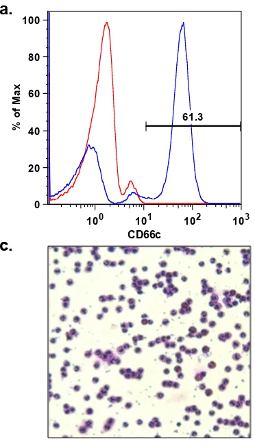
Neutrophils were initially isolated from whole blood using polymorphprep™ (Axis-Shield). Whole blood, taken directly into tubes containing 3.8% sodium citrate, an anti-coagulant, at a ratio of 1:9, was layered at 1:1 ratio over polymorphprep™. This was centrifuged at 1800rpm for 40 minutes, with the brake set to zero. Neutrophils were analysed by flow cytometry to establish the percentage of CD66c positive cells. CD66c is a granulocyte marker, which is highly expressed by neutrophils (130). Purity of the enriched neutrophil population was 61.3% (Figure 2-1). The majority of the contaminating cells were red blood cells but of more concern was the potential for monocyte contamination. It has been shown that the presence of monocytes, even in small numbers, can alter the behaviour of neutrophils (131). To ensure that all responses observed were neutrophil specific, magnetic immunoselection was adopted as the neutrophil purification technique (Figure 2-2). This method is described below and yielded 99.8% CD66c positive cells.
Antibody Concentration Host Supplier
Anti- human CD66c PE (monoclonal)
0.125μg per sample Mouse ebioscience
IgG1 isotype PE (monoclonal)
0.125μg per sample Mouse Beckman Coulter
43
Figure 2-1 Neutrophil purity using polymorph preparation method
a) Histogram showing percentage of cells that are CD66c positive measured by flow cytometry. Neutrophils were isolated using the polymorph preparation method. Blue line = neutrophils labelled with a monoclonal antibody to CD66c conjugated to PE. Red line = matched isotype control. In this sample 61.3% of neutrophils expressed CD66c. b) Flow cytometry scatter plot demonstrating the neutrophil population (A) and contaminating cell population (B) on the basis of forward and sidelight scatter properties. c) Neutrophils were spun onto a slide, then stained using a Romanowsky staining protocol, before being imaged using a Leiss microscope. Cells were histologically identified as neutrophils by their multi-lobed nucleus and stain uptake pattern.
0 20 40 60 80 100
SS Lin: SS Lin 0
50 100 150 200
FS
Lin:
FS
Lin
100 101 102 103 FL2 Log: FL2 Log
0 20 40 60 80 100
%
of
Max
61.3 a.
c.
b.
A"
B"
44 2.2.1 Negative immunoselection methodology
Antibody complexes and dextran-coated magnetic particles were used to isolate the neutrophils by removing unwanted cells from the cell suspension. The antibody complexes link targeted cell populations to the magnetic particles. Labelled cells are pulled to the sides of the tube when the sample is placed in the EasySep™ magnet. Magnetically labelled cells (the positive fraction) remain in the tube while untouched cells (the negative fraction) can be poured into a new tube. For neutrophil isolation, the tetrameric antibody complexes recognize CD2, CD3, CD9, CD19, CD36, CD56, glycophorin A and dextran coated magnetic particles.
2.2.2 Neutrophil ultra-purification by magnetic immunoselection
Neutrophils were isolated from the venous blood of healthy adult volunteers. Blood was taken directly into tubes containing 3.8% sodium citrate. Blood was centrifuged at 270g for 20 minutes at 18°c before plasma was aspirated from the top. Plasma was further centrifuged at 1155g for 10 minutes at 18°C to make platelet poor plasma (PPP). To the cell pellet, 6 ml of 6% dextran was added and total volume made up to 50ml with warmed 0.9% sodium chloride. After 25 minutes, the upper layer, which is red cell depleted following dextran sedimentation, was removed and cells pelleted by centrifugation at 185g for 6 minutes. Neutrophils were separated by discontinuous plasma:Percoll gradient centrifugation, pelleted at 420g for 6 minutes and resuspended in RoboSep™ buffer (Stemcell™ Technologies).
45
Figure 2-2 Neutrophil purity using negative immunoselection method
a) Histogram showing percentage of cells that are CD66c positive measured by flow cytometry. Blue line = neutrophils labelled with a monoclonal antibody to CD66c conjugated to PE. Red line = matched isotype control. Following negative immunoselection where antibody complexes link to CD2, CD3, CD9, CD19, CD36, CD56 and glycophorin A and bind to magnetic particles leaving a pure preparation of neutrophils, 99.8% of neutrophils expressed CD66c. b) Flow cytometry scatter plot demonstrating the one cell population on the basis of forward and sidelight scatter properties. c) Neutrophils were spun onto a slide then stained using a Romanowsky staining protocol, before being imaged using a Leiss microscope. Cells were histologically identified as neutrophils by their multi-lobed nucleus and stain uptake pattern.
0 50 100 150 200
FS Lin: FS Lin 0
20 40 60 80 100
SS
Lin:
SS
Lin
100 101 102 103
FL2 Log: FL2 Log 0
20 40 60 80 100
%
of
Max
99.8
a.
c.
b.
46 tube into a new tube, which was then placed into the magnet for a further 5 minutes. This was repeated once, leaving the negatively selected enriched cells in the new tube ready for use.
2.3 Romanowsky staining technique
Cells were spun onto slides using a cytospin and air-dried. Rapid Romanowsky Stain (HD Supplies) was used. 25 mls of each solution (A - fixative solution containing thiazine dye in methanol, B - Eosin Y in phosphate buffer, and C - Methylene blue in phosphate buffer) were placed in sequentially positioned Coplin jars. Slides were placed in solution A for 30 seconds, then solution B for 30 seconds and finally solution C for 30 seconds, with no rinsing between each immersion. Slides were washed gently in running water before being allowed to air dry for 5 minutes. Slides were then examined by Leica microscope (40x/0.5 objective) and images recorded using Leica IM50 Image Manager. Leukocytes show excellent nuclear and cytoplasmic staining. Immature granulocytes show bright azurophilic granules. Erythrocytes are orange-red.
2.4 Neutrophil culture
Neutrophils were counted using a manual haemocytometer and suspended at 5 million/ml in Dulbecco’s Modified Eagle Medium (DMEM) in 10ml falcons for experiments. If serum was required this was either 5% autologous serum, or 5% AB serum. Neutrophils were incubated at 37oC/5% CO2.
2.5 RSV preparation
30 000 Hep2 Cells/cm2 in a volume of 30ml were seeded into a T175 cm2 flask and left to grow to approximately 50% confluence overnight at 37oC/5% CO2 in DMEM and 10% foetal calf serum (FCS). Medium was removed from
the flask and cells washed twice with PBS. 1 vial of RSV (MOI 1) and 8mL of serum free medium were added to the flask which was left to rock gently at 37°C for 2hrs. After 2hrs, 22mL of DMEM containing 2% FCS was added and cells incubated for 24 hours at 37oC/5% CO
2. Media was replaced after
47 following infection. At this time, all but 4mls of media was removed. The cells were scraped from the bottom of the flask using a cell scraper and placed in a universal container on ice before being passed through a 25 gauge needle 10 times. Cells were then spun down at 4oC at 1500rpm for 5 minutes. The
supernatant was quickly transferred in 500ul aliquots into pre-labelled cyrovials before being snap frozen in liquid nitrogen and placed in the -80 oC
freezer for storage. As RSV is most stable when within cells and unbound virus degrades rapidly, work was carried out quickly when thawing and freezing down virus containing cryovials. For thawing, the cryovial was agitated in a water bath set to 37°C. When the pellet was almost completely thawed it was opened in a tissue culture or lamina flow hood and gently pipetted up and down to thaw remaining virus and used as quickly as possible. The RSV preparation used in the majority of the experiments was a gift from Dr. Paul Fitch (University of Edinburgh). This was 50 vials from 1 batch of propagated RSV A2 strain (PFU 1.79x10⁶).
2.6 Plaque assay
A plaque assay was used to quantify the amount of virus propagated. Hep2 cells were seeded into a 96 well plate in DMEM containing 10% FCS for 24 hours. A doubling dilution was made of virus stock staring at 1/100 and producing 8 dilutions in serum free DMEM. The Hep2 cells were washed with PBS and 50ul of virus was added to each well, with each dilution plated in triplicate and a virus free control included. After 2 hours at 37oC/5% CO2,
48 as were all replicates of that dilution. The numbers of plaques in the wells in the 2 dilutions below were also counted. Plaque forming units (PFU) for each virus batch were then calculated using the formula:
Number of plaques x dilution x 20 = PFU/ml
2.7 Phagocytosis assay
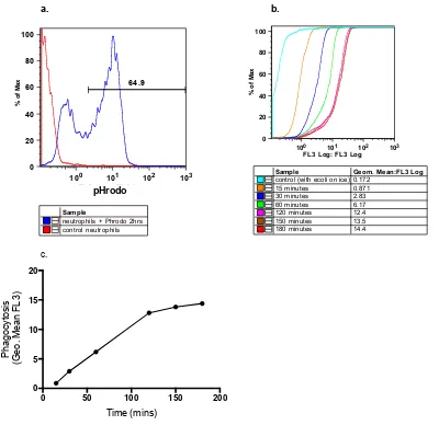
For this assay pHrodo™ E. coli BioParticles® Phagocytosis Kit for Flow Cytometry (Invitrogen) was used. Assays were carried out according to the manufacturer’s protocol except that 100ul of neutrophil preparation was used rather than whole blood. Lyophilized pHrodo E. coli BioParticles® Conjugate was stored in the freezer. When required, 2ml uptake buffer (HBSS + 20mM HEPES, pH 7.4 (NaOH added if necessary)) was placed into the vial containing the 2mg lyophilized product and vortexed to resuspend. Neutrophils were suspended at 5 million/ml in DMEM. 50ul of pHrodo was added to each tube, except the control. When required, 10-20% autologous serum was added. Cells were left to incubate at 37oC/5% CO2, except for the
ice control sample. At 15, 30, 60, 90, 120, 150 and 180 minutes, samples from each tube were collected, washed twice and then analysed by flow cytometry. Neutrophils that have phagocytosed bacteria emit fluorescence in the FL3 channel (Figure 2-3a). Neutrophils that have been incubated with pHrodo E. coli on ice were used as a negative control.
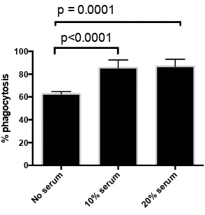
This technique was used to quantitatively assess the phagocytic activity of the isolated neutrophils and to ascertain the percentage of autologous serum needed to enhance phagocytosis.
2.7.1 Isolated neutrophils are capable of phagocytosis
49 (132). Phagocytosis plateaus at 2 hours as the maximal capacity of the neutrophil is reached.
2.7.2 Autologous serum enhances neutrophil phagocytosis
Neutrophils incubated with pHrodo E. coli for 2 hours were analysed by flow cytometry and percentage phagocytosis measured by percentage of neutrophils that were FL3 +ve. Comparison was made between incubations carried out with no serum, and in the presence of 10 and 20% autologous serum (Figure 2-4). Phagocytosis increased significantly in the presence of both 10% serum, from 62.72% (+/- 0.88) to 85.60% (+/- 3.06) p<0.0001 and 20% serum, to 86.85% (+/- 3.10) p = 0.0001. One-way ANOVA was performed with Bonferroni’s post hoc test to allow for multiple testing.