Developments in Cardiovascular Proteomics
Melissa A Noronha1, Christopher Linden2 and Parveen Sharma2*
1Children’s Hospital of Michigan, 3901 Beaubien St. Detroit, Michigan 48201, USA
2MRC Centre for Drug Safety Science, Department of Molecular and Clinical Pharmacology, University of Liverpool, Liverpool, L69 3GE, UK
*Corresponding author: Parveen Sharma, Department of Molecular and Clinical
Pharmacology, MRC Centre for Drug Safety Science, Institute of Translational Medicine, University of Liverpool, Sherrington Building, Liverpool, L69 3GE, UK, Tel: 0151 7950149; E-mail: [email protected]
Received April 14, 2016; Accepted May 12, 2016; Published May 18, 2016
Citation: Noronha MA, Linden C, Sharma P (2016) Developments in Cardiovascular
Proteomics. J Proteomics Bioinform 9: 144-150. doi:10.4172/jpb.1000400
Copyright: © 2016 Noronha MA, et al. This is an open-access article distributed
under the terms of the Creative Commons Attribution License, which permits unrestricted use, distribution, and reproduction in any medium, provided the original author and source are credited.
Keywords:
Cardiac; Disease; Cardiotoxicity; Sub-proteome; Mass spectrometry imagingIntroduction
Cardiovascular disease (CVD) is the leading cause of global morbidity and mortality, causing approximately 30% of all deaths in the United States [1] and the UK [2]. The on and off-target cardiovascular system (CVS) effects of a multitude of drugs for different of conditions; including beta blocking agents, chronic use of non-steroidal anti- inflammatory drugs (NSAIDs) [3-5] and anti-cancer agents add to the burden [6,7]. As the treatments for acute complications of CVD continue to improve, the epidemiology of CVD in our aging population is rapidly evolving from acute conditions to chronic disease.
An in-depth understanding of both the healthy and distressed CVS will allow greater understanding of the processes and mechanisms that may go awry in CVD. Many recent studies have used transcriptome and RNA profiling to describe CVD processes [8-10] and search for biomarkers of disease [11,12]. While these studies have shown vital aspects of molecular changes in CVD it is essential to understand the impact disease has at a protein level. Studies have shown in humans that not only do differences exist between DNA and the final mRNA product [13,14], but there is also marked variation in the expected proteome [15]. Consequently, a thorough understanding of the cardiovascular proteome is crucial to elucidating disease progression, biomarkers and potential therapeutic targets of CVD. Complicating things further, numerous studies point at post-translational modifications that not only regulate key processes in the CVS [16-18] but are also responsible for CVD pathologies and progression [19]. Recent innovative techniques using novel proteomic technologies to enrich and separate sub-proteomes have advanced our knowledge of disease progression and enhanced our ability to identify markers of disease and targets for new therapeutic strategies.
Gel based proteomics
Early examination of cardiac proteins were extensively carried out using two- dimensional gel electrophoresis (2DE) techniques and although these techniques have been predominantly replaced by gel-free methods, key studies still employ this well- established system [20]. In this technique, homogenized samples of cardiac tissue are solubilized and denatured, then separated based on isoelectric point and molecular mass using sodium dodecyl sulphate-polyacrylamide
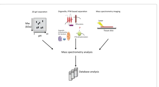
gel electrophoresis (SDS-PAGE) [21]. In this technique, which offers a method to reduce the complexity of the entire sample, protein bands or spots are excised out of the gel and further analyzed by tandem mass spectrometry. This approach has led to fundamental advances in healthy cardiac proteomics [22,23], stem cell derived cardiac maturation [24], in-depth organellular proteomes [25,26] as well as changes that occur upon disease [23,27-30] and those that are induced by drugs [31]. In particular this technique provides a simple method of detecting post-translational modifications such as acetylation, glycosylation, phosphorylation and deamidation, in which there has been a shift in molecular weight and isoelectric points of the protein [20]. Methods utilizing 2DE do however tend to be biased against proteins with hydrophobic regions such as membrane proteins and biased towards high abundance proteins. 2DE also has a limited dynamic range of 104 magnitude [32,33] compared with the very high dynamic range of protein abundance, estimated at 106 for cells and tissues [32] and 1012 for plasma [23,32,34]. In an effort to overcome these biases, new separation methodologies as well as advancements in mass spectrometry instrument technologies have reduced the need for separating proteins using a gel based system (Figure 1).
In-depth analysis of the cardiac sub-proteomes
In an effort to analyze the cardiac sub-proteomes and in-turn reduce the complexities of the samples, several subcellular fractionation methods exist which first fractionate the samples. These methods include differential centrifugation, flow cytometry, immune-based isolation, membrane protein enrichment strategies and/or density gradient isolation of organelles. These techniques have been applied to study the sub-proteomes of cardiac sarcomeric enriched fractions [35], myofilaments [36,37], mitochondria [38-41] and nuclei [41,42]. Abstract
One key sub-proteome however that often gets overlooked is cell-surface proteins. These proteins are difficult to isolate and enrich due to their low abundance and relatively low solubility in aqueous media. This challenge has prompted us and others to develop and modify cell surface enrichment methodologies that can be coupled to mass spectrometry techniques to identify existing and novel proteins in this key cellular compartment.
Cell surface proteomics
The cardiomyocyte plasma membrane is essential to the function of the heart, from the spread of an action potential to the regulation of inotropic and chronotropic changes of the heart. Analyzing cardiac membrane proteins is crucial to a better understanding of heart function especially when we consider that the majority of cardiac disorders including arrhythmias, cardiomyopathies and conduction defects can be attributed to altered expression, function, or subcellular localisation of cardiac ion channels and associated membrane proteins [43-46]. Thus the identification and characterization of membrane proteins is paramount to the understanding of cardiac dysfunction and the search for therapeutic targets. With this in mind, several techniques have been developed recently to isolate and purify plasma membrane proteins including: colloidal bead isolation, biotinylation and glycocapture, each having its own advantages and limitations which will be discussed in turn.
Colloidal silica bead membrane isolation: The silica bead plasma membrane isolation procedure uses positively charged colloidal silica beads to coat and bind to the anionic plasma membrane, and following homogenization and centrifugation, the plasma membrane is stripped from the rest of the cell [47] and then further purified in a discontinuous
nycodenz gradient. Recently we refined this protocol in order to couple it with shot-gun proteomics [48] and used it to identify plasma membrane proteins in vivo [49] and in vitro [50]. Our in vitro analysis of the mouse and human cardiomyocytes identified a total of 3033 mouse and 2762 cardiac proteins. This study method identified a total of 555 membrane associated proteins which included Tmem65, a novel cardiac membrane protein shown to regulate essential cardiomyocyte functionality [50]. This isolation procedure is however limited by its tendency to rupture cells, especially cells in early developmental stages which may have a dynamic membrane due to morphological immaturity and cause them to leak. This allows the beads to enter the cell and bind to internal organelle membranes thereby significantly contaminating the plasma membrane fraction [47,51].
Biotinylation: Biotinylation is used to isolate cell surface proteins by exploiting the strong affinity of biotin for avidin. Preparations of cell-impermeable, modified biotin allow it to bind to primary amines of exposed proteins in a cell culture system or using the in vivo application those accessible via the blood stream [52,53]. The proteins can then be isolated with avidin beads and following elution from the biotin-avidin complex these proteins can be identified using mass spectrometry [53]. We have previously utilized this technique to identify cell surface markers on linage specific stem cells, isolated from mouse embryos [54] which results in their complete separation using fluorescence assisted cell sorting (FACS). More recently, Strassberger et al. [55] used biotinylation combined with mass spectrometry to extract differentially expressed cell surface proteins in myeloid leukemia cell lines compared to normal human granulocytes. They identified a number of proteins found preferentially in myeloid leukemia cells and were able to target one of them, namely CD166/ALCAM, to kill these cells in vitro. This Mw
(kDa)
pH
3 10
Tissue slice Laser
P
2D gel separation Organelle, PTM based separation Mass spectrometry imaging
Mass spectrometry analysis PM proteins
Organelle (mitochondria, ER, Nucleus)
PTM phosphorylation
[image:2.595.43.574.100.392.2]Database analysis
Figure 1: Schematic of mass spectrometry workflow. Separation based on 2D gel electrophoresis and current organelle and post-translational modification
methodology is limited however in its ability to extract cell membrane proteins without an exposed extracellular domain thereby biasing against membrane proteins that are associated with the intracellular face of the plasma membrane and proteins fully enveloped inside the phospholipid bilayer.
Glycosylation: Glycosylation is the most common post-translational modification [56] and aberrant glycosylation has been shown to be early indicator of cellular changes in a multitude of human diseases including cancer, inflammation and neurodegeneration [57-59]. Alterations in plasma membrane glycoproteins have also been reported in ischemia reperfusion injuries [60] and cardiac hypertrophy [61]. Most membrane-bound and secretory proteins synthesized by mammalian cells are glycosylated and thereby may provide useful diagnostic tools as proteins that are shed from the cell surface during disease as well as offer mechanistic insights into disease progression and pathology. This understanding has thereby stemmed a wide variety of techniques to specifically isolate cell surface and plasma secreted glycoproteins. These include separation based on hydrophilic interactions [60,62,63], exploiting the high affinity of glycosylated proteins and peptides for lectins and the chemical capture of glycoproteins using hydrazide technology. A variety of lectins are available that selectively bind to different oligosaccharide epitopes and thereby can specifically isolate for a unique sub-set of glycoproteins containing a specific glycan moiety [59]. Although no one lectin is capable of isolating a cell’s complete glycoprotein complement, studies are now combining multiple lectins to achieve greater coverage [59].
Hydrazide technology aims to isolate a broader range of glycoproteins. This procedure involves oxidizing carbohydrates, to convert cis-diol groups into aldehydes so that they may be linked to hydrazide groups on solid supports, leading to their enrichment [56]. This technique was further refined for use in live cells by Wollscheid et al. [64]who showed that the gentle biocytin hydrazide labelling of oxidized carbohydrates is cell impermeable and can therefore be used to characterize changes in the cell surface glycoproteome. This procedure, termed cell surface capture (CSC), has recently been used to establish a cell surface protein atlas (CSPA) in which researchers have used CSC technology to isolate surface proteins from 41 human and 31 mouse cell types providing an invaluable cell surface resource [65]. A study conducted by Parker et al. [60] used multiple methods to enrich for glycoproteins in myocardial ischemia and reperfusion injury and identified extracellular and cell surface proteins involved in the various stages of cardiac remodeling. Their study suggested that cardiac remodeling following myocardial ischemia may begin much earlier than originally hypothesized. In a more recent study Yang et al.[66] identified 32 glycosite containing peptides elevated in a canine model of dyssynchronous heart failure of which 13 glycopeptides were reverted to normal levels after cardiac resynchronization therapy [66]. The major drawback of hydrazide based techniques is that efficient detection of glycosylated proteins requires the removal of the oligosaccharide moieties from the glycoprotein [67]. For N-glucosylation the β- aspartylglycosylamine linkage can be selectively cleaved using peptide-N-glycosidase F (PNGaseF) but until recent developments those moieties in which the root β-1,4- acetylglucosamine had been fucosylated were missed in the analysis [68]. Recent protocols however, have used Lens culinaris Agglutinin (LCA) to enrich for core fucosylated (CF) proteins, followed by Endo F3 partial deglycosylation and NanoLC- MS/MS. This identified CF proteins in human serum from healthy volunteers and patients with pancreatic cancer [69] and hepatocarcinoma [70]. This technique provides an essential tool to ensure greater coverage of the glycoproteome and indeed Parker
et al. [60] demonstrated that a combination of techniques to enrich for glycoproteins offers greater depth. O-linked glycosylation further lacks a common moiety, leaving its cleavage more challenging and to date a cocktail of glycosidases have been used [68]. A lack of consensus currently limits their detection and thus the majority of studies have focused on N-linked glycoprotein analysis [59].
Phosphoproteomics
When discussing cardiac sub-proteomes it is hard to ignore the phosphoproteome. Reversible protein phosphorylation is not only vital for the normal coordinated function of the heart but is also implicated in disease initiation and progression. This post- translation modification is essential for propagating cell signaling events in response to hormones, second messengers and pharmacological agents.
Several methods exist to subtract phosphorylated proteins from the cellular milieu which include both in-gel and gel-free methods. Using 2D-gel electrophoresis a visual migration of the protein can be monitored due to additional charge of the phosphorylated group which can then be specifically excised from the gel, purified and then identified by mass spectrometry methods. A recent study using 2D-gel electrophoresis identified 22 proteins with significantly changed phosphorylation states expressed during the acute off- target cardiotoxic effects of the chemotherapeutic agent doxorubicin. This study highlighted key signaling pathways involved in energy metabolism, sarcomeric function/structure and chaperone activity which could lead to identification of the early events involved in cardiotoxicity [71].
As previously mentioned, there has been a deviation from gel based methods and gel-free methods are predominantly used for phosphoprotein and peptide enrichment which include the use of titanium dioxide beads and immobilized metal affinity chromatography (IMAC) combined with mass spectrometry [72]. Titanium dioxide has a very high affinity for phosphopeptides and has been shown to be able to isolate femtomoles of phosphopeptides from complex protein mixtures [73,74]. Prior to the identification of proteins and signaling pathways involved in disease processes, it is essential to assess the healthy state. Lundby et al. [75] used titanium dioxide phospho- enrichment coupled to LC–MS/MS, to quantify the changes in the mouse cardiomyocyte phosphoproteome in vivo upon acute beta1-adrenergic and beta2-adrenergic stimulation. They uncovered 670 phosphorylation sites regulated by beta1-adrenergic stimulation which included previously unknown modifications of channels and transporters affecting cardiac function, highlighting the utility of phosphoproteomics analysis for elucidating cardiac signaling networks [75,76]. Wijeratne et al. [77] describe a novel workflow involving reductive alkylation by acetone to label peptides in tandem with titanium dioxide isolation followed by mass spectrometry based identification to effectively quantify phosphorylative changes in a mouse model of ischemia-reperfusion injury. Using this methodology they found increased cardioprotection in a mouse model of ischemia- reperfusion injury was due to low molecular weight fibroblast growth factor-2 (LMW FGF2) mediated phosphorylation of key downstream signaling pathways [77].
Quantification
free relative quantification techniques include spectral counting, peak intensity comparisons and more recently SWATH analysis [50,79,81-83] whereas labelled techniques include SILAC, iTRAQ, enzymatic 18O labelling and in vivo 2H
20 labelling [79,84-87]. Absolute techniques incorporate the use of internally spiked absolute quantification (AQUA) peptides to quantify absolute amounts using MRM, SRM, iTRAQ [88-91]. These techniques have been extensively used in cardiovascular proteomics [23,78,80] and can be combined with a number of the sub-proteome separation techniques in which quantification of differentially expressed proteins are informative about mechanism of disease progression, pathway analysis of potential drug targets and biomarkers. For example, the quantitative accuracy of SWATH analysis has been compared to that of SRM analysis [92] and has the ability to reproducibly identify low abundance proteins from small numbers of cells (50 000 cells) and from small amounts of tissue from biopsy samples (0.2-0.5 mg) including cardiac tissue [92]. SWATH and SRM were used in combination with CSC glycoprotein enrichment methods to provide quantitative and qualitative expression of 78 human and mouse tissue cell surface proteins [65]. Lau et al. combined in vivo 2H
20 labelling of drinking water with subcellular fractionation to quantitate protein dynamics and turnover in healthy mice and those with cardiac hypertrophy. This invaluable resource of cardiac disease progression provides quantification of the in vivo half-lives of 3,228 proteins and the expression of 8,064 proteins under healthy and diseased states [86]. Lam et al. [93] combined differential centrifugation to isolate mitochondria from human and mouse hearts with TiO2 purification of phosphoproteins with MRM quantification to establish 176 MRM transitions specific to protein modifications. These transitions can now be used to quantify alterations in the level of phosphorylation under conditions of disease or pharmacological intervention.
New mass spectrometry technologies
The advancement of mass spectrometry instrumentation and computational technologies has vastly improved the coverage and depth of the proteome. A comparison of generations of the Q Exactive instrument for example, showed that the latest iteration of the Q Exactive HF has the capability to detect > 4000 proteins in a 1 hour gradient from 1µg of Hela cells prior to any fractionation [94]. These powerful methods offer great versatility and depth when dealing with precious samples, in particular healthy human tissues which can not only be rare and difficult to obtain but also only available in small quantities retrieved from patient biopsies. These restrictions therefore limit the amount of sample manipulation and fractionation prior to analysis. Indeed, we have used these powerful methods to identify 2754 human atrial protein groups and 2825 human ventricular protein groups from which we were then able to identify and classify chamber unique proteins without prior fractionation [95]. If however the initial sample quantity does lend itself to prior fractionation, the possibilities are extensive as demonstrated by TiO2 enrichment of phosphoproteins which when analyzed gave 7600 unique phosphopeptides in a 1 hour gradient of elution [94].
Mass spectrometry imaging: One limitation of sub-fractionation is the loss of spatial distribution of proteins and drugs. Emerging technologies of mass spectrometry imaging (MSI) are now providing a powerful tool that allows the analysis, detection and mapping of the biodistribution of proteins and drugs within histological sections [96]. MSI can detect endogenous and exogenous analytes including low molecular weight drugs, lipids and proteins directly from tissue samples [96] (Figure 1). This technique has traditionally been linked to MALDI ionization and accomplished by using tissue sections
mounted onto a conductive slide in water or gelatin to which one of several MALDI matrices are sprayed depending on the type of analyte to be measured [96]. The matrix then co- crystalizes with the cell components and then the region of interest is subjected to MALDI ionization for detection by mass spectrometry [96,97]. More recently MSI has evolved to use desorption electrospray ionization (DESI) [98], which overcomes complications of matrix-analyte co-crystallisation issues [99]. The technical aspects of MSI and sample preparation are beyond the scope of this review but have previously been discussed in depth [96-99]. Using these techniques Martin-Loenzo et al. [100] pinpointed specific lipids and proteins to atherosclerotic aortic mouse cardiac sections. They showed that thymosin beta 4 is differentially overexpressed in the intima layer of atherosclerotic tissue compared to media layers providing new avenues of research in the area [100]. This technique further holds a lot of promise in drug distribution analysis.
Histological sections of models of drug dosed tissue allow the detection of drug and metabolite distribution. MSI has recently been used to monitor and quantify the release of celecoxib from packaging nanoparticles delivered to the healthy and ischemic myocardium of in vivo models. This study identified the angiogenic potential of this drug when delivered locally to a specific region, pointing to a potential new approach for treating ischemia [101]. Drug-induced cardiotoxicity remains the major contributor to drug attrition and withdrawal from patient distribution and a major complication for off- target toxicities of a multitude of drugs [6]. Whole body MSI offers the promise to determine localization of both parent and metabolite distribution within every organ or cell type of the body early in drug development stages, potentially aiding drug testing and reducing adverse off-target reactions [96,102,103]. One major drawback of MSI is the inability to directly identify proteins and the potential need to coordinate experiments with tandem mass spectrometry methods to carry out sequence identification [104,105]. However, with the continuing development of technologies, databases are now being established by the MSI community that allows the identification of molecules directly from the mass spectrometric measurement [106]. The identification of proteins [107,108] and glycoproteins [109,110] as well as lipids and cell metabolites [106] from MSI experiments of healthy versus diseased or pharmacologically treated tissue will undoubtedly reveal deeper insights into disease mechanisms and pathways at a tissue level.
Conclusions
This review highlights current technologies that have been developed and modified to further cardiovascular knowledge through proteomics. Taken together, these studies highlight a systems biology approach allowing the identification and quantification of potential targets, biomarkers, and mechanisms of disease progression including identification of post-translational modifications as well as methods to gain insights on the biodistribution of parent drugs and active metabolites. Applying the correct innovative techniques for a given system under healthy, diseased and drug-dosed conditions will allow us to gain further understanding into mechanisms that cause disease and drug-induced cardiotoxicity. These insights will in-turn help us to gain a deeper comprehension into cardiac disease and to reduce the number of drugs that can cause adverse cardiac reactions before they enter patient circulation.
Acknowledgment
References
1. Roger VL, Go AS, Lloyd-Jones DM, Benjamin EJ, Berry JD, et al. (2012) Heart disease and stroke statistics-2012 update: a report from the American Heart Association. Circulation 125: e2-2e220.
2. Bhatnagar P, Wickramasinghe K, Williams J, Rayner M, Townsend N (2015) The epidemiology of cardiovascular disease in the UK 2014. Heart 101: 1182-1189.
3. Anwar A, Anwar IJ, Delafontaine P3 (2015) Elevation of cardiovascular risk by
non-steroidal anti-inflammatory drugs. Trends Cardiovasc Med 25: 726-735.
4. Asghar W, Jamali F (2015) The effect of COX-2-selective meloxicam on the myocardial, vascular and renal risks: a systematic review.
Inflammopharmacology 23: 1-16.
5. García-Poza P, de Abajo FJ, Gil MJ, Chacón A, Bryant V, et al. (2015) Risk of ischemic stroke associated with non-steroidal anti-inflammatory drugs and
paracetamol: a population-based case-control study. J Thromb Haemost 13: 708-718.
6. Laverty H, Benson C, Cartwright E, Cross M, Garland C, et al. (2011) How can we improve our understanding of cardiovascular safety liabilities to develop safer medicines? British Journal of Pharmacology 163: 675-693.
7. Maxwell CB, Jenkins AT (2011) Drug-induced heart failure. Am J Health Syst Pharm 68: 1791-1804.
8. Ounzain S, Micheletti R, Beckmann T, Schroen B, Alexanian M, et al. (2015)
Genome-wide profiling of the cardiac transcriptome after myocardial infarction identifies novel heart-specific long non-coding RNAs. Eur Heart J 36: 353-368.
9. Gao C, Wang Y (2014) Transcriptome Complexity in Cardiac Development and Diseases: An Expanding Universe Between Genome and Phenome. Circ J 78: 1038-1047.
10. Liu Y, Morley M, Brandimarto J, Hannenhalli S, Hu Y, et al. (2015)
RNA-Seq identifies novel myocardial gene expression signatures of heart failure.
Genomics 105: 83-89.
11. Pedrotty DM, Morley MP, Cappola TP (2014) Transcriptomic biomarkers of cardiovascular disease. Prog Cardiovasc Dis 5: 64-69.
12. Costa Ade F, Franco OL (2015) Insights into RNA transcriptome profiling of cardiac
tissue in obesity and hypertension conditions. J Cell Physiol 230: 959-968. 13. Li M, Wang IX, Li Y, Bruzel A, Richards AL, et al. (2011) Widespread RNA and
DNA sequence differences in the human transcriptome. Science 333: 53-58. 14. Claydon AJ, Beynon R (2012) Proteome dynamics: revisiting turnover with a
global perspective. Mol Cell Proteomics 11: 1551-1565.
15. Lundberg E, Fagerberg L, Klevebring D, Matic I, Geiger T, et al. (2010) Defining
the transcriptome and proteome in three functionally different human cell lines. Mol Syst Biol 6: 450.
16. Portbury AL, Ronnebaum SM, Zungu M, Patterson C, Willis MS (2012) Back to your heart: ubiquitin proteasome system-regulated signal transduction. J Mol Cell Cardiol 52: 526-537.
17. Darley-Usmar VM, Ball LE, Chatham JC (2014) Protein O-linked beta-N- acetylglucosamine: a novel effector of cardiomyocyte metabolism and function. J Mol Cell Cardiol 52: 538-549.
18. Shao D, Oka S, Brady CD, Haendeler J, Eaton P, et al. (2012) Redox
modification of cell signaling in the cardiovascular system. J Mol Cell Cardiol
52: 550-558.
19. Murphy E, Kohr M, Sun J, Nguyen T, Steenbergen C (2012) S-nitrosylation: a radical way to protect the heart. J Mol Cell Cardiol 52: 568-577.
20. Rogowska-Wrzesinska A, Le Bihan MC, Thaysen-Andersen M, Roepstorff P (2013) 2D gels still have a niche in proteomics. J Proteomics 88: 4-13. 21. Gianazza E, Osio L, Grazioli G, Astrua-Testori S, Righetti PG, et al. (1987) An
examination of heart proteins by two-dimensional electrophoresis. Clin Chem 33: 2011-2018..
22. Scholten A, Heck AR (2013) Determining Protein Concentrations of the Human Ventricular Proteome. In: Vivanco F (Ed) Heart Proteomics, Humana Press. 23. Sharma P, Cosme J, Gramolini AO (2013) Recent advances in cardiovascular
proteomics. J Proteomics 81: 3-14.
24. Poon E, Keung W, Liang Y, Ramalingam R, Yan B, et al. (2015) Proteomic Analysis of Human Pluripotent Stem Cell–Derived, Fetal, and Adult Ventricular Cardiomyocytes Reveals Pathways Crucial for Cardiac Metabolism and Maturation. Circ Cardiovasc Genet 8: 427-436.
25. Giorgianni F, Koirala D, Weber KT, Beranova-Giorgianni S (2014) Proteome Analysis of Subsarcolemmal Cardiomyocyte Mitochondria: A Comparison of Different Analytical Platforms. Int J Mol Sci 1: 9285-9301.
26. Kim TY, Wang D, Kim AK, Lau E, Lin AJ, et al. (2012) Metabolic labeling reveals proteome dynamics of mouse mitochondria. Mol Cell Proteomics 11: 1586-1594. 27. Didangelos A, Yin X, Mandal K, Saje A, Smith A, et al. (2011) Extracellular
matrix composition and remodeling in human abdominal aortic aneurysms: a proteomics approach. Mol Cell Proteomics 10: M111.
28. Dietl A, Winkel I, Deutzmann R, Schröder J, Hupf J, et al. (2014) Interatrial differences of basal molecular set-up and changes in tachycardia-induced
heart failure–a proteomic profiling study. Eur J Heart Fail 16: 835-845.
29. Shekar KC, Li L, Dabkowski ER, Xu W, Ribeiro RF Jr, et al. (2014) Cardiac mitochondrial proteome dynamics with heavy water reveals stable rate of mitochondrial protein synthesis in heart failure despite decline in mitochondrial oxidative capacity. J Mol Cell Cardiol 7: 88-97.
30. Manakov D, Ujcikova H, Pravenec M, Novotny J4 (2016) Alterations in the cardiac proteome of the spontaneously hypertensive rat induced by transgenic expression of CD36. J Proteomics.
31. Yin X, Dwyer J, Langley SR, Mayr U, Xing Q, et al. (2013) Effects of perhexiline-induced fuel switch on the cardiac proteome and metabolome. J Mol Cell Cardiol 55: 27-30.
32. McGregor E, Dunn MJ (2006) Proteomics of the heart: unraveling disease. Circ Res 98: 309-321.
33. Rabilloud T (2002) Two-dimensional gel electrophoresis in proteomics: old, old fashioned, but it still climbs up the mountains. Proteomics 2: 3-10.
34. Anderson NL, Anderson NG, Pearson TW, Borchers CH, Paulovich AG, et al. (2009) A human proteome detection and quantitation project. Mol Cell Proteomics 8: 883-886.
35. Warren CM, Geenen DL, Helseth DL Jr, Xu H, Solaro RJ (2010) Sub-proteomic fractionation, iTRAQ, and OFFGEL-LC-MS/MS approaches to cardiac proteomics. J Proteomics 73: 1551-1561.
36. Yin X, Cuello F, Mayr U, Hao Z, Hornshaw M, et al. (2010) Proteomics Analysis
of the Cardiac Myofilament Subproteome Reveals Dynamic Alterations in
Phosphatase Subunit Distribution. Mol Cell Proteomics 9: 497-509.
37. Fert-Bober J, Giles JT, Holewinski RJ, Kirk JA, Uhrigshardt H, et al. (2015)
Citrullination of myofilament proteins in heart failure. Cardiovasc Res 108: 232-242.
38. Taylor SW, Fahy E, Zhang B, Glenn GM, Warnock DE, et al. (2003) Characterization of the human heart mitochondrial proteome. Nat Biotechnol 21: 281-286.
39. Elstner M, Andreoli C, Ahting U, Tetko I, Klopstock T, et al. (2008) MitoP2: an integrative tool for the analysis of the mitochondrial proteome. Mol Biotechnol 40: 306-315.
40. Pagliarini DJ, Calvo SE, Chang B, Sheth SA, Vafai SB, et al. (2008) A Mitochondrial Protein Compendium Elucidates Complex I Disease Biology. Cell 13: 112-123.
41. Kislinger T, Cox B, Kannan A, Chung C, Hu P, et al. (2006) Global survey of organ and organelle protein expression in mouse: combined proteomic and
transcriptomic profiling. Cell 12: 173-186.
42. Murray CI, Barrett M, Van Eyk JE (2009) Assessment of ProteoExtract subcellular fractionation kit reveals limited and incomplete enrichment of nuclear subproteome from frozen liver and heart tissue. Proteomics 9: 3934-3938. 43. Chen Q, Kirsch GE, Zhang D, Brugada R, Brugada J, et al. (1998) Genetic
basis and molecular mechanism for idiopathic ventricular fibrillation. Nature 39:
293-296.
44. Sanguinetti MC, Tristani-Firouzi M (2006) hERG potassium channels and cardiac arrhythmia. Nature 440: 463-469.
46. McNair WP, Ku L, Taylor MR, Fain PR, Dao D, et al. (2004) SCN5A mutation associated with dilated cardiomyopathy, conduction disorder, and arrhythmia. Circulation 110: 2163-2167.
47. Chaney LK, Jacobson BS (1983) Coating cells with colloidal silica for high yield
isolation of plasma membrane sheets and identification of transmembrane
proteins. J Biol Chem 258: 10062-10072.
48. Rast JP, Booth JWD (Eds.) Immune Receptors, Humana Press.
49. Cox B, Sharma P, Evangelou AI, Whiteley K, Ignatchenko V, et al. (2011) Translational Analysis of Mouse and Human Placental Protein and mRNA Reveals Distinct Molecular Pathologies in Human Preeclampsia. Mol Cell Proteomics 10.
50. Sharma P, Abbasi C, Lazic S, Teng AC, Wang D, et al. (2015) Evolutionarily conserved intercalated disc protein Tmem65 regulates cardiac conduction and connexin 43 function. Nat Commun 6: 8391.
51. Arjunan S, Reinartz M, Emde B, Zanger K, Schrader J (2009) Limitations of the colloidal silica method in mapping the endothelial plasma membrane proteome of the mouse heart. Cell Biochem Biophys 53: 135-143.
52. Elia G (2008) Biotinylation reagents for the study of cell surface proteins. Proteomics 8: 4012-4024.
53. Roesli C, Neri D, Rybak JN (2006) In vivo protein biotinylation and sample
preparation for the proteomic identification of organ- and disease-specific
antigens accessible from the vasculature. Nat Protoc 1: 192-199.
54. Rugg-Gunn PJ, Cox BJ, Lanner F, Sharma P, Ignatchenko V, et al. (2012)
Cell-Surface Proteomics Identifies Lineage-Specific Markers of Embryo-Derived
Stem Cells. Dev Cell 2: 887-901.
55. Strassberger V, Gutbrodt KL, Krall N, Roesli C, Takizawa H, et al. (2014) A comprehensive surface proteome analysis of myeloid leukemia cell lines for therapeutic antibody development. J Proteomics 99: 138-151.
56. Zhang H, Li XJ, Martin DB, Aebersold R (2003) Identification and quantification
of N-linked glycoproteins using hydrazide chemistry, stable isotope labeling and mass spectrometry. Nat Biotechnol 21: 660-666.
57. Dwek RA, Butters TD, Platt FM, Zitzmann N (2002) Targeting glycosylation as a therapeutic approach. Nat Rev Drug Discov 1: 65-75.
58. Christiansen MN, Chik J, Lee L, Anugraham M, Abrahams JL, et al. (2014) Cell surface protein glycosylation in cancer. Proteomics 14: 525-546.
59. Pan S, Chen R, Aebersold R, Brentnall TA (2011) Mass spectrometry based glycoproteomics--from a proteomics perspective. Mol Cell Proteomics 10: R110.
60. Parker BL, Palmisano G, Edwards AV, White MY, Engholm-Keller K, et al. (2011) Quantitative N- linked glycoproteomics of myocardial ischemia and reperfusion injury reveals early remodeling in the extracellular environment. Mol Cell Proteomics 10: M110 006833.
61. Yang S, Mishra S, Chen L, Zhou JY, Chan DW, et al. (2015) Integrated Glycoprotein Immobilization Method for Glycopeptide and Glycan Analysis of Cardiac Hypertrophy. Anal Chem 87: 9671-9678.
62. Takegawa Y, Deguchi K, Ito H, Keira T, Nakagawa H, et al. (2006) Simple separation of isomeric sialylated N-glycopeptides by a zwitterionic type of hydrophilic interaction chromatography. J Sep Sci 29: 2533-2540.
63. Di Palma S, Boersema PJ, Heck AJ, Mohammed S (2011) Zwitterionic Hydrophilic Interaction Liquid Chromatography (ZIC-HILIC and ZIC-cHILIC) Provide High Resolution Separation and Increase Sensitivity in Proteome Analysis. Anal Chem 83: 3440-3447.
64. Wollscheid B, Bausch-Fluck D, Henderson C, O’Brien R, Bibel M, et al. (2009)
Mass-spectrometric identification and relative quantification of N-linked cell
surface glycoproteins. Nat Biotechnol 27: 378-386.
65. Bausch-Fluck D, Hofmann A, Bock T, Frei AP, Cerciello F, et al. (2015) A mass spectrometric-derived cell surface protein atlas. PLoS One 10: e0121314. 66. Yang S, Chen L, Sun S, Shah P, Yang W, et al. (2015) Glycoproteins identified
from heart failure and treatment models. Proteomics 15: 567-579.
67. Kaji H, Yamauchi Y, Takahashi N, Isobe T (2007) Mass spectrometric
identification of N-linked glycopeptides using lectin-mediated affinity capture and glycosylation site- specific stable isotope tagging. Nat Protoc 1: 3019-3027.
68. Sun B, Hood L (2014) Protein-Centric N-Glycoproteomics Analysis of
Membrane and Plasma Membrane Proteins. J Proteome Res 13: 2705-2714. 69. Tan Z, Yin H, Nie S, Lin Z, Zhu J, et al. (2015) Large-scale identification of
core-fucosylated glycopeptide sites in pancreatic cancer serum using mass spectrometry. J Proteome Res 14: 1968-1978.
70. Yin H, Tan Z, Wu J, Zhu J, Shedden KA, et al. (2015) Mass-Selected
Site-Specific Core-Fucosylation of Serum Proteins in Hepatocellular Carcinoma. J
Proteome Res 14: 4876-4884.
71. Gratia S, Kay L, Michelland S, Sève M, Schlattner U, et al. (2012) Cardiac phosphoproteome reveals cell signaling events involved in doxorubicin cardiotoxicity. J Proteomics 75: 4705-4716.
72. Stensballe A, Andersen S, Jensen ON (2001) Characterization of
phosphoproteins from electrophoretic gels by nanoscale Fe(III) affinity
chromatography with off-line mass spectrometry analysis. Proteomics 1: 207-222.
73. Pinkse MW, Uitto PM, Hilhorst MJ, Ooms B, Heck AJ (2004) Selective isolation at the femtomole level of phosphopeptides from proteolytic digests using 2D-NanoLC-ESI-MS/MS and titanium oxide precolumns. Anal Chem 76: 3935-3943.
74. Larsen MR, Thingholm TE, Jensen ON, Roepstorff P, Jørgensen TJ (2005) Highly selective enrichment of phosphorylated peptides from peptide mixtures using titanium dioxide microcolumns. Mol Cell Proteomics 4: 873- 886. 75. Lundby A, Andersen MN, Steffensen AB, Horn H, Kelstrup CD, et al. (2013) In
vivo phosphoproteomics analysis reveals the cardiac targets of beta-adrenergic receptor signaling. Sci Signal 6: rs11.
76. Kuzmanov U, Emili A (2013) Using phosphoproteomics to monitor disregulated signaling networks in cardiac disease preceding heart failure. Bioanalysis 5: 2863- 2866.
77. Wijeratne AB, Manning JR, Schultz Jel J, Greis KD (2013) Quantitative phosphoproteomics using acetone-based Peptide labeling: method evaluation and application to a cardiac ischemia/reperfusion model. J Proteome Res 12: 4268-4279.
78. Mirza SP (2012) Quantitative mass spectrometry-based approaches in cardiovascular research. Circ Cardiovasc Genet 5: 477.
79. Meissner F, Mann M (2014) Quantitative shotgun proteomics: considerations
for a high-quality workflow in immunology. Nat Immunol 15: 112-117.
80. Chugh S, Sharma P, Kislinger T, Gramolini AO (2012) Clinical proteomics: getting to the heart of the matter. Circ Cardiovasc Genet 5: 377.
81. Rosenberger G, Koh CC, Guo T, Röst HL, Kouvonen P, et al. (2014) A repository of assays to quantify 10,000 human proteins by SWATH-MS. Sci Data 1: 140031.
82. Bourassa S, Fournier F, Nehmé B, Kelly I, Tremblay A, et al. (2015) Evaluation
of iTRAQ and SWATH-MS for the Quantification of Proteins Associated with
Insulin Resistance in Human Duodenal Biopsy Samples. PLoS One 10: e0125934.
83. Schubert OT, Gillet LC, Collins BC, Navarro P, Rosenberger G, et al. (2015) Building high-quality assay libraries for targeted analysis of SWATH MS data. Nat Protoc 10: 426-441.
84. Ong SE, Mann M (2006) A practical recipe for stable isotope labeling by amino acids in cell culture (SILAC). Nat Protoc 1: 2650-2660.
85. Wiese S, Reidegeld KA, Meyer HE, Warscheid B (2007) Protein labeling by iTRAQ: a new tool for quantitative mass spectrometry in proteome research. Proteomics 7: 340-350.
86. Lau E, Cao Q, Ng DC, Bleakley BJ, Dincer TU, et al. (2016) A large dataset of protein dynamics in the mammalian heart proteome. Sci Data 3: 160015. 87. Castillo MJ, Reynolds KJ, Gomes A, Fenselau C, Yao X, et al. (2014)
Quantitative Protein Analysis Using Enzymatic [(18)O]Water Labeling. Current protocols in protein science. Curr Protoc Protein Sci 76.
88. Desiere F, Deutsch EW, King NL, Nesvizhskii AI, Mallick P, et al. (2006) The PeptideAtlas project. Nucleic Acids Res 34: D655-658.
89. Kettenbach AN, Rush J, Gerber SA (2011) Absolute quantification of protein and post-translational modification abundance with stable isotope-labeled
90. Picotti P, Aebersold R (2012) Selected reaction monitoring-based proteomics:
workflows, potential, pitfalls and future directions. Nat Methods 9: 555-566.
91. Marx V (2013) Targeted proteomics. Nat Meth 10: 19-22.
92. Shao S, Guo T, Koh CC, Gillessen S, et al. (2015) Minimal sample requirement
for highly multiplexed protein quantification in cell lines and tissues by
PCT-SWATH mass spectrometry. Proteomics 15: 3711-3721.
93. Lam MP, Lau E, Scruggs SB, Wang D, Kim TY, et al. (2013) Site-specific
quantitative analysis of cardiac mitochondrial protein phosphorylation. J Proteomics 81: 15-23..
94. Kelstrup CD, Jersie-Christensen RR, Batth TS, Arrey TN, Kuehn A, et al. (2014) Rapid and deep proteomes by faster sequencing on a benchtop quadrupole
ultra-high-field Orbitrap mass spectrometer. J Proteome Res 13: 6187-6195.
95. Lu ZQ, Sinha A, Sharma P, Kislinger T, Gramolini AO (2014) Proteomic analysis of human fetal atria and ventricle. J Proteome Res 13: 5869-5878.
96. Cobice DF, Goodwin RJ, Andren PE, Nilsson A, Mackay CL, et al. (2015) Future technology insight: mass spectrometry imaging as a tool in drug research and development. Br J Pharmacol 172: 3266-3283.
97. Parker S, Raedschelders K, Van Eyk JE (2015) Emerging proteomic technologies for elucidating context-dependent cellular signaling events: A big challenge of tiny proportions. Proteomics 15: 1486-1502.
98. Cooks RG, Ouyang Z, Takats Z, Wiseman JM (2006) Detection Technologies. Ambient mass spectrometry. Science 311: 1566-1570.
99. Wiseman JM, Ifa DR, Venter A, Cooks RG (2008) Ambient molecular imaging by desorption electrospray ionization mass spectrometry. Nat Protoc 3: 517-524. 100. Martin-Lorenzo M, Balluff B, Maroto AS, Carreira RJ, van Zeijlb RJM et al.
(2015) Molecular anatomy of ascending aorta in atherosclerosis by MS
Imaging: Specific lipid and protein patterns reflect pathology. Journal of
Proteomics 126: 245-251.
101. Margulis K, Neofytou EA, Beygui RE, Zare RN (2015) Celecoxib Nanoparticles for Therapeutic Angiogenesis. ACS Nano 9: 9416-9426.
102. Stoeckli M, Staab D, Schweitzer A, Gardiner J, Seebach D (2007) Imaging of a beta-peptide distribution in whole-body mice sections by MALDI mass spectrometry. J Am Soc Mass Spectrom 18: 1921-1924.
103. Goodwin RJ, Macintyre L, Watson DG, Scullion SP, Pitt AR (2010) A solvent-free matrix application method for matrix-assisted laser desorption/ionization imaging of small molecules. Rapid Commun Mass Spectrom 24: 1682-1686. 104. Irie M, Fujimura Y, Yamato M, Miura D, Wariishi H (2014) Integrated MALDI- MS
imaging and LC–MS techniques for visualizing spatiotemporal metabolomic dynamics in a rat stroke model. Metabolomics 10: 473-483.
105. Shobo A, Bratkowska D, Baijnath S, Naiker S, Bester LA, et al. (2015) Visualization of Time- Dependent Distribution of Rifampicin in Rat Brain Using MALDI MSI and Quantitative LCMS/MS. Assay Drug Dev Technol 13: 277-284.
106. Aichler M, Walch A (2015) MALDI Imaging mass spectrometry: current frontiers and perspectives in pathology research and practice. Lab Invest 95: 422-431. 107. Maier SK, Hahne H, Gholami AM, Balluff B, Meding S, et al. (2013)
Comprehensive identification of proteins from MALDI imaging. Mol Cell
Proteomics 12: 2901-2910.
108. McDonnell LA, Walch A, Stoeckli M, Corthals GL (2014) MSiMass list: a public
database of identifications for protein MALDI MS imaging. J Proteome Res
13: 1138-1142.
109. Powers TW, Neely BA, Shao Y, Tang H, Troyer DA, et al. (2014) MALDI imaging
mass spectrometry profiling of N-glycans in formalin-fixed paraffin embedded
clinical tissue blocks and tissue microarrays. PLoS One 9: e106255. 110. Jones EE, Powers TW, Neely BA, Cazares LH, Troyer DA, et al. (2014) MALDI
imaging mass spectrometry profiling of proteins and lipids in clear cell renal
cell carcinoma. Proteomics 14: 924-935.
Citation: Noronha MA, Linden C, Sharma P (2016) Developments in