Analysis of the XPA and ssDNA-binding surfaces
on the central domain of human ERCC1 reveals
evidence for subfunctionalization
Konstantinos Tripsianes, Gert E. Folkers, Chao Zheng, Devashish Das,
Jeffrey S. Grinstead, Robert Kaptein and Rolf Boelens*
Department of NMR spectroscopy, Bijvoet Center for Biomolecular Research, Utrecht University, Padualaan 8, 3584 CH Utrecht, The Netherlands
Received May 11, 2007; Revised June 6, 2007; Accepted June 10, 2007
ABSTRACT
Human ERCC1/XPF is a structure-specific endonu-clease involved in multiple DNA repair pathways. We present the solution structure of the non-catalytic ERCC1 central domain. Although this domain shows structural homology with the catalytically active XPF nuclease domain, functional investigation reveals a completely distinct function for the ERCC1 central domain by performing interactions with both XPA and single-stranded DNA. These interactions are non-competitive and can occur simultaneously through distinct interaction surfaces. Interestingly, the XPA binding by ERCC1 and the catalytic function of XPF are dependent on a structurally homologous region of the two proteins. Although these regions are strictly conserved in each protein family, amino acid composition and surface characteristics are distinct. We discuss the possibility that after XPF gene duplication, the redundant ERCC1 central domain acquired novel functions, thereby increas-ing the fidelity of eukaryotic DNA repair.
INTRODUCTION
During DNA replication, recombination and repair, double-stranded DNA inevitably forms three- or four-way junctions, bubbles, flaps or broken ends with single-stranded extensions. These irregular structures must be processed correctly in order to successfully complete DNA metabolism and thereby maintain genome integrity. This task is accomplished by structure-specific endonu-cleases specialized in pruning downstream of branch, flap or bubble structures by incision at junctions between double- and single-stranded DNA (1). Inactivation or malfunctioning of these enzymes causes genetic defects or cancer, underlying their importance in genome stability.
A remarkable class of structure-specific endonucleases in humans is XPF. The protein family is characterized by the presence of the ERCC4 domain and consists of seven members (XPF, MUS81, ERCC1, EME1, EME2, FANCM and FAAP24) (2). Only XPF and MUS81 have nuclease activity, which is mediated by the conserved
core nuclease motif (ERKX3D) (3–5). Their catalytic
function depends on heterodimer formation with the non-catalytic family members. XPF forms an obligate complex with ERCC1 and functions primarily in nucleo-tide excision repair (NER), a versatile pathway able to detect and remove a variety of DNA lesions induced
by UV light and environmental carcinogens. The
ERCC1/XPF heterodimer has additional roles in DNA interstrand cross-link (ICL) repair (6) and telomere maintenance (7). The symptoms of the first patient with inherited ERCC1 deficiency (8) and of a patient with a novel XPF mutation (9) are distinct from the classical NER phenotype, and underscore the pleiotropic function of ERCC1/XPF.
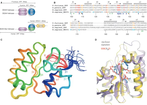
In contrast to eukaryotes, archaea have a single homolog of the XPF endonuclease that forms homo-dimers. The archaeal XPF minimally consists of the catalytic nuclease domain followed by a DNA-binding domain containing two consecutive helix-hairpin-helix
motifs (HhH2 domain). Dimerization occurs between
both the nuclease and HhH2 domains of each subunit
(Figure 1A) (3). The strong preference of the archaeal endonucleases for 30 flap DNA substrates is explained by
the function of their individual domains. Structural data
suggest a model for DNA binding where one HhH2
domain of the dimer binds to an upstream and the other to a downstream DNA duplex, sharply bending the DNA substrate and thereby allowing one active site of the dimeric nuclease domain to cleave the 30protruding single
strand (10). Combination of mutations in nuclease and HhH2domains that do not disrupt the dimeric interfaces
but impair the individual functions (DNA cleavage or DNA binding), provide evidence for this model (11).
*To whom correspondence should be addressed. Tel: +31 30 2534035; Fax: +31 30 2537623; Email: [email protected]
ß2007 The Author(s)
The DNA substrate specificity is different for the human ERCC1/XPF heterodimer. The human enzyme is known to specifically incise hairpin, bubble or splayed-arm DNA substrates (12,13), which consist of only one duplex (upstream). Previous data indicated the role of the
C-terminal ERCC1 ‘canonical’ HhH2domain in engaging
the upstream DNA duplex of a hairpin substrate (14).
This function requires heterodimerization with the
C-terminal HhH domain of the human XPF partner to
form a complex analogous to the archaeal HhH2 dimer
interface (12,14). Previous structural data (12) and the data presented here illustrate the structural similarity between the human ERCC1 central domain and the archaeal XPF nuclease domain. Unlike the archaeal nuclease dimer interface, there is no evidence that the corresponding domains of human ERCC1 and XPF interact with each other in solution (Figure 1A) (12).
The catalytic function of the ERCC1/XPF endonu-clease is crucial for NER. NER operates through a ‘cut and patch’ mechanism by excising and removing a short stretch of DNA containing the lesion, and subsequently restoring the genetic information by repair synthesis using the undamaged strand as the template. The incision step involves the sequential and coordinated assembly of the DNA damage sensor XPC-HR23B, the transcription/ repair factor TFIIH, the architectural protein XPA, the ubiquitous ssDNA-binding protein RPA, and the two structure-specific endonucleases ERCC1/XPF and XPG, responsible for the incisions 50and 30to the damaged site,
respectively (15). A main role in the progress of the reaction is attributed to XPA, which in complex with RPA probes the DNA helix conformations (16) and participates in multiple interactions with the other NER factors (15). XPA is required for the recruitment of ERCC1/XPF in the
DEAH helicase
HhH2 HhH2
HhH2 nuclease nuclease DEAH helicase
A.pernix XPF 248aa P.furiosus XPF 764aa
Human XPF 916aa
nuclease HhH
central DEAH-like helicase
Human ERCC1 293aa
A
C
B
D
R->F
E->L K->L D->Y
nuclease signature
ERKX3D N
C
P.furiosus_XPF VKVVVDSRELRSEVVKRLKLLGVKLEVKTLDVGDYIISEDVAIERKSANDFIQSIIDGRLFDQVKRLKEA
A.pernix_XPF PRVYVDVREERSPVPSILESLGVQVIPKQLPMGDYLVSDSIIVERKTSSDFAKSLFDGRLFEQASRLAEH
H.sapiens_XPF QSIVVDMREFRSELPSLIHRRGIDIEPVTLEVGDYILTPEMCVERKSISDLIGSLNNGRLYSQCISMSRY
H.sapiens_ERCC1 NSIIVSPRQRGNPVLKFVR--NVPWEFGDV-IPDYVLGQSTCALFLSLRYHNLHP--DYIHGRLQSLGKN
P.furiosus_XPF YSRPIMIVEGSL-YGIRNVHP---NAIRGAIAAVTVDFGVPIIFSSTPEETAQYIFLIAKREQ
A.pernix_XPF YETVFIIVEGPPVPRRYRGRE---RSLYAAMAALQLDYGIRLMNTMDPKGTALVIESLARLST
H.sapiens_XPF YKRPVLLIEFDPSKPFSLTSRGALFQEISSNDISSKLTLLTLHFPRLRILWCPSPHATAELFEELKQSKP
H.sapiens_ERCC1 FALRVLLVQVDVKDP---QQALKELAKMCILADCTLILAWSPEEAGRYLETYKAYEQ
100 110 120 140 150 160
170
130
[image:2.612.51.552.69.426.2]180 190 200 210
NER pre-incision complex (17,18). Consistent with this
regulatory function, in vitro studies with recombinant
proteins and in vivo studies using a yeast two-hybrid
system have demonstrated interactions between XPA and ERCC1 (19).
Here, we report the solution structure of the ERCC1 central domain (cERCC1) and investigate its interactions with XPA and DNA at the molecular level. Using biochemical studies and NMR titration experiments, we have identified two distinct interaction surfaces of cERCC1 that mediate XPA and DNA binding. Interestingly, the two interactions can take place simulta-neously to yield a cERCC1/DNA/XPA ternary complex, which in turn explains the important role of ERCC1 in targeting its catalytic XPF partner to the NER pre-incision complex.
MATERIALS AND METHODS
Cloning, protein expression and protein purification
ERCC1 central domain (cERCC1, residues 96 to 219) was PCR-amplified from a vector containing the full-length ERCC1 gene and subcloned into a pET28b (Novagen) expression vector between BamHI and XhoI sites. Downstream of the XhoI site a linker together with a his-tag has been engineered. XPA constructs were derived from a full-length XPA vector purchased from RZPD (clone ID IRAUp969B1273D6) and subcloned into a pLICHIS vector using the enzyme free cloning (EFC) strategy (20). The same procedure was followed for the GST fusion proteins, either XPA or cERCC1 (pLICHISGST vector). As a result all the EFC constructs contain an N-terminal his-tag, while the cERCC1 construct bears a C-terminal his-tag. The nucleotide sequences of the cloned DNAs were confirmed by sequencing. The cERCC1-HIS, HIS-XPA, HIS-GST-XPA and HIS-GST-cERCC1 proteins were expressed in BL21(DE3) Rosetta cells (Novagen) and were subject to two-step purification as has been described (21). When appropriate, HIS-GST-cERCC1 was cleaved with
thrombin in the presence of 1 mM Ca2+, 50 mM Tris
(pH 8.0) and 100 mM NaCl in order to obtain the untagged protein. Finally, the protein samples were exchanged either to 50 mM sodium phosphate (pH 5.5), 100 mM NaCl for the cERCC1 structure determination, or to 50 mM Tris (pH 8.0), 100 mM NaCl for the binding studies.
NMR spectroscopy
Multidimensional NMR experiments were carried out at 290 K on Bruker AVANCE 600 and 900 NMR
spectrometers equipped with TXI triple-resonance
probes in 50 mM sodium phosphate (pH 5.5), 100 mM NaCl by using the cERCC1-HIS protein. Spectra were processed using the NMRPipe software package (22) and analyzed with Sparky (23). The1H,15N and13C resonance assignments are 97% complete and were made using standard triple resonance techniques, 3D
NOESY-(15N, 1H)-HSQC spectra, and 3D NOESY-(13C, 1
H)-HSQC spectra (both with a mixing time of 60 ms) (24).
The chemical shifts have been deposited at the BMRB with accession number 15240.
Structure calculations
Automatic NOE assignment and structure calculations were performed using the program CYANA version 2.0 (25). Hydrogen bond restraints were defined when they were consistent with the secondary chemical shift data and expected NOE contacts and only for the helical parts of the protein. The set of 2669 NOE restraints determined by CYANA, together with restraints for 31 hydrogen bonds andfandc torsion angle restraints (174) derived from TALOS (26) were used in a water refinement run according to the standard RECOORD protocol (27) utilizing CNS (28). Molecular images were generated with PyMol (29). Coordinates have been deposited at the PDB with accession code 2jpd and the structural restraints at the BMRB with entry number 15240.
EMSAs
All the DNA substrates were 50 32
P-labeled with T4 polynucleotide kinase and purified on a polyacrylamide gel under native conditions. The nucleotide sequences for ss20, b10 and ds30 have been described before (30). Per reaction, 100 fmol of substrate was incubated with the appropriate amount of ERCC1 protein in binding buffer (50 mM Tris, pH 8.0, 100 mM NaCl, 10% glycerol and
1 mg/ml BSA). After incubation for 30 min at 48C,
samples were loaded onto 7.5% native polyacrylamide gels containing 0.5 TBE and run at 48C. Alternatively Electrophoretic mobility shift assay (EMSA) reactions were loaded on a 3.5% agarose gel containing 0.5% TBE, run at 4 or 208C yielding essentially identical results. Gels were visualized and quantified using a phosphor imager (BioRad) as described before (30). For depletion
experiments, 20ml of MagneHis beads (Promega) or GST
agarose beads (Sigma), were extensively washed with binding buffer, and after buffer removal the EMSA reaction was added to the beads. After 15 min incubation with regular mixing, the beads were removed from the EMSA reaction using a magnet or centrifugation and the reaction mixture was loaded on acrylamide gel or, when appropriate, the substrate was added to the depleted reaction mixture.
GST pull-down assay
GST pull-down assay was performed and quantified as described before (31) using the indicated cERCC1 concentrations and 3–5mg of GST proteins in 150ml of 50 mM Tris, 100 mM NaCl, 10% glycerol, 1 mM DTT, 0.2 mM PMSF and 20 mg/ml of BSA (pH 8.0). The semi-quantitative experiments were performed in 50, 150, 500,
1500 and 5000ml buffer with a constant amount of
cERCC1 and GST-XPA.
NMR titrations
cERCC1 titrations with XPA and DNA were performed on a Bruker AVANCE 700 NMR spectrometer and were
cases, the concentration of cERCC1 was 0.2 mM in 50 mM Tris (pH 8.0), 100 mM NaCl. XPA and DNA were dissolved in the same buffer and therefore salt and pH did not vary throughout the experiments. DNA binding was feasible only with the cERCC1 construct lacking the C-terminal his-tag, while XPA binding was identical, regardless of the his-tag presence. Normalized chemical shift changes were calculated by using the equation:= ([HN]2+ [N/6]2)0.5.
RESULTS AND DISCUSSION
Structure analysis
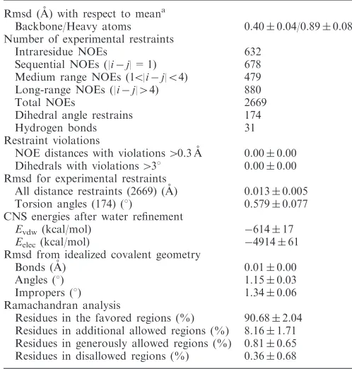
We have determined the solution structure of the ERCC1 central domain (cERCC1) to elucidate the molecular details of its proposed functions. We have collected a large number of distance and dihedral angle restraints that yielded an ensemble of 20 conformers with very good convergence (Figure 1C). The quality of the structure can be judged by the summary of the structural and restraint statistics given in Table 1. All the secondary structure elements are well defined including the short loops that join them. cERCC1 comprises a compact architecture and folds as a six-stranded b-sheet flanked by five a-helices on both sides. As described before (12), this fold is reminiscent of the type II restriction endonucleases, to which the catalytic domain of XPF also belongs.
Overall, the solution NMR structure of cERCC1 is in very good agreement with the crystal structure of the same
domain (2a1i) (rmsd 1.1 A˚ for 108 Ca atoms). The only
noticeable difference relates to the last few residues of
helix a3 and the loop connecting this structure element withb5. The presence of a mercury atom (linked to C137) in the crystal structure may account for the substantial side-chain rearrangements within this loop. When com-pared to the crystal structures of the archaeal nucleases, helicesa1,a2, and strandsb1,b2 appear to be shorter in the solution structure of cERCC1. Solution cERCC1 and the crystal structures of archaeal XPF nucleases (2bgw and 1j23) contain the same number of secondary structure elements arranged in a very similar manner (rmsd 1.8
and 2.0 A˚ for Aeropyrum pernixand Pyrococcus furiosus
nucleases, respectively, for 108 Ca atoms), although in both cases the primary sequence homology is very low (Figure 1B and D).
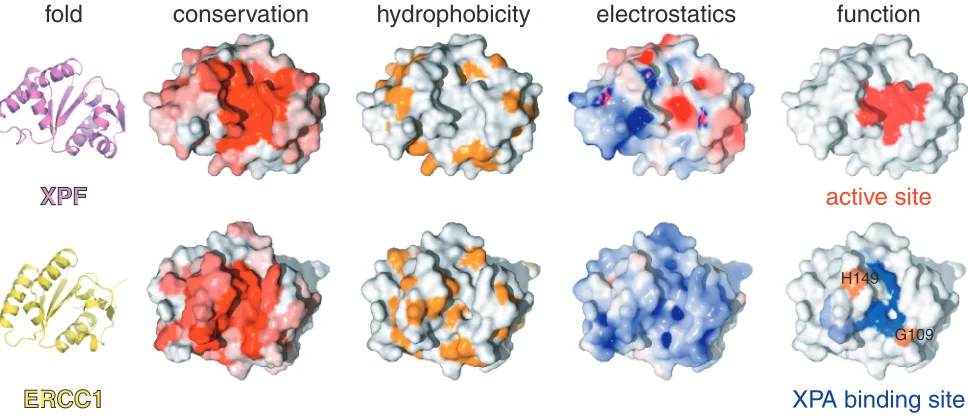
Remarkably, conserved residues of XPF nuclease scattered in the primary sequence superimpose structurally with residues conserved in the ERCC1 sequence family (Figures 1 and 3). Whereas the conservation in XPF is directly related to the catalytic function, ERCC1 has preserved the same fold but lacks the essential residues for catalysis. The fold similarities, coupled with the obligate nature of the heterodimerization (32), are in full agreement with the common origin of the two proteins (33).
Minimal domains for ERCC1–XPA interaction
Truncation studies of ERCC1 have mapped the XPA interaction site to a region between residues 91 and 118 (34). From the cERCC1 structure and its compact fold, we predict that such truncations will have a devastating effect on the structural integrity of the central domain (96–219). For XPA, the ERCC1 interaction region seems to be located in a small stretch containing two highly conserved motifs rich in glycine and glutamic acid residues
(72-GGGFILEEEEEEE-84, conserved residues are
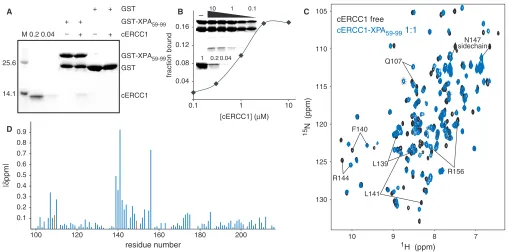
underlined) (19). We have prepared a HIS-GST-XPA construct (59–99) containing the conserved stretch, and examined its ability to interact with cERCC1 in a GST pull-down assay. Indeed, the GST XPA fusion protein was able to specifically bind cERCC1 (Figure 2A), confirming that this domain of ERCC1 is sufficient for interactions with XPA. No binding of cERCC1 to GST bound agarose
beads or uncharged agarose beads was observed
(Figure 2A, data not shown). This XPA peptide is unstructured (35) and appeared very sensitive to proteo-lysis both during overexpression inEscherichia coliand as pure protein after extensive purification (Figure 2A and B). We next performed a semi-quantitative GST
pull-down assay (31) and estimated an apparent Kd of 1mM.
Since we reached only 20% binding saturation at the two
highest protein concentrations (3 and 10mM of cERCC1)
under these experimental conditions, we could not
determine the binding constant more accurately
(Figure 2B).
Functional disparity of the XPF nuclease and ERCC1 central domains
[image:4.612.40.289.96.357.2]The cERCC1–XPA interaction specificity has been explored using NMR titration experiments. Titration with the HIS-XPA peptide causes extensive specific
Table 1. Structural statistics of the structure ensemble of human cERCC1 (residues 96–219)
Rmsd (A˚) with respect to meana
Backbone/Heavy atoms 0.400.04/0.890.08 Number of experimental restraints
Intraresidue NOEs 632
Sequential NOEs (|ij| = 1) 678 Medium range NOEs (15|ij|54) 479 Long-range NOEs (|ij|44) 880
Total NOEs 2669
Dihedral angle restrains 174
Hydrogen bonds 31
Restraint violations
NOE distances with violations40.3 A˚ 0.000.00 Dihedrals with violations438 0.000.00 Rmsd for experimental restraints
All distance restraints (2669) (A˚) 0.0130.005 Torsion angles (174) (8) 0.5790.077 CNS energies after water refinement
Evdw(kcal/mol) 61417
Eelec(kcal/mol) 491461
Rmsd from idealized covalent geometry
Bonds (A˚) 0.010.00
Angles (8) 1.150.03
Impropers (8) 1.340.06
Ramachandran analysis
Residues in the favored regions (%) 90.682.04 Residues in additional allowed regions (%) 8.161.71 Residues in generously allowed regions (%) 0.810.65 Residues in disallowed regions (%) 0.360.68
a
changes in the cERCC1 1H-15N HSQC NMR spectra. The titrations show slow exchange regime with respect to the NMR chemical shift timescale, in agreement with the determined affinity (Figure 2C). We have been able to assign all the amide resonances of cERCC1 in the bound form (1:1 complex stoichiometry) by using 3D
NOESY-(15N, 1H)-HSQC spectra. The pattern of the backbone
amide NOEs does not change substantially compared to the free cERCC1 protein. This indicates that the overall structure is maintained, and led us to conclude that the observed chemical shift perturbations arise from specific contacts with the XPA peptide.
The most significant perturbations map to the surface of the V-shaped groove of the cERCC1 structure and involve many residues that compose the corresponding DNA cleavage site of the archaeal XPF endonucleases (Figures 2D and 3). Interestingly, among the largest chemical shifts of cERCC1 upon XPA binding are those of L139, F140 and L141. These ERCC1 residues correspond to the characteristic strongly conserved ERK catalytic triad of the XPF nuclease domain (Figure 1). The rest of the significantly perturbed residues are polar or positively charged (Q107, N110, S142, R144, N147 and R156), are competent for hydrogen bonding with the XPA peptide and are more conserved than the surround-ing regions of the cERCC1 V-shaped groove (Figure 3). Additionally, of special interest are the amide signals of G109 and H149, which are not observed in the free protein spectrum due to fast exchange with the solvent at pH 8
(these amide resonances are observable at pH 5.5). However, both give sharp signals at the end of the titration, an indication that the solvent exchange in the free protein is quenched in the complex. These residues reside at the rim of the V-shaped binding site and may be involved in hydrogen bonding with the XPA peptide (Figure 3).
Although ERCC1 residues forming the XPA-binding site are different from the acidic and basic residues which form the active site in XPF (5), there are no significant changes in the backbone fold and the spatial side-chain orientations in the structure remain unaffected. However, their different chemical properties result in a distinct charge and hydrophobicity distribution on the structure surface (Figure 3). While XPF displays negative charge at the DNA cleavage site (important for divalent cation coordination), the equivalent XPA-binding site in ERCC1 is neutral or slightly positively charged. Therefore, the ERCC1–XPA interaction is a combination of hydrophobic and electrostatic interactions possibly involving the glutamic acid stretch of XPA (19) and two conserved positively charged residues (R106 and R156) of cERCC1.
DNA binding by the ERCC1 central domain
Given the proposed common origin of ERCC1 and XPF (33), it can be expected that the degenerated catalytic site of ERCC1 could still bind DNA in a similar fashion as the archaeal XPF active site (12). Because our ERCC1–XPA
cERCC1 GST GST-XPA59-99
GST-XPA59-99 + +
M 0.2 0.04 + + cERCC1 GST + +
25.6
14.1
A B – 10 1 0.1 C
0.04 0.2 1
1 10
0.1 0.04 0.08 0.12 0.16
fraction bound
[cERCC1] (µM)
0.1 0.2 0.3 0.4 0.5 0.6 0.7 0.8 0.9
100 120 140 160 180 200
|
δ
ppm|
residue number
D
cERCC1 free
cERCC1-XPA59-99 1:1
1H (ppm)
15
N (ppm)
10 9 8 7
130 125 120 115 110 105
F140
Q107
L139
L141
R144 R156
[image:5.612.59.568.68.320.2]N147 sidechain
interaction data render this unlikely, we explored whether cERCC1 is able to bind DNA as has been suggested previously (12).
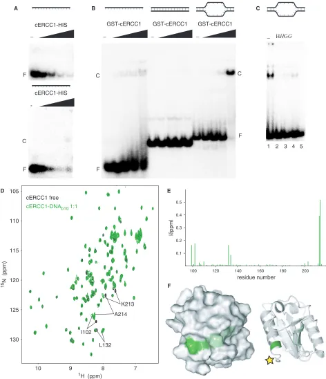
We performed an electrophoretic mobility shift assay (EMSA) where increasing amounts of cERCC1-HIS protein were added to ssDNA. While we saw a loss of free probe, we failed to see any complex formation under these conditions. If the same protein–DNA complex was loaded on a 3.5% agarose gel, we detected a weak smeary complex at the highest protein concentration (Figure 4A). However, by using a HIS-GST-cERCC1 protein, we
observed formation of the complex with ssDNA
(Figure 4B). This suggests that the C-terminal his-tag interferes with DNA binding. Therefore, we used the HIS-GST-cERCC1 protein, which we cleaved with thrombin to obtain the untagged protein. DNA binding with the untagged cERCC1 demonstrated a faster migrating complex with various ssDNA substrates (data not shown). HIS-GST-cERCC1/DNA complex formation is prevented by incubation of the reaction mixture with MagneHIS magnetic beads or glutathione agarose beads, which depletes HIS-GST-cERCC1 from the reaction mixture either prior to or after the addition of DNA. If beads were added after complex formation, a small but significant decrease in the amount of free probe was observed. Depletion of the HIS-GST tagged cERCC1
protein from the binding reaction confirms that
the protein–DNA complex is indeed formed by the HIS-GST-cERCC1 protein (Figure 4C).
In agreement with earlier studies, under these condi-tions no binding was observed for dsDNA (12), while fairly comparable binding affinities were found for both ssDNA and bubble substrates with 10 or 20 unpaired bases. We calculated an apparentKd of 2.50.7mM for
the HIS-GST-cERCC1 protein bound to a bubble10 (b10)
substrate in the presence of 100 mM NaCl (Figure 4B). Corroborating our results, an equilibrium binding titra-tion experiment using fluorescence anisotropy has demon-strated ssDNA binding for the same domain with an apparentKdof 10mM (12). The relatively small difference
in affinity can be well explained by different salt conditions or substrate used.
For the NMR titrations we performed a thrombin cleavage on HIS-GST-cERCC1, to remove the his-tag.
The untagged cERCC1 1H-15N HSQC spectrum is
identical to that of cERCC1-HIS, except for the two amides preceding the artificial his-tag (Supplementary Figure 1). Addition of the b10 substrate to this cERCC1 protein caused a limited number of specific perturbations that were unambiguously assigned. Again, the perturbed resonances exhibit slow exchange behavior in the NMR titrations and indicate contacts with the DNA. The affected resonances include the backbone amides of N99, I102, L132, K213, A214 and the side-chain amide of Q134 (Figure 4D and E). Compared to the free-protein spectrum we only miss T211, which cannot be identified. The perturbation induced by the b10 DNA was complete at equimolar concentration with cERCC1. Chemical shift mapping reveals that all perturbations are in the vicinity of the C-terminus of the cERCC1 construct, consistent with the presence of a flexible tag interfering with DNA binding. The DNA-binding region resides at the surface of the structure where the N- and C-termini meet (Figure 4F). The perturbed residues belong to well-defined structure elements. The only positively charged residue we identified (K213) is fully conserved in the ERCC1 proteins and may explain the dependence of DNA binding on salt concentration. All the other affected residues are less conserved than the residues involved in XPA binding. The composition of the affected residues together with the
XPF
XPF
active site
XPA binding site
H149
G109
ERCC1
ERCC1
hydrophobicity
conservation
electrostatics
function
[image:6.612.55.539.72.280.2]fold
1H (ppm)
15
N (ppm)
10 9 8 7
130 125 120 115 110 105
cERCC1 free
cERCC1-DNAb10 1:1
0.1 0.2 0.3 0.4 0.5
100 120 140 160 180 200
|
δ
ppm|
residue number C
F F
-−
cERCC1-HIS
cERCC1-HIS
A
D E
B C
F
GST-cERCC1
C C
F
− − −
GST-cERCC1 GST-cERCC1
1 2 3 4 5
HHGG
−
K213
A214
I102
L132
[image:7.612.77.549.69.617.2]F
slow exchange behavior suggests a large contribution to the binding by hydrophobic interactions with the DNA bases.
Summarizing, both the biochemical and the NMR experiments show that the ERCC1 central domain binds to DNA. The DNA-binding site we identified on cERCC1 by the NMR titrations suggests that cERCC1 contacts only a small part of the ssDNA, probably three to four bases. The situation is different in the full-length heterodimer, where other DNA-binding surfaces, such as previously established for the C-terminal ERCC1/XPF domains (12,14), assist in DNA recognition.
cERCC1 can bind simultaneously to XPA and ssDNA
Chemical shift perturbation experiments revealed distinct binding surfaces for ssDNA and XPA, and therefore, it is probable that both can bind simultaneously to cERCC1. We performed EMSA experiments in the presence of an equimolar or 5-fold excess of the HIS-XPA peptide. Addition of XPA did not influence binding of cERCC1 to ssDNA, while the binding affinity of XPA for the cERCC1 domain under these conditions would suggest that the vast majority of cERCC1 is in complex with XPA. We did not observe formation of a slower migrating HIS-GST-cERCC1/ssDNA/XPA super complex, possibly because the increase in mass by the addition of the HIS-XPA peptide was too small to discern (data not shown).
To independently confirm the formation of a ternary cERCC1/ssDNA/XPA complex, we added the HIS-XPA peptide to the cERCC1-bubble10 protein–DNA complex and followed the chemical shift perturbations in the
1
H-15N HSQC spectra. As shown in Figure 5, addition of an equimolar amount of the XPA peptide causes the same
amide displacements as in the titration with the free protein. On the contrary, most amides influenced by the DNA binding remain unaffected by the presence of XPA (Figure 5A). I102 and N110 were perturbed by both XPA and b10 DNA when the titrations were performed independently. In the ternary complex, however, I102 remains close to the position as in the DNA bound form. Conversely, the side-chain amide of N110, although affected by the DNA, adopts the distinct XPA bound position upon XPA addition. These data strongly indicate the formation of the ternary ERCC1/DNA/XPA complex. DNA and XPA-binding sites are distinct, and the two interactions can happen simultaneously (Figure 5B).
Functional role of the ERCC1 central domain in the heterodimer
Since the catalytic activity has been preserved in the human XPF protein through strong conservation of the nuclease signature (5), the human nuclease fold should be identical to that of the archaeal counterparts. In that sense, human XPF nuclease and human ERCC1 central domains are expected to exhibit the same fold. Moreover,
the ERCC1 and XPF HhH2 domains feature a common
architecture and come into tight association exactly in the same way that the HhH2domains of both archaeal species
do (10,11,14). Therefore, in structural terms the human ERCC1 and XPF proteins share the essential architectural subunits observed in the short XPF homodimer of
A. pernix. The structural similarities within the XPF family provide additional evidence for the proposed common origin of the human ERCC1 and XPF proteins (33). Because ERCC1 is absent in archaea, this gene is thought to have been acquired from an ancient XPF gene
180°
7.8 7.6 7.4 110
108 106
Y130N-H S201N-H
N110ND2-QD2 N147ND2-HD21 Q172NE2-HE21 G161N-H
G155N-H
7.8 7.6 7.4 Y130N-H
S201N-H N147ND2-HD21
N110ND2-QD2 Q172NE2-HE21 G161N-H
G155N-H
130 128 126
L141N-H D174N-H
L132N-H I102N-H A165N-H
Q172N-H
L141N-H
L132N-H D174N-H
I102N-H A165N-H
Q172N-H A214N-H 7.8 7.6 7.4
Y130N-H N147ND2-HD21 S201N-H
Q172NE2-HE21 G161N-H
G155N-H
7.8 7.6 7.4 G161N-H
G155N-H
Q172NE2-HE21
Y130N-H N147ND2-HD21 S201N-H
L143N-H
L132N-H D174N-H
I102N-H Q172N-H A165N-H
132
8.4 8.2 8.0 8.4 8.2 8.0 8.4 8.2 8.0 8.4 8.2 8.0 A165N-H
I102N-H
A214N-H
Q172N-H
D174N-H
L132N-H L143N-H
cERCC1 free cERCC1-XPA59-99 cERCC1-DNAb10 cERCC1-DNAb10-XPA59-99
1H (ppm)
15
N (ppm)
[image:8.612.51.548.70.312.2]A B
Figure 5. Dual function of cERCC1. (A) Indicative portions of the1H-15N HSQC spectrum for the free cERCC1, bound to either XPA (1:1) or
duplication in the eukaryal lineage. The ERCC1 and XPF genes have subsequently evolved by the process called subfunctionalization (36,37). This model suggests that after gene duplication, both copies may be reciprocally preserved through the fixation of complementary loss-of-subfunction mutations, which results in a partitioning of the tasks of the ancestral gene. From the functions present in the ancestral XPF protein (archaea), human ERCC1
retained the canonical HhH2domain that acts as a
DNA-binding domain (14), while XPF retained the catalytic activity (5). Once the separation of the ancestral subfunc-tions occurred in ERCC1 and XPF genes, only the heterodimeric protein complex could restore the original function. Additionally, due to genetic alterations, the second helix-hairpin-helix motif of human XPF degener-ated, yet the fold remained crucial for stabilizing the corresponding intact domain of human ERCC1 (14). Similarly, the central domain of ERCC1 lost the catalytic activity by sequence drift, but despite adoption of a novel dual function (XPA interaction and DNA binding), the 3D fold was preserved.
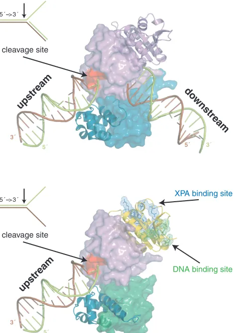
We suggest a mechanistic model for the heterodimeric function based on the model for the homodimeric archaeal homolog (Figure 6) (10,38). Human ERCC1 corresponds
to the archaeal protomer that binds with the HhH2
domain to the upstream duplex. Human XPF corresponds to the archaeal protomer that recognizes the downstream duplex. Since it cannot make contacts with the minor groove (14), the specificity of the human heterodimer has shifted to splayed-arms substrates consisting of only one duplex. This does not exclude contribution of the XPF HhH domain to ssDNA interactions, as reported before (12). We show here that the central domain of ERCC1 is also involved in ssDNA binding. Additional DNA interactions are likely for the nuclease of XPF, analogous to the archaeal case (38). Furthermore, ssDNA binding by cERCC1 will be stimulated by specific interactions with XPA, already present in the NER pre-incision complex and possibly bound to the DNA via its own DNA-binding site. Most importantly, through these coordinated inter-actions of the ERCC1 domains the XPF protein will be
positioned to cleave the 30 protruding strand (limon),
thereby retaining the polarity present in the archaeal homodimer.
Our model underlines the significant role of ERCC1 in the context of the full-length heterodimer. XPF is the catalytic module but the ERCC1 domains guarantee that the enzymatic activity is targeted properly. The presence of multiple distinct DNA-binding surfaces within the
ERCC1/XPF/XPA/RPA repair protein intermediate
coordinates cleavage to occur only when the DNA damage is recognized correctly by the NER machinery. The duplication of the ancestral XPF gene within the eukaryal kingdom resulted in an obligate heterodimer through loss of function (with an altered HhH2domain of
XPF and a degenerated catalytic domain of ERCC1) and adoption of novel functions (ssDNA and XPA binding of cERCC1). This permitted additional quality control mechanisms through a more complicated molecular interaction network, mediated by the novel functional
domains, and thereby improving the fidelity of DNA damage repair.
SUPPLEMENTARY DATA
Supplementary Data are available at NAR Online.
ACKNOWLEDGEMENTS
We would like to thank Koos Jaspers and Jan
Hoeijmakers for providing the full-length vectors of ERCC1 and XPF, and for many fruitful discussions. This work was financially supported by the Netherlands Organization for Scientific Research, Chemistry Council (NWO-CW), by the Center of Biomedical Genetics, the Netherlands, and by the EU project Spine2-Complexes (Contract no 031220). Funding to pay the Open Access publication charges for this article was provided by NWO-CW.
Conflict of interest statement. None declared.
5 ´ --> 3 ´
3´
5´ 5´ 3´
upstream
do wnstream
cleavage site
5 ´ --> 3 ´
3´ 5´ upstream cleavage site
XPA binding site
[image:9.612.331.564.71.402.2]DNA binding site
REFERENCES
1. Nishino,T. and Morikawa,K. (2002) Structure and function of nucleases in DNA repair: shape, grip and blade of the DNA scissors.Oncogene,21, 9022–9032.
2. Ciccia,A., Ling,C., Coulthard,R., Yan,Z., Xue,Y., Meetei,A.R., Laghmani el,H., Joenje,H., McDonald,N.et al. (2007) Identification of FAAP24, a Fanconi anemia core complex protein that interacts with FANCM.Mol. Cell,25, 331–343.
3. Nishino,T., Komori,K., Ishino,Y. and Morikawa,K. (2003) X-ray and biochemical anatomy of an archaeal XPF/Rad1/Mus81 family nuclease: similarity between its endonuclease domain and restriction enzymes.Structure (Camb.),11, 445–457.
4. Heyer,W.D., Ehmsen,K.T. and Solinger,J.A. (2003) Holliday junctions in the eukaryotic nucleus: resolution in sight?
Trends Biochem. Sci.,28, 548–557.
5. Enzlin,J.H. and Scharer,O.D. (2002) The active site of the DNA repair endonuclease XPF-ERCC1 forms a highly conserved nuclease motif.EMBO J.,21, 2045–2053.
6. Niedernhofer,L.J., Odijk,H., Budzowska,M., van Drunen,E., Maas,A., Theil,A.F., de Wit,J., Jaspers,N.G.J., Beverloo,H.B.et al. (2004) The structure-specific endonuclease Ercc1-Xpf is required to resolve DNA interstrand cross-link-induced double-strand breaks.
Mol. Cell. Biol.,24, 5776–5787.
7. Zhu,X.D., Niedernhofer,L., Kuster,B., Mann,M.,
Hoeijmakers,J.H.J. and de Lange,T. (2003) ERCC1/XPF removes the 30overhang from uncapped telomeres and represses formation
of telomeric DNA-containing double minute chromosomes.
Mol. Cell,12, 1489–1498.
8. Jaspers,N.G.J., Raams,A., Silengo,M.C., Wijgers,N.,
Niedernhofer,L.J., Robinson,A.R., Giglia-Mari,G., Hoogstraten,D. and Kleijer,W.J. (2007) First reported patient with human ERCC1 deficiency has cerebro-oculo-facio-skeletal syndrome with a mild defect in nucleotide excision repair and severe developmental failure.Am. J. Hum. Genet.,80, 457–466.
9. Niedernhofer,L.J., Garinis,G.A., Raams,A., Lalai,A.S., Robinson,A.R., Appeldoorn,E., Odijk,H., Oostendorp,R., Ahmad,A.et al. (2006) A new progeroid syndrome reveals that genotoxic stress suppresses the somatotroph axis.Nature,444, 1038–1043.
10. Newman,M., Murray-Rust,J., Lally,J., Rudolf,J., Fadden,A., Knowles,P.P., White,M.F. and McDonald,N.Q. (2005) Structure of an XPF endonuclease with and without DNA suggests a model for substrate recognition.EMBO J.,24, 895–905.
11. Nishino,T., Komori,K., Ishino,Y. and Morikawa,K. (2005) Structural and functional analyses of an archaeal XPF/Rad1/Mus81 nuclease: asymmetric DNA binding and cleavage mechanisms.
Structure,13, 1183–1192.
12. Tsodikov,O.V., Enzlin,J.H., Scharer,O.D. and Ellenberger,T. (2005) Crystal structure and DNA binding functions of ERCC1, a subunit of the DNA structure-specific endonuclease XPF-ERCC1.
Proc. Natl Acad. Sci. USA,102, 11236–11241. 13. de Laat,W.L., Appeldoorn,E., Jaspers,N.G.J. and
Hoeijmakers,J.H.J. (1998) DNA structural elements required for ERCC1-XPF endonuclease activity.J. Biol. Chem.,273, 7835–7842. 14. Tripsianes,K., Folkers,G.E., AB,E., Das,D., Odijk,H.,
Jaspers,N.G.J., Hoeijmakers,J.H.J., Kaptein,R. and Boelens,R. (2005) The structure of the human ERCC1/XPF interaction domains reveals a complementary role for the two proteins in nucleotide excision repair.Structure,13, 1849–1858.
15. Gillet,L.C. and Scharer,O.D. (2006) Molecular mechanisms of mammalian global genome nucleotide excision repair.Chem. Rev.,
106, 253–276.
16. Missura,M., Buterin,T., Hindges,R., Hubscher,U., Kasparkova,J., Brabec,V. and Naegeli,H. (2001) Double-check probing of DNA bending and unwinding by XPA-RPA: an architectural function in DNA repair.EMBO J.,20, 3554–3564.
17. Riedl,T., Hanaoka,F. and Egly,J.M. (2003) The comings and goings of nucleotide excision repair factors on damaged DNA.EMBO J.,
22, 5293–5303.
18. Volker,M., Mone,M.J., Karmakar,P., van Hoffen,A., Schul,W., Vermeulen,W., Hoeijmakers,J.H.J., van Driel,R., van Zeeland,A.A.
et al. (2001) Sequential assembly of the nucleotide excision repair factors in vivo.Mol. Cell,8, 213–224.
19. Li,L., Peterson,C.A., Lu,X. and Legerski,R.J. (1995) Mutations in XPA that prevent association with ERCC1 are defective in nucleotide excision repair. Mol. Cell. Biol.,15, 1993–1998. 20. de Jong,R.N., Daniels,M.A., Kaptein,R. and Folkers,G.E. (2007)
Enzyme free cloning for high throughput gene cloning and expression. J. Struct. Funct. Genomics[Epub ahead of print]. 21. Folkers,G.E., van Buuren,B.N. and Kaptein,R. (2004) Expression
screening, protein purification and NMR analysis of human protein domains for structural genomics.J. Struct. Funct. Genomics,5, 119–131.
22. Delaglio,F., Grzesiek,S., Vuister,G.W., Zhu,G., Pfeifer,J. and Bax,A. (1995) NMRPipe: a multidimensional spectral processing system based on UNIX pipes.J. Biomol. NMR,6, 277–293.
23. Goddard,T.D. and Kneller,D.G. (2001)SPARKY 3University of California, San Francisco.
24. Cavanagh,J., Fairbrother,J.W., Palmer,G.A. III and Skelton,J.N. (1996)Protein NMR Spectroscopy.Academic Press, San Diego, CA, USA.
25. Herrmann,T., Gu¨ntert,P. and Wu¨thrich,K. (2002) Protein NMR structure determination with automated NOE assignment using the new software CANDID and the torsion angle dynamics algorithm DYANA. J. Mol. Biol.,319, 209–227.
26. Cornilescu,G., Delaglio,F. and Bax,A. (1999) Protein backbone angle restraints from searching a database for chemical shift and sequence homology.J. Biomol. NMR,13, 289–302.
27. Nederveen,A.J., Doreleijers,J.F., Vranken,W., Miller,Z., Spronk,C.A.E.M., Nabuurs,S.B., Gu¨ntert,P., Livny,M.,
Markley,J.L. et al. (2005) RECOORD: a recalculated coordinate database of 500+ proteins from the PDB using restraints from the BioMagResBank.Proteins,59, 662–762.
28. Bru¨nger,A.T., Adams,P.D., Clore,G.M., DeLano,W.L., Gros,P., Grosse-Kunstleve,R.W., Jiang,J.S., Kuszewski,J., Nilges,M.et al. (1998) Crystallography & NMR system: a new software suite for macromolecular structure determination.Acta. Crystallogr. D Biol. Crystallogr.,54(Pt 5), 905–921.
29. DeLano,W. (2002)The PyMOL Molecular Graphics System.
DeLano Scientific, San Carlos, CA, USA.
30. Singh,S., Folkers,G.E., Bonvin,A.M.J.J., Boelens,R.,
Wechselberger,R., Niztayev,A. and Kaptein,R. (2002) Solution structure and DNA-binding properties of the C-terminal domain of UvrC from E.coli. EMBO J., 21, 6257–6266.
31. Jonker,H.R., Wechselberger,R.W., Boelens,R., Folkers,G.E. and Kaptein,R. (2005) Structural properties of the promiscuous VP16 activation domain.Biochemistry,44, 827–839.
32. Sijbers,A.M., de Laat,W.L., Ariza,R.R., Biggerstaff,M., Wei,Y.F., Moggs,J.G., Carter,K.C., Shell,B.K., Evans,E.et al. (1996) Xeroderma pigmentosum group F caused by a defect in a structure-specific DNA repair endonuclease.Cell,86, 811–822. 33. Gaillard,P.H. and Wood,R.D. (2001) Activity of individual ERCC1
and XPF subunits in DNA nucleotide excision repair.Nucleic Acids Res.,29, 872–879.
34. Li,L., Elledge,S.J., Peterson,C.A., Bales,E.S. and Legerski,R.J. (1994) Specific association between the human DNA repair proteins XPA and ERCC1.Proc. Natl Acad. Sci. USA,91, 5012–5016.
35. Buchko,G.W., Isern,N.G., Spicer,L.D. and Kennedy,M.A. (2001) Human nucleotide excision repair protein XPA: NMR
spectroscopic studies of an XPA fragment containing the ERCC1-binding region and the minimal DNA-binding domain (M59-F219).Mutat. Res., 486, 1–10.
36. Force,A., Lynch,M., Pickett,F.B., Amores,A., Yan,Y.L. and Postlethwait,J. (1999) Preservation of duplicate genes by complementary, degenerative mutations.Genetics,151, 1531–1545.
37. Lynch,M. and Conery,J.S. (2003) The evolutionary demography of duplicate genes. J. Struct. Funct. Genomics,3, 35–44.
38. Roberts,J.A. and White,M.F. (2005) An archaeal endonuclease displays key properties of both eukaryal XPF-ERCC1 and Mus81.
J. Biol. Chem.,280, 5924–5928.
39. Baker,N.A., Sept,D., Joseph,S., Holst,M.J. and McCammon,J.A. (2001) Electrostatics of nanosystems: application to