ii
Acute Respiratory Infection in
Pre-School Children
Thesis submitted in accordance with the requirements of the University of
Liverpool for the degree of Master in Philosophy
by
Katie Rose
i
Acknowledgments
ii
Abbreviations
% Percentage
oC Degrees Celcius
µl Microlitre
A&E Accident & Emergency
AdV Adenovirus
APC Antigen presenting cell
ARI Acute respiratory infection
ASF Airway Surface Fluid
ATL Animal tissue lysis
BAL Bronchoalveolar lavage
B-cells B-lymphocytes
BCG Bacillus Calmette-Guerin
BSA Bovine serum albumin
Caspase cysteine-aspartic protease
CD Cluster of differentiation
cDNA Complimentary deoxyribonucleic acid
CoV Coronavirus
CpG Cytosine-phosphate-guanine
Cpp Chlamydia pneumoniae
DALY Disability adjusted life year
DAMP Damage-associated molecular pattern
DNA Deoxyribonucleic acid
ELISA Enzyme-linked Immunosorbent Assay
EVW Episodic viral wheeze
Flu Influenza virus
Flu A Influenza virus type A
Flu B Influenza virus type B
G-CSF Granulocyte colony-stimulating factor
GM-CSF Granulocyte macrophage colony-stimulating factor
hBoV human Bocavirus
Hib Haemophilus influenzae type B
HIV Human immunodeficiency virus
HLA Human leukocyte antigen
hMPV human Metapneumovirus
hRSV human respiratory syncytial virus
hRV human rhinovirus
IFN Interferon
Ig Immunoglobulin
IL- Interleukin
IMIP Instituto de Medicina Integral Professor Fernando Figueira
iii
LPS Lipopolysaccharide
LRTI Lower respiratory tract infection
MAC Membrane attack complex
MASP Mannan binding lectin-associated serine protease
MBL Mannan binding lectin
MHC Major histocompatibility complex
MIP Macrophage inflammatory protein
Mpp Mycoplasma pneumoniae
NaCl Sodium Chloride
NF-κB Nuclear factor-Kappa B
NK Natural Killer
NPA Nasopharyngeal aspirate
PAMP Pathogen associated molecular pattern
PBS Phosphate buffered saline
PCR Polymerase chain reaction
PCV7 Heptavalent pneumococcal conjugate vaccine
pg Picogram
PICU Paediatric intensive care unit
PIV Parainfluenza virus
PRR Pattern recognition receptor
R$ Brazilian Reais
RNA Ribonucleic acid
RPM Revolutions per minute
RT-PCR Reverse transcription-polymerase chain reaction SARS Severe acute respiratory syndrome
SD Standard deviation
S. pneumoniae Streptococcus pneumoniae
TB Tuberculosis
Th T-helper
TLR Toll-like receptors
TNF Tumour necrosis factor
T-cells Thymus-derived lymphocytes
UK United Kingdom
URTI Upper respiratory tract infection
USA United States of America
US$ United States dollar
iv
Abstract
Acute Respiratory Infection in Pre-School Children
Katie Rose
Introduction: Acute respiratory infection (ARI) is a major cause of morbidity and mortality worldwide, being responsible for 3.5 million deaths annually. Despite this the precise aetiology of ARI is often unclear, and the pathogenesis remains poorly understood. The aims of this study were i) to investigate the viral and atypical bacterial causes of ARI in a clinically and demographically well defined population of children less than five years of age and ii) to investigate host immunological response to ARI caused by different pathogens and in different severities of disease.
Methods: Clinical and demographic data, along with nasopharyngeal aspirate samples were collected from 407 pre-school children who presented with ARI to the IMIP Children’s Hospital, Recife, Brazil over a 12 month period. Multiplex PCR was used to detect 17 different respiratory pathogens.
The concentrations of nine cytokines were determined in a selection of samples from children with ARI caused by different pathogens and in differing severities of human respiratory syncytial virus (hRSV) disease.
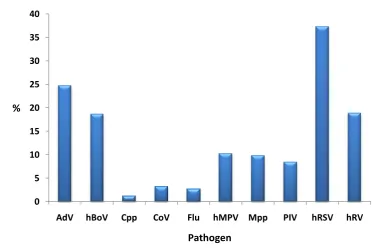
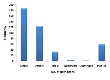
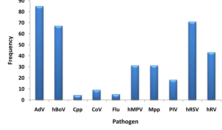
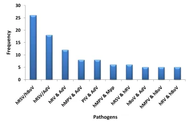
Results: At least one pathogen was detected in 85.5% of samples. The pathogens detected most frequently were hRSV (in 37.3% of samples), adenovirus (AdV; 24.8%), human rhinovirus (hRV; 18.9%), human bocavirus (hBoV; 18.7%), human metapneumovirus (hMPV; 10.3%) and Mycoplasma pneumoniae (Mpp; 9.8%). Co-infection with multiple pathogens was detected in 39.6% of samples, with the pathogens most frequently detected in co-infection being AdV (in 52.8% of co-infections), hRSV (44.1%), hBoV (41.6%) and hRV (26.7%). Infection with Mpp or hRSV was associated with hospital admission and more severe disease.
Cytokine profiles were similar in ARI caused by different pathogens and in different severities of disease.
Conclusions: We have described high rates of pathogen detection and co-infection, with particularly high rates of Mpp, AdV and hBoV infection. Our results may reflect the type of samples collected, the number of pathogens tested for, and/or the population demographics. The high prevalence of Mpp is more commonly associated with ARI in older children, and our findings may have implications for antibiotic use in this age group.
v
Table of Contents
Acknowledgments ... i
Abbreviations ... ii
Abstract ... iv
Table of Contents...v
List of Tables ... x
List of Figures ... xi
1
Introduction ... 1
1.1 Acute Respiratory Infection ... 1
1.1.1 Definition ... 1
1.1.2 Burden ... 1
1.1.3 Epidemiology ... 2
1.1.4 Risk Factors ... 3
1.1.5 Aetiology ... 5
1.1.5.1 ‘Typical’ Bacterial Pathogens ... 5
1.1.5.2 ‘Atypical’ Bacterial Pathogens ... 6
1.1.5.3 Viral infection ... 7
1.1.5.4 Tuberculosis ... 11
1.1.5.5 Previous studies into aetiology ... 12
1.1.6 Clinical Manifestations of ARI ... 14
1.1.6.1 Upper Respiratory Tract Infection ... 14
1.1.6.2 Bronchiolitis ... 15
1.1.6.3 Pneumonia ... 15
1.1.6.4 Episodic Viral Wheeze/Asthma ... 15
vi
1.1.8 Management of the Clinical Manifestations of ARI... 19
1.1.8.1 Upper Respiratory Tract Infection ... 19
1.1.8.2 Bronchiolitis ... 21
1.1.8.3 Pneumonia ... 22
1.1.8.4 Episodic Viral Wheeze/asthma ... 22
1.1.9 Prevention ... 23
1.1.9.1 Prophylaxis ... 23
1.1.10 Sequelae of ARI ... 25
1.1.10.1 Recurrent Wheeze ... 26
1.1.10.2 Invasive pneumococcal disease ... 27
1.1.10.3 Bronchiolitis obliterans ... 27
1.1.10.4 Bronchiectasis ... 28
1.2 Immunological responses to ARI ... 29
1.2.1 Innate Response ... 29
1.2.1.1 Physical Barriers to Infection... 30
1.2.1.2 Pathogen recognition/detection ... 31
1.2.1.3 Soluble mediators of the innate immune system ... 34
1.2.1.4 Effectors of innate Immunity ... 36
1.2.1.5 Antigen-Presenting Cells ... 41
1.2.2 Adaptive Response ... 42
1.2.2.1 Humoral Immunity ... 42
1.2.2.2 Cell-mediated Immunity ... 44
1.3 Aims of this study ... 49
2
Methodology ... 50
2.1 Study Design ... 50
2.2 Recruitment ... 50
vii
2.4 Sample collection & processing ... 54
2.4.1 Nasopharyngeal Aspirate collection ... 54
2.4.2 RNA Extraction ... 54
2.5 PCR Analysis ... 56
2.5.1 Introduction to PCR... 56
2.5.2 PCR Methods ... 58
2.6 Cytokine analysis ... 59
2.6.1 Protein Assay ... 59
2.6.2 Background – Bio-Plex technology ... 59
2.6.3 Cytokine Analysis ... 60
2.7 Statistical Analysis... 62
3
Demographic and Clinical Characteristics ... 64
3.1 Introduction ... 64
3.2 Methodology ... 64
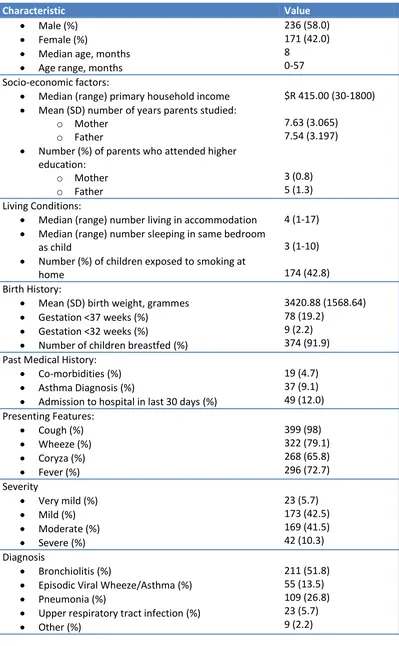
3.3 Results ... 65
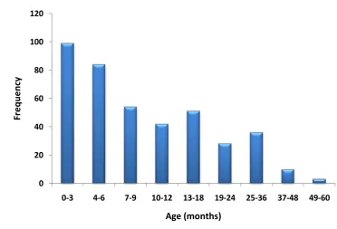
3.3.1 Demographics ... 65
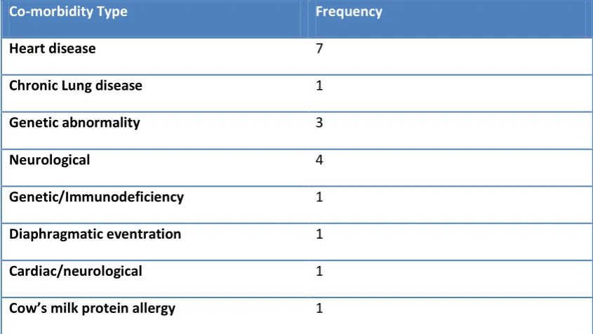
3.3.2 Clinical Information ... 72
3.4 Discussion ... 75
4
PCR Results ... 79
4.1 Introduction ... 79
4.2 Methods ... 79
4.3 Results ... 80
4.3.1 Co-infection ... 82
4.3.2 Pathogen detection and disease severity ... 86
4.3.3 Pathogen detection and clinical condition ... 87
viii
4.3.5 Relationships with age ... 90
4.3.6 PCR Negative Samples ... 90
4.4 Discussion ... 90
5
Cytokine Analysis ... 94
5.1 Introduction ... 94
5.2 Methods ... 95
5.2.1 Patient Samples ... 95
5.2.2 Cytokine Analysis ... 96
5.2.3 Statistics ... 96
5.3 Results ... 97
5.3.1 hRSV severity samples ... 97
5.3.2 Cytokine Responses to Various Pathogens ... 101
5.3.3 Cytokine responses to co-infection ... 105
5.4 Discussion ... 109
5.4.1 Cytokine response to varying severities of hRSV infection ... 109
5.4.2 Cytokine responses to various pathogens ... 113
5.4.3 Cytokine responses to co-infection ... 114
6
Discussion ... 116
6.1 Strengths of the study ... 116
6.2 Limitations ... 117
6.2.1 Demographics and clinical information ... 117
6.2.2 PCR Results ... 119
6.2.3 Cytokine analysis... 121
6.3 Conclusions ... 122
6.4 Further work ... 123
ix
Appendix 1: Reagents ... 138
Appendix 2: Equipment ... 140
Appendix 3: Questionnaire Details ... 141
Appendix 4: PCR Cycling Conditions ... 142
x
List of Tables
xi
List of Figures
Figure 1: Map detailing the location of Recife ... 52
Figure 2: Photograph of one of Recife's favelas ... 52
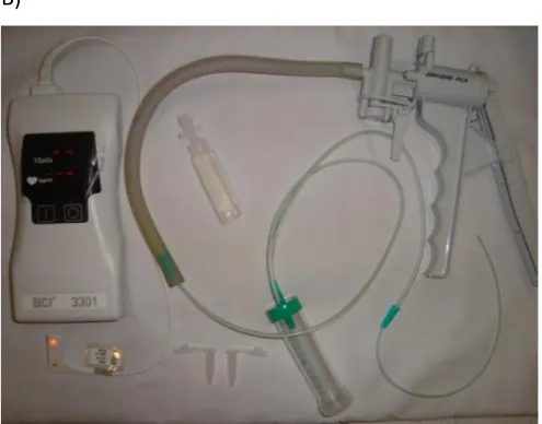
Figure 3: Nasopharyngeal aspirate collection ... 55
Figure 4: Distribution of age in study population. ... 67
Figure 5: Distribution of severity of disease in study population. ... 74
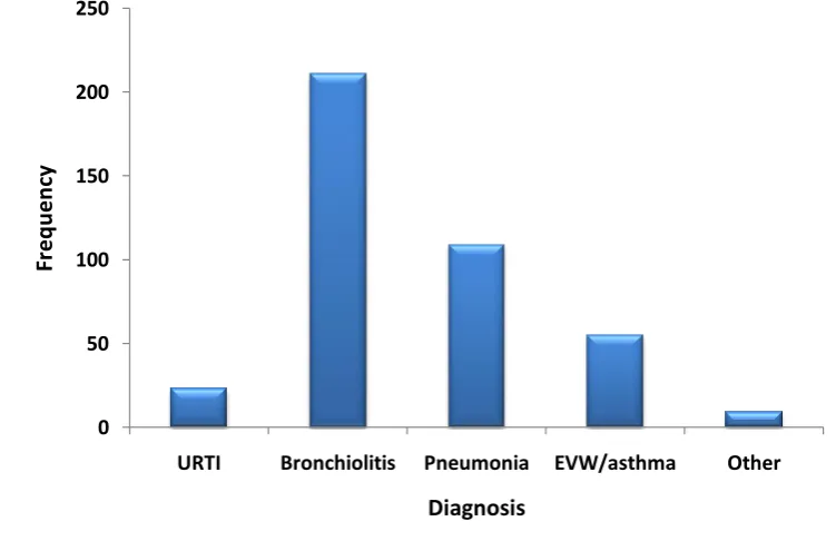
Figure 6: Clinical diagnoses recorded in study population. ... 74
Figure 7: Pathogen prevalence ... 81
Figure 8: Number of pathogens detected per sample... 83
Figure 9: Pathogen frequency in single infection ... 84
Figure 10: Pathogen frequency in co-infection ... 84
Figure 11: Most frequent co-infections ... 85
Figure 12: Seasonal distribution ... 89
Figure 13: Cytokine concentrations in varying severities of single hRSV infection. ... 100
Figure 14: Cytokine concentrations according to pathogen. ... 104
1
1
Introduction
1.1
Acute Respiratory Infection
1.1.1 Definition
Acute respiratory infection (ARI) describes a multitude of signs and symptoms, essentially including all types of infection of the respiratory tract. It is characterised by cough or wheeze, with or without the presence of fever, acute rhinitis, otitis media and pharyngitis1.
1.1.2 Burden
The World Health Organisation (WHO) estimates that respiratory infections account for 6% of the total global burden of disease – a higher percentage than diarrhoeal disease, cancer, human immunodeficiency virus (HIV) infection, ischaemic heart disease or malaria2. ARI is the third most common cause of death worldwide, and the most important in developing countries3 4. Worldwide, ARI is responsible for 3.5 million deaths each year, and just under two million of these are children under the age of five5.
Pneumonia is the most common cause of death in children under the age of five, causing an estimated 19-21% of all deaths in this age group6.
2
In developed countries, ARI are the largest contributor to the total infectious disease burden. In the United States of America (USA) it has been estimated that ARI cause between 545,000 and 840,000 admissions in under 18 year olds each year, accounting for between 14 and 20% of all admissions in this age group7. Statistics show that for the majority of health problems, advancement in management or prevention is decreasing the burden of particular diseases. However, in ARI there has been very little change in the number of DALYs lost per 100,000 population between 1990 and 20022. Firstly, this may be explained by the ageing population seen in so many developed countries. Along with young children, the elderly are more likely to develop an ARI in the first place, and more likely once they have an ARI to develop a severe infection requiring hospitalisation8. Secondly new respiratory pathogens, or strains of already known pathogens, are emerging frequently, which can be increasingly virulent causing widespread problems worldwide, or increasingly severe and more difficult to manage2.
1.1.3 Epidemiology
Age groups
Acute respiratory infections affect all age groups, although the very young and the elderly are most likely to suffer severe disease and require hospital admission8.
Seasonal Distribution
3
however, none have such a distinct seasonality as hRSV infection, and patterns of particular pathogens tend to vary from year to year. Human metapneumovirus (hMPV) for example, has been found to peak in some years at the same time as hRSV infection12, while other years it may peak immediately after the hRSV peak, or months later13 14. Other pathogens such as human rhinovirus (hRV) and adenovirus (AdV) are prevalent throughout the year10 15.
1.1.4 Risk Factors
Risk factors for ARI can be split into clinical and demographic factors. Clinical risk factors include male sex; prematurity; low birth weight; co-existing cardiac or respiratory problems; immunodeficiency and birth during the first half of the hRSV season. Demographic risk factors include a lack of breastfeeding; multiple siblings/crowded living conditions; a low socioeconomic status and smokers in the household16.
Breastfeeding
Many studies have found associations between breast-feeding and protection against ARI, specifically against ARI hospitalisation, and with a greater effect in developing countries17-19. Breast-feeding is beneficial due to colostrum containing hRSV-IgA and lactoferrin, both of which have antiviral properties, and it is also thought to promote lung maturation, having a particularly positive effect in the very young, and premature infants16.
Overcrowding
4 Socio-economic status
Low socioeconomic status has itself been found to be an independent risk factor for bronchiolitis, with one study finding children in the most deprived area of one particular city in Sweden to be almost twice as likely to need hospitalisation for bronchiolitis than those in the rest of the city24. Explanations for low socioeconomic status leading to increased risk of ARI are probably due to previously mentioned risk factors, including an increased likelihood of over-crowding, and parental smoking, as several studies have found increased rates of smokers in lower socioeconomic groups25-27.
Smoking
Both passive smoking and maternal smoking during pregnancy have been linked to an increased risk of the development of ARI, and increased risk of severe ARI requiring admission to hospital28 29.
Air Pollution
Air pollution has been reported as a risk factor for ARI for many years, with data from the 1950’s during the ‘London fog’ suggesting increased mortality due to respiratory causes during that period30. Indoor air pollutants such as the use of particular cooking fuels have also been shown to be risk factors of ARI in children. This is particularly relevant in developing countries, where biomass fuels are still commonly used, often in poorly ventilated settings, and may also be a contributing factor to the increased morbidity and mortality from ARI in developing countries31 32.
HIV infection
5 1.1.5 Aetiology
Causative organisms are predominately bacterial or viral, with Streptococcus pneumoniae (S. pneumoniae) and Haemophilus influenzae type b (Hib) being the most common bacterial pathogens and hRSV, hRV, AdV, and parainfluenza virus (PIV) being the most commonly identified viruses33.
1.1.5.1 ‘Typical’ Bacterial Pathogens
Streptococcus pneumoniae
Streptococcus pneumoniae (S. pneumoniae) is the most common cause of community-acquired pneumonia worldwide34. It also causes meningitis and otitis media. S. pneumoniae is commonly carried in the nasopharynx of healthy individuals, with one particular study finding 26% of two month old infants, and 62% of two year olds to be carriers35. Problems arise when S. pneumoniae spreads from the nasopharynx to the lower respiratory tract causing pneumonia, or other sites such as the meninges, blood or middle ear, causing meningitis, bacteraemia or otitis media – these together are termed invasive pneumococcal disease34. Risk factors for carriage of S. pneumoniae include a young age, young siblings, day care attendance, asthma and, in adults, smoking34. Invasive pneumococcal disease is a huge problem, causing an estimated one million deaths in children under the age of five annually34. Human immunodeficiency virus (HIV) infection is an important risk factor for invasive pneumococcal disease, particularly in Sub-Saharan Africa. Those with HIV have a 10-100 times increased risk of developing invasive pneumococcal disease when compared to the general population36.
Haemophilus influenzae type b
6
and 371,000 deaths in the year 200037. Successful Hib vaccines have been available for a number of years, and have led to dramatically reduced levels of Hib disease. A study published in 2009 estimated that in 2006, 55% of children in the world were included in Hib vaccination programmes. However, it is in a number of less developed countries where the vaccine is not routinely given, where Hib is still a major problem. It is estimated that 61% of all Hib childhood deaths occur in just 10 countries in Asia or Africa38.
1.1.5.2 ‘Atypical’ Bacterial Pathogens
‘Atypical’ bacterial pathogens are so called due to their ability to cause atypical pneumonia – pneumonia with symptoms different from those caused by S. pneumonia, including a sub-acute onset, low-moderate grade fever, dry cough, prominent constitutional symptoms, absent or moderate leukocytosis and more extensive radiographic involvement than would be expected after physical examination39. The three most important atypical bacteria are Mycoplasma pneumoniae (Mpp), Chlamydia pneumoniae (Cpp), and Legionella40.
Mycoplasma pneumoniae
Mycoplasma was first described in 194441, but was classified as Mycoplasma pneumoniae (Mpp) by Chanock et al in 196342. It is estimated that Mpp may cause between 14 and 34% of lower respiratory tract infections in children, but it is generally accepted that it is more commonly associated with disease in older children and adolescents43 44, and is thought to be the most common cause of community acquired pneumonia in the 5-20 age group40. Studies specifically investigating children under the age of five with ARI have found Mpp rates to be between 0 and 1.5%10 45 46.
Chlamydia pneumoniae
7
pneumonia40. Few studies have investigated the role of Cpp in children under the age of five, but those that have found rates of 0-1.3%10 45.
Legionella
Legionella is a Gram negative bacterium that was first named after an outbreak at an American Legion convention in 197647, although after identification previous outbreaks without known aetiology were discovered to have been caused by species of Legionella48. Legionella is usually transmitted by direct inhalation from contaminated water supplies, usually man-made reservoirs, where the bacterium thrives40. It causes a wide variety of clinical manifestations, from mild respiratory disease, to Legionnaires disease, with severe pneumonia and multisystem involvement, with neurological, hepatic and haematological abnormalities. Legionella is thought to be associated with between 1 and 27% of community acquired pneumonias40, however it is very rare in young children, with most cases occurring in the immunocompromised, or those with risk factors such as prematurity or bronchopulmonary dysplasia48.
1.1.5.3 Viral infection
Human Respiratory Syncytial Virus
8
There are many other respiratory viruses which have been implicated in the pathogenesis of ARI. Influenza (flu), PIV, AdV, coronaviruses (CoV) and hRV have all been accepted as common aetiological agents, and have been found in varying prevalences in populations of children and adults with ARI.
Rhinoviruses
Rhinoviruses have been found to cause about two-thirds of common colds and asthma exacerbations in adults and older children52. Previous work has shown rhinoviruses have optimum replication temperatures of 33oC – the temperature in the nasal passages, as opposed to the higher temperatures in the lower respiratory tract, another factor in support of rhinoviruses being associated with upper respiratory tract infections (URTI) and the common cold. However, further research has shown that although the optimum temperature for hRV replication is 33oC, they are still able to replicate effectively at 37oC, proving it is feasible that they are a cause of lower respiratory tract infection53.
Kusel et al. in 2006 detected hRV in 48% of nasopharyngeal aspirates from children under the age of one year with ARI10, however this study population comprised children who were all at high risk of atopy (i.e. had at least one parent with asthma, eczema or hayfever), so results may not apply to general populations. Other studies looking specifically at children with ARI found the prevalence of hRV to be between 3.6 and 24.7%1 15 45 54.
Adenovirus
9
with one particular serotype (serotype 14) being associated with particularly severe disease and a high mortality rate56.
Parainfluenza Viruses
Parainfluenza viruses (PIV) are common causes of ARI, especially in children, with studies estimating that most children will have evidence of infection with multiple serotypes by the age of 557.
Influenza Viruses
Influenza (flu) viruses are responsible for between 250,000 and 500,000 deaths each year. Influenza A (flu A) viruses commonly cause pandemics, with the most recent being the novel H1N1 pandemic (commonly referred to as ‘Swine flu’), which was declared a public health emergency by the WHO on the 25th April 200958. H1N1 influenza presented in a similar way to seasonal influenza, but had increased occurrences of vomiting and diarrhea, and an increased number of hospitalisations and deaths, with young people being particularly affected58. Influenza B (flu B) is also an important cause of morbidity and mortality, however it generally causes milder disease, and is responsible for 75% fewer hospital admissions than flu A59.
Coronaviruses
Coronaviruses (CoV) are also a common cause of ARI, usually causing similar clinical manifestations to other previously mentioned viruses, with mild URTI being common. However, severe associated respiratory syndrome (SARS)-associated coronavirus was identified in 2002, which had particularly severe clinical manifestations, with some patients developing respiratory distress syndrome, requiring intensive care and ventilation57.
Recently discovered viruses
10
two new coronaviruses, NL-63 (CoV NL-63), discovered in the Netherlands in 2004, and HKU-1 (CoV-HKU-1), discovered in Hong Kong in 200561 62; as well as human bocavirus (hBoV) discovered in Sweden in 200563.
Human metapneumovirus
11 Human Bocavirus
Human bocavirus (hBoV) was discovered by random polymerase chain reaction (PCR) amplification of respiratory samples from children63. Since its discovery it has been described by several groups worldwide, with prevalences in those with ARI being found to be between 1.5 and 18%, with detection most frequent in young children46 65. However, proving causality of hBoV in ARI has not been fully achieved, and it is still uncertain as to whether or not the detection of hBoV is clinically significant65-67.
1.1.5.4 Tuberculosis
12
1.1.5.5 Previous studies into aetiology
Many previous studies have investigated the aetiology of ARI, looking at various populations and a varying number of pathogens. Table 1 outlines the findings of a selection of studies that have investigated the prevalence of a range of pathogens in ARI. The studies in the table have been included due to their populations being restricted to pre-school children.
As well as indicating the % prevalence of each pathogen, the table highlights co-infection rates described in the studies. Since the introduction of multiplex PCR and its ability to simultaneously detect multiple pathogens within one sample, many studies have included co-infection rates in their analysis, with findings ranging from 4% to 33%10 15 33 45 46.
13
Table 1: Summary of a selection of studies investigating the epidemiology of ARI in pre-school children
Note: All values shown are % prevalence. X indicates that the particular study did not test for that pathogen. (AdV, adenovirus; hBoV, human bocavirus; CoV, coronavirus; Cpp, Chlamydia
pneumoniae; Flu, influenza virus; hMPV, human metapneumovirus; Mpp, Mycoplasma
pneumoniae; PIV, parainfluenza virus; hRSV, human respiratory syncytial virus; hRV, human
rhinovirus)
Study Bharaj
et al13
2009
Kaplan
et al46
Sung
et al45
2009
Lambert
et al71
2007
Souza
et al15
2003
Canducci
et al33
2008
Regamey
et al1
2008
Kusel
et al10
2006
Age Group (years)
<5 <5 <5 <5 <2 <2 <1 <1
Total +ve 35.2 78 47 74 43 46.6 79 69
Co-infection 6.6 33 4 9.9 4.8 14 20 10.3
AdV X 37 4.8 3.1 5.9 X 3 1.5
hBoV X 18 X X X 2.2 5 X
CoV X 1.2 3.8 1.5 X 8.7 18 5.5
Cpp X 4.5 0 X X X X 1.3
Flu 3.0 0.6 10.6 3.7 3.0 X 4 4.5
hMPV 3.7 2.5 1.5 3.7 X 14.3 13 1.8
Mpp X 0 1.5 X X X X 1.4
PIV 16.3 0 8.8 4.1 5.1 X 17 5.2
hRSV 20.3 43 8.4 6.6 1.8 28 16 10.9
14 1.1.6 Clinical Manifestations of ARI
The term ‘acute respiratory infection’ encompasses all infections of the respiratory tract, from a mild upper respiratory tract infection (URTI), to severe bronchiolitis or pneumonia. Diagnoses are usually made on clinical grounds, which are subjective, being dependant on the clinician involved, with lots of overlaps between different clinical diagnoses.
1.1.6.1 Upper Respiratory Tract Infection
URTI are very common, and usually mild, with the common cold being the most common manifestation. Other manifestations classed as URTI include otitis media, croup, whooping cough and epiglottitis.
Otitis Media
Otitis media describes infection or inflammation of the middle ear, and is very common, affecting over 80% of children by the age of three years72. Symptoms of otitis media include ear-ache, which in a young child may present with them being irritable and tugging at one or both ears, fever, loss of balance and a degree of hearing loss. It is thought that up to one third of cases of otitis media are caused by S. pneumoniae73.
Croup
Laryngotracheobronchitis, or croup, describes inflammation of the larynx and trachea. It causes hoarseness and a loud, harsh, barking cough. It can also cause stridor, commonly when the child becomes upset, but also in more severe disease. The most common cause of croup is PIV infection, but it can also be caused by other respiratory pathogens74.
Whooping cough
15
ending in vomiting. The illness is usually mild, although it can be severe in young children, and can last for a prolonged period of time74.
Epiglottitis
Epiglottitis is a particularly serious manifestation of URTI. It describes inflammation of the supraglottic larynx, usually caused by Haemophilus influenzae type b. It is characterised by acute onset stridor, and can rapidly cause obstruction of the trachea, meaning prompt management is vital74.
1.1.6.2 Bronchiolitis
Bronchiolitis predominately affects infants and young children, with the main clinical features being initial coryza, developing into cough, with the presence of wheezing and/or crackles and signs of respiratory distress, such as tachypnoea, hyperinflation and recession, commonly complicated by acute otitis media75 76.
1.1.6.3 Pneumonia
Pneumonia describes inflammation of the parenchyma of the lung77. In children the signs and symptoms include fever, tachypnoea and respiratory distress, often with focal or diffuse crackles on auscultation. In young children pneumonia is commonly caused by both bacteria and viruses, with S. pneumoniae being the most common bacterial cause78.
1.1.6.4 Episodic Viral Wheeze/Asthma
16
wheeze is most common in children under five, and usually disappears by the age of six, although it may continue79. The Global Initiative for Asthma (GINA) has described asthma as ‘a syndrome with a highly variable clinical spectrum, characterised by airway inflammation’. However, the presence of inflammation is difficult to determine in very young children, so EVW is a more accurate description, until symptoms can be more accurately assessed when the child is older, and if necessary a diagnosis of asthma can be made79.
1.1.7 Investigations
The diagnosis of ARI is a clinical one, based on history and physical examination findings. Specific diagnoses such as URTI, bronchiolitis, pneumonia, and episodic viral wheeze are used and do have an impact on management, with children diagnosed with pneumonia being more likely to be given antibiotics than children diagnosed with bronchiolitis, which is presumed to be viral.
17 Sampling Techniques
The British Thoracic Society guidelines on community acquired pneumonia state that all children suspected of having bacterial pneumonia should have blood cultures taken, and all children under the age of 18 months should have a nasopharyngeal aspirate (NPA) taken for viral antigen detection with or without viral culture78.
Obtaining a nasopharyngeal aspirate involves inserting a catheter between five and seven centimeters into the child’s nostril, then pulling back on the catheter while applying gentle suction82. An alternative method involves taking a nasal swab, where a cotton swab is inserted around 1.5 centimeters into the nostril and rotated against the nasal cavity to obtain a sample83. Taking a nasal swab is quicker, less invasive and less uncomfortable for the child. However, several studies have found that the sensitivity of detection of respiratory viruses is higher in NPA samples when compared to nasal swabs82-84.
The other problem with sampling using NPAs or nasal swab samples, is that in both cases, the sample is being taken from the upper respiratory tract, rather than the lower respiratory tract where the infection is causing the clinical manifestations of disease. Bronchoalveolar lavage (BAL) is a way of obtaining fluid from the lower respiratory tract, and although this would make it ideal for the detection of pathogens which are causing lower respiratory tract infection, it is invasive and requires sedation, so is not a practical option for the majority of those with ARI. It does, however, have a place in severely ill children who are already ventilated, and non-bronchoscopic BAL may then be a practical way of detecting pathogens in the lower respiratory tract85.
Pathogen detection
18
described. More traditional methods include virus culture and antigen detection. However, virus culture can take between 2 and 14 days to obtain results86, so in most cases would not be of much use in guiding the management of a patient. Antigen detection methods such as enzyme immunoassay are quicker than virus culture and results can be available in less than 24 hours. However, the sensitivity and specificity of PCR has been found to be greater than that of other methods86 87. PCR will be discussed in more detail in the methods section. Chest Radiographs
In ARI decisions about antibiotic use are often based on radiographic findings, with signs of consolidation on chest x-ray being presumed to be caused by bacteria and thus treated with antibiotics. Studies have shown that the use of chest radiographs are associated with increased likelihood of antibiotics and longer stays in hospital. One study randomised children with ARI to receive or not receive a chest x-ray, and found that children who received a chest x-ray were more likely to be diagnosed with having bacterial pneumonia or lower respiratory tract infection and treated with antibiotics, although the median time to recovery was the same in both groups88. This study concluded that chest x-rays did not affect clinical outcome, and were not beneficial, in children over two months of age with acute lower respiratory infection88.
Indicators of disease severity
19
1.1.8 Management of the Clinical Manifestations of ARI
The majority of children with ARI will have a mild, self-limiting illness and do not require any treatment. However, ARI can cause very severe infections, especially in the very young and those with other co-morbidities, leading to many hospital admissions each year. The mainstay of treatment is supportive, and includes the use of supplemental fluids and oxygen.
1.1.8.1 Upper Respiratory Tract Infection
Upper respiratory tract infection (URTI) is the most common manifestation of ARI, covering everything from the common cold to otitis media, croup, whooping cough and epiglottitis. The common cold requires supportive treatment, making sure the child is kept well hydrated, and the use of anti-pyretics such as paracetamol for fever.
Otitis Media
20
recommended that in children without any severe signs or symptoms, a ‘watch and wait’ strategy should be used, which will eliminate any adverse effects from antibiotic use, and it may help reduce antibiotic resistance91.
Croup
The management of croup is supportive, ensuring the parents are aware of signs of deterioration such as increasing respiratory rate, fatigue, chest-wall indrawing and a softening stridor. Severe disease may require intubation under general anaesthesia to avoid airway obstruction74.
Whooping Cough
The macrolide antibiotic erythromycin may be given to children in the early stages of Bordetella pertussis infection, although it is of little use once the paroxysmal cough has begun. It does, however, have a prophylactic use in contacts of someone with Bordetella pertussis infection. Treatment is otherwise supportive, with admission necessary in young children who are at risk from apnoea, and may require ventilation74.
Epiglottitis
21
1.1.8.2 Bronchiolitis
The mainstay of treatment for bronchiolitis is supportive. Oxygen should be given if saturations are below 93%, if the child has severe respiratory distress or if the child is cyanosed. Adequate hydration should be maintained, and if this is not possible through oral feeding nasogastric feeding or the use of intravenous fluids should be considered92.
Bronchodilators are widely used in the treatment of ARI76. However, a Cochrane review investigating evidence for the use of bronchodilators in bronchiolitis found that although bronchodilator therapy led to a significant improvement in clinical scores, it recognised that the studies had included children with recurrent wheeze or asthma, which may have skewed the results93. This study also found no difference in rates of hospitalisation or oxygen saturations in those who had been given bronchodilators compared to controls93. A more recent systematic review found similar results, with studies finding improvements in short-term clinical measurements in children receiving bronchodilators, but no difference in the need for hospital admission or duration of hospital stay94. As with all pharmacological treatments, bronchodilator therapy is not without adverse effects, and these include increased heart rate and temporarily decreased oxygen saturations95. There is therefore little evidence supporting the use of bronchodilators for bronchiolitis, with only short term improvements in clinical measures being demonstrated.
22
1.1.8.3 Pneumonia
Pneumonia may be viral or bacterial, and while viral pneumonia requires only supportive treatment, the correct treatment of bacterial infection is important. The British Thoracic Society guidelines suggest bacterial pneumonia should be considered in children less than three years if there is fever over 38.5oC, along with a respiratory rate greater than 50 breaths per minute and chest recession, and that bacterial pneumonia is unlikely if wheeze is present78. Antibiotics are given according to local or national guidelines, which should take into account patterns of particular pathogens and resistance in that area. Current British Thoracic Society guidelines suggest that the most appropriate treatment for suspected bacterial infection in children under five is with amoxicillin, and in those with severe pneumonia co-amoxiclav, cefuroxime and cefotaxime are appropriate78. Macrolide antibiotics are only advised as first line treatment in children over the age of five, in whom infection with Mpp is more common78. Overuse of antibiotics, especially in viral infection when they are completely unnecessary, can lead to increased antibiotic resistance of bacteria, which is an important clinical problem. There is significant penicillin resistance to S. pneumoniae worldwide with the main risk factor being previous antibiotic use, and a further risk factor for penicillin resistant invasive pneumococcal disease being recent respiratory tract infection34.
1.1.8.4 Episodic Viral Wheeze/asthma
23
Corticosteroids are often used during acute episodes of wheeze in children due to their proven role in acute exacerbations of asthma in adults and older children97. However, a recent review article looking into the results of several trials has found conflicting evidence on the use of corticosteroids in this age group, and recommended that a trial of oral corticosteroids should be given only children admitted to hospital with severe wheeze79.
1.1.9 Prevention
The burden of ARI is great on many levels, from its effects on individuals, to the large hospital costs from the high number of admissions that it causes each year. Due to the lack of effective treatment for ARI, with management being largely supportive, prevention may be key to decreasing the burden. Prevention of ARI can be viewed from two perspectives. Firstly, prevention can be attempted by addressing some of the risk factors mentioned in section 1.1.4. The biggest problem faced here is that many of the issues are already large health problems, which many countries are already attempting to address. Smoking cessation, for example, is the focus of many government schemes in both developed and developing countries. Although this may have a positive impact on the prevalence of ARI, the schemes are already large and costly and to further them specifically for the purpose of preventing ARI would not be feasible. Poverty is the other major risk factor that contributes to ARI. Again, this is a global problem for which there is not an easy solution.
1.1.9.1 Prophylaxis
24
vaccinations, or vaccinate against a large number of pathogens that in most cases cause mild, self-limiting illnesses.
hRSV prophylaxis
It is not surprising that most efforts to develop a vaccine have been focused on hRSV infection, as it has been shown to cause 50-90% of hospitalisations for bronchiolitis and 20-50% of hospitalisations for winter pneumonia. The development of hRSV vaccination has been slowed by safety issues following the administration of a formalin-inactivated hRSV vaccine to 13 residents of a children’s home in 196698. An outbreak of hRSV infection at the centre approximately nine months following the vaccinations, demonstrated that the children who had received the vaccinations were not only not protected against hRSV, but developed more severe disease, with significantly higher rates of hospitalisation, higher rates of pneumonia, and longer duration of illness98. A breakthrough in immunisations against hRSV occurred with the development of Palivizumab, a humanised monoclonal antibody, which has been shown to significantly reduce hospitalisations in children at high risk of hRSV infection99 100. Palivizumab is given monthly as an intramuscular injection to high risk infants during the hRSV season. Although Palivizumab has been shown to be clinically effective at reducing hospitalisations in high-risk infants, the cost of vaccinating these infants is very high, and studies have found the use of palivizumab is not cost effective in all high risk children101.
Pneumococcal conjugate vaccine
25
the bacteria in both children and adults102-104. Large studies have found reduced rates of meningitis and invasive pneumonia that have been sustained in the years since the vaccine was introduced105. Although the vaccine has been shown to be effective at preventing cases of invasive pneumococcal disease caused by the seven common serotypes, since the introduction of the vaccine, the presence of invasive disease caused by other non PCV7 serotypes has increased dramatically, with a higher percentage of these being resistant to penicillin as well34. Similarly, a large study in Massachusetts found that after introduction of the vaccine there was very little carriage of any of the PCV7 serotypes, but greatly increased carriage of the non-PCV7, often penicillin resistant serotypes106. Invasive pneumococcal disease therefore remains an important problem causing significant mortality worldwide. More recently, a 13-valent pnumococcal conjugate vaccine (PCV13) has been introduced into the vaccination schedules, replacing PCV7. It was introduced in the US in February 2010, shortly followed by the UK in April 2010, protecting against 13 strains of the bacteria, which are responsible for three-quarters of invasive disease in young children, although as with PCV7, the prevalence of non-PCV13 serotypes is likely to increase107.
1.1.10 Sequelae of ARI
26 1.1.10.1 Recurrent Wheeze
Several studies have investigated the development of wheezing and asthma in children who had a viral respiratory illness, often focusing on particular viruses or particularly severe disease. One large study by Jackson et al in 2008, found that rhinovirus infection causing wheezing in the first three years of life, but especially in the third year, was strongly associated with an increased risk of asthma by the age of six (odds ratio = 9.8)108. They also found increased asthma at age six in children who had had wheezy illness caused by hRSV infection in the first three years of life, although this association was not as strong (odds ratio = 2.6)108. A limitation to this study is that all study participants were at high risk of asthma development – in order to be eligible for inclusion into the study the child must have one parent with proven allergen sensitivity or diagnosed asthma108. Sigurs et al in 2009 reported asthma/recurrent wheezing, allergic rhinoconjunctivitis and sensitisation to common inhaled allergens were all significantly more frequent in a group of children who were hospitalised with hRSV bronchiolitis in infancy, when compared to matched controls109. Stein et al in 1999 found an association between hRSV LRTI before the age of three, and wheeze up until the age of 11. However, after age 11 the risk decreased and unlike the previously mentioned studies, was not significant by the age of 13110.
27
developing recurrent wheeze or asthma and, for the same reason, are at higher risk of having a viral respiratory illness.
1.1.10.2 Invasive pneumococcal disease
A study by Ampofo et al found that viral infection with hRSV, influenza or hMPV was associated with increased risk of admission with invasive pneumococcal disease in the weeks following the viral infection. The increased risk existed for four weeks after hRSV infection, and two weeks after influenza or hMPV infection111. In a large study of adults and children with invasive pneumococcal disease in New Zealand, Murdoch et al. found statistically significant correlations between incidence rates of invasive pneumococcal disease and detection rates of flu viruses, AdV and PIV3, and in the under 5’s RSV112.
Madhi et al found that a pneumococcal conjugate vaccine introduced in South Africa prevented nearly one third of ARI caused by influenza A, RSV, PIV 1-3 and AdV. This highlighted the importance of viral-bacterial co-infection, and its role in causing disease, as well as the role of pneumococcal vaccination in the prevention of viral ARI as well as those caused directly by Streptococcus pneumoniae 113.
1.1.10.3 Bronchiolitis obliterans
28
with some patients having a rapidly declining lung function with a fatal outcome, while some patients develop chronic lung disease and others have actually improved over time, with gradual resorption of the fibrovascular tissue and improvement to almost normal airways and lung function114.
1.1.10.4 Bronchiectasis
29
1.2
Immunological responses to ARI
The lung makes up the largest epithelial surface in the body, with each breath exposing the airways to new micro-organisms which may give rise to acute respiratory infection117. As well as the need for a rapid acting, efficient immune system to deal with these micro-organisms, the complex physiology of the respiratory system, with its ability to facilitate gas exchange, is a delicate, balanced environment, in which an inflammatory response, aimed at destroying a potentially harmful pathogen, may itself create problems and affect the normal functioning of the lung118.
The immune response can be divided into the innate and the adaptive responses. The innate response comprises rapid, first line, non-specific mechanisms in the defence against infection by pathogens. The adaptive response is in contrast, a much more complex system, with specialised cells mounting specific responses against the particular pathogen involved in each ‘attack’, and producing an immunological ‘memory’ to enable the rapid elimination of that particular pathogen should it come into contact with the host immune system on a subsequent occasion. However, the adaptive immune system’s specificity, although essential, can take three to five days to produce an adequate response to effectively deal with a pathogen, a time when the innate immune system plays a vital role in controlling the replication of the pathogen, and limiting its damaging effects119.
1.2.1 Innate Response
30
Innate immunity can be divided into three groups, the physical barriers to infection, the non-cellular response and the cellular response.
1.2.1.1 Physical Barriers to Infection
Physical barriers to infection are the very first line of defence any invading organisms will encounter, and include the cough reflex, the barrier function of respiratory epithelium and the muco-ciliary escalator. The respiratory epithelium is highly specialised, with numerous roles in the protection of the respiratory tract from invasion by pathogens, but firstly, it provides a physical barrier to foreign particles and pathogens. The epithelium also plays a pivotal role in the muco-ciliary escalator, with pseudostratified columnar epithelial cells, which form a large part of the epithelial surface of the conducting airways, being ciliated cells. The co-ordinated beating of the cilia on these cells provides a mechanism for clearing mucus and with it any trapped material, out of the airways, to the epiglottis, where it can be safely swallowed, clearing any potential pathogens away from the delicate respiratory surfaces120.
31
to influence and increase mucus production, leading to some of the clinical manifestations associated with respiratory illness.
For pathogens that get past the initial physical barriers of the respiratory tract, the next line of defence comprises soluble mediators of the immune system that can be found in the fluid lining the respiratory tract – the airway surface fluid (ASF)117.
1.2.1.2 Pathogen recognition/detection
The ability to recognise a pathogen in order to initiate an immune response is the first, vital role of the immune system. The innate immune system uses a range of genetically pre-programmed receptors, which, instead of being able to recognise specific pathogens, like the adaptive immune system, are able to recognise structures or patterns common to many pathogens, which, importantly, are absent in self molecules. These structures are called ‘pattern-associated molecular patterns’ or PAMP’s, and the receptors are called ‘pattern recognition receptors’ (PRR’s)119. Examples of PAMP’s are lipopolysaccharide (LPS), an endotoxin and component of the cell membrane of gram negative bacteria; forms of bacterial DNA – unmethylated cytosine-guanosine-rich areas – CpG sequences; and double stranded RNA117 119.
Pattern Recognition Receptors
32
Signalling receptors recognise PAMP’s and activate pathways that lead to the transcription of many genes vital to an effective immune response119.
Toll like receptors
33
Several TLRs are involved in the recognition of pathogens which cause ARI. TLR-2 is known to recognise bacteria, including Mpp, as well as viruses, and is able to initiate different responses depending on the particular ligand it comes into contact with by forming heterodimers with other TLRs123. TLR-4 has also been shown to be important in the recognition of respiratory pathogens. As well as recognising bacteria, it has been found to be important in the recognition of hRSV, as well as being implicated in the pathogenesis of H5N1 avian influenza. It is thought that TLR4 recognises one of the molecules released from cells damaged by H5N1 (a damage-associated molecular pattern, DAMP), and mice deficient in TLR-4 were found to be resistant to the H5N1 induced lethality123. This illustrates one example of the immune response being damaging rather than protective. Collectins
Collectins are secreted pattern recognition receptors which bind to carbohydrates on pathogens including gram negative and gram positive bacteria, and some viruses, parasites and yeasts. Examples of collectins include mannan-binding lectin (MBL) and surfactant proteins A and D. The main functions of these are to alert and recruit other cells of the immune system, such as macrophages and lymphocytes, and to activate the ‘lectin pathway’ in which the activation of a series of proteases leads to the activation of the complement pathway – one of the effectors of the innate immune system119. The surfactant proteins are also particularly important in the regulation of inflammation in the lungs, inhibiting the activation of alveolar macrophages until they themselves come into contact with a pathogen118.
Lysosome
34
pathogen are then processed and displayed on the surface of the phagocyte by major histocompatability complex (MHC) molecules, in order to alert the adaptive immune system, which will be described later119.
1.2.1.3 Soluble mediators of the innate immune system
Defensins
The defensins are small peptides found in the respiratory tract, as well as the gastro-intestinal and reproductive tracts. The α-defensins are produced by neutrophils, and the β-defensins are produced by epithelial cells124. They have broad antimicrobial activity against bacteria, fungi and some viruses117. The antimicrobial action of defensins relies upon attachment to the cell membrane, where it is thought they then form pores in the membrane124. They are also able to activate the alternative complement cascade, which will be discussed in more detail in section 1.2.1.4. The defensins have been shown to be up-regulated in response to epithelial cell exposure to pathogens, TLR agonists and inflammatory cytokines125. Murine studies have found that mice deficient in β-defensin had delayed clearance of Haemophilus influenzae from the lung after infection126, and also that mouse β-defensin inhibits influenza A replication127, both studies suggesting defensins may have an important role in the defence against ARI.
Lysozyme
35 Lactoferrin
Lactoferrin is another broad-spectrum, anti-microbial protein found in high concentrations in lavage fluid120, but is probably better known for its high concentration in colostrum and breast milk, where it provides important anti-microbial activity to infants. In the lungs it is produced by serous cells and neutrophils. Lactoferrin’s antimicrobial activity is based on its ability to cause agglutination of bacteria, upon recognising them by their carbohydrate motifs117, as well as its ability to remove iron from pathogens125 .
Increased amounts of both lysozyme and lactoferrin have been detected in the lungs of mice after treatment with Haemophilus influenzae, as well as other infectious agents, implicating them in the immune response to ARI, although further details on specific modes of action against specific pathogens are not known125.
Immunoglobulin A
Immunoglobulins are secreted by B-lymphocytes, and while these are usually associated with the adaptive immune response, immunoglobulin A (IgA) release has been shown to be stimulated by the release of stimulatory cytokines from epithelial cells. IgA is found in the secretions of the respiratory tract, as well as the gastro-intestinal tract, genito-urinary tract, breast milk and colostrum, and is important in the rapid response, especially to viruses117.
Type I Interferon Response
36
macrophages, in response to viral infection117. Mice deficient in receptors for type I interferons have shown increased viral replication in the lung, although the infection was still cleared by other mechanisms, illustrating the complexity of the immune response with various different levels of defence117.
1.2.1.4 Effectors of innate Immunity
The complement system
The complement system consists of a number of plasma proteins that may be activated by one of three pathways: the classical pathway, the alternative pathway, and the lectin pathway. Once activated these pathways lead to a number of steps which eventually all result in destruction of the pathogen with the formation of membrane attack complexes (MAC); or opsonisation of the pathogen for destruction by other parts of the immune system.
The classical pathway, named due to its earlier discovery than the other two pathways, is activated by certain antibody-antigen complexes, and is a major component of the adaptive immune system.
37
The lectin pathway was briefly mentioned earlier in the chapter. Lectins such as the mannan binding lectin (MBL), bind to polysaccharides on bacteria, as well as MBL-associated serine proteases (MASPs), including MASP-1, MASP-2 and MASP-3. The subsequent complexes activate the lectin pathway by initiating the cleavage of C4 to C4b and C2 to C2b, forming the complex C4bC2b – C3 convertase, which, as in the alternative pathway, cleaves C3 to C3b. The C3b this time forms a C4b2b3b complex – another (classical) form of C5 convertase129.
Both these pathways, as well as the classical pathway, lead to the formation of C5 convertase. C5 convertase initiates the activation of further components of the complement cascade, including C6, C7, C8 and C9, which ultimately lead to the formation of the membrane attack complex (MAC). The MAC has the ability to form pores in the cell membrane, causing cell lysis129.
Other functions of complement activation include opsonisation and stimulation of the inflammatory response. Opsonisation occurs when the pathogen becomes coated in C3b or C4b, both of which bind to receptors on neutrophils and macrophages, and initiate phagocytosis of the pathogen128.
The C3a, C4a and C5a fragments released during the cleavage of C3, C4 and C5, respectively, have their own function in the stimulation of the immune response. C5 is the most potent, causing mast cell degranulation and stimulating neutrophil motility and adhesion to endothelial cells. C3b and C4b have similar effects but are less potent128 130. Phagocytes
38
important antigen presenting cells (APC’s), having a vital role in the activation of the adaptive immune system.
Neutrophils
Neutrophils are the most numerous circulating leukocytes, and are constantly produced in the bone marrow. Production and release of neutrophils is stimulated by granulocyte colony-stimulating factor (G-CSF). Neutrophils circulate in the blood, and in the absence of infection they will undergo apoptosis and phagocytosis after a period of around six hours130. However, when infection is present, neutrophils migrate to the site of infection, in response to the release of mediators such as IL-8, particular complement proteins and chemokines. This is very true in the lung during hRSV infection, with neutrophils accounting for 93% of cells taken from the upper respiratory tract, and 76% in the lower respiratory tract131. The process of neutrophil recruitment can be divided into a number of steps and begins with adhesion of the neutrophils to the endothelial surface, followed by migration through the endothelial cells towards the site of infection. Once reaching the site of infection, the neutrophils are kept in place, fulfilling their function as important phagocytes130.
39
During infection there is rapid proliferation and migration of neutrophils to the affected tissue, in response to the release of a variety of mediators, one of the most important being IL-8. IL-8 is released by macrophages and epithelial cells in response to infection, with initial responses being incredibly rapid. Studies of respiratory epithelial cells have shown IL-8 responses initially peaking within two hours of infection with hRSV132. IL-8 release by macrophages and epithelial cells is stimulated by TNF-α and IL-1133, both of which are produced by macrophages in the early immune response to pathogens. IL-8 causes a rapid accumulation and activation of neutrophils, with neutrophils being abundant and IL-8 concentrations being increased in respiratory samples from patients with ARI. Recent studies, however, have implicated IL-8 in the pathogenesis of ARI, particularly in hRSV disease. An association between severe hRSV bronchiolitis and a polymorphism near the IL-8 gene was detected, with those with the polymorphism having increased IL-IL-8 levels, and increased severity of disease134. This will be discussed further in section 5.4.1.
Macrophages
40
no infection is present, and inhibit their activation. When a pathogen is detected, the surfactant proteins bind to the pathogen, no longer inhibiting the macrophage, which will then be activated and begin to produce its inflammatory mediators which will alert other aspects of the immune system118. Macrophages are also important antigen presenting cells (APC’s), which will be discussed further in section 1.2.1.5.
Natural Killer Cells
Natural killer (NK) cells are important in the innate response to viral infection. They are derived from the same lineage as T-cells in the bone marrow, but do not need to mature in the thymus. NK cells recognise cells that have abnormal expression of human leukocyte antigen (HLA) class I tissue antigens117. These antigens are present on the surface of cells, and function to present peptides from pathogens, in order to alert the immune system. If a virus is present inside the cell, the expression of HLA molecules will be altered, and this will be recognised by the NK cell. HLA recognition is very important in the adaptive response to infection. However, some viruses, instead of altering the HLA molecules, are able to down-regulate them in order to avoid recognition by the adaptive immune system. In this respect, NK cells are very important, as they are able to recognise these cells with low HLA expression which are missed by other parts of the immune system130.
41
come into contact with infected cells, and binds to Fas on the infected cell, and again induces apoptosis through activation of caspases130.
As well as causing apoptosis, NK cells, like many of the other effectors of the innate immune system, secrete cytokines, specifically large amounts of interferon-gamma (IFN-γ), which is important in the activation and recruitment of cells of the adaptive immune system130.
1.2.1.5 Antigen-Presenting Cells
Macrophages have already been discussed in terms of their role as phagocytes, and it was mentioned that a further role of theirs is as antigen presenting cells (APCs). APCs provide a vital link between the innate immune response and activation of the adaptive immune response. Antigens are presented with major histocompatibility complexes, which enable recognition by T-cells, and the subsequent development of a specific response to the antigen, as well as the development of immunological memory.
Dendritic Cells
42 1.2.2 Adaptive Response
The adaptive immune system has two main functions. Firstly, if the innate immune system isn’t able to destroy pathogens immediately, an adaptive immune response develops, specific for that particular pathogen, enabling complete clearance of the invading organism. Secondly, the adaptive immune system is responsible for immunological memory, whereby the immune system is able to ‘remember’ a particular pathogen from a prior infection. When the same pathogen is subsequently encountered on a later occasion, immunological memory allows a very rapid, specific, response to be mounted against the pathogen130.
The adaptive immune system can be divided into two parts – the humoral response, and the cell-mediated response.
1.2.2.1 Humoral Immunity
Humoral immunity describes the antibody response. Antibodies are proteins produced by B-lymphocytes (B-cells), which use various mechanisms to destroy pathogens. Antibodies are particularly useful in the defence against extracellular pathogens, as the antibodies are able to bind to the free pathogens, and support the destruction by other mechanisms128.
B-43
cell undergoes clonal expansion, with production of the same antigen specific B-cells, with differentiation to antibody secreting plasma cells, and memory B-cells128. The proliferation and differentiation of B-cells is stimulated by a range of cytokines, produced by other parts of the immune system. IL-2, IL-4 and IL-21 produced by helper T-cells, as well as IL-6 produced by helper T-cells, macrophages and many other cells, are all important in B-cell proliferation, and also have roles in determining the differentiation. For example, IL-6 has been found to have a role in promoting the differentiation of B-cells into antibody secreting plasma cells135, while IL-4 has been found to promote the development of memory B-cells136. Different immunoglobulins have different effects, and are released at different times in the response. IgM is released early in the immune response and is particularly good in the defence against bacteria. It has 10 potential binding sites, and is able to attach to and cause agglutination of bacteria, as well as activating the complement system and opsonising the bacteria for phagocytosis130. Viruses and bacteria promote the release of IFN-γ from T-helper cells, which then promotes the differentiation of B-cells into IgG secreting plasma cells. IgG also opsonises pathogens for phagocytosis by macrophages128. IgD has already been mentioned as a receptor on B-cells, where it has a role in the identification of antigens and the activation of the B-cells. IgA was mentioned in the previous chapter and is found on mucosal surfaces in the respiratory, gastrointestinal and reproductive tracts, as well as in saliva, breast milk and tears. It is thought to be one of the earlier defences against pathogens, preventing their movement across mucosal surfaces130.
44
pathogenesis of hRSV disease. Conversely, it has also been shown that infants with low placental hRSV antibodies are more likely to suffer severe hRSV disease, indicating a protective role132.
1.2.2.2 Cell-mediated Immunity
Cell-mediated immunity is based on the response by T-lymphocytes (T-cells). Cell-mediated immunity, in contrast to humoral immunity which is only effective against extracellular pathogens, is effective against intracellular pathogens, which may escape other parts of the immune system, as well as pathogens within other immune cells, such as those that have been phagocytosed by macrophages as part of the innate immune response128.