Open Access
Primary research
Epithelial Na, K-ATPase expression is down-regulated in canine
prostate cancer; a possible consequence of metabolic
transformation in the process of prostate malignancy
Ali Mobasheri*
1, Richard Fox
2, Iain Evans
1, Fay Cullingham
1, Pablo
Martín-Vasallo
3and Christopher S Foster
4Address: 1Molecular Pathogenesis Research Group, Department of Veterinary Preclinical Sciences, Faculty of Veterinary Science, University of Liverpool, Liverpool L69 7ZJ, United Kingdom, 2Department of Veterinary Pathology, Faculty of Veterinary Science, University of Liverpool, Liverpool L69 7ZJ, United Kingdom, 3Labratorio de Biología del Desarollo, Departamento de Bioquímica y Biología Molecular, Universidad de La Laguna, 38201 La Laguna, Tenerife, Spain and 4Department of Cellular and Molecular Pathology, Faculty of Medicine, University of Liverpool, Liverpool L69 3GA, United Kingdom
Email: Ali Mobasheri* - [email protected]; Richard Fox - [email protected]; Iain Evans - [email protected];
Fay Cullingham - [email protected]; Pablo Martín-Vasallo - [email protected]; Christopher S Foster - [email protected] * Corresponding author
NaK-ATPaseisoformcanine prostatebenign prostatic hyperplasiaprostate cancercitrate metabolismmetabolic transformationmalignancy
Abstract
Background: An important physiological function of the normal prostate gland is the synthesis and secretion of a citrate rich prostatic fluid. In prostate cancer, citrate production levels are reduced as a result of altered cellular metabolism and bioenergetics. Na, K-ATPase is essential for citrate production since the inward Na+ gradients it generates are utilized for the Na+ dependent uptake of aspartate, a major substrate for citrate synthesis. The objective of this study was to compare the expression of previously identified Na, K-ATPase isoforms in normal canine prostate, benign prostatic hyperplasia (BPH) and prostatic adenocarcinoma (PCa) using immunohistochemistry in order to determine whether reduced citrate levels in PCa are also accompanied by changes in Na, K-ATPase expression.
Results: Expression of Na, K-ATPase α1 and β1 isoforms was observed in the lateral and basolateral plasma membrane domains of prostatic epithelial cells in normal and BPH prostates. Canine kidney was used as positive control for expression of Na, K-ATPase α1 and γ isoforms. The α1 isoform was detected in abundance in prostatic epithelial cells but there was no evidence of α2, α3 or γ subunit expression. In advanced PCa, Na, K-ATPase α1 isoform expression was significantly lower compared to normal and BPH glands. The abundant basolateral immunostaining observed in normal and BPH tissue was significantly attenuated in PCa.
Conclusion: The loss of epithelial structure and function and the transformation of normal epithelial cells to malignant cells in the canine prostate have important implications for cellular metabolism and are accompanied by a down regulation of Na, K-ATPase.
Published: 13 June 2003
Cancer Cell International 2003, 3:8
Received: 25 November 2002 Accepted: 13 June 2003
This article is available from: http://www.cancerci.com/content/3/1/8
Cancer Cell International 2003, 3 http://www.cancerci.com/content/3/1/8
Background
The principal physiological function of the prostate gland is the synthesis, accumulation and secretion of the anion citrate [1]. Citrate may be used as an important energy source for spermatozoa, or involved as a buffer or chelator of cations in seminal fluid [2]. Na+ dependent uptake of
aspartate from plasma is achieved by two kinetically dis-tinct Na+-dependent transport systems to create a high
cytosolic aspartate concentration [3]. Aspartate provides the intra-mitochondrial source of oxaloacetate while glu-cose provides the source of acetyl-CoA for citrate biosyn-thesis [4]. Ouabain-sensitive Na, K-ATPase-mediated transport is critical for aspartate uptake, citrate production and prostatic fluid formation since the inward Na+
gradi-ents generated by Na, K-ATPase are utilized for the Na+
dependent uptake of aspartate. Na+ and K+ also represent
a large component of prostatic fluid osmolarity [5] and their levels are finely regulated by plasma membrane transport systems that have yet to be identified. Further-more, androgen activation of Na, K-ATPase serves as a metabolic pacemaker in the prostate [6,7] exerting tran-scriptional control over the expression of Na, K-ATPase subunits, which play a critical role in the biogenesis of Na, K-ATPase in prostate cancer [8,9]. In normal prostate, cit-rate concentrations in prostatic fluid range from 40 to 150 mM. In prostate cancer however, citrate production levels are significantly reduced as a result of altered cellular metabolism and bioenergetics [4].
Na, K-ATPase is an important regulator of intracellular electrolyte levels in almost all mammalian cells [10,11]. It is a Mg2+-dependent P-type transport pump responsible
for maintaining the low intracellular Na+:K+ ratio that is
essential for cell homeostasis and physiological function. It catalyzes the active uptake of K+ and extrusion of Na+ at
the expense of hydrolyzing ATP with a stoichiometry of 3Na+ for 2K+. The active form of Na, K-ATPase is an
inte-gral membrane protein complex primarily composed of two non-covalently attached subunits; a 110-kDa catalytic
α subunit and a 45–55-kDa glycosylated β subunit. The α subunit has binding sites for Na+, K+, ATP and cardiac
gly-cosides (digitalis and ouabain) [12]. Four α isoforms encoded by different genes have been identified which are ~85% identical at the protein level [13,14]. The β subunit is a complex type II glycoprotein with a short cytoplasmic
NH2 terminus, a single transmembrane domain and a
large globular COOH ectodomain containing three disulfide bridges and sites for N-linked glycosylation.
Renal Na, K-ATPase consists of an additional small com-ponent known as the γ subunit [15,16]. The γ subunit is a member of the FXYD family of small ion transport tors [17] and is believed to be responsible for fine regula-tion of Na+ transport in the nephron by modulating the
transport function of renal Na, K-ATPase [18,19].
We have previously shown that human and rat prostatic epithelial cells express the α1, β1 and β2 isoforms of Na, ATPase [20,11]. Despite the importance of Na, K-ATPase function for citrate production there is no infor-mation about its expression patterns in hyperplastic or neoplastic prostate. The objective of this study was to determine the localization of Na, K-ATPase and to com-pare expression of Na, K-ATPase isoforms in normal canine prostate, benign prostatic hyperplasia (BPH) and prostatic adenocarcinoma (PCa) in order to determine whether reduced citrate levels in PCa are also accompa-nied by changes in Na, K-ATPase expression.
Results
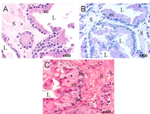
In normal prostatic tissue (Fig. 1, panel A) glands are lined with a relatively continuous sheet of simple cuboidal to low columnar/pseudostratified epithelial cells which are separated by stromal connective tissue consisting of smooth muscle, blood vessels and neuroendocrine cells. Normal glands exhibit large lumens without evidence of tufting or micropapillae. The tissue in panel B exhibits all the classic morphological features of BPH; the tubuloalve-oli vary in size and form with many of the individual
[image:2.612.311.554.392.576.2]Histological appearance of normal (A), BPH (B) and PCa canine prostate tissues incorporated in this study
Figure 1
glands appearing cystically dilated or collapsed. Epithelia consist of columnar cells but some basal cells are also present. Panel C shows sheets of neoplastic epithelial cells that are morphologically typical of high-grade adenocarci-noma. Epithelial and stromal cells are densely packed and disorganized. There is marked cellular and nuclear pleo-morphism and numerous mitotic figures are observed particularly in the epithelial cells layer. Enlarged hyper-chromatic nuclei display marked anisokaryosis. Nucleoli are large and prominently multiplied. The basal cell layer is often irregular in contour but basement membranes appear confluent. Micro-glands exhibit ill-defined lumens significantly constricted by the geometric progression of proliferating cells.
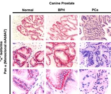
Expression of Na, K-ATPase α1 and β1 isoforms was observed in the lateral and basolateral plasma membrane domains of prostatic epithelial cells in normal and BPH prostates (Fig. 2). In advanced PCa, Na, K-ATPase expres-sion was significantly lower compared to normal and BPH glands (Fig. 2). The expression observed in high grade (poorly differentiated) adenocarcinomas was significantly lower than low grade (well differentiated tumours); the abundant basolateral immunostaining observed in nor-mal and BPH tissue was significantly reduced in high grade PCa as determined by immunohistochemistry using
a monoclonal pan α antibody (mAb9A7) that has been
shown to recognize the α1 isoform in addition to all other known α isoforms [21] (Fig. 3). Identical results were
obtained using the α5 monoclonal pan α antibody
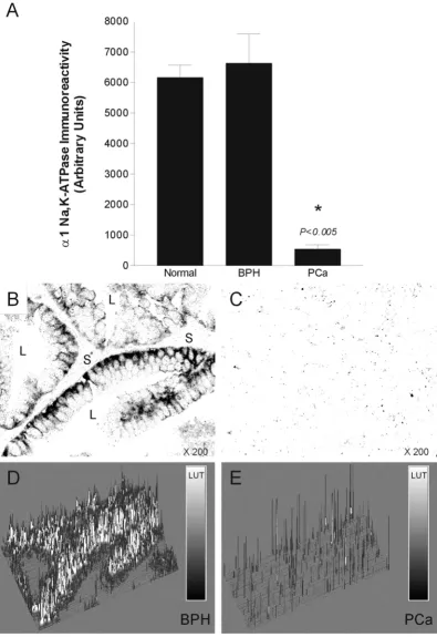
(results not shown). Image analysis confirmed the down-regulation of Na, K-ATPase (Fig. 5).
The α1 isoform was detected in abundance but there was no evidence of α2 or α3 isoform expression (Fig. 4A). The
γ isoform was not detected in canine prostate (Fig. 4B). Canine kidney was used as a positive control to confirm that the polyclonal antibodies raised against the rodent γ isoform recognize the canine γ isoform expressed in the
nephron (Fig. 4B). Although the γ isoform was not
detected in all nephron segments, it was found in low lev-els in basolateral membranes of proximal convoluted tubules and in very high levels in the medullary thick ascending limbs, distal convoluted tubules and connect-ing tubules. The γ isoform was not present in the medul-lary and papilmedul-lary collecting ducts.
Discussion
The results of this study reveal that the α1 and β1 subunits are the dominant Na, K-ATPase isoforms expressed in the basolateral membranes of epithelial cells in normal and BPH canine prostate. The α2, α3 and γ subunits of Na, K-ATPase are not expressed in this tissue. Immunohisto-chemical and image analyses performed in this study sug-gest that Na, K-ATPase expression is significantly reduced in canine PCa. The cause of this down-regulation is not known at present but it may be associated with the loss of epithelial polarity and function in prostate cancer. It may Immunohistochemical staining of Na, K-ATPase α1 and β1 subunits in canine normal, BPH and PCa tissues
Figure 2
Cancer Cell International 2003, 3 http://www.cancerci.com/content/3/1/8
also be related to the reduced citrate production and secre-tion that accompanies neoplastic development in the prostate [4]. There is, however, an argument against the latter scenario: unlike the human prostate, the canine counterpart does not produce huge quantities of citrate (L.C. Costello, personal communication). Therefore, the down-regulation of Na, K-ATPase could be intricately involved in a series of other metabolic changes that occur during the progression of prostate malignancy, or it could merely be a consequence of such changes, which has little effect in the process of neoplastic transformation.
Androgen ablation therapy is often used to treat advanced prostate cancer – a treatment that is successful until the malignant growth evolves resistance to this and becomes
androgen-independent [22]. Previous studies have indi-cated that the β-subunit of Na, K-ATPase is down-regu-lated in the prolonged presence of a synthetic androgen at a transcriptional level, resulting in a reduction of func-tional Na+, K+-ATPase in androgen-dependant prostate
[image:4.612.115.495.102.424.2]cell-lines [9]. Studies have also shown that voltage acti-vated sodium channel (VASC) activity and expression is altered in prostate cancer cell lines [23] and VASC protein expression has been shown to enhance the invasive, met-astatic properties of rat and human prostate cancer cells [24]. Taken together, these results suggest that neoplastic development in the prostate is accompanied by changes in cell homeostasis and expression levels of ion transport-ers including Na, ATPase and VASC. Whether Na, K-Comparing the immunohistochemical localization of Na, K-ATPase "α " subunits in normal, BPH and PCa tissues using a pan α monoclonal antibody (mAb9A7) that recognizes all known α isoforms
Figure 3
ATPase expression is also reduced in human PCa remains to be determined.
The metabolic transformation of zinc-accumulating cit-rate-producing normal prostate epithelial cells to citrate-oxidizing malignant neoplastic epithelial cells has impor-tant implications for cell metabolism and appears to be accompanied by a down regulation of Na, K-ATPase. Given the importance of the altered intermediary metab-olism of prostatic cells in the pathogenesis of malignant prostatic adenocarcinoma and the progression of malig-nancy [4,25], the focus of future studies will be to address the role of Na, K-ATPase and other ion and metabolite transporters as potential epigenetic factors which may
contribute to the metabolic transformation of sane cells to neoplastic cells. Sodium and potassium flux pathways (including Na, K-ATPase, Na/K/2Cl co-transport and volt-age-gated Na+ channels) have already been studied in
prostate cancer cells lines in terms of their potential value as targets for cytotoxic anti-neoplastic therapy [26–28]. Our results suggest that down-regulation of Na, K-ATPase in prostate cancer may accompany the up-regulation of voltage-gated sodium channels that has been observed by other investigators, contributing to the hyper-excitability of prostate cancer cells. These phenotypic changes will influence citrate biosynthesis, zinc uptake and energy metabolism (Fig. 6) and may promote neoplastic devel-opment, galvanotaxis and metastasis [29]. Collectively Panel A: Absence of Na, K-ATPase α2, α3 and γ isoform expression and comparison with α1 expression in canine prostate
Figure 4
Cancer Cell International 2003, 3 http://www.cancerci.com/content/3/1/8
[image:6.612.106.502.92.668.2]Quantitative analysis of Na, K-ATPase immunoreactivity in normal, BPH and PCa canine prostate
Figure 5
Proposed scheme for the role of Na, K-ATPase in maintaining prostatic epithelial cell polarity and citrate-related energy metabolism (adapted from a concept first proposed by Costello and Franklin, 2000[4])
Figure 6
Cancer Cell International 2003, 3 http://www.cancerci.com/content/3/1/8
these observations have important implications for cellu-lar homeostasis, bioenergetics and metabolism and justify further functional (i.e. Na+ and K+ flux measurements)
and molecular studies to evaluate the role of ion transport pathways and the regulation of their expression during neoplastic development and metastasis in the prostate.
Methods
Tissues
Normal (2 prostates), BPH (2 prostates) and PCa (3 pros-tates) were dissected from the cadaver of canines follow-ing euthanasia. Normal and BPH tissue was obtained from animals euthanased for non-related clinical reasons. Canine kidneys were used as positive controls for expres-sion of Na, K-ATPase γ subunit. All the procedures were carried out in accordance with current local guidelines. Tissues were fixed for 48 hrs in neutral buffered formalin before being embedded in paraffin wax. The sections of prostate tissue were histologically and morphologically analyzed using established histpathological criteria (i.e. cellular and nuclear pleomorphisms as evidence of dysplastic and neoplastic alterations [30]) by two inde-pendent veterinary pathologists and were categorized into normal, BPH, well differentiated (low grade) and poorly differentiated (high grade) adenocarcinomas.
Chemicals
All chemicals and secondary antibodies used were pur-chased from Sigma Biosciences (Poole, Dorset, UK). Fast-Red alkaline phosphatase precipitating agent was pur-chased from Sigma/Aldrich (Poole, Dorset, UK).
Immunohistochemistry
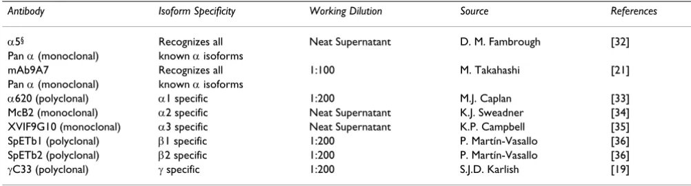
[image:8.612.53.554.110.245.2]Immunohistochemical studies were carried out on 8µm paraffin sections mounted on 3-aminopropyl-triethoxysi-lane (APES) treated slides. Normal, BPH and PCa tissues were incubated for identical periods of time with antibod-ies and the precipitating agent throughout the studantibod-ies. Sections were dewaxed in 100% xylene for 5 minutes and taken through a graded series (100%, 70 % and 50%) of alcohol baths for 1 min each before antigen retrieval in the microwave oven in the presence of 10 mM citrate buffer (pH 6.0) for 12 minutes. The sections were allowed to cool before washing in phosphate buffered saline solu-tion (PBS). Endogenous alkaline phosphatase was blocked for one hour at room temperature (RT) by treat-ment with 1.25 mM Levamisole solution (Vector Labora-tories, Peterborough, UK). Non-specific protein binding sites were blocked by addition of 10% normal goat serum in PBS (1 hour RT). A panel of monoclonal and polyclo-nal antibodies to the α and β subunits of Na, K-ATPase was used (Table 1). Sections were incubated with primary antibodies (various dilutions in PBS) for 24 hrs at 4°C and subsequently washed in PBS before a two-hour incu-bation with goat anti-rabbit IgG or goat anti-mouse IgG conjugated to alkaline phosphatase. Tissue sections were washed in PBS and alkaline phosphatase active sites were developed using Fast-Red TR/Naphthol AS-MX as precipi-tating agent for 10–15 minutes. Sections were counter-stained with haematoxylin for 1 min and washed in distilled water before mounting in aqueous medium and visualization under a light microscope. Photographs were taken using a Nikon Microphot-FX microscope fitted with a Nikon DXM1200 digital camera or goat anti-mouse IgG. Table 1: Isoform specific antibodies used to detect the isoforms of Na, K-ATPase in canine prostatic tissue by immunohistochemical analysis.
Antibody Isoform Specificity Working Dilution Source References
α5§ Recognizes all Neat Supernatant D. M. Fambrough [32] Pan α (monoclonal) known α isoforms
mAb9A7 Recognizes all 1:100 M. Takahashi [21]
Pan α (monoclonal) known α isoforms
α620 (polyclonal) α1 specific 1:200 M.J. Caplan [33]
McB2 (monoclonal) α2 specific Neat Supernatant K.J. Sweadner [34] XVIF9G10 (monoclonal) α3 specific Neat Supernatant K.P. Campbell [35] SpETb1 (polyclonal) β1 specific 1:200 P. Martín-Vasallo [36] SpETb2 (polyclonal) β2 specific 1:200 P. Martín-Vasallo [36]
γC33 (polyclonal) γ specific 1:200 S.J.D. Karlish [19]
Image Analysis
Image analysis was carried out using Scion Image for Win-dows (version 4.0.2 http://www.scioncorp.com/) based on NIH Image for Macintosh. Sections used for image analysis were only exposed to the Fast-Red precipitating agent and not counterstained with hematoxylin.
Statistical analysis
The results are expressed as the means +/-SD of a repre-sentative experiment performed in triplicate. The means were compared using student's t-test assuming equal vari-ances. p < 0.05 was considered statistically significant.
Note added in proof
In a very recent study of Na, K-ATPase α and β subunit expression in bladder tumours, human urothelial cancer tissue microarrays have been successfully used to demon-strate that the mean protein expression for both α and β subunits of Na, K-ATPase is reduced in invasive bladder tumours compared to benign and dysplastic tissue [31]. These recent findings partially confirm our results in canine prostate cancer. The authors have suggested that Na, K-ATPase α and β subunit expression levels may be useful predictors of clinical outcomes.
Acknowledgements
Research grants from the Pet Plan Charitable Trust (Grant no. 02–11) and the Wellcome Trust (U.K.) supported this work. We wish to thank the pathologists of the Department of Veterinary Pathology at the University of Liverpool for supplying tissues and original histopathological interpreta-tions. We are particularly grateful to Dr M. Takahashi and Dr M.W. McEn-ery for the pan α specific monoclonal mAb 9A7. We express our gratitude to Dr. D.M. Fambrough (Johns Hopkins University), Dr. K.J. Sweadner (Harvard University), Dr. S.J.D. Karlish (Weizmann Institute of Science, Rehovot, Israel) and Dr. M.J. Caplan (Yale University School of Medicine) for their continued generosity in provision of antibodies. We would also like to acknowledge Dr. D. Alvarez de la Rosa (Yale University School of Medicine) for critical comments on the manuscript and Dr. L.C. Costello (University of Maryland) for useful discussions and invaluable advice.
References
1. Costello LC and Franklin RB: Citrate metabolism of normal and malignant prostate epithelial cellsUrology 1997, 50:3-12. 2. Kavanagh JP: Sodium, potassium, calcium, magnesium, zinc,
citrate and chloride content of human prostatic and seminal fluidJ Reprod Fertil 1985, 75:35-41.
3. Franklin RB, Lao LX and Costello LC: Evidence for two aspartate transport systems in prostate epithelial cellsProstate 1990, 16:137-45.
4. Costello LC and Franklin RB: The intermediary metabolism of the prostate: a key to understanding the pathogenesis and progression of prostate malignancyOncology 2000, 59:269-82. 5. Smith ER: The secretion of electrolytes by the
pilocarpine-stimulated canine prostate glandProc Soc Exp Biol Med 1969, 132:223-6.
6. Farnsworth WE: Androgen regulation of prostatic membrane ATPaseBiol Reprod 1970, 3:218-22.
7. Farnsworth WE: Na+, K(+)-ATPase: the actual androgen receptor of the prostate?Med Hypotheses 1993, 41:358-62. 8. Blok LJ, de Ruiter PE and Brinkmann AO: Forskolin-induced
dephosphorylation of the androgen receptor impairs ligand bindingBiochemistry 1998, 37:3850-7.
9. Blok LJ, Chang GT, Steenbeek-Slotboom M, van Weerden WM, Swarts HG, De Pont JJ, van Steenbrugge GJ and Brinkmann AO: Reg-ulation of expression of Na+, K+-ATPase in androgen-dependent and androgen-inandrogen-dependent prostate cancerBr J Cancer 1999, 81:28-36.
10. Blanco G and Mercer RW: Isozymes of the Na-K-ATPase: het-erogeneity in structure, diversity in functionAm J Physiol 1998, 275:F633-50.
11. Mobasheri A, Avila J, Cozar-Castellano I, Brownleader MD, Trevan M, Francis MJ, Lamb JF and Martin-Vasallo P: Na+, K+-ATPase iso-zyme diversity; comparative biochemistry and physiological implications of novel functional interactionsBiosci Rep 2000, 20:51-91.
12. Scheiner-Bobis G, Meyer zu Heringdorf D, Christ M and Habermann E: Palytoxin induces K+ efflux from yeast cells expressing the mammalian sodium pumpMol Pharmacol 1994, 45:1132-6. 13. Shamraj OI and Lingrel JB: A putative fourth Na+, K(+)-ATPase
alpha-subunit gene is expressed in testisProc Natl Acad Sci U S A
1994, 91:12952-6.
14. Woo AL, James PF and Lingrel JB: Sperm motility is dependent on a unique isoform of the Na, K-ATPaseJ Biol Chem 2000, 275:20693-9.
15. Mercer RW, Biemesderfer D, Bliss DP Jr, Collins JH and Forbush B 3rd: Molecular cloning and immunological characterization of the gamma polypeptide, a small protein associated with the Na, K-ATPaseJ Cell Biol 1993, 121:579-86.
16. Beguin P, Wang X, Firsov D, Puoti A, Claeys D, Horisberger JD and Geering K: The gamma subunit is a specific component of the Na, K-ATPase and modulates its transport functionEmbo J
1997, 16:4250-60.
17. Sweadner KJ and Rael E: The FXYD gene family of small ion transport regulators or channels: cDNA sequence, protein signature sequence, and expressionGenomics 2000, 68:41-56. 18. Arystarkhova E, Wetzel RK, Asinovski NK and Sweadner KJ: The
gamma subunit modulates Na(+) and K(+) affinity of the renal Na, K-ATPaseJ Biol Chem 1999, 274:33183-5.
19. Kuster B, Shainskaya A, Pu HX, Goldshleger R, Blostein R, Mann M and Karlish SJ: A new variant of the gamma subunit of renal Na, K-ATPase. Identification by mass spectrometry, anti-body binding, and expression in cultured cellsJ Biol Chem 2000, 275:18441-6.
20. Mobasheri A, Oukrif D, Dawodu SP, Sinha M, Greenwell P, Stewart D, Djamgoz MB, Foster CS, Martin-Vasallo P and Mobasheri R: Iso-forms of Na+, K+-ATPase in human prostate; specificity of expression and apical membrane polarizationHistol Histopathol
2001, 16:141-54.
21. Choi Y, Dubel SJ, Pacioaiou ML, Omori A, Ito T, Copeland TD, Taka-hashi M and McEnery MW: Parallel detection of Na, K-ATPase alpha subunit isoforms by pan-specific monoclonal mAb 9A7 Arch Biochem Biophys 1997, 344:165-75.
22. Schmidt JD, Gibbons RP, Murphy GP and Bartolucci A: Adjuvant therapy for localized prostate cancerCancer 1993, 71:1005-13. 23. Laniado ME, Lalani EN, Fraser SP, Grimes JA, Bhangal G, Djamgoz MB and Abel PD: Expression and functional analysis of voltage-activated Na+ channels in human prostate cancer cell lines and their contribution to invasion in vitroAm J Pathol 1997, 150:1213-21.
24. Smith P, Rhodes NP, Shortland AP, Fraser SP, Djamgoz MB, Ke Y and Foster CS: Sodium channel protein expression enhances the invasiveness of rat and human prostate cancer cellsFEBS Lett
1998, 423:19-24.
25. Foster CS, Cornford P, Forsyth L, Djamgoz MB and Ke Y: The cel-lular and molecular basis of prostate cancer BJU Int 1999, 83:171-94.
26. Sandstrom PE, Jonsson O, Grankvist K and Henriksson R: Identifica-tion of potassium flux pathways and their role in the cytotox-icity of estramustine in human malignant glioma, prostatic carcinoma and pulmonary carcinoma cell linesEur J Cancer
1994, 30A:1822-6.
27. Fraser SP, Ding Y, Liu A, Foster CS and Djamgoz MB: Tetrodotoxin suppresses morphological enhancement of the metastatic MAT-LyLu rat prostate cancer cell lineCell Tissue Res 1999, 295:505-12.
Publish with BioMed Central and every scientist can read your work free of charge "BioMed Central will be the most significant development for disseminating the results of biomedical researc h in our lifetime."
Sir Paul Nurse, Cancer Research UK
Your research papers will be:
available free of charge to the entire biomedical community
peer reviewed and published immediately upon acceptance
cited in PubMed and archived on PubMed Central
yours — you keep the copyright
Submit your manuscript here:
http://www.biomedcentral.com/info/publishing_adv.asp
BioMedcentral
Cancer Cell International 2003, 3 http://www.cancerci.com/content/3/1/8
29. Djamgoz MBA, Mycielska M, Madeja Z, Fraser SP and Korohoda W: Directional movement of rat prostate cancer cells in direct-current electric field: involvement of voltagegated Na+ channel activityJ Cell Sci 2001, 114:2697-705.
30. Foster CS and Ke Y: Stem cells in prostatic epitheliaInt J Exp Pathol 1997, 78:311-29.
31. Espineda C, Seligson DB, James Ball W Jr, Rao J, Palotie A, Horvath S, Huang Y, Shi T and Rajasekaran AK: Analysis of the Na, K-ATPase alpha- and beta-subunit expression profiles of bladder cancer using tissue microarraysCancer 2003, 97:1859-68.
32. Takeyasu K, Tamkun MM, Renaud KJ and Fambrough DM: Ouabain-sensitive (Na+ + K+)-ATPase activity expressed in mouse L cells by transfection with DNA encoding the alpha-subunit of an avian sodium pumpJ Biol Chem 1988, 263:4347-54.
33. Gottardi CJ and Caplan MJ: Molecular requirements for the cell-surface expression of multisubunit ion-transporting ATPases. Identification of protein domains that participate in Na, K-ATPase and H, K-ATPase subunit assemblyJ Biol Chem 1993, 268:14342-7.
34. Urayama O, Shutt H and Sweadner KJ: Identification of three iso-zyme proteins of the catalytic subunit of the Na, K-ATPase in rat brainJ Biol Chem 1989, 264:8271-80.
35. Arystarkhova E and Sweadner KJ: Isoform-specific monoclonal antibodies to Na, K-ATPase alpha subunits. Evidence for a tissue-specific post-translational modification of the alpha subunitJ Biol Chem 1996, 271:23407-17.