18-Fluorodeoxyglucose Positron Emission
Tomography/Computed Tomography
in the Management of Aggressive
Non-Hodgkin's B-Cell Lymphoma
The Harvard community has made this
article openly available. Please share how
this access benefits you. Your story matters
Citation
Shelly, M. J., S. McDermott, O. J. O'Connor, and M. A. Blake. 2012.
18-fluorodeoxyglucose positron emission tomography/computed
tomography in the management of aggressive non-Hodgkin's B-cell
lymphoma. ISRN Hematology 2012:456706.
Published Version
doi:10.5402/2012/456706
Citable link
http://nrs.harvard.edu/urn-3:HUL.InstRepos:10341907
Terms of Use
This article was downloaded from Harvard University’s DASH
repository, and is made available under the terms and conditions
applicable to Other Posted Material, as set forth at
http://
Volume 2012, Article ID 456706,9pages doi:10.5402/2012/456706
Review Article
18-Fluorodeoxyglucose Positron Emission
Tomography/Computed Tomography in the Management of
Aggressive Non-Hodgkin’s B-Cell Lymphoma
M. J. Shelly,
1, 2S. McDermott,
1, 2O. J. O’Connor,
1, 2and M. A. Blake
1, 21Department of Radiology, Massachusetts General Hospital, Boston, MA 02114, USA 2Division of Abdominal Imaging & Interventional Radiology, Massachusetts General Hospital,
Harvard Medical School, Boston, MA 02114, USA
Correspondence should be addressed to M. J. Shelly,mjshelly@partners.org
Received 8 December 2011; Accepted 29 December 2011
Academic Editors: S. Daenen, M. Gobbi, and S. Koschmieder
Copyright © 2012 M. J. Shelly et al. This is an open access article distributed under the Creative Commons Attribution License, which permits unrestricted use, distribution, and reproduction in any medium, provided the original work is properly cited.
18-Fluorodeoxyglucose (FDG-PET/CT) is an established imaging modality that has been proven to be of benefit in the management of aggressive B-cell non-Hodgkin’s lymphoma, such as diffuse large B-cell lymphoma and advanced stage follicular lymphoma. The combination of anatomic and functional imaging afforded by FDG-PET/CT has led to superior sensitivity and specificity in the primary staging, restaging, and assessment of response to treatment of hematological malignancies when compared to FDG-PET and CT alone. The use of FDG-PET/CT for posttreatment surveillance imaging remains controversial, and further study is needed to ascertain whether this modality is cost effective and appropriate for use in this setting.
1. Introduction
The lymphoproliferative disorders, which can be broadly divided into Hodgkin’s (HL) and non-Hodgkin’s (NHL) lymphoma, represent a heterogeneous group of lymphoid malignancies that display varying patterns of biological behavior and response to treatment [1]. It is estimated that over 65,000 Americans will be diagnosed with NHL with over 20,000 deaths in 2011, making it the eighth and sixth most common cause of cancer death in men and women, respectively [2]. The annual incidence of NHL is trending upwards year on year; however, the overall 5-year survival rate has been steadily improving due in part to refinements in treatment regimens which minimize patient toxicity and maximize remission rates. Classification of lym-phomas according to the 2008 World Health Organization classification system groups lymphomas by cell type (i.e., the normal cell type that most resembles the tumor) and defines phenotypic, molecular, or cytogenetic characteristics with three major groupings, namely, the B-cell, T-cell, and natural killer cell neoplasms [3]. The most common subtypes of NHL affecting adults are follicular lymphoma (FL) and
diffuse large B-cell lymphoma (DLBCL), which together account for greater than 50% of NHL [4,5]. Prognosis of patients with NHL is affected by the stage and grade of disease as well as the histological subtype. Physicians often classify NHL into indolent, aggressive, and highly aggressive histologic subtypes for the purpose of estimating prognosis and determining treatment options. This paper will discuss the role of FDG-PET/CT in the pretreatment staging, restag-ing, treatment monitorrestag-ing, assessment of transformation, and posttherapy surveillance of patients with aggressive non-Hodgkin’s lymphoma of B-cell origin. The discussion of the role of FDG-PET/CT in the management of Hodgkin’s disease is beyond the scope of this paper.
2. Imaging Considerations in Lymphoma
2 ISRN Hematology
response, and prognosis in patients with aggressive NHL [8]. There are a number of caveats that one must be aware of when interpreting FDG-PET/CT in the setting of lymphoma. Certain subtypes of B-cell lymphoma, par-ticularly extranodal marginal zone lymphoma and periph-eral T-cell lymphoma, display variable FDG uptake and can also be sometimes difficult to identify on contrast enhanced CT (CECT) [9]. In these cases, a baseline FDG-PET/CT is essential to facilitate meaningful interpretation of posttreatment imaging in terms of treatment response. A number of subtypes of lymphoma may be difficult or almost impossible to detect with FDG-PET/CT such as primary CNS lymphoma, testicular lymphoma, and gastric lymphoma. The normal brain and testes demonstrate intense FDG avidity and against this high background FDG activity, primary CNS and testicular lymphoma may be very difficult to detect without adjusting the image intensity thresholds [10]. Gastric lymphoma displays an extremely variable FDG uptake that may be difficult or impossible to differentiate from normal physiologic FDG uptake in the gastric mucosa [11]. Diffuse or heterogeneous bone marrow uptake of radiotracer in a pretreatment scan should not immediately suggest bone marrow involvement by lymphoma as this pattern of uptake can be seen in reactive myeloid hyperplasia [5,12]. Focally increased FDG uptake can also be seen in the musculoskeletal system due to trauma, muscle activity, and degenerative change in synovial joints and also in brown fat which has a higher basal metabolic rate than adjacent tissues and may be mistaken for a lymphoma deposit. However, brown fat has a characteristic distribution and its detection is usually reasonably straightforward on FDG-PET/CT [13] (Figure 1). If patients with lymphoma are treated with granulocyte-colony stimulating factors, their bone marrow may show a diffuse increase in FDG uptake. It is very important to recognize the aforementioned pitfalls to avoid false-positive interpretation of FDG-PET/CT scans and hence potential overstaging of patients.
2.1. FDG-PET/CT Technique. There is a wide variance in
the protocols used to obtain FDG-PET/CT images with variations in the use of intravenous contrast and the tube current (and hence radiation dose) used in the CT compo-nent of the study. In general, a patient who is undergoing an FDG-PET/CT scan will have their blood glucose level measured prior to the intravenous administration of FDG and if the serum glucose concentration is too high (above 140 mg/dL), a subcutaneous injection of rapid acting insulin may be considered [14]. The patient is placed in a recumbent position and instructed to limit movement as this will reduce muscle activity and prevent unwanted FDG uptake in skeletal muscle. An intravenous dose of FDG is administered (the dose is dependent on the patient’s weight) and imaging is performed 1-2 hours (usually 1 hour) after injection as this allows a steady state of FDG uptake. Imaging is performed using an FDG-PET/CT scanner which integrates a multidetector CT scanner with a high-resolution PET scanner, facilitating the acquisition of three-dimensional images. Axial whole-body FDG-PET/CT images are acquired from the vertex or skull base to the upper thigh [8].
The “whole-body” nature of the imaging technique makes FDG-PET/CT ideal for evaluating systemic diseases such as lymphoma.
2.2. Standardized Uptake Value (SUV). SUVs represent
reproducible estimates of tissue glucose metabolic activity and can be used for semiquantitative analysis of findings on FDG-PET/CT in patients with lymphoma [15]. This is very useful in clinical practice as it allows accurate comparison of lesions on subsequent scans (i.e., to guide response to treatment) and can be used to evaluate the degree of residual metabolic activity in a treated mass. This helps physicians make clinical decisions regarding the use of salvage chemotherapy [16]. While the SUV is semiquantitative, a number of studies have demonstrated that visual assessment of treatment response in patients with lymphoma is adequate for determining whether an FDG-PET/CT scan is positive for disease [6,7].
2.3. FDG-PET/CT versus CT. FDG-PET/CT is often not the
first-line imaging modality in the assessment of a patient with suspected lymphoma. CT is widely available and, in part due to ease of access and cost factors, it was the dominant radiologic tool for the assessment of lymphoma over the past three decades. Lymphoma displays homogenous attenuation on CT with characteristic patterns of disease such as vessel encasement and displacement (the so-called “sandwich sign” [17]) and spread across existing anatomic structures which indicates an aggressive, permeative disease process [8] (Figure 2).
However, the fundamental limitation of CT in imaging lymphoma is that recognition of nodal involvement by disease is based solely on size criteria and the detection of bone marrow and extranodal involvement is limited. PET/CT incorporates a CT component in addition to a FDG-PET scan, and it has been demonstrated to have a higher sensitivity and specificity for the detection of nodal and extranodal sites of disease when compared to CT alone, thus improving baseline staging [8, 18–20]. It has long been recognized that CT is limited in its ability to assess the response of lymphoma to treatment as CT cannot differentiate between viable tumor and necrosis or fibrosis in patients presenting with persistent masses posttreatment [21]. The sensitivity and specificity of FDG-PET/CT has been shown to be superior to that of CT alone in the posttreatment assessment of lymphoma due to the addition of information regarding the metabolic activity of residual masses (RMs) detected on CT [8,20].
2.4. FDG-PET/CT versus Gallium-67 Citrate Scintigraphy.
The development and clinical utilization of the first molec-ular imaging agent for lymphoma assessment, namely, Gallium-67 citrate (67Ga), greatly improved the accuracy of
treatment response assessment in patients with lymphoma [22–24].67Ga scanning is based on the accumulation of the
(a) (b) (c)
Figure 1: Coronal PET maximum intensity projection (MIP) image (a) displaying foci of increased radiotracer uptake bilaterally in the neck
in a patient with treated DLBSL. The corresponding coronal CT (b) and fused FDG-PET/CT (c) demonstrate no abnormality and the areas with increased FDG uptake correspond to regions of fat density (arrows). This is the characteristic appearance of hypermetabolic brown fat on FDG-PET/CT.
(a) (b) (c)
Figure 2: Axial (a) and coronal (b) contrast enhanced CT of the abdomen in a patient with chronic lymphocytic leukemia displaying
encasement of vessels such as the left renal vein (white arrow) and right renal artery (black arrow) by lymph nodes masses, the so-called “sandwich sign.” Axial contrast enhanced CT (c) of the abdomen at a lower level in the same patient displaying elevation of the abdominal aorta offthe vertebral column (white arrow) which is a characteristic feature of lymphoma and helps to differentiate lymphoma from other retroperitoneal masses, such as retroperitoneal fibrosis. Note the encasement of the superior mesenteric artery (SMA) by the lymph node mass and increased separation of the SMA from the aorta (black arrow).
negative 67Ga scan; however, the positive and negative
predictive values of67Ga imaging are suboptimal [22,23].
There are a number of technical and clinical factors that have limited the use and acceptance of67Ga imaging, namely,
its low spatial resolution, low sensitivity and specificity, its limited use for imaging intra-abdominal disease (due to physiologic bowel uptake), and the time involved in performing the scan, which can take up to a week after67Ga
injection [25,26].67Ga imaging has been largely replaced
by FDG-PET, which is a radiolabelled isomer of glucose and is hence a very sensitive marker of glucose metabolism and metabolic activity [27]. FDG-PET is superior to 67Ga
scintigraphy in terms of sensitivity for the detection of and specificity for malignant lymphoma [27–30]. The use of FDG-PET/CT tends to upstage patients due to the detection of additional sites of lymphomatous involvement such as the liver, spleen, and lung which can result in a change of treatment strategy [30–32].
3. FDG-PET/CT for Primary Staging of
Aggressive B-Cell Lymphoma
4 ISRN Hematology
[image:5.600.114.474.69.214.2](a) (b) (c)
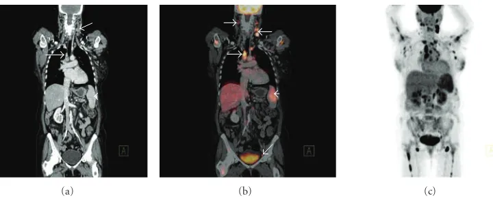
Figure 3: Coronal CT (a) demonstrating left cervical and right paratracheal lymphadenopathy (white arrows) and mild splenomegaly
(white arrowhead) consistent with high-grade follicular lymphoma. Corresponding coronal fused FDG-PET/CT (b) demonstrates increased radiotracer uptake in the pathologically enlarged left cervical and right paratracheal lymphadenopathy (white arrows) and spleen (white arrowhead), but also abnormal FDG accumulation in right cervical and left external iliac lymph nodes (white arrows) that appeared normal by size criteria on the corresponding CT scan. Coronal PET MIP (c) in the same patient demonstrating widespread increased radiotracer uptake throughout the neck, thorax, abdomen, and proximal humeri and femora bilaterally; the widespread bone marrow involvement was not apparent on CT.
and CT alone, with equal sensitivity and better specificity [30]. Pretreatment FDG-PET/CT imaging acts as a baseline study to facilitate comparison to posttreatment scans to assess response to treatment and evaluate RMs for metabolic activity [6, 7]. FDG-PET/CT has been demonstrated to be effective in the detection of focal or multifocal bone marrow infiltration in patients with a negative iliac crest bone marrow biopsy (BMB) [43]. However, it is important to stress that FDG-PET/CT alone is not completely reliable in detecting limited bone marrow involvement by lymphoma, and, hence, it cannot entirely replace BMB in the initial diagnosis of lymphomas of any type as a negative FDG-PET/CT scan cannot definitively outrule involvement of the bone marrow by lymphoma [44] (Figure 3).
Combined FDG-PET/CT imaging represents a func-tional and anatomical approach to lymphoma assessment which exploits the strengths of both modalities while at the same time minimizing the shortcomings of each individual modality. A number of studies have demonstrated that a staging FDG-PET/CT scan provides diagnostic information that is at least equal but likely superior to that which is provided by a separate FDG-PET and intravenous contrast enhanced CT (CECT) scan [7,12]. FDG-PET provides the functional information lacking on CT images that may represent the only indication of disease (e.g., abnormal FDG uptake in a normal-sized lymph node on CT). CT provides superior anatomic detail and spatial resolution that is lacking in FDG-PET which enables better localization of FDG uptake and, importantly, may identify false-negative FDG-PET findings due to the limited spatial resolution of PET scanners or lack of FDG avidity of some subtypes of lymphoma. The anatomic correlation of FDG uptake that is afforded by FDG-PET/CT is also very useful in reducing the rate of false-positive findings due to physiologic FDG uptake in, for example, muscle or brown fat [45]. There is debate in the published literature regarding the benefits of using intravenous contrast during the CT portion of a
FDG-PET/CT scan. Some authors suggest that the use of intravenous contrast reduces the number of indeterminate findings on FDG-PET/CT and results in the detection of a higher number of extranodal sites compared with FDG-PET/CT performed without intravenous contrast [46]. However, a number of studies suggest that an FDG-PET/CT scan performed without intravenous contrast and using a low radiation dose CT technique is an acceptable alternative to separate CECT and FDG-PET scans even when the known limitations of the unenhanced CT in the evaluation of the liver and spleen are taken into account [19, 47]. It is the opinion of the authors that, for initial staging of patients with lymphoma, an FDG-PET/CT scan using a standard radiation dose CT protocol and intravenous contrast is the optimal choice as a single imaging modality.
4. FDG-PET/CT for Restaging of
Aggressive B-Cell Lymphoma
[6,7] in lymphoma made a number of recommendations intended to reduce the rate of false-positive FDG-PET scan interpretation after completion of treatment.
(1) FDG-PET/CT should not be performed for at least 3 weeks after chemotherapy and 8–12 weeks after radiotherapy to minimize confounding FDG uptake secondary to posttreatment inflammation.
(2) RMs≥2 cm in maximum transverse diameter should only be considered positive for residual disease on FDG-PET if their FDG avidity visually exceeds that of the mediastinal blood pool structures.
(3) RMs with a maximum transverse diameter<2 cm are considered FDG-PET positive if their FDG uptake is higher than the surrounding background tissue.
(4) In the liver and spleen, focal lesions with FDG avidity greater than in surrounding liver or spleen parenchyma are considered positive for viable lym-phoma.
(5) Single or multiple foci of distinctly elevated FDG uptake in the bone marrow is positive for residual disease.
(6) Visual assessment alone is adequate for determining whether FDG-PET scans are positive or negative at the conclusion of treatment and SUV measurement is not necessary.
The use of FDG-PET/CT in restaging lymphoma is supported by a large body of evidence that shows that FDG-PET/CT has a high negative predictive value (NPV) that exceeds 80% in practically all reported studies for aggressive NHL, with the reported 10–20% false-negative rate mainly attributed to the inability of FDG-PET/CT to detect micro-scopic disease which results in future relapse [6,7,12,50– 53] (Figure 4). However, the positive predictive value (PPV) of FDG-PET/CT is reported in the moderate range of 70– 80% which is significantly lower and more variable than its NPV and is due to the recognized false-positive rate on FDG-PET due to persistent metabolic activity in RMs posttreatment that often represents inflammatory change rather than residual disease. The PPV of FDG-PET/CT is substantially higher than that of CT alone which has a reported PPV in the range of 40–50% in posttreatment aggressive NHL leading to a considerably higher accuracy of FDG-PET/CT for response assessment when compared to CT alone [6,7,12,50–53]. The significantly higher accuracy of FDG-PET/CT for response assessment when compared to CT alone justifies the increased cost associated with dual modality imaging when compared to CT or PET alone [54, 55].
5. FDG-PET/CT for Interim
Treatment Monitoring
The use of FDG-PET/CT for treatment monitoring is based on the assumption that interim imaging, typically performed
after two to three cycles of chemotherapy, provides accurate prediction of response to current treatment and ultimate patient outcome [30,56]. The application of FDG-PET/CT in interim treatment monitoring of lymphoma has been highly successful in Hodgkin’s disease [57,58]. However, the benefits of FDG-PET/CT in the management of aggressive NHL are less clear cut with conflicting reports in the literature regarding the usefulness of interim FDG-PET/CT in predicting treatment response and patient outcome with most reports recommending further prospective studies to ascertain the role of FDG-PET/CT in this respect [59–61]. There are a number of reported studies in the literature that support this premise, particularly with regard to the high NPV of interim FDG-PET/CT in patients with aggressive NHL [62–66]. It is interesting to note that the PPV and, hence, accuracy of interim FDG-PET scans that are visually interpreted (i.e., without the use of semiquantitative SUV) appear to depend on the “strictness” of criteria used to define a scan as positive with a number of studies demonstrating that a more “liberal” interpretation of FDG-PET findings at the site of RMs on interim FDG-PET results in higher PPV and accuracy with no compromise in NPV [66–68]. This “liberal” visual interpretation involves considering an FDG-PET scan as negative if it displays only minimal FDG uptake that is similar or only slightly greater in intensity than FDG uptake in normal liver [66–68]. If these “liberal” criteria for defining FDG-PET negativity are used, interim FDG-PET was found to be at least as accurate for predicting patient outcome as end of treatment FDG-PET interpreted using “strict” criteria that does not allow any residual FDG uptake in RMs [68].
6 ISRN Hematology
(a) (b)
Figure 4: Coronal fused FDG-PET/CT image (a) demonstrating increased radiotracer uptake in multiple intra-abdominal lymph node
masses in a patient with DLBCL (white arrows). Note how a large soft tissue mass displaces the small bowel and the mesenteric vessels (white arrowhead). Coronal fused FDG-PET/CT image (b) in the same patient demonstrating complete resolution of the previously described soft tissue masses. The observed residual FDG avidity is within bowel and is normal. This FDG-PET/CT confirms complete response to treatment.
(a) (b)
Figure 5: Coronal fused FDG-PET/CT image (a) of a patient with nodular sclerosis type Hodgkin’s lymphoma prechemotherapy
demonstrating an FDG-avid lymph node mass in the superior mediastinum (white arrowhead) and hypermetabolic bilateral axillary lymphadenopathy (white arrows). Interim staging coronal fused FDG-PET/CT image (b) in the same patient after 3 cycles of chemotherapy demonstrating a significant interval decrease in both the degree of FDG uptake and the size of the lymph node mass in the superior mediastinum (white arrow) with resolution of the axillary lymphadenopathy.
6. FDG-PET/CT for Posttreatment Surveillance
The use of FDG-PET/CT for posttreatment surveillance in patients with aggressive NHL is performed following completion of treatment and in the absence of clinical, biochemical, or radiologic evidence of disease recurrence (i.e., a complete response (CR) to treatment) with the aim of early detection of disease relapse and hence earlier treatment to improve patient outcome [25]. A recent study reported positive results with the use of surveillance FDG-PET scanning in 421 lymphoma patients (160 HL, 183 aggressive NHL, and 78 indolent NHL) who were imaged at 6-month intervals for 2 years and then yearly thereafter following initial CR. This study demonstrated that the detection rate of proven relapses was higher with FDG-PET compared with CT alone or clinical assessment in aggressive NHL (31% versus 25% and 22%, resp.) and
posttreatment surveillance, and thus, until large prospective trials address these issues, the use of FDG-PET/CT for disease surveillance will remain controversial with some authors suggesting that its use should be limited to a clinical trial setting [69, 72]. If there is a low clinical suspicion for recurrence of patients with treated initially FDG-avid lymphomas, then FDG-PET/CT scan using a low radiation dose CT protocol without intravenous contrast may be a reasonable choice of a single lower-dose imaging modality for follow-up assessment. The routine use of FDG-PET/CT in disease surveillance is not yet supported by a sufficient body of evidence to justify its cost and associated radiation dose, and there are also some issues in relation to false-positive FDG-PET/CT studies during disease surveillance which could cause unwarranted patient distress and further add to the cost of management.
7. FDG-PET/CT for Assessment
of Transformation
The transformation of indolent lymphoma to a higher grade of lymphoma can occur, most commonly with DLBCL, and is associated with a very poor prognosis and is an indication for aggressive, high-dose chemotherapy and postremission stem cell transplantation [73,74]. FDG-PET/CT is useful to confirm the clinical suspicion of transformation of an indo-lent lymphoma to a more aggressive histology by identifying sites of abnormally high FDG uptake (indolent lymphoma normally has low-grade FDG uptake) and also guiding selec-tion of an optimal biopsy site for pathologic confirmaselec-tion of transformation [41,73]. Due to the significant overlap in the degree of FDG uptake between indolent and aggressive lymphomas, FDG-PET/CT cannot entirely replace biopsy in the assessment of transformation of lymphoma but rather may be a useful alternative when a biopsy is not practical due to technical or clinical reasons [41,75].
8. Conclusion
FDG-PET/CT is an established imaging modality that has been proven to be of benefit in the management of aggressive B-cell NHL, such as DLBCL and advanced stage FL. The combination of anatomic and functional imaging afforded by FDG-PET/CT has led to superior sensitivity and speci-ficity in the primary staging, restaging, and assessment of response to treatment of hematological malignancies when compared to PET and CT alone. The use of FDG-PET/CT for posttreatment surveillance imaging remains controversial, and further study is needed to ascertain whether this modality is cost effective and appropriate for use in this setting.
References
[1] C. R. Flowers and J. O. Armitage, “A decade of progress in lymphoma: advances and continuing challenges,” Clinical
Lymphoma, Myeloma and Leukemia, vol. 10, no. 6, pp. 414–
423, 2010.
[2] A. Jemal, R. Siegel, J. Xu, and E. Ward, “Cancer statistics, 2010,” CA Cancer Journal for Clinicians, vol. 60, no. 5, pp. 277– 300, 2010.
[3] E. Campo, S. H. Swerdlow, N. L. Harris, S. Pileri, H. Stein, and E. S. Jaffe, “The 2008 WHO classification of lymphoid neoplasms and beyond: evolving concepts and practical applications,” Blood, vol. 117, no. 19, pp. 5019–5032, 2011. [4] E. S. Jaffe, “The 2008 WHO classification of lymphomas:
implications for clinical practice and translational research,”
Hematology, pp. 523–531, 2009.
[5] C. G. Cronin, R. Swords, M. T. Truong et al., “Clinical utility of PET/CT in lymphoma,” American Journal of Roentgenology, vol. 194, no. 1, pp. W91–W103, 2010.
[6] B. D. Cheson, B. Pfistner, M. E. Juweid et al., “Revised response criteria for malignant lymphoma,” Journal of Clinical
Oncology, vol. 25, no. 5, pp. 579–586, 2007.
[7] M. E. Juweid, S. Stroobants, O. S. Hoekstra et al., “Use of positron emission tomography for response assessment of lymphoma: consensus of the imaging subcommittee of international harmonization project in lymphoma,” Journal of
Clinical Oncology, vol. 25, no. 5, pp. 571–578, 2007.
[8] M. Okada, N. Sato, K. Ishii, K. Matsumura, M. Hosono, and T. Murakami, “FDG PET/CT versus CT, MR imaging, and 67Ga scintigraphy in the posttherapy evaluation of Malignant lymphoma,” Radiographics, vol. 30, no. 4, pp. 939–957, 2010. [9] M. Hoffmann, K. Kletter, A. Becherer, U. J¨ager, A. Chott,
and M. Raderer, “18F-fluorodeoxyglucose positron emission
tomography (18F-FDG-PET) for staging and follow-up of
marginal zone B-cell lymphoma,” Oncology, vol. 64, no. 4, pp. 336–340, 2003.
[10] S. C. Chua, F. I. Rozalli, and S. R. O’Connor, “Imaging features of primary extranodal lymphomas,” Clinical Radiology, vol. 64, no. 6, pp. 574–588, 2009.
[11] W. K. Lee, E. W. F. Lau, V. A. Duddalwar, A. J. Stanley, and Y. Y. Ho, “Abdominal manifestations of extranodal lymphoma: spectrum of imaging findings,” American Journal
of Roentgenology, vol. 191, no. 1, pp. 198–206, 2008.
[12] P. Seam, M. E. Juweid, and B. D. Cheson, “The role of FDG-PET scans in patients with lymphoma,” Blood, vol. 110, no. 10, pp. 3507–3516, 2007.
[13] F. M. Paes, D. G. Kalkanis, P. A. Sideras, and A. N. Serafini, “FDG PET/CT of extranodal involvement in non-Hodgkin lymphoma and Hodgkin disease,” Radiographics, vol. 30, no. 1, pp. 269–291, 2010.
[14] G. K. von Schulthess, H. C. Steinert, and T. F. Hany, “Inte-grated PET/CT: current applications and future directions,”
Radiology, vol. 238, no. 2, pp. 405–422, 2006.
[15] S. C. Huang, “Anatomy of SUV. Standardized uptake value,”
Nuclear Medicine and Biology, vol. 27, pp. 643–646, 2000.
[16] M. Allen-Auerbach, S. de Vos, and J. Czernin, “The impact of fluorodeoxyglucose-positron emission tomography in pri-mary staging and patient management in lymphoma patients,”
Radiologic Clinics of North America, vol. 46, no. 2, pp. 199–211,
2008.
[17] P. R. Mueller, J. T. Ferrucci Jr., and W. P. Harbin, “Appearance of lymphomatous involvement of the mesentery by ultra-sonography and body computed tomography: the “sandwich sign”,” Radiology, vol. 134, no. 2, pp. 467–473, 1980.
[18] M. Tatsumi, C. Cohade, Y. Nakamoto, E. K. Fishman, and R. L. Wahl, “Direct comparison of FDG PET and CT findings in patients with lymphoma: initial experience,” Radiology, vol. 237, no. 3, pp. 1038–1045, 2005.
8 ISRN Hematology
Nuclear Medicine and Molecular Imaging, vol. 31, no. 3, pp.
325–329, 2004.
[20] L. Le Dortz, S. De Guibert, S. Bayat et al., “Diagnostic and prognostic impact of 18F-FDG PET/CT in follicular
lym-phoma,” European Journal of Nuclear Medicine and Molecular
Imaging, vol. 37, no. 12, pp. 2307–2314, 2010.
[21] B. D. Cheson, S. J. Horning, B. Coiffier et al., “Report of an international workshop to standardize response criteria for non-Hodgkin’s lymphomas,” Journal of Clinical Oncology, vol. 17, no. 4, pp. 1244–1253, 1999.
[22] W. D. Kaplan, M. S. Jochelson, T. S. Herman et al., “Gallium-67 imaging: a predictor of residual tumor viability and clinical outcome in patients with diffuse large-cell lymphoma,”
Journal of Clinical Oncology, vol. 8, no. 12, pp. 1966–1970,
1990.
[23] D. Front, R. Bar-Shalom, M. Mor et al., “Aggressive non-Hodgkin lymphoma: early prediction of outcome with 67Ga scintigraphy,” Radiology, vol. 214, no. 1, pp. 253–257, 2000. [24] D. Front, R. Bar-Shalom, and O. Israel, “The continuing
clinical role of gallium 67 scintigraphy in the age of receptor imaging,” Seminars in Nuclear Medicine, vol. 27, no. 1, pp. 68– 74, 1997.
[25] B. D. Cheson, “Role of functional imaging in the management of lymphoma,” Journal of Clinical Oncology, vol. 29, no. 14, pp. 1844–1854, 2011.
[26] E. Even-Sapir and O. Israel, “Gallium-67 scintigraphy: a cornerstone in functional imaging of lymphoma,” European
Journal of Nuclear Medicine and Molecular Imaging, vol. 30, no.
1, pp. S65–S81, 2003.
[27] A. Wirth, J. F. Seymour, R. J. Hicks et al., “Fluorine-18 fluorodeoxyglucose positron emission tomography, gallium-67 scintigraphy, and conventional staging for Hodgkin’s disease and non-Hodgkin’s lymphoma,” American Journal of
Medicine, vol. 112, no. 4, pp. 262–268, 2002.
[28] N. Tsukamoto, M. Kojima, M. Hasegawa et al., “The use-fulness of18F-fluorodeoxyglucose positron emission
tomog-raphy (18F-FDG-PET) and a comparison of 18F-FDG-PET
with67gallium scintigraphy in the evaluation of lymphoma:
relation to histologic subtypes based on the World Health Organization classification,” Cancer, vol. 110, no. 3, pp. 652– 659, 2007.
[29] L. Kostakoglu, J. P. Leonard, I. Kuji, M. Coleman, S. Vallab-hajosula, and S. J. Goldsmith, “Comparison of fluorine-18 fluorodeoxyglucose positron emission tomography and Ga-67 scintigraphy in evaluation of lymphoma,” Cancer, vol. 94, no. 4, pp. 879–888, 2002.
[30] M. Hutchings and L. Specht, “PET/CT in the management of haematological malignancies,” European Journal of
Haematol-ogy, vol. 80, no. 5, pp. 369–380, 2008.
[31] P. Raanani, Y. Shasha, C. Perry et al., “Is CT scan still necessary for staging in Hodgkin and non-Hodgkin lymphoma patients in the PET/CT era?” Annals of Oncology, vol. 17, no. 1, pp. 117– 122, 2006.
[32] E. Miller, U. Metser, G. Avrahami et al., “Role of 18F-FDG
PET/CT in staging and follow-up of lymphoma in pediatric and young adult patients,” Journal of Computer Assisted
Tomography, vol. 30, no. 4, pp. 689–694, 2006.
[33] F. Moog, M. Bangerter, C. G. Diederichs et al., “Lymphoma: role of whole-body 2-deoxy-2-[F-18]fluoro-D-glucose (FDG) PET in nodal staging,” Radiology, vol. 203, no. 3, pp. 795–800, 1997.
[34] F. Moog, M. Bangerter, C. G. Diederichs et al., “Extranodal malignant lymphoma: detection with FDG PET versus CT,”
Radiology, vol. 206, no. 2, pp. 475–481, 1998.
[35] G. Jerusalem and Y. Beguin, “The place of positron emission tomography imaging in the management of patients with malignant lymphoma,” Haematologica, vol. 91, no. 4, pp. 442– 444, 2006.
[36] G. Jerusalem, Y. Beguin, M. F. Fassotte et al., “Whole-body positron emission tomography using18F-fluorodeoxyglucose
compared to standard procedures for staging patients with Hodgkin’s disease,” Haematologica, vol. 86, no. 3, pp. 266–273, 2001.
[37] N. G. Schaefer, T. F. Hany, C. Taverna et al., “Non-Hodgkin lymphoma and Hodgkin disease: coregistered FDG PET and CT at staging and restaging—do we need contrast-enhanced CT?” Radiology, vol. 232, no. 3, pp. 823–829, 2004.
[38] R. Naumann, B. Beuthien-Baumann, A. Reiß et al., “Sub-stantial impact of FDG PET imaging on the therapy decision in patients with early-stage Hodgkin’s lymphoma,” British
Journal of Cancer, vol. 90, no. 3, pp. 620–625, 2004.
[39] M. A. Pizzichetta, G. Argenziano, R. Talamini et al., “2-(fluorine-18)fluoro-2-deoxy-D-glucose positron emission tomography in the detection and staging of malignant lym-phoma: a bicenter trial,” Cancer, vol. 91, no. 5, pp. 889–899, 2001.
[40] H. Sch¨oder, J. Meta, C. Yap et al., “Effect of whole-body
18F-FDG PET imaging on clinical staging and management
of patients with malignant lymphoma,” Journal of Nuclear
Medicine, vol. 42, no. 8, pp. 1139–1143, 2001.
[41] H. Sch¨oder, A. Noy, M. G¨onen et al., “Intensity of
18fluorodeoxyglucose uptake in positron emission
tomog-raphy distinguishes between indolent and aggressive non-Hodgkin’s lymphoma,” Journal of Clinical Oncology, vol. 23, no. 21, pp. 4643–4651, 2005.
[42] M. Hutchings, A. Loft, M. Hansen et al., “Position emission tomography with or without computed tomography in the primary staging of Hodgkin’s lymphoma,” Haematologica, vol. 91, no. 4, pp. 482–489, 2006.
[43] F. Moog, M. Bangerter, J. Kotzerke, A. Guhlmann, N. Frick-hofen, and S. N. Reske, “18F-fluorodeoxyglucose-positron
emission tomography as a new approach to detect lymphoma-tous bone marrow,” Journal of Clinical Oncology, vol. 16, no. 2, pp. 603–609, 1998.
[44] E. E. Pakos, A. D. Fotopoulos, and J. P.A. Ioannidis, “18F-FDG
PET for evaluation of bone marrow infiltration in staging of lymphoma: a meta-analysis,” Journal of Nuclear Medicine, vol. 46, no. 6, pp. 958–963, 2005.
[45] M. Allen-Auerbach, A. Quon, W. A. Weber et al., “Comparison between 2-deoxy-2-[18F]fluoro-D-glucose positron emission
tomography and positron emission tomography/computed tomography hardware fusion for staging of patients with lymphoma,” Molecular Imaging and Biology, vol. 6, no. 6, pp. 411–416, 2004.
[46] B. Rodr´ıguez-Vigil, N. G ´omez-Le ´on, I. Pinilla et al., “PET/CT in lymphoma: prospective study of enhanced full-dose PET/CT versus unenhanced low-dose PET/CT,” Journal of
Nuclear Medicine, vol. 47, no. 10, pp. 1643–1648, 2006.
[47] R. L. Elstrom, J. P. Leonard, M. Coleman, and R. K. J. Brown, “Combined PET and low-dose, noncontrast CT scanning obviates the need for additional diagnostic contrast-enhanced CT scans in patients undergoing staging or restaging for lymphoma,” Annals of Oncology, vol. 19, no. 10, pp. 1770– 1773, 2008.
[48] G. Jerusalem, Y. Beguin, M. F. Fassotte et al., “Whole-body positron emission tomography using18F-fluorodeoxyglucose
value than classical computed tomography scan imaging,”
Blood, vol. 94, no. 2, pp. 429–433, 1999.
[49] J. M. Zijlstra, G. Lindauer-Van Der Werf, O. S. Hoek-stra, L. Hooft, I. I. Riphagen, and P. C. Huijgens, “18
F-fluoro-deoxyglucose positron emission tomography for post-treatment evaluation of malignant lymphoma: a systematic review,” Haematologica, vol. 91, no. 4, pp. 522–529, 2006. [50] N. G. Mikhaeel, A. R. Timothy, S. F. Hain, and M. J.
O’Doherty, “18-FDG-PET for the assessment of residual masses on CT following treatment of lymphomas,” Annals of
Oncology, vol. 11, no. 1, supplement, pp. S147–S150, 2000.
[51] N. G. Mikhaeel, A. R. Timothy, M. J. O’Doherty, S. Hain, and M. N. Maisey, “18-FDG-PET as a prognostic indicator in the treatment of aggressive non-Hodgkin’s lymphoma-comparison with CT,” Leukemia and Lymphoma, vol. 39, no. 5-6, pp. 543–553, 2000.
[52] P. L. Zinzani, M. Magagnoli, F. Chierichetti et al., “The role of positron emission tomography (PET) in the management of lymphoma patients,” Annals of Oncology, vol. 10, no. 10, pp. 1181–1184, 1999.
[53] M. E. Juweid, “18F-FDG PET as a routine test for
post-therapy assessment of Hodgkin’s disease and aggressive non-Hodgkin’s lymphoma: where is the evidence?” Journal of
Nuclear Medicine, vol. 49, no. 1, pp. 9–12, 2008.
[54] C. K. Hoh, J. Glaspy, P. Rosen et al., “Whole-body FDG-PET imaging for staging of Hodgkin’s disease and lymphoma,”
Journal of Nuclear Medicine, vol. 38, no. 3, pp. 343–348, 1997.
[55] T. Klose, R. Leidl, I. Buchmann, H. J. Brambs, and S. N. Reske, “Primary staging of lymphomas: cost-effectiveness of FDG-PET versus computed tomography,” European Journal of
Nuclear Medicine, vol. 27, no. 10, pp. 1457–1464, 2000.
[56] N. G. Mikhaeel, M. Hutchings, P. A. Fields, M. J. O’Doherty, and A. R. Timothy, “FDG-PET after two to three cycles of chemotherapy predicts progression-free and overall survival in high-grade non-Hodgkin lymphoma,” Annals of Oncology, vol. 16, no. 9, pp. 1514–1523, 2005.
[57] A. Gallamini, F. Fiore, R. Sorasio, and M. Meignan, “Interim positron emission tomography scan in Hodgkin lymphoma: definitions, interpretation rules, and clinical validation,”
Leukemia and Lymphoma, vol. 50, no. 11, pp. 1761–1764, 2009.
[58] A. Gallamini, C. Patti, S. Viviani et al., “Early chemotherapy intensification with BEACOPP in advanced-stage Hodgkin lymphoma patients with a interim-PET positive after two ABVD courses,” British Journal of Haematology, vol. 152, no. 5, pp. 551–560, 2011.
[59] D.-H. Yang, J.-J. Min, H.-C. Song et al., “Prognostic signifi-cance of interim18F-FDG PET/CT after three or four cycles
of R-CHOP chemotherapy in the treatment of diffuse large B-cell lymphoma,” European Journal of Cancer, vol. 47, no. 9, pp. 1312–1318, 2011.
[60] C. Yoo, D. H. Lee, J. E. Kim et al., “Limited role of interim PET/CT in patients with diffuse large B-cell lymphoma treated with R-CHOP,” Annals of Hematology, vol. 90, pp. 797–802, 2011.
[61] A. S. Michallet, J. Trotman, and C. Tychyj-Pinel, “Role of early PET in the management of diffuse large B-cell lymphoma,”
Current Opinion in Oncology, vol. 22, no. 5, pp. 414–418, 2010.
[62] G. Jerusalem, Y. Beguin, M. F. Fassotte et al., “Persistent tumor
18F-FDG uptake after a few cycles of polychemotherapy is
predictive of treatment failure in non-Hodgkin’s lymphoma,”
Haematologica, vol. 85, no. 6, pp. 613–618, 2000.
[63] K. Spaepen, S. Stroobants, P. Dupont et al., “Early restaging positron emission tomography with 18F-fluorodeoxyglucose
predicts outcome in patients with aggressive non-Hodgkin’s
lymphoma,” Annals of Oncology, vol. 13, no. 9, pp. 1356–1363, 2002.
[64] K. Spaepen, S. Stroobants, G. Verhoef, and L. Mortelmans, “Positron emission tomography with [18F]FDG for therapy
response monitoring in lymphoma patients,” European
Jour-nal of Nuclear Medicine and Molecular Imaging, vol. 30, no. 1,
supplement, pp. S97–S105, 2003.
[65] L. Kostakoglu, M. Coleman, J. P. Leonard, I. Kuji, H. Zoe, and S. J. Goldsmith, “Pet predicts prognosis after 1 cycle of chemotherapy in aggressive lymphoma and Hodgkin’s disease,” Journal of Nuclear Medicine, vol. 43, no. 8, pp. 1018– 1027, 2002.
[66] C. Lin, E. Itti, C. Haioun et al., “Early 18F-FDG PET for
prediction of prognosis in patients with diffuse large B-cell lymphoma: SUV-based assessment versus visual analysis,”
Journal of Nuclear Medicine, vol. 48, no. 10, pp. 1626–1632,
2007.
[67] W. A. Weber, “18F-FDG PET in non-Hodgkin’s lymphoma:
qualitative or quantitative?” Journal of Nuclear Medicine, vol. 48, no. 10, pp. 1580–1582, 2007.
[68] M. Hutchings, N. G. Mikhaeel, P. A. Fields, T. Nunan, and A. R. Timothy, “Prognostic value of interim FDG-PET after two or three cycles of chemotherapy in Hodgkin lymphoma,”
Annals of Oncology, vol. 16, no. 7, pp. 1160–1168, 2005.
[69] B. Cheson, “The case against heavy PETing,” Journal of Clinical
Oncology, vol. 27, no. 11, pp. 1742–1743, 2009.
[70] P. L. Zinzani, V. Stefoni, M. Tani et al., “Role of [18F]fluorodeoxyglucose positron emission tomography scan
in the follow-up of lymphoma,” Journal of Clinical Oncology, vol. 27, no. 11, pp. 1781–1787, 2009.
[71] U. Petrausch, P. Samaras, S. R. Haile et al., “Risk-adapted FDG-PET/CT-based follow-up in patients with diffuse large B-cell lymphoma after first-line therapy,” Annals of Oncology, vol. 21, no. 8, pp. 1694–1698, 2010.
[72] G. Jerusalem, Y. Beguin, M. F. Fassotte et al., “Early detection of relapse by whole-body positron emission tomography in the follow-up of patients with Hodgkin’s disease,” Annals of
Oncology, vol. 14, no. 1, pp. 123–130, 2003.
[73] C. Bodet-Milin, F. Kraeber-Bod´er´e, P. Moreau, L. Campion, B. Dupas, and S. Le Gouill, “Investigation of FDG-PET/CT imaging to guide biopsies in the detection of histological transformation of indolent lymphoma,” Haematologica, vol. 93, no. 3, pp. 471–472, 2008.
[74] A. M. Tsimberidou, W. G. Wierda, W. Plunkett et al., “Phase I-II study of oxaliplatin, fludarabine, cytarabine, and rituximab combination therapy in patients with Richter’s syndrome or fludarabine- refractory chronic lymphocytic leukemia,”
Journal of Clinical Oncology, vol. 26, no. 2, pp. 196–203, 2008.
[75] A. Noy, H. Sch¨oder, M. G¨onen et al., “The majority of transformed lymphomas have high standardized uptake values (SUVs) on positron emission tomography (PET) scanning similar to diffuse large B-cell lymphoma (DLBCL),” Annals of