Admission vitamin D status is
associated with discharge destination
in critically ill surgical patients
The Harvard community has made this
article openly available.
Please share
how
this access benefits you. Your story matters
Citation
Brook, Karolina, Carlos A. Camargo, Kenneth B. Christopher, and
Sadeq A. Quraishi. 2015. “Admission vitamin D status is associated
with discharge destination in critically ill surgical patients.” Annals
of Intensive Care 5 (1): 23. doi:10.1186/s13613-015-0065-9. http://
dx.doi.org/10.1186/s13613-015-0065-9.
Published Version
doi:10.1186/s13613-015-0065-9
Citable link
http://nrs.harvard.edu/urn-3:HUL.InstRepos:22856845
Terms of Use
This article was downloaded from Harvard University’s DASH
repository, and is made available under the terms and conditions
applicable to Other Posted Material, as set forth at
http://
RESEARCH
Admission vitamin D status is associated
with discharge destination in critically ill surgical
patients
Karolina Brook
1, Carlos A. Camargo
2,3,4, Kenneth B. Christopher
3,5and Sadeq A. Quraishi
1,6*Abstract
Background: Discharge destination after critical illness is increasingly recognized as a valuable patient-centered outcome. Recently, vitamin D status has been shown to be associated with important outcomes such as length of stay (LOS) and mortality in intensive care unit (ICU) patients. Our goal was to investigate whether vitamin D status on ICU admission is associated with discharge destination.
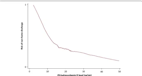
Methods: We performed a retrospective analysis from an ongoing prospective cohort study of vitamin D status in critical illness. Patients were recruited from two surgical ICUs at a single teaching hospital in Boston, Massachusetts. All patients had 25-hydroxyvitamin D (25OHD) levels measured within 24 h of ICU admission. Discharge destination was dichotomized as non-home or home. Locally weighted scatterplot smoothing (LOWESS) was used to graph the relationship between 25OHD levels and discharge destination. To investigate the association between 25OHD level and discharge destination, we performed logistic regression analyses, controlling for age, sex, race, body mass index, socioeconomic status, acute physiology and chronic health evaluation II score, need for emergent vs. non-emergent surgery, vitamin D supplementation status, and hospital LOS.
Results: 300 patients comprised the analytic cohort. Mean 25OHD level was 19 (standard deviation 8) ng/mL and 41 % of patients had a non-home discharge destination. LOWESS analysis demonstrated a near-inverse linear relation-ship between vitamin D status and non-home discharge destination to 25OHD levels around 10 ng/mL, with rapid flattening of the curve between levels of 10 and 20 ng/mL. Overall, 25OHD level at the outset of critical illness was inversely associated with non-home discharge destination (adjusted OR, 0.88; 95 % CI 0.82–0.95). When vitamin D status was dichotomized, patients with 25OHD levels <20 ng/mL had an almost 3-fold risk of a non-home discharge destination (adjusted OR, 2.74; 95 % CI 1.23–6.14) compared to patients with 25OHD levels ≥20 ng/mL.
Conclusions: Our results suggest that vitamin D status may be a modifiable risk factor for non-home discharge destination in surgical ICU patients. Future randomized, controlled trials are needed to determine whether vitamin D supplementation in surgical ICU patients can improve clinical outcomes such as the successful rate of discharge to home after critical illness
Keywords: Vitamin D, 25-hydroxyvitamin D, Critical illness, Discharge destination, Patient-centered outcome
© 2015 Brook et al. This article is distributed under the terms of the Creative Commons Attribution 4.0 International License (http://creativecommons.org/licenses/by/4.0/), which permits unrestricted use, distribution, and reproduction in any medium, provided you give appropriate credit to the original author(s) and the source, provide a link to the Creative Commons license, and indicate if changes were made.
Background
Advances in acute care medicine have resulted in declining mortality rates after critical illness [1–4].
Nonetheless, survival, while evidently a positive outcome, does not truly reflect the health condition of a patient upon discharge from the intensive care unit (ICU). Among survivors, 5–10 % of ICU patients transition to chronic critical illness, with estimates of between 100,000 and 250,000 such patients in the United States at any given point in time [5–8]. These chronically critically ill patients continue to depend on myriad intensive care
Open Access
*Correspondence: squraishi@mgh.harvard.edu
1 Department of Anesthesia, Critical Care, and Pain Medicine,
Massachusetts General Hospital, Harvard Medical School, 55 Fruit Street, GRJ 402, Boston, MA 02114, USA
Page 2 of 9 Brook et al. Ann. Intensive Care (2015) 5:23
resources outside of the acute care setting [7–9]. As such, within the context of a healthcare system gravitating towards patient-centered outcomes, the concept of dis-ability-free survival is gaining popularity [10]. In hospi-talized patients, discharge destination is a potential early indicator of disability-free survival [11–13], and the abil-ity to predict or identify modifiable risk factors for dis-charge destination may improve patient-centered health outcomes.
Discharge destination is known to be affected by a vari-ety of factors, which not only arise during the course of hospitalization, but may also be influenced by patient-related factors that pre-date hospital admission. In particular, socioeconomic factors, such as insurance coverage [14], the level of support at home, and baseline functional status [15], have been shown to influence dis-charge destination. Additional factors, including older age, use of mechanical ventilation or the need for tra-cheotomy (in the setting of chronic respiratory failure), the presence of severe pressure ulcers, declines in cogni-tive function, and metabolic or nutritional derangements (e.g., hypoalbuminemia) may influence discharge destina-tion after ICU admission [16–18]. Notably, while many of these identified risk factors are potentially modifiable, easily and/or rapidly correcting them can be a challenge in the clinical setting.
Although vitamin D is typically recognized for its role in promoting optimal musculoskeletal health [19, 20], emerging data suggests that vitamin D status, as deter-mined by measuring its most widely accepted proxy, circulating 25-hydroxyvitamin D (25OHD) [21], may also influence various ICU-related outcomes, includ-ing length of stay (LOS), readmission, overall costs, and mortality [22–24]. Moreover, low vitamin D status has an estimated prevalence of 40–80 % among critically ill patients [25–29] and can be rapidly corrected [24,
30, 31]. And while 25OHD levels may influence several provider-centered health outcomes, its role in patient-centered outcomes among survivors of critical illness is unclear. Therefore, the goal of our study was to investi-gate whether vitamin D status on admission to the ICU is associated with discharge destination in critically ill sur-gical patients.
Methods
We performed a retrospective analysis of the data from an ongoing prospective cohort study designed to assess vitamin D status in critically ill patients. A subset of these patients was previously described in studies that investigated the association of vitamin D status with duration of mechanical ventilation and 90-day mortality in ICU patients [22, 32]. For the present study, subjects were recruited from two, 18-bed surgical ICUs at the
Massachusetts General Hospital (MGH), in Boston, USA. Both ICUs received admissions from all surgical services except for Cardiac Surgery. All subjects were enrolled between 06/01/2012 and 05/30/2015. MGH is a 1052-bed, teaching hospital and a level-one trauma center, which serves a diverse population in and around Eastern Massachusetts. The Partners Human Research Commit-tee (local Institutional Review Board) approved the study protocol.
Inclusion and exclusion criteria
All adult males and females, ≥18 years of age, and who were expected to require at least 48 h of critical care (as determined by the treating ICU team), were deemed eligible to participate. Informed consent was obtained either directly from subjects or appropriate healthcare surrogates. Subjects were only included in the study if blood samples to assess vitamin D status could be obtained within 24 h of admission to the ICU. Exclusion criteria included a known history of anemia at the time of ICU admission (defined as hematocrit <25 %), pregnancy or immediate post-partum status, and history of vitamin D supplementation ≥4000 IU/day. To minimize con-founding from either partially treated, new-onset illness, or chronic illnesses, subjects were also excluded if they were transferred from another ICU or had been in an ICU within year of the most current admission. Patients expected to transition to “comfort only measures” were also excluded.
Blood sample processing and biomarker assays
Following informed consent, fresh blood was acquired from an indwelling vascular catheter and was collected directly into an ethylenediaminetetraacetic acid-con-taining tube (lavender top). The sample was immedi-ately stored on ice and then centrifuged within 30 min to separate out plasma. All samples were centrifuged at 2,300 rpm for 15 min at a temperature of 4 °C. The sepa-rated plasma was immediately transferred to polypropyl-ene tubes and stored at −80 °C until biomarker testing was ready to be initiated. Assays were performed at the Harvard Medical School Clinical and Translational Sci-ence Award core laboratory at MGH. Plasma 25OHD (combined D2 and D3) levels were measured by
enzyme-linked immunoabsorbent assay (ELISA), using commer-cially available kits (Abbott Laboratories, Abbott Park, IL, USA). Intra- and inter-assay coefficients of variation were both <10 %.
Demographic and clinical data collection
(SES), acute physiology and chronic health evaluation (APACHE) II score, type of surgical patient, vitamin D supplementation status, hospital LOS, and discharge des-tination. We estimated SES by abstracting the residential zip code from each medical record and cross-referencing it with the US Census Bureau per capita income data [33] specific to each locality. Zip codes in the top third of the per capita income rankings for Massachusetts were con-sidered high SES, middle third were concon-sidered moder-ate SES, and those in the bottom third were considered low SES. Sex, race, type of surgical patient, and in-hos-pital vitamin D supplementation status were dichoto-mized as female vs. male, non-white vs. white, emergent vs. non-emergent, and ≤1000 IU vs. >1000 to <4000 IU per day throughout the hospitalization, respectively. Dis-charge destination was also dichotomized as non-home vs. home. A non-home discharge included all in-hos-pital deaths, transfers to another healthcare facility for patients who lived at home before ICU admission, and/ or transition to a higher level of care compared to base-line (e.g., nursing home resident who was discharged to an acute rehabilitation hospital after ICU admission and acute care on the general wards at MGH). A home dis-charge included all patients who returned home (with or without home health services) or returned to the same level of care as baseline (e.g., nursing home resident who was discharged back to his/her nursing home after ICU admission and acute care on the general wards at MGH). All other variables were considered as continuous data.
Locally weighted scatterplot smoothing analysis
Locally weighted scatter plot smoothing (LOWESS) was used to graphically represent the relationship between 25OHD levels and the risk of non-home discharge. LOWESS curves are a form of nonparametric regression, which summarize a relationship between two variables in a fashion that initially relies on limited assumptions about the form or strength of the relationship [34]. The rationale and methods underlying the use of LOWESS for depicting the local relationship between measure-ments of interest across parts of their ranges have previ-ously been described [35].
Statistical analysis
Descriptive statistics were calculated for subjects with plasma 25OHD levels <20 ng/mL vs. those with levels ≥20 ng/mL. Continuous data were reported as means with standard deviations (SDs) or medians with inter-quartile ranges (IQRs). Comparison of characteristics was performed using t test and Mann–Whitney analy-ses for normally distributed variables and nonparametric variables, respectively. Categorical values were expressed as proportions and compared using Chi-squared tests.
Since we considered discharge destination as a dichot-omous variable, logistic regression analysis was used to model the relationship between plasma 25OHD levels and discharge destination, while controlling for biologi-cally plausible covariates. In this approach, we controlled for: (1) age; (2) sex; (3) race; (4) BMI; (5) SES; (6) type of surgical patient; (7) APACHE II score; (8) hospital LOS; and (9) vitamin D supplementation. We then repeated the analysis with 25OHD levels dichotomized as <20 vs. ≥20 ng/mL. This threshold was selected based on exist-ing conservative guidelines regardexist-ing optimal vitamin D status [36] and observed relationship between 25OHD levels and discharge destination on the LOWESS curve. Results are reported as odds ratios (ORs) with 95 % confi-dence intervals (CIs).
We performed an a priori sample size calculation using the previously described biologically plausible model for the association between plasma 25OHD levels and discharge destination. Based on previous studies per-formed by our group and others [37–39] on discharge destination, we assumed a 40 % discharge to home rate in patients with plasma 25OHD levels ≥20 ng/mL and a 20 % discharge to home rate in patients with levels <20 ng/mL. To detect this difference in discharge rate between groups with a power of 0.8 and with alpha set at 0.05, a minimum sample size of 82 patients in each group would be required for the present study. All analyses were performed in STATA 13.0 (StataCorp LP, College Station, TX). A two-tailed P < 0.05, or 95 % CI that did not span 1, was considered statistically significant.
Results
A total of 300 patients comprised the final analytic cohort (Table 1). The mean age was 66 (SD 16) years and most subjects were male (58 %) as well as white (90 %). Overall, mean BMI was 29 (SD 7) kg/m2. Approximately 26, 40,
and 35 % of patients were from low, moderate, and high SES neighborhoods, respectively, while 14 % of patients had emergency surgery. The mean APACHE II score was 17 (SD 9), median hospital LOS was 9 (IQR 6–14) days, and mean 25OHD level was 19 (SD 8) ng/mL. The overall in-hospital mortality rate was 16 and 41 % of patients had a non-home discharge destination. Among patients who survived to hospital discharge, the non-home destination rate was 30 %.
Page 4 of 9 Brook et al. Ann. Intensive Care (2015) 5:23
plausible covariates, demonstrated an inverse associa-tion between admission plasma 25OHD levels and non-home discharge destination (OR per 1 ng/mL, 0.88; 95 % CI 0.82–0.95). When vitamin D status was considered as a dichotomous variable, patients with 25OHD <20 ng/ mL were more than 2.5 times more likely to have a non-home discharge destination compared to patients with 25OHD ≥20 ng/mL (OR, 2.74; 95 % CI 1.23–6.14). Addi-tional variables found to be independently associated with non-home discharge destination in these models were APACHE II score, hospital LOS, and vitamin D supplementation.
Discussion
In this retrospective cohort study, we investigated whether vitamin D status determined at initiation of critical care was associated with subsequent discharge destination from the hospital. We demonstrated that plasma 25OHD levels measured on admission to the surgical ICU were inversely associated with the risk of a non-home discharge destination (i.e., patients with low 25OHD levels are more likely to have a non-home dis-charge destination). This relationship was most promi-nent when comparing patients with 25OHD levels <20 ng/mL to those with levels ≥20 ng/mL. However, due to the observational nature of this study, a causal inference about the effect of vitamin D status on dis-charge destination in critically ill surgical patients is lim-ited. And while the association between vitamin D status and various provider-centered outcomes, such as hospital LOS and readmission rates, has previously been reported [22–24], its impact on a patient-centered outcome, such as discharge destination after hospitalization, has not been well explored. Nonetheless, the biologic plausibil-ity of the relationship between vitamin D status and dis-charge destination is undeniable.
The wide-ranging role of vitamin D in musculoskel-etal health, epithelial barrier site integrity, and regulation of both innate as well as adaptive immunity, in addition to calcium homeostasis, may explain how optimizing 25OHD levels in critically ill patients may improve out-comes [40]. Proximal muscle group weakness and atro-phy, particularly of type II fibers, are associated with low vitamin D status [41–43], and profound deconditioning associated with muscle wasting in the ICU is associated with long-term physical as well as functional disabil-ity [44–46]. Vitamin D metabolites have been shown to affect muscle metabolism through a variety of pathways [43]. Most importantly, the vitamin D membrane recep-tor (VDR), which binds the most biologically active vita-min D metabolite 1,25-dihydroxyvitavita-min D (1,25OHD), is expressed in human skeletal muscle cells [47, 48]. Mod-ulated both by genetic transcription after transportation to the nucleus [49], and by direct interaction with muscle cell membranes resulting in second-messenger pathways [50], the 1,25OHD–VDR interaction results in increased calcium uptake within muscle cells [51].
Additionally, vitamin D has been shown to be impor-tant for the maintenance of epithelial and mucosal cells, in particular preserving the barrier functions of these cells [52]. Activity of the 1-α-hydroxylase that converts
25OHD–1,25OHD has been demonstrated in epithelial cells of the respiratory system [53], intestine [54, 55], skin [56, 57], and endothelium [58]. Vitamin D supplementa-tion has been shown to increase the growth and differ-entiation of respiratory epithelial cells [59], while low
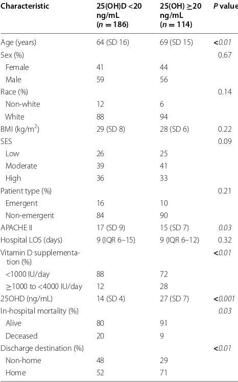
Table 1 Demographic factors, baseline information, and clinical outcomes in surgical intensive care unit patients according to vitamin D status at initiation of care (n = 300)
BMI body mass index, SES socioeconomic status, APACHE II acute physiology and chronic health evaluation II, LOS length of stay, 25OHD 25-hydroxyvitamin D Data presented as either mean with standard deviation (SD), median with interquartile range (IQR), or proportions and compared using t tests, Mann– Whitney tests, and Chi-squared tests, respectively. Significant P values (<0.05) are shown in italic. To convert ng/mL to nmol/L, please multiply by 2.496
Characteristic 25(OH)D <20 ng/mL (n = 186)
25(OH) ≥20 ng/mL (n = 114)
P value
Age (years) 64 (SD 16) 69 (SD 15) <0.01
Sex (%) 0.67
Female 41 44
Male 59 56
Race (%) 0.14
Non-white 12 6
White 88 94
BMI (kg/m2) 29 (SD 8) 28 (SD 6) 0.22
SES 0.09
Low 26 25
Moderate 39 41
High 36 33
Patient type (%) 0.21
Emergent 16 10
Non-emergent 84 90
APACHE II 17 (SD 9) 15 (SD 7) 0.03 Hospital LOS (days) 9 (IQR 6–15) 9 (IQR 6–12) 0.32 Vitamin D
supplementa-tion (%) <0.01
<1000 IU/day 88 72 ≥1000 to <4000 IU/day 12 28
25OHD (ng/mL) 14 (SD 4) 27 (SD 7) <0.001 In-hospital mortality (%) 0.03
Alive 80 91
Deceased 20 9
Discharge destination (%) <0.01
Non-home 48 29
[image:5.595.56.289.138.512.2]vitamin D status has been shown to cause pulmonary epithelial dysfunction in mice [60]. Vitamin D metabo-lites have also been shown to promote colonic epithelial barrier integrity and function through the preservation of intercellular tight junctions in both in vitro and murine studies [61, 62], with low vitamin D status increasing the risk of colitis in animal models [62]. Furthermore, keratinocytes not only produce and metabolize the vita-min D precursor [63], but keratinocyte differentiation is in turn modulated by vitamin D and is essential for main-taining healthy skin [64, 65]. And finally, in vitro studies have shown that in endothelium, supplementing with vitamin D reduces the generation of anion superoxides, prevents free radical release [66], and promotes endothe-lial cell viability through the production of nitric oxide [66, 67]. Therefore, compromised natural barriers in the setting of low vitamin D status likely increases suscep-tibility to nosocomial infections, including central line-associated blood stream infections, hospital-acquired pneumonia, and Clostridium difficile infections [40, 68,
69], which all not only increase the risk of mortality, but also lead to prolonged hospitalization and likely com-pound the functional decline associated with critical illness.
VDR is also expressed in cells of the innate and adap-tive immune system [70], including T cells, activated B cells, and dendritic cells [71–75]. Low vitamin D status
is linked to the Toll-like receptor stimulation of mac-rophages that leads to increased expression of VDR [76], assists with conversion of 25OHD–1,25OHD [77], and also upregulates the expression of endogenous antimi-crobial peptides, cathelicidin (LL-37) and β-defensin
[20, 76, 78]. These peptides have known activity against Gram-positive and Gram-negative bacteria, mycobacte-ria, viruses, and fungi [79], thereby suggesting an impor-tant role of optimal vitamin D status in prevention and potentially treatment of serious infections [80]. Indeed, recent evidence suggests that high-dose cholecalciferol (vitamin D3) supplementation in patients with severe sepsis or septic shock increases systemic cathelicidin lev-els, while modulating immunoregulatory cytokines such as interleukin (IL)-1β and IL-6 [31]. Previous studies have
also suggested that vitamin D is a key regulator of inflam-matory cytokines [81–83].
While our findings are intriguing, it is important to dis-cuss the limitations of our study. We performed a retro-spective, observational analysis, and therefore, causality cannot be inferred. And despite our efforts to adjust for multiple covariates, the presence of residual confound-ing that may have contributed to the observed outcomes cannot be ruled out. Indeed, we are unable to fully adjust for baseline functional status, exposure to sunlight, as well as nutritional status before and through hospitalization. Additionally, hospital LOS is often influenced by several
[image:6.595.59.538.87.346.2]Page 6 of 9 Brook et al. Ann. Intensive Care (2015) 5:23
factors, such as social and financial issues, that we could not fully adjust for in our model. Of note, we elected to use hospital LOS instead of ICU LOS, since patients may often remain in the ICU due to a lack of available beds on the general wards. Furthermore, discharge destina-tion may be affected by financial and insurance consid-erations, the availability of home help, and family/social support—all factors that we are unable to adjust for in the present study. Moreover, we only recruited a limited num-ber of patients from two surgical ICUs at a single, large referral center, which may decrease the generalizability of our results to all ICU patients. For the most part, patients recruited into the study were also racially homogenous, and this limited diversity may further affect the generaliz-ability of our findings. In the present study, only 25OHD levels assessed within 24 h of ICU admission were con-sidered; it is possible that this single data point does not reflect the variation in vitamin D status with inflamma-tion, metabolic changes, fluid shifts, and systemic micro and macronutrient needs for the entire duration of critical illness [22, 84].
It is worth noting that more patients in our ana-lytic cohort with admission 25OHD levels ≥20 ng/mL received vitamin D supplementation between ≥1000 and <4000 IU/day during hospitalization, as compared to patients with levels <20 ng/mL (28 vs. 12 %, respec-tively; P < 0.01). This is due to the fact that patients with higher initial 25OHD levels were more likely to be taking vitamin D supplementation before hospitalization and as a result, it was more likely to be continued after ICU admission. Measurement of vitamin D status is not part of routine care in our surgical ICUs and all admission 25OHD levels were measured from frozen plasma sam-ples several weeks after hospitalization (and therefore not temporally related to clinical decision-making). Since only 18 % of the entire study cohort received vitamin D supplementation during hospitalization, we were likely underpowered to detect an independent association between supplementation status and discharge destina-tion (the primary outcome) when the exposure of interest in our regression model (i.e., 25OHD) was considered as a continuous variable. Dichotomizing the exposure (<20
Table 2 Biologically plausible models to test the association of admission 25-hydroxyvitamin D level with non-home dis-charge destination in surgical intensive care unit patients (n = 300)
(–) represents each referent variable
Statistically significant variables are highlighted in italic
OR odds ratio, CI confidence interval, BMI body mass index, SES socioeconomic status, APACHE II acute physiology and chronic health evaluation II, LOS length of stay,
25OHD 25-hydroxyvitamin D
Covariate OR (95 % CI)
25OHD (continuous variable) OR (95 % CI)25OHD (<20 vs. ≥20 ng/mL)
Age (years) 1.01 (0.98–1.04) 1.00 (0.98–1.03)
Sex
Female – –
Male 0.81 (0.36–1.83) 1.08 (0.51–2.31)
Race
Non-white – –
White 1.57 (0.96–2.56) 2.12 (0.52–8.68)
BMI (kg/m2) 2.07 (0.58–8.47) 0.97 (0.91–1.02) SES
Low – –
Moderate 1.09 (0.38–3.09) 1.08 (0.39–3.02)
High 1.07 (0.36–3.18) 0.92 (0.32–2.65)
Patient type
Emergent – –
Non-emergent 1.73 (0.32–9.32) 1.66 (0.32–8.66)
APACHE II 1.33 (1.22–1.45) 1.32 (1.22–1.43)
Hospital LOS (days) 1.18 (1.11–1.25) 1.18 (1.11–1.25) Vitamin D supplementation (IU/day)
<1000 – –
[image:7.595.57.539.114.407.2]vs. ≥20 ng/mL) improved the ability of our model to criminate between patients who were and were not dis-charged home, and in doing so, there was likely sufficient power to also detect an independent association of sup-plementation status with discharge destination. Notable also, was the fact that the proportion of patients in each SES group did not differ by vitamin D status. This may be related to the generally high prevalence of low vitamin D status in New England (due to geography and weather) and the enrollment of patients more likely to be indoors during the day (e.g., elderly, retirees, office workers). As such, these issues will need to be addressed in larger future studies to replicate and extend the results of our current study.
Conclusion
Our results suggest that vitamin D status may be a modi-fiable risk factor for discharge destination in surgical ICU patients. Given that suboptimal 25OHD levels are highly prevalent in ICU patients [25–29], further prospective studies are needed to validate our findings, to assess the potential benefits of optimizing 25OHD levels in criti-cal illness, and to identify the mechanism by which vita-min D may impact patient-oriented outcomes in ICU patients.
Abbreviations
LOS: length of stay; ICU: intensive care unit; 25OHD: 25-hydroxyvitamin D; LOWESS: locally weighted scatterplot smoothing; MGH: Massachusetts Gen-eral Hospital; ELISA: enzyme-linked immunoabsorbent assay; BMI: body mass index; SES: socioeconomic status; APACHE II: acute physiology and chronic health evaluation; SD: standard deviation; IQR: interquartile range; OR: odds ratio; CI: confidence interval; VDR: vitamin D membrane receptor; 1,25OHD: 1,25-dihydroxyvitamin D; IL: interleukin.
Authors’ contributions
KB and SAQ jointly conceived the study and drafted the manuscript. CAC and KBC critically revised the manuscript. SAQ assembled the data and performed all statistical analyses. All authors read and approved the final manuscript.
Author details
1 Department of Anesthesia, Critical Care, and Pain Medicine, Massachusetts
General Hospital, Harvard Medical School, 55 Fruit Street, GRJ 402, Boston, MA 02114, USA. 2 Department of Emergency Medicine, Massachusetts General
Hospital, Boston, MA, USA. 3 Department of Medicine, Harvard Medical School,
Boston, MA, USA. 4 Department of Epidemiology, Harvard School of Public
Health, Boston, MA, USA. 5 Department of Medicine, Brigham and Women’s
Hospital, Boston, MA, USA. 6 Department of Anaesthesia, Harvard Medical
School, Boston, MA, USA.
Acknowledgements
CAC received support from the United States National Institutes of Health grants R01 AI093723 and U01 AI087881. SAQ received support from the United States National Institutes of Health grants T32 GM007592, UL1 RR025758, and L30 TR001257 as well as from Harvard Medical School.
Compliance with ethical guidelines
Competing interests
The authors declare that they have no competing interests.
Received: 21 July 2015 Accepted: 28 August 2015
References
1. Kaukonen KM, Bailey M, Suzuki S, Pilcher D, Bellomo R. Mortality related to severe sepsis and septic shock among critically ill patients in Australia and New Zealand, 2000–2012. JAMA. 2014;311(13):1308–16.
2. Gaieski DF, Edwards JM, Kallan MJ, Carr BG. Benchmarking the incidence and mortality of severe sepsis in the United States. Crit Care Med. 2013;41(5):1167–74.
3. Stevenson EK, Rubenstein AR, Radin GT, Wiener RS, Walkey AJ. Two dec-ades of mortality trends among patients with severe sepsis: a compara-tive meta-analysis. Crit Care Med. 2014;42(3):625–31.
4. Klein MB, Goverman J, Hayden DL, Fagan SP, McDonald-Smith GP, et al. Benchmarking outcomes in the critically injured burn patient. Ann Surg. 2014;259(5):833–41.
5. Girard K, Raffin TA. The chronically critically ill: To save or let die? Respir Care. 1985;30(5):339–47.
6. Carson SS. Definitions and epidemiology of the chronically critically ill. Respir Care. 2012;57(6):848–56 (discussion 856–8).
7. Macintyre NR. Chronic critical illness: the growing challenge to health care. Respir Care. 2012;57(6):1021–7.
8. Nelson JE, Cox CE, Hope AA, Carson SS. Chronic critical illness. Am J Respir Crit Care Med. 2010;182(4):446–54.
9. Donahoe MP. Current venues of care and related costs for the chronically critically ill. Respir Care. 2012;57(6):867–86 (discussion 886–8). 10. Shulman MA, Myles PS, Chan MTV, McIlroy DR, Wallace S, Ponsford P.
Measurement of disability-free survival after surgery. Anesthesiology. 2015;122(3):524–36.
11. Bala M, Kashuk JL, Willner D, Kaluzhni D, Bdolah-Abram T, Almogy G. Looking beyond discharge: clinical variables at trauma admission predict long term survival in the older severely injured patient. World J Emerg Surg WJES. 2014;9:10. doi:10.1186/1749-7922-9-10.
12. Chen C, Sia I, Ma H, et al. The synergistic effect of functional status and comorbidity burden on mortality: a 16-year survival analysis. Laks J (ed). PLoS One. 2014;9(8):e106248.
13. Fried TR, Bradley EH, Towle VR, Allore H. Understanding the treatment preferences of the seriously ill. N Engl J Med. 2002;346:1061–6.
14. Lane-Fall MB, Iwashyna TJ, Cooke CR, Benson NM, Kahn JM. Insurance and racial differences in long-term acute care utilization after critical illness. Crit Care Med. 2012;40(4):1143–9.
15. Rudberg MA, Sager MA, Zhang J. Risk factors for nursing home use after hospitalization for medical illness. J Gerontol A Biol Sci Med Sci. 1996;51:M189–94.
16. Rady MY, Johnson DJ. Hospital discharge to care facility. A patient-centered outcome for the evaluation of intensive care for octogenarians. Chest. 2004;126:1583–91.
17. Gehlbach BK, Salamanca VR, Levitt JE, et al. Patient factors associated with hospital discharge to care facility following critical illness. Am J Crit Care. 2011;20(5):378–86. doi:10.4037/ajcc2011827.
18. Szubski CR, Tellez A, Klika AK, et al. Predicting discharge to a long-term acute care hospital after admission to an intensive care unit. Am J Crit Care. 2014;23:e46–53.
19. DeLuca HF. Overview of general physiologic features and functions of vitamin D. Am J Clin Nutr. 2004;80(Suppl):1689S–96S.
20. Holick MF. Vitamin D deficiency. N Engl J Med. 2007;357(3):266–81. 21. Quraishi SA, Camargo CA Jr. Vitamin D in acute stress and critical illness.
Curr Opin Clin Nutr Metab Care. 2012;15(6):625–34.
22. Quraishi SA, Bittner EA, Blum L, McCarthy CM, Bhan I, Camargo CA Jr. Prospective study of vitamin D status at initiation of care in criti-cally ill surgical patients and risk of 90-day mortality. Crit Care Med. 2014;42(6):1365–71.
Page 8 of 9 Brook et al. Ann. Intensive Care (2015) 5:23
24. Amrein K, Schnedl C, Holl A, et al. Effect of high-dose vitamin D3 on hospital length of stay in critically ill patients with vitamin D deficiency: the VITdAL-ICU randomized clinical trial. JAMA. 2014;312(15):1520–30. 25. Nair P, Lee P, Reynolds C, Nguyen ND, Myburgh J, Eisman JA, Center
JR. Significant perturbation of vitamin D-parathyroid-calcium axis and adverse clinical outcomes in critically ill patients. Intensive Care Med. 2013;39(2):267–74.
26. Venkatram S, Chilimuri S, Adrish M, Salako A, Patel M, Diaz-Fuentes G. Vitamin D deficiency is associated with mortality in the medical intensive care unit. Crit Care. 2011;15:R292.
27. Lucidarme O, Messai E, Mazzoni T, Arcade M, du Cheyron D. Incidence and risk factors of vitamin D deficiency in critically ill patients: results from a prospective observational study. Intensive Care Med. 2010;36:1609–11. 28. Flynn L, Zimmerman LH, McNorton K, et al. Effects of vitamin D deficiency
in critically ill surgical patients. Am J Surg. 2012;203:379–82.
29. Lee P, Eisman JA, Center JR. Vitamin D deficiency in critically ill patients. N Engl J Med. 2009;360(18):1912–4. doi:10.1056/NEJMc0809996. 30. Amrein K, Sourij H, Wagner G, et al. Short-term effects of high-dose oral
vitamin D3 in critically ill vitamin D deficient patients: a randomized, double-blind, placebo-controlled pilot study. Crit Care. 2011;15(2):R104. 31. Quraishi SA, De Pascale G, Needleman JS, Nakazawa H, Kaneki M, Bajwa EK, Camargo CA Jr, Bhan I. Effect of cholecalciferol supplementation on vitamin d status and cathelicidin levels in sepsis: a randomized, Placebo-Controlled Trial. Crit Care Med. 2015;43:1928–37.
32. Quraishi SA, McCarthy C, Blum L, Cobb JP, Camargo CA Jr. Plasma 25-Hydroxyvitamin D Levels at Initiation of Care and Duration of Mechanical Ventilation in Critically Ill Surgical Patients. JPEN J Parenter Enteral Nutr. 2015. doi:10.1177/0148607114566276.
33. U.S. Census Bureau. “SELECTED ECONOMIC CHARACTERISTICS 2009–2013 American Community Survey 5-Year Estimates”.
34. Cleveland WS. Robust locally weighted regression and smoothing scat-terplots. J Am Stat Assoc. 1979;74(368):829–36.
35. Cleveland WS, Devlin SJ. Locally weighted regression: an approach to regression analysis by local fitting. J Am Stat Assoc. 1988;83(403):596–610. 36. Ross AC, Manson JE, Abrams SA, Aloia JF, Brannon PM, Clinton SK, Durazo-Arvizu RA, Gallagher JC, Gallo RL, Jones G, Kovacs CS, Mayne ST, Rosen CJ, Shapses SA. The 2011 report on dietary reference intakes for calcium and vitamin D from the Institute of Medicine: what clinicians need to know. J Clin Endocrinol Metab. 2011;96(1):53–8.
37. Yeh DD, Fuentes E, Quraishi SA, Cropano C, Kaafarani H, Lee J, King DR, DeMoya M, Fagenholz P, Butler K, Chang Y, Velmahos G. Adequate nutri-tion may get you home: effect of caloric/protein deficits on the discharge destination of critically ill surgical patients. JPEN J Parenter Enteral Nutr. 2015. doi:10.1177/0148607115585142.
38. Gehlbach BK, Salamanca VR, Levitt JE, Sachs GA, Sweeney MK, Pohlman AS, Charbeneau J, Krishnan JA, Hall JB. Patient-related factors associated with hospital discharge to a care facility after critical illness. Am J Crit Care. 2011;20(5):378–86. doi:10.4037/ajcc2011827.
39. Wunsch H, Guerra C, Barnato AE, Angus DC, Li G, Linde-Zwirble WT. Three-year outcomes for Medicare beneficiaries who survive intensive care. JAMA. 2010;303(9):849–56.
40. Lee P, Nair P, Eisman JA, Center JR. Vitamin D deficiency in the intensive care unit: an invisible accomplice to morbidity and mortality? Intensive Care Med. 2009;35(12):2028–32. doi:10.1007/s00134-009-1642-x(Epub 2009 Sep 15).
41. Janssen HC, Samson MM, Verhaar HJ. Vitamin D deficiency, muscle func-tion, and falls in elderly people. Am J Clin Nutr. 2002;75(4):611–5. 42. Ceglia L, Niramitmahapanya S, da Silva Morais M, Rivas DA, Harris SS,
Bischoff-Ferrari H, Fielding RA, Dawson-Hughes B. A randomized study on the effect of vitamin D3 supplementation on skeletal muscle morphology
and vitamin D receptor concentration in older women. J Clin Endocrinol Metab. 2013;98(12):E1927–35.
43. Boland R. Role of vitamin D in skeletal muscle function. Endocr Rev. 1986;7:434–48.
44. Connolly B, Salisbury L, O’Neill B, Geneen L, Douiri A, Grocott MP, Hart N, Walsh TS, Blackwood B, ERACIP Group. Exercise rehabilitation following intensive care unit discharge for recover from critical illness. Cochrane Database Syst Rev. 2015;6:CD008632 (Epub ahead of print). 45. Needham DM, Davidson J, Cohen H, Hopkins RO, Weinert C, Wunsch H,
Zawistowski C, Bemis-Dougherty A, Berney SC, Bienvenu OJ, Brady SL, Brodsky MB, Denehy L, Elliott D, Flatley C, Harabin AL, Jones C, Louis D,
Meltzer W, Muldoon SR, Palmer JB, Perme C, Robinson M, Schmidt DM, Scruth E, Spill GR, Storey CP, Render M, Votto J, Harvey MA. Improving long-term outcomes after discharge from intensive care unit: report from a stakeholders’ conference. Crit Care Med. 2012;40(2):502–9. doi:10.1097/ CCM.0b013e318232da75.
46. Connolly B, MacBean V, Crowley C, Lunt A, Moxham J, Rafferty GF, Hart N. Ultrasound for the assessment of peripheral skeletal muscle architecture in critical illness: a systematic review. Crit Care Med. 2015;43(4):897–905. doi:10.1097/CCM.0000000000000821.
47. Bischoff HA, Borchers M, Gudat F, et al. In situ detection of 1,25-dihy-droxyvitamin D3 receptor in human skeletal muscle tissue. Histochem J. 2001;33:19–24.
48. Costa EM, Blau HM, Feldman D. 1,25-Dihydroxyvitamin D3 receptors and hormonal responses in cloned human skeletal muscle cells. Endocrinol-ogy. 1986;119:2214–20.
49. Simpson RU, Thomas GA, Arnold AJ. Identification of 1,25-dihydroxyvita-min D3 receptors and activities in muscle. J Biol Chem. 1985;260:8882–91. 50. Nemere I, Schwartz Z, Pedrozo H, Sylvia VL, Dean DD, Boyan BD.
Identification of a membrane receptor for 1,25-dihydroxyvitamin D3 which mediates rapid activation of protein kinase C. J Bone Miner Res. 1998;13:1353–9.
51. Vazquez G, de Boland AR, Boland R. Stimulation of Ca2+ release-activated
Ca2+ channels as a potential mechanism involved in non-genomic
1,25(OH)2-vitamin D3-induced Ca2+ entry in skeletal muscle cells.
Bio-chem Biophys Res Commun. 1997;239:562–5.
52. Zhang YG, Wu S, Sun J. Vitamin D, vitamin D receptor, and tissue barriers. Tissue Barriers. 2013;1(1) (pii: e23118).
53. Hansdottir S, Monick MM, Hinde SL, Lovan N, Look DC, Hunninghake GW. Respiratory epithelial cells convert inactive vitamin D to its active form: potential effects on host defense. J Immunol. 2008;181:7090–9. 54. Tangpricha V, Flanagan JN, Whitlatch LW, Tseng CC, Chen TC, Holt PR,
Lipkin MS, Holick MF. 25-Hydroxyvitamin D-1-hydroxylase in normal and malignant colon tissue. Lancet. 2001;357:1673–4.
55. Ogunkolade BW, Boucher BJ, Fairclough PD, Hitman GA, Dorudi S, Jenkins PJ, Bustin SA. Expression of 25-hydroxyvitamin D-1-hydroxylase mRNA in individuals with colorectal cancer. Lancet. 2002;359:1831–2.
56. Bikle DD, Nemanic MK, Gee E, Elias P. 1,25-Dihydroxyvitamin D3 produc-tion by human keratinocytes: kinetics and regulaproduc-tion. J Clin Invest. 1986;78:557–66.
57. Schauber J, Dorschner RA, Coda AB, Buchau AS, Liu PT, Kiken D, Helfrich YR, Kang S, Elalieh HZ, Steinmeyer A, et al. Injury enhances TLR2 function and antimicrobial peptide expression through a vitamin D-dependent mechanism. J Clin Invest. 2007;117:803–11.
58. Zehnder D, Bland R, Chana RS, Wheeler DC, Howie AJ, Williams MC, Stewart PM, Hewison M. Synthesis of 1,25-dihydroxyvitamin D(3) by human endothelial cells is regulated by inflammatory cytokines: a novel autocrine determinant of vascular cell adhesion. J Am Soc Nephrol. 2002;13(3):621–9.
59. Brockman-Schneider RA, Pickles RJ, Gern JE. Effects of vitamin D on airway epithelial cell morphology and rhinovirus replication. PLoS One. 2014;9(1):e86755. doi:10.1371/journal.pone.0086755(eCollection 2014). 60. Zosky GR, Berry LJ, Elliot JG, James AL, Gorman S, et al. Vitamin d
defi-ciency causes deficits in lung function and alters lung structure. Am J Respir Crit Care Med. 2011;183:1336–43.
61. Zhao H, Zhang H, Wu H, et al. Protective role of 1,25(OH)2 vitamin D3 in the mucosal injury and epithelial barrier disruption in DSS-induced acute colitis in mice. BMC Gastroenterol. 2012;12:57.
62. Assa A, Vong L, Pinnell LJ, Avitzur N, Johnson-Henry KC, Sherman PM. Vitamin D deficiency promotes epithelial barrier dysfunction and intesti-nal inflammation. J Infect Dis. 2014;210(8):1296–305. doi:10.1093/infdis/ jiu235(Epub 2014 Apr 21).
63. Bikle DD, Nemanic MK, Gee E, Elias P. 1,25- Dihydroxyvitamin D3 produc-tion by human keratinocytes. Kinetics and regulaproduc-tion. J Clin Invest. 1986;78:557–66.
64. Bikle DD. Vitamin D regulated keratinocyte differentiation. J Cell Biochem. 2004;92(3):436–44.
65. Bikle DD, Chang S, Crumrine D, et al. 25 hydroxyvitamin D 1 [alpha]-hydroxylase is required for optimal epidermal differentiation and perme-ability barrier homeostasis. J Invest Dermatol. 2004;122:984–92. 66. Uberti F, Lattuada D, Morsanuto V, Nava U, Bolis G, Vacca G, Squarzanti
oxidative stress through the autophagic and survival pathways. J Clin Endocrinol Metab. 2014;99(4):1367–74. doi:10.1210/jc.2013-2103(Epub 2013 Nov 27).
67. Molinari C, Uberti F, Grossini E, Vacca G, Carda S, Invernizzi M, Cisari C. 1α,25-dihydroxycholecalciferol induces nitric oxide production in cul-tured endothelial cells. Cell Physiol Biochem. 2011;27(6):661–8. doi:10.1159/000330075(Epub 2011 Jun 17).
68. Quraishi SA, Litonjua AA, Moromizato T, Gibbons FK, Camargo CA Jr, Giovannucci E, Christopher KB. Association between prehospital vitamin D status and hospital-acquired Clostridium difficile infections. JPEN J Parenter Enteral Nutr. 2015;39(1):47–55. doi:10.1177/0148607113511991 (Epub 2014 Feb 3).
69. Quraishi SA, Litonjua AA, Moromizato T, Gibbons FK, Camargo CA Jr, Giovannucci E, Christopher KB. Association between prehospital vitamin D status and hospital-acquired bloodstream infections. Am J Clin Nutr. 2013;98(4):952–9. doi:10.3945/ajcn.113.058909(Epub 2013 Aug 14). 70. Adams JS, Hewison M. Unexpected actions of vitamin D: new
perspec-tives on the regulation of innate and adaptive immunity. Nat Clin Pract Endocrinol Metab. 2008;4:80–90.
71. Dickson I. New approaches to vitamin D. Nature. 1987;325:18. 72. Minghetti PP, Norman AW. 1,25(OH)2-vitamin D3 receptors: gene
regula-tion and genetic circuitry. FASEB J. 1988;2:3043–53.
73. Mahon BD, Wittke A, Weaver V, Cantorna MT. The targets of vitamin D depend on the differentiation and activation status of CD4 positive T cells. J Cell Biochem. 2003;89:922–32.
74. Heine G, Anton K, Henz BM, Worm M. 1alpha,25-dihydroxyvitamin D3 inhibits anti-CD40 plus IL-4-mediated IgE production in vitro. Eur J Immu-nol. 2002;32:3395–404.
75. Adorini L, Penna G, Giarratana N, Roncari A, Amuchastegui S, Daniel KC, Uskokovic M. Dendritic cells as key targets for immunomodula-tion by vitamin D receptor ligands. J Steroid Biochem Mol Biol. 2004;89–90:437–41.
76. Liu PT, Stenger S, Li H, Wenzel L, Tan BH, Krutzik SR, Ochoa MT, Schau-ber J, Wu K, Meinken C, et al. Toll-like receptor triggering of a vitamin D-mediated human antimicrobial response. Science. 2006;311:1770–3. 77. Bhalla AK, Amento EP, Krane SM. Differential effects of
1,25-dihydroxyvita-min D3 on human lymphocytes and monocyte/macrophages: Inhibition of interleukin-2 and augmentation of interleukin-1 production. Cell Immunol. 1986;98:311–22.
78. Wang TT, Nestel FP, Bourdeau V, Nagai Y, Wang Q, Liao J, Tavera-Mendoza L, Lin R, Hanrahan JW, Mader S, White JH. Cutting edge: 1,25-dihydroxyvi-tamin D3 is a direct inducer of antimicrobial peptide gene expression. J Immunol. 2004;173(5):2909–12.
79. Durr UH, Sudheendra US, Ramamoorthy A. LL-37, the only human mem-ber of the cathelicidin family of antimicrobial peptides. Biochim Biophys Acta. 2006;1758:1408–25.
80. Jeng L, Yamshchikov AV, Judd SE, Blumberg HM, Martin GS, Ziegler TR, Tangpricha V. Alterations in vitamin D status and anti-microbial peptide levels in patients in the intensive care unit with sepsis. J Transl Med. 2009;23:28.
81. Baeke F, Takiishi T, Korf H, Gysemans C, Mathieu C. Vitamin D: modula-tor of the immune system. Curr Opin Pharmacol. 2010;10(4):482–96. doi:10.1016/j.coph.2010.04.001(Epub 2010 Apr 27).
82. Baeke F, Gysemans C, Korf H, Mathieu C. Vitamin D insufficiency: implica-tions for the immune system. Pediatr Nephrol. 2010;25(9):1597–606. doi:10.1007/s00467-010-1452-y(Epub 2010 Feb 24).
83. Hewison M. Vitamin D and the immune system: new perspectives on an old theme. Endocrinol Metab Clin North Am. 2010;39(2):365–79. doi:10.1016/j.ecl.2010.02.010(table of contents).