White Rose Research Online URL for this paper:
http://eprints.whiterose.ac.uk/83764/
Version: Published Version
Article:
Torode, TA, Marcus, SE, Jam, M et al. (4 more authors) (2015) Monoclonal antibodies
directed to fucoidan preparations from brown algae. PLoS One, 10 (2). e0118366. ISSN
1932-6203
https://doi.org/10.1371/journal.pone.0118366
eprints@whiterose.ac.uk https://eprints.whiterose.ac.uk/
Reuse
Unless indicated otherwise, fulltext items are protected by copyright with all rights reserved. The copyright exception in section 29 of the Copyright, Designs and Patents Act 1988 allows the making of a single copy solely for the purpose of non-commercial research or private study within the limits of fair dealing. The publisher or other rights-holder may allow further reproduction and re-use of this version - refer to the White Rose Research Online record for this item. Where records identify the publisher as the copyright holder, users can verify any specific terms of use on the publisher’s website.
Takedown
If you consider content in White Rose Research Online to be in breach of UK law, please notify us by
Monoclonal Antibodies Directed to Fucoidan
Preparations from Brown Algae
Thomas A. Torode1, Susan E. Marcus1, Murielle Jam2,3, Thierry Tonon2,3, Richard S. Blackburn4, Cécile Hervé2,3, J. Paul Knox1*
1Centre for Plant Sciences, Faculty of Biological Sciences, University of Leeds, Leeds, United Kingdom,
2Sorbonne Universités, UPMC Univ Paris 06, UMR 8227, Integrative Biology of Marine Models, Station Biologique de Roscoff, CS 90074, Roscoff, France,3CNRS, UMR 8227, Integrative Biology of Marine Models, Station Biologique de Roscoff, CS 90074, Roscoff, France,4Sustainable Materials Research Group, Centre for Technical Textiles, University of Leeds, Leeds, United Kingdom
*j.p.knox@leeds.ac.uk
Abstract
Cell walls of the brown algae contain a diverse range of polysaccharides with useful bioac-tivities. The precise structures of the sulfated fucan/fucoidan group of polysaccharides and their roles in generating cell wall architectures and cell properties are not known in detail. Four rat monoclonal antibodies, BAM1 to BAM4, directed to sulfated fucan preparations, have been generated and used to dissect the heterogeneity of brown algal cell wall polysac-charides. BAM1 and BAM4, respectively, bind to a non-sulfated epitope and a sulfated epi-tope present in the sulfated fucan preparations. BAM2 and BAM3 identified additional distinct epitopes present in the fucoidan preparations. All four epitopes, not yet fully charac-terised, occur widely within the major brown algal taxonomic groups and show divergent dis-tribution patterns in tissues. The analysis of cell wall extractions and fluorescence imaging reveal differences in the occurrence of the BAM1 to BAM4 epitopes in various tissues of
Fucus vesiculosus. InEctocarpus subulatus, a species closely related to the brown algal modelEctocarpus siliculosus, the BAM4 sulfated epitope was modulated in relation to salin-ity levels. This new set of monoclonal antibodies will be useful for the dissection of the highly complex and yet poorly resolved sulfated polysaccharides in the brown algae in relation to their ecological and economic significance.
Introduction
Brown algae are a large and diverse class of organisms which dominate most temperate coastal environments. They fulfil important roles as primary producers within intertidal zones, and are key ecological players in some marine environments by being the first recruited species for the organization and structuring of ecosystems. Brown algae vary in size from small filamen-tous species such as those belonging to the Ectocarpales which can be grownin vitroin labora-tories, to giant kelps of the Laminariales which can reach 60 m in length [1]. Previous research, including studies on early embryogenesis, has focused on species of the Fucales, which grow in
a11111
OPEN ACCESS
Citation:Torode TA, Marcus SE, Jam M, Tonon T, Blackburn RS, Hervé C, et al. (2015) Monoclonal Antibodies Directed to Fucoidan Preparations from Brown Algae. PLoS ONE 10(2): e0118366. doi:10.1371/journal.pone.0118366
Academic Editor:Tilmann Harder, University of New South Wales, AUSTRALIA
Received:September 8, 2014
Accepted:January 15, 2015
Published:February 18, 2015
Copyright:© 2015 Torode et al. This is an open access article distributed under the terms of the
Creative Commons Attribution License, which permits unrestricted use, distribution, and reproduction in any medium, provided the original author and source are credited.
Data Availability Statement:All relevant data are within the paper and its Supporting Information files.
Funding:This work was supported by a UK Biotechnology and Biological Research Council (http://www.bbsrc.ac.uk) studentship to TAT. TAT also received two awards from Assemble (#227799), a European Union FP7 research network (http://www. assemblemarine.org/). The funders had no role in study design, data collection and analysis, decision to publish, or preparation of the manuscript.
the intertidal regions of most coasts in the northern hemisphere [2]. More recently, the devel-opment of the filamentousEctocarpus siliculosusas a genetic model organism for brown algae [3] has paved the way for studies on different aspects of brown algal biology including early morphogenesis and life cycles [4,5], response to abiotic change [6] and evolution of species [7,8]. Furthermore, the divergent evolution of brown algae when compared to plants and ani-mals has led to unique biochemical pathways resulting in a range of novel bioactive com-pounds and polymers including those in cell walls [9]. Hence brown algae have received a renewed interest as a source of biomass that does not compete with arable land. Indeed, brown algal polymers have been used in high-capacity lithium ion batteries [10], to produce nanopar-ticles with enhanced delivery efficiency for gene and drug delivery [11] in addition to processes for the production of ethanol [12–14].
Brown algal cell walls are composed predominantly of polysaccharides together with lower amounts of phenolic substances, proteins and halide compounds such as iodide. The polyanio-nic polysaccharides alginates and sulfated fucans are prevalent over neutral and crystalline polysaccharides including cellulose [15]. Alginates are linear polymers of two 1,4-linked uronic acids:β-D-mannuronic acid andα-L-guluronic acid [16]. Sulfated fucans or fucoidans are col-lective terms that group a highly diverse spectrum of sulfated polysaccharides containing α-L-fucose residues. They can generally be divided into homopolymers called homofucans or heteropolymers [9,15–19]. Backbones of homofucans are invariably made of 1,3- or 1,3– 1,4-linkedα-L-fucose, while backbones of heterofucans are more diverse and can be based on neutral sugars and/or uronic acid residues (i.e. glycuronofucogalactans, xylofucoglycuronans, fucomannoglucuronans) [16,20,21]. The fucose residues are commonly sulfated at positions 2, 3 and/or 4. Alternatively they can be substituted by methyl or acetyl groups, or branched with additional fucose, xylose or uronic acid residues. Some prokaryotes and most eukaryotic organ-isms produce sulfated carbohydrates, and this ability is likely to be of ancestral origin [9,22]. Exceptions are the freshwater and land plants which have probably lost such an ability or re-quirement during the conquest of land, as a functional adaptation to sulfate-depleted habitats. Marine angiosperms however do produce sulfated polysaccharides as a result of their second-ary exploitation of marine environments and polysaccharide sulfation is positively correlated with increasing saline conditions [23–25]. In the green macroalgaeLamprothamnium papulo-sum, the extracellular sulfated mucilage increases in thickness and sulfate content with increas-ing salinity [24,25]. In brown algae the concentrations of sulfate groups on fucans positively correlate with increasing exposure to the atmosphere in the intertidal zone, suggesting a role in desiccation resistance [26]. A strain of the speciesEctocarpus subulatus, isolated from a true freshwater environment [27] undergoes major morphological, transcriptomic and metabolic changes under variable salinities, including alteration of the expression of genes encoding en-zymes potentially involved in the sulfation or de-sulfation of cell wall polysaccharides [28].
that bind to sulfated fucan/fucoidan preparations. These probes are used in assays and proce-dures to dissect the heterogeneity of sulfated fucan preparations and of brown algal cell walls.
Materials and Methods
Animals
The use of rats to generate monoclonal antibodies was carried out in strict accordance with the guidelines and licence of United Kingdom Home Office under Project Licence PL4003426 / Animals (Scientific Procedures) Act 1986. Procedures included injection and withdrawal of blood and were performed by the same trained staff and all efforts were made to minimize suf-fering. An inhalational agent (isoflurane) was used for the initial induction and the mainte-nance of general anaesthesia via an anaesthetic delivery system. The animals were sacrificed by exposure to carbon dioxide gas in a rising concentration which is an approved method listed in the Schedule 1 of the UK Animals (Scientific Procedures) Act 1986.
Algal samples
Samples of algae, except theEctocarpussamples, were collected from natural environments as follows: Guernsey, Channel Islands GPS coordinates: 49.43–2.66 (Laminaria digitata,Pelvetia canaliculata); Shetland Islands, UK GPS: 60.42–1.09 (Chorda filum); Scarborough, UK GPS: 54.27–0.39 (Halidrys siliquosa,Laminaria hyperborea,Saccharina latissima); Roscoff, France GPS: 48.72–3.98 (Ascophyllum nodosum,Fucus serratus,Fucus spiralis,Himanthalia elongata, Laminaria digitata,Laminaria hyperborea,Laminaria ochroleuca,Saccharina latissima, Sargas-sum muticum,Pelvetia canaliculata,Undaria pinnatifida). No permissions were required for these locations/activities. The collection of brown algae samples did not involve endangered or protected species.Ectocarpus siliculosus(marine strain, Ec32) andEctocarpus subulatus (fresh-water strain, Ec371) were both cultured at Station Biologique de Roscoff as described [28].
Generation of sulfated fucan-directed rat monoclonal antibodies
Four rat monoclonal antibodies (MAbs), designated BAM1 to BAM4, were derived subsequent to immunisation with a neoglycoprotein immunogen, prepared by coupling of fucoidan (Sigma-Aldrich F5631) to bovine serum albumin (BSA) by activation with 1-cyano-4-dimethylamino-pyridium tetrafluroborate (CDAP) [36]. Two male Wistar rats were each injected with 250μg
fucoidan-BSA in complete Freund’s adjuvant administered subcutaneously on day 0, and the same was administered with incomplete Freund’s adjuvant on days 31, 59, 125 and 158. A pre-fusion boost of 100μg fucoidan-BSA in 1 ml PBS was administered on day 215 prior to spleen
removal. Hybridoma production and cloning procedures were performed as described [37].
Polysaccharides
Fucoidan (F5631), alginate (A7003), laminaran (L9634), carrageenan (C1013), sulfated Dex-tran (D6001), chondroitin (C4384), oat spelt xylan (X0627), gum Arabic (G9752) and citrus pectin (P9135) were obtained from Sigma-Aldrich. Tamarind xyloglucan, potato galactan, guar galactomannan, sugar beet arabinan and citrus polygalacturonan were obtained from Mega-zyme International (Bray, Ireland).
Enzyme-linked immunosorbent assays
ELISAs were performed in 96-well microtitre plates (Maxisorb, NUNC) coated with 100μL per
well of antigen (50μg/mL) in phosphate-buffered saline (PBS, 137 mM NaCl) overnight at 4°C.
powder in PBS (MP/PBS) were added per well. After 1 h at room temperature (RT), plates were rinsed in tap water and 100μL of hybridoma supernatant in MP-PBS were added at desired
lution. Unless otherwise stated, BAM MAb hybridoma cell supernatants were used at 25-fold di-lution, except for BAM1, which was used at 50-fold dilution to generate equivalent signals. Plates were incubated at RT for 1.5 h, washed with tap water, and then incubated with secondary antibody (rabbit anti-rat IgG, whole molecule, coupled to horseradish peroxidase (HRP), Sigma-Aldrich) at a dilution of 1:1000 for 1.5 h and at RT. Plates were washed in tap water, and antibody binding was detected by the addition of 150μL per well of HRP substrate (0.1 M
sodi-um acetate buffer, pH 6.0, 1% tetramethyl benzidine, 0.006% (v/v) H2O2) and allowed to
devel-op for 5 min before the reaction was stdevel-opped by the addition of 30μL 2.5 M H2SO4.The
absorbance at 450 nm was read with a microtitre plate reader. For Azure A inhibition studies, coated and blocked microtitre plates were incubated with 1 mg/ml of Azure A (Sigma-Aldrich) for 1 h, washed, probed with antibodies, and developed as for standard ELISA techniques.
De-sulfation and sulfate assay
Sulfated fucan fraction FS28 [38] (100 mg) was passed through a Dowex 50-W cation exchange resin (H+200–400 mesh, Sigma-Aldrich), eluted with water, collected over 20% (v/v) pyridine, dialysed against distilled water (MCWO 6–8 kDa) and then freeze-dried. The pyridine salt was then re-suspended at 1% (w/v) in anhydrous pyridine, and 10% (v/v) total volume of chlorotri-methysilane (CTMS) was added. The solution was incubated at 100°C for 3 h. Samples were taken at different incubation times (DS0, before incubation; DS1 and DS2, at 1 and 2 h into the incubation, respectively). The reaction was ended at sampling time points by dropwise addition of distilled water to destroy excess CTMS. All solutions were then dialysed (MWCO 3.5 kDa) sequentially against running tap water, distilled water, 0.1 M NaCl (to remove any unreactive pyridine salts) and distilled water and freeze-dried. Sulfate concentrations were measured as equivalents of FS28, by colorimetric quantification using Azure A (Sigma). Polysaccharide samples (50μl, concentrations ranging from 0.3 to 5 mg/ml) were mixed with 50μl 2.5 M
H2SO4, before addition of 900μl of Azure A (20μg/ml) and determination of absorbance at
620 nm in a microtitre plate reader (200μl/well). Absorbances were measured against a
stan-dard curve of FS28 with concentrations ranging from 0 to 1 mg.
Treatment of glycans with sodium metaperiodate
Assessment of the impact of periodate treatment on antigens followed a published method [39]. Antigen-coated microtitre plates were incubated with 200μl per well of 25 mM sodium
metaperiodate (Sigma-Aldrich) in 50 mM sodium acetate buffer (pH 4.5), or with buffer only for controls, and kept in the dark at 4°C for the required time. Plates were then washed, blocked and developed as for standard ELISA.
Epitope detection chromatography (EDC)
EDC analysis of sulfated fucans was based on a technique described for plant glycans [40]. Samples (400μg) in 50 mM sodium acetate buffer pH 5.5 were loaded onto a 1 ml
anion-ex-change column (Hi Trap, GE Healthcare), and eluted with a gradient 0 to 5 M of NaCl in 50 mM sodium acetate pH 5.0 buffer. Fractions (1 ml) were collected, and neutralised with 50μl
of 1 M sodium carbonate and 100μl aliquots of fractions were then used to coat microtitre
Algal materials, cell wall extractions and preparation for microscopy
All samples of algae, except theEctocarpussamples, were collected from natural environments (see statement above). Algae were cleaned of epiphytes and washed in tap water.Ectocarpus samples used for the preparation of the alcohol insoluble residues (AIRs) were grown in 10 L flasks, while the algae used forin situfluorescence imaging were grown in 140 mm Petri dishes.
Samples for cell wall extractions were oven-dried, blended finely and exhaustively washed sequentially in 70, 80, 90, 100% ethanol, acetone, and chloroform:methanol (3:2, v/v). The re-sulting alcohol insoluble residues (AIRs) were air-dried overnight and stored for further use. AIRs were extracted sequentially with 2% (w/v) CaCl2, 3% (w/v) Na2CO3and 4 M KOH.
Sam-ples were centrifuged between each extraction (1500g, 30 min) and the supernatants collected, dialysed (MWCO of 6–8 kDa) against distilled water and freeze-dried. KOH extracts were neu-tralised with glacial acetic acid prior to dialysis.
TheE.subulatussamples used for microscopy were fixed for 1 h in seawater containing 4% paraformaldehyde and 10% glycerol. Samples were then washed twice in PBS before immuno-labelling. Other algae used for microscopy were fixed overnight in PEM buffer (50 mM PIPES, 5 mM EGTA, 5 mM MgSO4, pH 6.9) containing 4% glutaraldehyde and 1% (w/v) caffeine.
Samples were then washed twice in PBS before being dehydrated in an ethanol series of 5, 10, 15, 20, 30, 40, 50, 60, 70, 80, 90, and twice 100% at 4°C. Samples were then embedded in LR White resin, and polymerised at 37°C. Resin-embedded material was then sectioned (1μm
thickness). Fluorescence imaging of BAM probe binding to sections was achieved with indirect immunofluorescence labelling procedures with 5-fold dilutions of hybridoma supernatants fol-lowed by 100-fold dilution of anti-rat-IgG-whole molecule-FITC (Sigma-Aldrich, F1763) as described for other rat monoclonal probes [41]. Sections were stained prior to analysis with 0.1% (w/v) Toluidine Blue O in 0.2 M sodium phosphate buffer, pH 5.5.
Results
Comparative analysis of monoclonal antibody binding to brown algal
polysaccharides
Four novel fucan-binding rat monoclonal antibodies (MAbs), designated Brown Alga Monoclo-nal antibodies (BAM), are described in this study. All were generated subsequent to immuniza-tion with commercially available fucoidan fromF.vesiculosus(Sigma-Aldrich, F5631) coupled to BSA to generate an immunogen. Together with BAM1 to BAM4, two previously described an-tibodies were found to bind to cell wall materials derived from brown algae. MAb LM7 is direct-ed to pectic homogalacturonan [42] but also binds to alginate, and cross-reactivity of alginate antibodies and pectin has previously been reported [43]. MAb LM23 binds to xylosyl residues in various glycans [44,45] and was found to also bind to brown algal cell wall materials. Analysis of the binding of all MAbs to the fucoidan used as the immunogen, to an isolated sulfated fucan FS28 [38], to brown algal polysaccharides alginate and laminaran, and to a range of other sulfat-ed polysaccharides and polysaccharides from land plant cell walls is shown inFig. 1. MAbs BAM1, BAM2 and BAM4 display a strong and specific binding to both fucan samples. MAb BAM3 shows a preference for the fucoidan sample over FS28 and, although at low antibody dilu-tion it does not produce as high a signal as the other fucan-directed MAbs, it has a 50% of maxi-mum signal titre that is>600. Titration of MAb binding to the fucan and alginate samples is
The role of sulfate in BAM epitope recognition
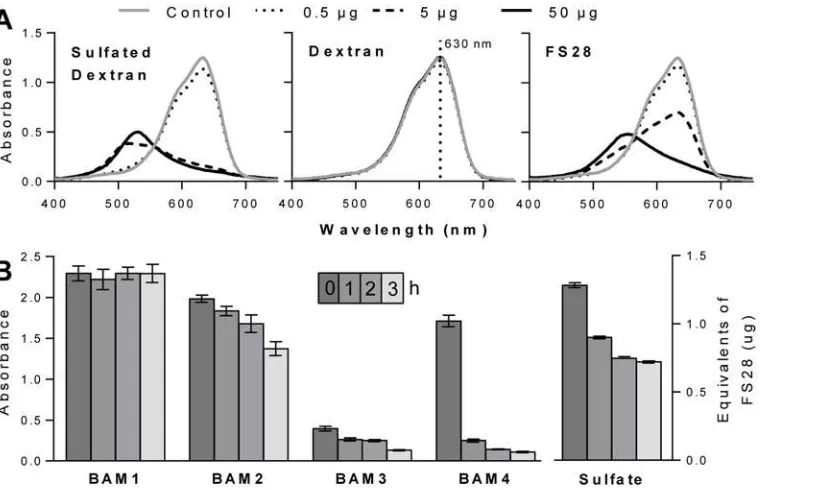
Due to the potential for variation in the density and patterning of sulfate groups on the fucan family of polysaccharides, it was required to determine if any of the MAbs had a sulfate-requiring epitope. Azure A is a cationic dye capable of binding to sulfate residues on polysac-charides, and is widely used to measure the sulfate content of heparin [46]. Addition of a sulfat-ed polysaccharide to an Azure A solution causes a colour change from blue to purple, as seen by sulfated dextran and FS28 (Fig. 2A). For comparison addition of a non-sulfated dextran has no effect on the spectrum of Azure A. Therefore, increasing sulfate levels can be measured by a decrease in absorbance at 630 nm.
The sulfated fucan fraction FS28 was de-sulfated using chlorotrimethylsilane (CTMS) in an-hydrous pyridine [47]. Sulfate analysis revealed that the majority of de-sulfation occurred in the first hour of the reaction, with only small decreases occurring in the following 2 h (Fig. 2B). ELISA analysis of the samples for alterations of MAb binding revealed no change for BAM1 recognition and a slow decline for BAM2 and BAM3 over 3 h. In the case of BAM4 there was a large decrease in binding to FS28 (>80%) in the first hour of the de-sulfation procedure. These
[image:7.612.200.475.78.376.2]observations suggest that BAM1 binds to a non-sulfate containing epitope and that BAM4 binds to a sulfated epitope. The BAM2 and BAM3 epitope may require sulfate groups for Fig 1. Heat map showing the relative binding of the BAM1 to BAM4, LM7 and LM23 MAbs to a range of brown algal, sulfated, red algal and land plant polysaccharides.Binding was determined by ELISA with
50μg/ml of polysaccharide samples and 25-fold dilutions of antibody hybridoma supernatants. S indicates
polysaccharides containing sulfate residues. The colour scale in relation to absorbance values is shown top left. Values shown are means of 4 replicates and in all cases standard deviations (SDs) were<0.1 absorbance units.
recognition but it is also possible that the de-sulfation process results in other changes to glycan structures that impact on binding.
To further explore the role of sulfate in epitope recognition the potential of the sulfate-binding dye Azure A to interfere with MAb recognition of the fucoidan sample was assessed. After coating and blocking of microtitre plates, Azure A (1 mg/ml) was incubated with the immobilised fucan for 1 h and plates were washed prior to the antibody incubation steps. As a control to assess non-specific inhibition by Azure A in MAb recognition of a non-sulfated glycan, Azure A was also in-cubated with oat spelt xylan prior to probing with xylan MAb LM11 [48]. The results shown in Fig. 3Aindicate that BAM1 and LM11 binding to glycans was unaffected by Azure A whereas BAM4, BAM3 and BAM2 displayed inhibition of binding by the presence of the sulfate-binding dye, and the binding of BAM2 was reduced by over 70% (Fig. 3A). For BAM1 recognition, the non-inhibition by Azure A is consistent with the previous interpretation of BAM1 binding to a non-sulfate containing epitope. For BAM2, BAM3 and BAM4, clear conclusions cannot be made from this assay alone. Indeed, one interpretation could be that sulfate groups may be an element of the BAM2, BAM3, and BAM4 epitopes. However, the loss in BAM4 binding upon Azure A competition is not as strong as one may expect for a MAb recognizing a sulfated epitope (Fig. 2B). It is possible that Azure A and BAM4 might both compete for a sulfated epitope, leading to a lower apparent inhibition. On the other hand a sulfate group bound by Azure A and present in the close vicinity to BAM2 and BAM3 epitopes might also impair their binding ability, making it impossible to conclude from this assay alone if sulfate groups are elements of their epitopes.
[image:8.612.41.453.76.321.2]Sodium metaperiodate is capable of cleaving the bond between two carbon atoms that both have associated hydroxyl groups (vicinal diols) and in homofucans these are present in fucose residues linked by 1,4-linkages, but not in the case of 1,3-linkages. Sodium metaperiodate sen-sitivity can be used to confirm antibody recognition of some glycan structures [39]. However, Fig 2. Sulfate assay and de-sulfation.(A) Azure A absorbance spectra changes in the presence of differing levels (0 to 50μg) of sulfated dextran, dextran and FS28. (B) Effect of sulfation on BAM MAb recognition of FS28. Samples of FS28 were collected over 4 time points (0, 1, 2 and 3 h) during a de-sulfation treatment with chlorotrimethylsilane and assayed using ELISA. Absorbance values are means of 4 replicates and error bars indicate SD. Sulfate levels as equivalents to native FS28 (w/w).
sulfate residues inhibit the action of sodium metaperiodate, due to the sulfation at the required OH groups [49]. Fucoidan-coated ELISA plates were treated for 2, 4, 8 and 24 h with sodium metaperiodate, and then the impact on the binding of the BAM MAbs determined. As shown inFig. 3Bthe BAM4 epitope is the most susceptible to sodium metaperiodate treatment and this might be indicative of the presence of 1,4-glycosyl residues in the BAM4 epitope. Those of BAM1 and BAM3 display a moderate sensitivity and BAM2 is largely unaffected even after 24 h. The basis of periodate insensitivity in the latter case is not known, but it may be indicative of the presence of 1,3-glycosyl residues and/or of sulfate groups in the epitope, or alternatively of an epitope composed of residues other than fucose but with no vicinal diols.
We propose, based on the accumulated evidence outlined above, that the BAM1 MAb binds to an un-sulfated epitope present in sulfated fucans and that BAM4, with the highest sensitivity to de-sulfation, binds to a sulfate-containing epitope. BAM2 and BAM3 MAbs are specific to sulfated fucan/fucoidan preparations but their specificities in relation to the role of sulfation in their epitope structure are less clear. The panel of four BAM MAbs will therefore be useful for the study of fucan polymers in brown algal cell walls.
Epitope detection chromatography analysis of sulfated fucan
heterogeneity
[image:9.612.204.484.77.338.2]In order to further explore the heterogeneity of sulfated fucans, two extracts obtained from FS28, one with native sulfation (DS0) and the other produced by 3 h of de-sulfation (DS3), were analysed by epitope detection chromatography (EDC) [40] in anion-exchange mode. This chromatographic separation is based on charge, and fucans will be retained on the column Fig 3. Effect of Azure A inhibition and sodium metaperiodate treatment on BAM antibody recognition of fucoidan.ELISA plates were coated with fucoidan and (A) pre-incubated with Azure A for 1 h, or equivalent control plates with buffer only, or (B) exposed to sodium metaperiodate for 0 to 24 h at 4°C in the dark. Absorbance values are means of 4 replicates and error bars indicate SD. LM11 binding to non-sulfated xylan was used as a control for Azure A inhibition.
and separated predominantly due to the number of sulfate residues and the presence of uronic acids which can also be abundantly found in fucan preparations. EDC involves the use of gly-can MAbs as detection tools (using ELISAs) for the chromatographically-separated glygly-cans. Both samples were run through an anion-exchange column, and fractions collected by applying a NaCl gradient. After their neutralisation, equivalent aliquots from the same chromatographic run were used to coat microtitre plate wells that were probed with MAbs BAM1, BAM2, BAM3, BAM4, LM7 and LM23. Both samples contained a small subset of detected BAM1 and LM23 epitopes that were not retained by the column (Fig. 4). The major epitope peaks were eluted in fractions 33 to 65 for DS0 and fractions 26 to 66 for DS3. The LM23 xylosyl epitope was detected at an equivalent level across these fractions, albeit eluting earlier in the DS3 sam-ple, indicating little disruption to the xylosyl-containing domains in the fucan. In contrast, the BAM1, BAM2 and BAM4 epitopes were differentially detected in both extracts tested. The first BAM epitope to be detected in both samples after the initiation of the salt gradient (fraction 26) is the non-sulfate-requiring BAM1 that peaks in fraction 42 in DS0 and 37 in DS3. This confirms that the BAM1 epitope is carried by an acidic polymer and that de-sulfation reduces the number of charges on this polymer. In both profiles the BAM1 epitope is eluted in a range of separated peaks, indicating the heterogeneity in the charges of fucans carrying this epitope. The BAM2 epitope is detected in later eluting fractions and peaks around fraction 54 in both FS28 extracts tested. The BAM2 EDC profiles are therefore complementary to the BAM1 patterns and indicate that the BAM1 and BAM2 epitopes are carried by less acidic and more acidic poly-mers respectively. In the de-sulfated sample the BAM2 peak is in the same fractions and shows lit-tle shift due to de-sulfation which may indicate an acidic but non-sulfated minor component of the preparation. In contrast the de-sulfation-sensitive epitope BAM4 was detected in two peaks in the DS0 sample, the less acidic broadly coinciding with that of the BAM1 epitope and also a late-eluting peak around fraction 58. The BAM4 epitope, as anticipated, shows the greatest loss in sig-nal in DS3 compared to DS0, although it is detected across the fucan region of the chromatogram (Fig. 4). Equivalent EDC profiles of the BAM3 and LM7 epitopes, which only bind weakly to the samples, indicate recognition of the same regions of the chromatograms (Fig. C inS1 File).
Taken together the EDC experiments provide evidence that all four BAM epitopes are carried by molecules that are acidic. They support the conclusions that BAM1 binds to a non-sulfated epitope that is present on a sulfated glycan, and that BAM4 recognizes a sulfated epitope. BAM2 and BAM3 identified distinct and additional epitopes present in the fucoidan preparation but within polymers showing little elution shifts due to de-sulfation. These results also indicate that the FS28 fucan preparation contains a spectrum of molecules with highly variable patterns/den-sities of sulfate groups, and that aspects of this diversity can be assessed by the BAM MAbs.
In situ
fluorescence imaging of BAM epitopes reveals changes in
cell-wall epitopes in relation to
Fucus vesiculosus
development
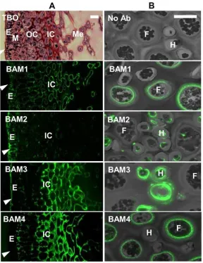
locations within theF.vesiculosusreceptacle, and thus suggests variations of the cell-wall struc-tures including sulfated fucans across tissues. Examination of BAM probe binding to hyphal and filament cells in the medulla of receptacles where cells are less closely packed revealed de-tection of the BAM1 and BAM4 epitopes in the cell walls of the larger filament cells whereas the BAM2 and BAM3 epitopes were detected in the smaller hyphal cells (Fig. 5B).
[image:11.612.40.435.77.375.2]F.vesiculosusis a dioecious species and the male (antheridia) and female (oogonia) repro-ductive structures are found in separate individuals. During development the conceptacle forms as a sphere which matures and releases the gametes through an opening or ostiole (Fig. 6).In situfluorescence imaging with the MAbs across sections of reproductive receptacles ofF.vesiculosuscontaining the female conceptacles provides contrasting images of unopened (no ostiole formed—spherical shaped) and opened conceptacles (including ostiole—teardrop shaped) after immunolabelling with BAM2 and BAM3 (Fig. 6). The BAM2 epitope is only weakly detected in paraphyses (sterile hairs) in unopened conceptacles, whereas after matura-tion of the conceptacle, it is detected in the meristoderm, and is also abundantly present inside the conceptacle around the paraphyses cells protruding from the ostiole. In contrast, the BAM3 epitope is detected strongly in cells surrounding the conceptacle and the oogonia in the conceptacle, but does not accumulate around the ostiole opening.
Fig 4. Epitope detection chromatographic (EDC) anion-exchange analysis.EDC analysis of A, sulfated (DS0) and B, de-sulfated (DS3) derivatives of FS28 using BAM1, BAM2, BAM4 and LM23 as detection tools. The elution gradient was 0 to 4 M NaCl from 26 ml to 80 ml elution volume. Blue arrows indicate shift in peak of BAM1 epitope elution associated with de-sulfation. Green arrows indicate loss of BAM4 epitope peak height but no shift in elution time. EDC profiles shown are representative of two chromatographic runs.
In situ
fluorescence imaging of the BAM4 epitope in
E
.
subulatus
reveals
physiological changes in sulfate patterning during acclimation to salinity
[image:12.612.201.489.76.450.2]The genusEctocarpusgroups several species of filamentous brown algae from environments exhibiting different levels of salinities. A strain ofE.subulatus(Ec371), isolated from a freshwa-ter environment, has been shown to be able to tolerate medium of various salinities [28]. Fur-thermore, acclimation of this strain to undiluted (100%) seawater (conductivity 4.8 S/m) or diluted (5%) seawater (conductivity 0.5 S/m) is associated with morphological changes. The BAM4 sulfated epitope was found to be the most abundant of the epitopes at the surface of in-tact, whole mount preparations ofE.subulatus.In situfluorescence imaging of the strain grown in 100% seawater indicates that the BAM4 epitope is present at the surface of most fila-ments, while it is not detected at the surface of the same strain when grown in 5% diluted sea-water (Fig. 7A). Preparations of alcohol insoluble residues (AIRs) ofE.subulatusgrown in 100% and 5% seawater conditions were sequentially extracted with CaCl2, Na2CO3and KOH
Fig 5. Indirect immunofluorescence analysis ofF.vesiculosusreceptacles.(A) Bright field image of Toluidine Blue O (TBO) stained resin-embedded section and fluorescence imaging of the BAM1 to BAM4 epitopes in equivalent sections. E, epidermis; M, meristoderm; OC, outer cortex; IC, Inner cortex; Me, Medulla. Arrowheads indicate outer surface. (B) Combined bright field/fluorescence of BAM epitopes in relation to hyphal cells (H) and filament cells (F) of the medulla region. Scale bars = 30μm.
Fig 6. Indirect immunofluorescence analysis ofF.vesiculosusfemale conceptacles.Bright field images of TBO-stained sections and fluorescence imaging of the BAM2 and BAM3 epitopes in equivalent sections. (A) Conceptacles prior to ostiole opening and (B) conceptacles after ostiole opening. Asterisks indicate oogonia and arrowheads indicate paraphyses. Scale bars = 100μm.
to solubilize cell wall fractions for analysis of relative BAM4 epitope and sulfate levels. As shown inFig. 7B, the BAM4 epitope was most abundantly detected in the Na2CO3fractions
with higher levels in the extracts forE.subulatusgrown in 100% seawater. Sulfate levels, in terms of FS28 equivalents, were also higher in the 100% seawater samples with greater differen-tials between the 100% / 5% samples in the Na2CO3and KOH extracts (Fig. 7C).
Distribution of BAM sulfated fucan/fucoidan epitopes in brown algae
[image:14.612.201.466.81.482.2]To explore the distribution of the BAM sulfated fucan/fucoidan epitopes, and associated LM7 and LM23 epitopes, across an extended range of brown algae, samples were collected from eight species belonging to the Fucales (Ascophyllum nodosum,Fucus serratus,F.spiralis,F.vesiculosus, Fig 7. Analysis ofEctocarpus subulatus.Analysis ofE.subulatusgrown in seawater (100%) or in 20-fold diluted seawater (5%). (A) Indirect immunofluorescence detection of the BAM4 epitope at the surface of whole mount preparations, scale = 25μm. (B) ELISA analysis of BAM4 epitope levels in extracts of AIR. C = CaCl2extract, N = Na2CO3extract, K = KOH extract. (C) Sulfate levels (FS28 equivalents w/w) in C, N and K
extracts of AIR. Error bars indicate SD of 4 replicates.
Halidrys siliquosa,Himanthalia elongata,Pelvetia canaliculata,and Sargassum muticum) and six species belonging to the Laminiarales (Chorda filum,Laminaria digitata,L.hyperborea,L. ochroleuca,Saccharina latissimaandUndaria pinnatifida) in addition to an Ectocarpale, Ecto-carpus siliculosus. Alcohol insoluble residues (AIRs) were prepared from whole algae and ex-tracted sequentially as described above. Treatment with CaCl2causes cross linking of the
alginates rendering them insoluble, thus fractions obtained through this treatment should con-tain polysaccharides free from alginates. Extraction with Na2CO3solubilises the alginate
net-work and any associated polysaccharides, whilst 4 M KOH solubilise glycans more tightly bound within the cell walls. The relative abundances of the BAM1 to BAM4, LM7 and LM23 epitopes assessed by ELISA are shown inFig. 8. All epitopes were present but at different levels in all the algal species examined. The BAM3 epitope was most abundant in three species of the Fucales. The BAM1 and BAM2 epitopes were consistently abundant in both the CaCl2and
Na2CO3extracts, apart from the BAM2 epitope inChorda filumextracts, whereas the BAM4
epitope occurred most often in the Na2CO3extract along with alginates which are detected by
LM7. The LM23 xylosyl epitope was most abundant in the CaCl2and Na2CO3extracts and
showed wide variation in levels of detection contrasting with the BAM1 and BAM2 epitopes.
Discussion
[image:15.612.38.494.80.306.2]MAbs BAM1 to BAM4 all bind specifically to preparations of fucoidans and to brown algal cell wallsin situ. Evidence indicates that BAM1 binds to a non-sulfated fucan epitope and that BAM4 binds to a sulfated fucan epitope. The role of sulfation in the BAM2 and BAM3 epitopes is not yet determined. The epitopes recognised by the MAbs are not yet entirely characterized and this will require future work with isolated oligosaccharides and enzymatic deconstructions. MAbs that can distinguish spatially and temporally distributed epitopes in cell walls of brown Fig 8. Heat map of antibody binding to cell wall extracts of a range of brown algae.Heat map of relative BAM1 to BAM4, LM7 and LM23 epitope levels as determined by ELISA absorbance obtained from 25-fold dilution of MAbs with 50μg/ml of extracts from 8 species of the Fucales (F), 6 species of Laminariales (L) and 1 species of Ectocarpales (E). Fucale and Laminariale species are listed in relation to approximate occurrence from upper to lower regions of intertidal zones. Extracts of AIR with CaCl2(C), Na2CO3(N) and KOH (K) after dialysis and freeze-drying. The colour scale in relation to
absorbance values is shown top left. Values shown are means of 4 replicates and in all cases SDs were<0.1 absorbance units.
algae are useful tools for the dissection of polysaccharide structure, sulfation patterns and bio-activities in addition to their use in the study of cell wall architectures and functions.
Use of MAbs in sulfated fucan/fucoidan analyses and potential of EDC
It is clear that the sulfated fucan/fucoidan group of glycans found in brown algae are highly heterogeneous in structural terms [16–20] and this adds a level of complexity to any assessment of the bioactivity of sulfated fucan preparations. The heterogeneity of a fucan preparation is di-rectly addressed in the EDC analysis shown inFig. 4, using three of the BAM MAbs. Acidic pectins extracted from land plant cell walls are readily eluted by<0.5 M NaCl [40], however,
the FS28 fucan required in the region of 4 M NaCl for full elution from a similar anion-ex-change column. In the process of this elution the BAM epitopes were differentially eluted, indi-cating a spectrum of acidic molecules with likely varying sulfation densities. The observation that the BAM2 epitope peak was not altered in mobility after partial de-sulfation is suggestive of a core component with a high acidic group density that is largely unaffected by the de-sulfation procedure used. In contrast, following de-de-sulfation, the earlier elution of the unsul-fated BAM1 epitope, which co-eluted with the BAM4 epitope, suggests a less acidic component that is more sensitive to the de-sulfation process. Analysis of epitope presence in cell walls and tissues—discussed below—clearly indicates that epitopes can occur on separate molecules—as for example the differential occurrence of the BAM1/BAM4 epitopes in filament cells and the BAM2/BAM3 epitopes in hyphal cells ofF.vesiculosusreceptacles (Fig. 5). Such analytical ap-proaches to dissect the distinct polymers that occur within a fucan preparation have consider-able potential to directly address issues of heterogeneity and to elucidate the spectrum of molecules within a preparation or extract that is being applied to a particular end use. More-over, it should also be possible using epitope tagging/affinity techniques to isolate specific sub-fractions of sulfated fucans for assessments of both structures and specific bioactivities. Poten-tial links between particular oligosaccharide structural features/epitopes within molecules, for example the LM23 xylosyl epitope and the BAM epitopes, could also be explored by enzymatic interventions prior to EDC analyses or sandwich ELISA approaches [50].
Use of MAbs in developmental and ecophysiological studies
In addition to the capacities of the BAM antibodies to discriminate components of fucan preparations, the BAM epitopes are clearly differentially regulatedin situ. Modulations in epitope levels are observed during development ofF.vesiculosusand in response to changes in salinity forE.subulatus.
Distinct cell morphologies are found in the tissue zones ofF.vesiculosusreceptacles. Cells in the medulla have heavily thickened walls and are further interspaced by an abundant intercellu-lar matrix. Cell walls in the outer cortex and epidermis remain rather thin, except the epidermis outer wall. The four BAM epitopes have different patterns of detection across these tissues with the BAM2 and BAM3 epitopes being evident at the outer surface of the organ, and the BAM1/ BAM4 epitopes being absent from this region (Fig. 5). The BAM2 epitope is mostly restricted to the thallus surface, in direct contact with the external environment. At this location cell wall components may act as an ionic barrier important for the osmotic adjustment during changes in salinity. It has been proposed [51–52] that cells are shielded by fucans of increasing sulfation toward the outer walls. The abundance of the BAM2 epitope (carried by the most acidic fraction of FS28) at the ostiole opening ofF.vesiculosusfemale conceptacles (Fig. 6) may also reflect a role of a specific form of fucoidan in algal cell interactions with saline environments.
extracellular matrices may contribute to cell ionic regulation by means of cation-binding to the nega-tively-charged polysaccharides, notably sulfated fucans in brown algae [21,51]. TheE.subulatus Ec371 strain can grow in the laboratory under a broad range of salinities with all cells in its filamen-tous structure in contact with the environment [28]. It is of interest that the de-sulfation sensitive BAM4 epitope was the most abundant at the surface of intact, whole mount preparations of E.subulatuswhen grown in 100% seawater but not detected when grown in 5% seawater. This re-sponsiveness to variations in salinity was also confirmed by assessment of total BAM4 epitope levels (Fig. 7). This difference demonstrates that the sulfation patterning at the surface of this brown alga can be actively regulated in relation to external salt concentrations. These cellular observations corre-late well with transcriptomic analysis of genes encoding sulfotransferases (for the de-novo sulfated fucan assembly) which were induced under growth ofE.subulatusin 100% seawater while the ex-pression of genes coding for sulfatases involved in de-sulfation was reduced under low salinity [28]. These results further support the idea that sulfation density in cell walls, which would directly impact on the ion composition and cation-binding activity, would influence water potential in the vicinity of cell membranes and be an important factor in the osmotic adjustment to salinity stress.
The mapping of the occurrence of the four BAM epitopes across 15 species of brown algae (Fig. 8) demonstrates the wide abundance of the BAM1 and BAM2 epitopes in both the calcium chloride and sodium carbonate extracts, indicating that the carrying polymers are held within cell wall structures by a range of mechanisms. It is of interest that the BAM3 and BAM4 epitopes are more varied in distribution between species and taxonomic groups. The Fucales have a character-istic distribution of species in intertidal zones, ranging fromPelvetia canaliculataat the upper limit, toSargassum muticumat the lower limit of the intertidal zone. Organising the species in this order (Fig. 8) shows the trend of increased detection of the BAM3 and BAM4 epitopes in ex-tracts of Fucales in relation to algal zonation in the intertidal zone. The BAM3 epitope is most abundant in three Fucale species:Pelvetia canaliculata,F.spiralisandAscophyllum nodosumand least abundant in brown algae which are always submerged such asSargassum muticum. The Laminariales also have a distribution of species from the low water mark into the sublittoral zone; however, this distribution depends greatly on the exposure of the shore to wave action and the op-tical clarity of water. Hence these species are harder to order in this manner. The BAM3 epitope is less abundant in this group whereas the BAM4 epitope has a similar detection pattern in the Fucales and Laminariales with a low abundance inChorda filum. Alginate (LM7) is consistently detected in the sodium carbonate fraction in all species, and a highly varied pattern of occurrence is seen for the LM23 xylosyl epitope. It remains to be elucidated how xylosyl-containing structural elements are arranged or linked to sub-fractions of the sulfated fucan group of polymers.
Conclusion
The MAbs described in the present study are highly versatile molecular tools for the analysis and dissection of fucoidan preparations, including sulfated fucan polysaccharides. They also have the potential to generate defined sub-fractions in relation to anionic charges/sulfation which may be of importance in industrial applications. MAb use in conjunction with molecular tools such as defined oligosaccharides and enzymes directed to sulfated fucans will allow more precision in defining epitope and glycan structures. Moreover, the MAbs will be important mo-lecular tools to integrate with cell biological, genomic and genetic approaches to understand the physiological and developmental mechanisms of brown algae.
Supporting Information
LM23, binding to fucoidan, sulfated fucan FS28 and alginate. Polymers were coated on microtitre plates at 50μg/ml and probed with a 5-fold dilution series of MAb hybridoma cell culture
super-natants. Results shown are means of 4 replicates and SD are in all cases<0.1 absorbance units.
Fig. B. ELISA analysis of the four sulfated fucan-directed MAbs (BAM1, BAM2, BAM3 and BAM4) binding to fucoidan in the presence of varying NaCl concentrations in phosphate buffer. The 137 mM NaCl concentration is equivalent to the PBS used for antibody experiments else-where in the study. Error bars indicate SD of 4 replicates.Fig. C. Epitope detection chro-matographic (EDC) anion-exchange analysis of sulfated (DS0) and de-sulfated (DS3) derivatives of FS28 using BAM3, and LM7 as detection tools. Elution gradient was 0 to 4 M NaCl from 26 ml to 80 ml elution volume. EDC profiles shown are representative of two chromatographic runs. (PDF)
Acknowledgments
We thank Margaret Blance and Böd Ayre for provision of algal materials.
Author Contributions
Conceived and designed the experiments: TAT RSB CH JPK. Performed the experiments: TAT SEM MJ CH. Analyzed the data: TAT CH JPK. Contributed reagents/materials/analysis tools: TAT SEM MJ TT CH. Wrote the paper: TAT CH JPK.
References
1. Seaweeds TD. Natural History Museum, Life Series, London; 2002.
2. Brownlee C, Bouget FY, Corellou F. Choosing sides: establishment of polarity in zygotes of fucoid algae. Semin Cell Dev Biol. 2001; 12: 345–351. PMID:11535041
3. Cock M, Sterck L, Rouzé P, Scornet D, Allen A, Amoutzias G, et al. TheEctocarpusgenome and the in-dependent evolution of multicellularity in brown algae. Nature. 2010; 465: 617–621. doi:10.1038/ nature09016PMID:20520714
4. Coelho SM, Godfroy O, Arun A, Le Corguille G, Peters AF, Cock JM. OUROBOROS is a master regula-tor of the gametophyte to sporophyte life cycle transition in the brown algaEctocarpus. Proc Natl Acad Sci USA. 2011; 108: 11518–11523. doi:10.1073/pnas.1102274108PMID:21709217
5. Le Bail A, Billoud B, Le Panse S, Chenivesse S, Charrier B. ETOILE regulates developmental pattern-ing in the filamentous brown algaEctocarpus siliculosus. Plant Cell. 2011; 23: 1666–1678. doi:10. 1105/tpc.110.081919PMID:21478443
6. Dittami SM, Gravot A, Renault D, Goulitquer S, Eggert A, Bouchereau A, et al. Integrative analysis of metabolite and transcript abundance during the short-term response to saline and oxidative stress in the brown algaEctocarpus siliculosus. Plant Cell Environ. 2011; 34: 629–642. doi: 10.1111/j.1365-3040.2010.02268.xPMID:21281312
7. Peters AF, Wijk SJ Van, Cho GY, Scornet D, Hanyuda T, Kawai H, et al. Reinstatement ofEctocarpus crouaniorumThuret in Le Jolis as a third common species ofEctocarpus(Ectocarpales, Phaeophy-ceae) in Western Europe, and its phenology at Roscoff, Brittany. Phycol Res. 2010; 58: 157–170. 8. Dittami SM, Proux C, Rousvoal S, Peters AF, Cock JM, Coppee J-Y, et al. Microarray estimation of
ge-nomic inter-strain variability in the genusEctocarpus(Phaeophyceae). BMC Mol Biol. 2011; 12: 2. doi: 10.1186/1471-2199-12-2PMID:21226968
9. Michel G, Tonon T, Scornet D, Cock JM, Kloareg B. The cell wall polysaccharide metabolism of the brown algaEctocarpus siliculosus. Insights into the evolution of extracellular matrix polysaccharides in Eukaryotes. New Phytol. 2010; 188: 82–97. doi:10.1111/j.1469-8137.2010.03374.xPMID:20618907 10. Kovalenko I, Zdyrko B, Magasinski A, Hertzberg B, Milicev Z, Burtovyy R et al. A major constituent of
brown algae for use in high-capacity Li-ion batteries. Science. 2011; 334: 75–79. doi:10.1126/science. 1209150PMID:21903777
12. Wargacki AJ, Leonard E, Win MN, Regitsky D, Santos CNS, Kim PB et al. An engineered microbial plat-form for direct biofuel production from brown macroalgae. Science. 2012; 335: 308–313. doi:10.1126/ science.1214547PMID:22267807
13. Wei N, Quaterman J, Jin Y-S. Marine macroalgae: an untapped resource for producing fuels and chem-icals. Trends Biotechnol. 2013; 31: 70–77. doi:10.1016/j.tibtech.2012.10.009PMID:23245657 14. Enquist-Newman M, Faust AM, Bravo DD, Santos CN, Raisner RM, Hanel A et al. Efficient ethanol
pro-duction from brown macroalgae sugars by a synthetic yeast platform. Nature. 2014; 505: 239–243. doi: 10.1038/nature12771PMID:24291791
15. Deniaud-Bouët E, Kervarec N, Michel G, Tonon T, Kloareg B, Hervé C. Chemical and enzymatic frac-tionation of cell walls from Fucales: insights into the structure of the extracellular matrix of brown algae. Ann Bot. 2014; 114: 1203–1216. doi:10.1093/aob/mcu096PMID:24875633
16. Kloareg B, Quatrano RS. Structure of the cell walls of marine algae and ecophysiological functions of the matrix polysaccharides. Oceanogr Mar Biol Annu Rev. 1988; 26: 259–315.
17. Anastyuk SD, Shevchenko NM, Nazarenko EL, Imbs TI, Gorbach VI, Dmitrenok PS, Zvyagintseva TN. Structural analysis of a highly sulfated fucan from the brown algaLaminaria cichorioidesby tandem MALDI and ESI mass spectrometry. Carbohydr Res. 2010; 345:2206–2212. doi:10.1016/j.carres. 2010.07.043PMID:20813351
18. Ale MT, Mikkelsen JD, Meyer AS. Important determinants for fucoidan bioactivity: a critical review of structure-function relations and extraction methods for fucose-containing sulfated polysaccharides from brown seaweeds. Mar Drugs. 2011; 9: 2106–2130. doi:10.3390/md9102106PMID:22073012 19. Pomin VH, Mourão PAS. Structure, biology, evolution, and medical importance of sulfated fucans and
galactans. Glycobiol. 2008; 18: 1016–1027.
20. Bilan MI, Grachev AA, Shashkov AS, Kelly M, Sanderson CJ, Nifantiev NE, et al. Further studies on the composition and structure of a fucoidan preparation from the brown algaSaccharina latissima. Carbo-hydr Res. 2010; 345: 2038–2047. doi:10.1016/j.carres.2010.07.009PMID:20701899
21. Kloareg B, Demarty M, Mabeau S. Polyanionic characteristics of purified sulphated homofucans from brown algae. Int J Biol Macromol. 1986; 8: 380–386.
22. Popper ZA, Michel G, Hervé C, Domozych DS, Willats WGT, Tuohy MG, et al. Evolution and diversity of plant cell walls: from algae to flowering plants. Ann Rev Plant Biol. 2011; 62: 567–590. doi:10.1146/ annurev-arplant-042110-103809PMID:21351878
23. Aquino RS, Grativol C, Mourão PAS. Rising from the sea: Correlations between sulfated polysaccha-rides and salinity in plants. PLOS ONE. 2011; 6, e18862. doi:10.1371/journal.pone.0018862PMID: 21552557
24. Shepherd VA, Beilby MJ. The effect of an extracellular mucilage on the response to osmotic shock in the charophyte algaLamprothamnium papulosum. J Membr Biol. 1999; 170: 229–242. PMID: 10441666
25. Shepherd VA, Beilby MJ, Heslop DJ. Ecophysiology of the hypotonic response in the salt-tolerant char-ophyte algaLamprothamnium papulosum. Plant Cell Environ. 1999; 22: 333–346.
26. Mabeau S, Kloareg B. Isolation and analysis of the cell walls of brown algae:Fucus spiralis,F. cera-noides,F.vesiculosus,F.serratus,Bifurcaria bifurcataandLaminaria digitata. J Exp Bot. 1987; 38: 1573–1580.
27. West J, Kraft G.Ectocarpus siliculosus(Dillwyn) Lyngb. from Hopkins River Falls, Victoria—the first re-cord of a freshwater brown alga in Australia. Muelleria. 1996; 9: 29–33.
28. Dittami SM, Gravot A, Goulitquer S, Rousvoal S, Peters AF, Bouchereau A, et al. Towards deciphering dynamic changes and evolutionary mechanisms involved in the adaptation to low salinities in Ectocar-pus(brown algae). Plant J. 2012; 71: 366–377. doi:10.1111/j.1365-313X.2012.04982.xPMID: 22394375
29. Senni K, Pereira J, Gueniche F, Delbarre-Ladrat C, Sinquin C, Ratiskol J et al. Marine polysaccharides: a source of bioactive molecules for cell therapy and tissue engineering. Mar Drugs. 2011; 9:
1664–1681. doi:10.3390/md9091664PMID:22131964
30. Larsen B, Haug A, Painter TJ. Sulphated polysaccharides in brown algae. I. Isolation and preliminary characterisation of three sulphated polysaccharides fromAscophyllum nodosum(L.) Le Jol. Acta Chem Scand. 1966; 20: 219–230.
31. Hahn T, Lang S, Ulber R, Muffler K. Novel procedures for the extraction of fucoidan from brown algae. Process Biochem. 2012; 47: 1691–1698.
32. Vreeland V, Slomich M, Laetsch WM. Monoclonal antibodies as molecular probes for cell wall antigens of the brown algaFucus. Planta. 1984; 162: 506–517. doi:10.1007/BF00399916PMID:24253267 33. Green JR, Stafford CJ, Jones JL, Wright PJ, Callow JA. Binding of monoclonal antibodies to vegetative
34. Jones JL, Callow JA, Green JR. Monoclonal antibodies to sperm surface antigens of the brown alga Fucus serratus exhibit region-, gamete-, species- and genus-preferential binding. Planta. 1988; 176: 298–306. doi:10.1007/BF00395410PMID:24220858
35. Eardley DD, Sutton CW, Hempel WM, Reed DC, Eberling AW. Monoclonal antibodies specific for sul-fated polysaccharides on the surface ofMacrocystis pyrifera(Phaeophyceae). J Phycol. 1990; 26: 54–62.
36. Lees A, Nelson BL, Mond JJ. Activation of soluble polysaccharides with 1-cyano-4-dimethylaminopyri-dinium tetrafluoroborate for use in protein polysaccharide conjugate vaccines and immunological re-agents. Vaccine. 1996; 14: 190–198. PMID:8920699
37. Willats WGT, Marcus SE, Knox JP. Generation of a monoclonal antibody specific to (1!5)-alpha-L-ara-binan. Carbohydr Res. 1998; 308: 149–152. PMID:9675359
38. Descamps V, Colin S, Lahaye M, Jam M, Richard C, Potin P, et al. Isolation and culture of a marine bac-terium degrading the sulfated fucans from marine brown algae. Mar Biotechnol. 2006; 8: 27–39. PMID: 16222488
39. Woodward MP, Young WW Jr, Bloodgood RA. Detection of monoclonal antibodies specific for carbohy-drate epitopes using periodate oxidation. J Immun Meth. 1985; 78: 143–153.
40. Cornuault V, Manfield IW. Ralet M-C, Knox JP. Epitope Detection Chromatography (EDC): a method to dissect the structural heterogeneity and interconnections of plant cell wall matrix glycans. Plant J. 2014; 78: 715–722. doi:10.1111/tpj.12504PMID:24621270
41. Marcus S, Verhertbruggen Y, Herve C, Ordaz-Ortiz J, Farkas V, Pedersen H, et al. Pectic homogalac-turonan masks abundant sets of xyloglucan epitopes in plant cell walls. BMC Plant Biol. 2008; 8: 60. doi:10.1186/1471-2229-8-60PMID:18498625
42. Willats WGT, Orfila C, Limberg G, Buchholt HC, van Alebeek G-JWM, Voragen AGJ, et al. Modulation of the degree and pattern of methyl-esterification of pectic homogalacturonan in plant cell walls: implica-tions for pectin methyl esterase action, matrix properties and cell adhesion. J Biol Chem. 2001; 276: 19404–19413. PMID:11278866
43. Larsen B, Vreeland V, Laetsch WM. Assay-dependent specificity of a monoclonal antibody with algi-nate. Carbohydr Res. 1985; 143: 221–227.
44. Manabe Y, Nafisi M, Verhertbruggen Y, Orfila C, Gille S, Rautengarten C, et al. Loss-of-function muta-tion of REDUCED WALL ACETYLATION2 inArabidopsisleads to reduced cell wall acetylation and in-creased resistance toBotrytis cinerea. Plant Physiol. 2011; 155: 1068–1078. doi:10.1104/pp.110. 168989PMID:21212300
45. Pedersen HL, Fangel JU, McCleary B, Ruzanski C, Rydahl MG, Ralet M-C et al. Versatile high-resolu-tion oligosaccharide microarrays for plant glycobiology and cell wall research. J Biol Chem. 2012; 287: 39429–39438. doi:10.1074/jbc.M112.396598PMID:22988248
46. Klein MD, Drongowski RA, Linhardt RJ, Langer RS. A colorimetric assay for chemical heparin in plas-ma. Anal Biochem. 1982; 124: 59–64. PMID:7125227
47. Kolender AA, Matulewicz MAC. Desulfation of sulfated galactans with chlorotrimethylsilane. Characteri-zation ofβ-carrageenan by 1H NMR spectroscopy. Carbohydr Res. 2004; 339: 1619–1629. PMID: 15183736
48. McCartney L, Marcus SE, Knox JP. Monoclonal antibodies to plant cell wall xylans and arabinoxylans. J Histochem Cytochem. 2005; 53: 543–546. PMID:15805428
49. Parsons TF, Pierce JG. Oligosaccharide moieties of glycoprotein hormones: bovine lutropin resists en-zymatic deglycosylation because of terminal O-sulfated N-acetylhexosamines. Proc Natl Acad Sci USA. 1980; 77: 7089–7093. PMID:6938955
50. Lee KJD, Cornuault V, Manfield I, Ralet M-C, Knox JP. Multiscale spatial heterogeneity of pectic rham-nogalacturonan-I (RG-I) in tobacco seed endosperm cell walls. Plant J. 2013; 75: 1018–1027. doi:10. 1111/tpj.12263PMID:23789903
51. Mariani P, Tolomio C, Braghetta P. An ultrastructural approach to the adaptive role of the cell wall in the intertidal algaFucus virsoides. Protoplasma. 1985; 128: 208–217.