This is a repository copy of
Phase transitions in shock compressed bismuth identified
using single photon energy dispersive X-ray diffraction (SPEDX)
.
White Rose Research Online URL for this paper:
http://eprints.whiterose.ac.uk/125901/
Version: Published Version
Article:
Briggs, R., Suggit, Matthew J., Gorman, M. G. et al. (6 more authors) (2017) Phase
transitions in shock compressed bismuth identified using single photon energy dispersive
X-ray diffraction (SPEDX). Journal of Physics: Conference Series. 042038. ISSN
1742-6596
https://doi.org/10.1088/1742-6596/950/4/042038
[email protected] https://eprints.whiterose.ac.uk/
Reuse
This article is distributed under the terms of the Creative Commons Attribution (CC BY) licence. This licence allows you to distribute, remix, tweak, and build upon the work, even commercially, as long as you credit the authors for the original work. More information and the full terms of the licence here:
https://creativecommons.org/licenses/
Takedown
If you consider content in White Rose Research Online to be in breach of UK law, please notify us by
PAPER • OPEN ACCESS
Phase transitions in shock compressed bismuth
identified using single photon energy dispersive
X-ray diffraction (SPEDX)
To cite this article: R Briggs et al 2017 J. Phys.: Conf. Ser. 950 042038
View the article online for updates and enhancements.
Related content
Structural Study of Molten Silicon by Energy Dispersive X-Ray Diffraction Method
Susumu Takeda
-Angle Dispersive X-ray Diffraction Beamline on Indus-2 Synchrotron Radiation Source: Commissioning and First Results
A K Sinha, Archna Sagdeo, Pooja Gupta et al.
-Phase Transitions in Silicon-Germanium Alloys under Pressure
G. Queisser, W. A. Grosshans and W. B. Holzapfel
1
Content from this work may be used under the terms of theCreative Commons Attribution 3.0 licence. Any further distribution of this work must maintain attribution to the author(s) and the title of the work, journal citation and DOI.
Published under licence by IOP Publishing Ltd
1234567890
AIRAPT IOP Publishing
IOP Conf. Series: Journal of Physics: Conf. Series 950 (2017) 042038 doi :10.1088/1742-6596/950/4/042038
Phase transitions in shock compressed bismuth
identified using single photon energy dispersive
X-ray diffraction (SPEDX)
R Briggs1, MJ Suggit2, MG Gorman1, A Coleman1, R Heathcote3, A
Higginbotham4
, S Patel2
, JS Wark2
and MI McMahon1
1
SUPA, School of Physics and Astronomy, and Centre for Science at Extreme Conditions, The University of Edinburgh, Edinburgh, EH9 3FD, UK
2
Department of Physics, Clarendon Laboratory, University of Oxford, Parks Road, Oxford, OX1 3PU, UK
3
Department of Physics, University of York, Heslington, York, YO10 5DD, UK
4
Science and Technology Facilities Council, Rutherford Appleton Laboratory, Harwell, Didcot, OX11 0QX, UK
E-mail: [email protected]
Abstract. We present evidence for phase transitions in shock-compressed bismuth using the
SPEDX x-ray diffraction technique. Experiments were performed on the Vulcan laser at the Central Laser Facility, RAL, Didcot, UK. We observed diffraction from the (110) bcc peak of Bi-V, and from its calculated lattice parameter the pressure was determined to be approximately 17 GPa. Upon further compression (higher laser intensities), no further diffraction from solid phases was observed. Shock melting of bismuth is thought to occur between 18 and 27 GPa. Diffraction results at lower pressures as a function of delay time are also presented.
1. Introduction
Dynamic compression of materials using high-power lasers allows access to extreme P-T states that lie well beyond the current limits of diamond anvil cell techniques. Laser facilities such as the National Ignition Facility are capable of compressing samples to 10s of megabars (> 1 TPa), whilst ensuring that the sample remains sufficiently cool to investigate solid-solid phase transitions [1]. However, collecting X-ray diffraction data from such samples is difficult, as the very high laser intensities used to produce the nanosecond-duration plasma X-ray source create a hostile environment in which the X-ray background from the drive lasers can eclipse any diffraction signal from the sample [2]. Obtaining X-ray diffraction from dynamically-compressed samples is therefore challenging at even modest pressures. Single photon energy dispersive X-ray diffraction (SPEDX) is a relatively new diffraction technique that uses CCD cameras in single photon counting mode to directly record the energy of X-rays diffracted from a laser-compressed sample [3]. Two X-ray CCDs record the diffraction at different Bragg angles, with each camera covering a wide range of k-space, allowing for recording of a significant number of Bragg peaks arising from the sample’s crystal structure.
2
peak compression to the Bi-V phase (body centered cubic; bcc) at ∼ 10 GPa, the pressure in
the sample then released to ambient pressure via three successive phase transitions – Bi-V (bcc)
→Bi-III (host-guest) → Bi-II → Bi-I – within 30 ns. More recently, we showed that on shock
release from pressures above 11 GPa Bi-V melts within 3 ns [5].
Here we present evidence for phase transitions in shock-compressed bismuth using the SPEDX diffraction technique. Experiments were performed on the Vulcan laser at the Central Laser Facility, RAL, Didcot, UK. We observe diffraction from the (110) bcc peak of Bi-V, and from the measured lattice parameter the sample’s pressure was determined to be∼17 GPa. Upon further
compression (higher laser intensities) no diffraction from crystalline Bi was observed, consistent with the incipient melting thought to occur between 18 and 27 GPa [6]. Also presented and discussed are lower-pressure diffraction data on compressed Bi to< 10 GPa.
2. Experimental methods
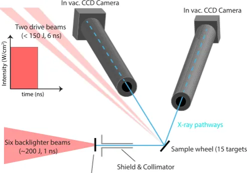
The experiment was carried out at the TAW, Vulcan facility (Central Laser Facility, Rutherford Appleton Laboratory, Didcot, UK). Two laser beam pathways were used to either drive the sample target package (with 1 or 2 drive beams) or to generate a broadband X-ray spectrum from a backlighter target foil (Fig. 1). The drive lasers delivered up to 200 J of 527 nm light onto target using a flat top laser pulse with pulse length of 6 ns, which shock-compressed samples to pressures of ∼ 20 GPa and below. The drive spot was 3.2 mm in diameter, generating
intensities of ∼ 1011-1012 W/cm2. Similar laser intensities were shown to shock compress Bi
to peak pressures of ∼ 14 GPa using the same ablating material [5]. Hydrocode simulations in
Gorman et al. revealed a steady shock in 15µm of Bi with a 20 ns pulse length. The flat top laser pulse of 6 ns used in these experiments ensures a steady shock within the diffracting layers of the sample (since X-ray diffraction here is collected in reflection geometry).
Six beams were used to drive the x-ray backlighter, delivering up to 600 J of 1053 nm light in 1 ns with a spot size of <1.0 mm and intensities of∼1014 W/cm2.
In vac. CCD Camera
Sample wheel (15 targets)
Backlighter wheel (15 targets)
Shield & Collimator
In vac. CCD Camera
Two drive beams (< 150 J, 6 ns)
Six backlighter beams (~200 J, 1 ns)
X-ray pathways
time (ns)
In
tensit
y (
W/cm
[image:4.595.167.414.472.645.2]2)
Figure 1. Experimental setup within the TAW target chamber.
3 1234567890
AIRAPT IOP Publishing
IOP Conf. Series: Journal of Physics: Conf. Series 950 (2017) 042038 doi :10.1088/1742-6596/950/4/042038
Both sample and backlighter targets were mounted on a multi-target wheel that was remotely controlled to rotate and align new targets after each shot, a technique developed to increase the shot rate during each experimental shift.
Two in-vacuum X-ray CCD cameras were used to collect scattered X-rays at two different Bragg angles. The experimental setup is shown in Fig. 1. The two cameras were placed approximately 50 cm from the sample wheel and were shielded with thick Al tubes to reduce the background noise. Plastic filtering was also added to the end of the Al tubes to preferentially reduce the number of background photons near 3 keV that were generated from the mid-Z backlighter samples. Both cameras were water-cooled via an external chiller to minimize the number of dark counts recorded by the CCDs. The X-rays were collimated using a Mo collimator with Pb shielding, thereby ensuring that only an X-ray spot size of ∼ 0.5 mm2 was incident
upon the driven area of the sample.
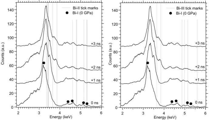
The two drive laser beams were timed with respect to each other such that the samples could be studied before, during and after the shock wave reached the bismuth sample. This allowed us to collect ambient diffraction from undriven targets at early times, diffraction from driven samples at the peak compression, and observe possible phases on shock release from the peak state, such as liquid-Bi or Bi-III / Bi-II, as observed by Hu et al. in their synchrotron-based study [4]. 140 120 100 80 60 40 20 0 C o u n ts (a .u .) 6 5 4 3 2 Energy (keV) 140 120 100 80 60 40 20 0 C o u n ts (a .u .) 6 5 4 3 2 Energy (keV) 0 ns +1 ns +2 ns +3 ns
Bi-II tick marks Bi-III tick marks
[image:5.595.119.464.363.561.2]0 ns +1 ns +2 ns +3 ns Bi-I (0 GPa) Bi-I (0 GPa)
Figure 2. SPEDX diffraction data collected at low pressure P <5 GPa with tick marks (grey
shaded lines) of the Bi-II (left) and Bi-III (right) crystal structures. Filled circles identify the ambient Bi-I diffraction peaks.
3. Results
We first investigated shock-compressed Bi at low pressures (P<10 GPa) by using laser intensities of∼1011W/cm2. From a previous experiment on Bi we were able to generate a pressure versus
4
positions at ∼2.7 GPa [7]. Whilst there is a reasonable agreement with the observed peaks at
+1 ns (Fig. 2 left), at +2 ns a new peak appears at ∼ 3.75 keV that does not fit very well
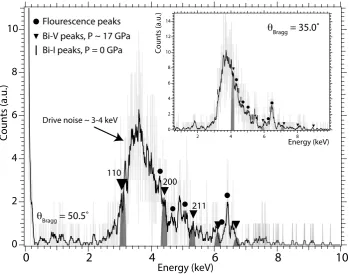
to the Bi-II structure. Fits to the Bi-III crystal structure are shown in Fig. 2 right. Both the Bi-II and Bi-III crystal structures have some agreement to the position of this peak (Bi-II peak within 0.6 keV, Bi-III within 0.3 keV), but it should be noted that the Bi-II peak (-201) has a calculated intensity that is significantly weaker than the strong Bi-III (310) peak at this energy. When the laser intensity was increased to beyond ∼ 1012 W/cm2 heavy filtering of the
CCD cameras was required in order to reduce the large drive noise at 3-4 keV (Fig. 3). For data collected using such drive laser intensities, the total number of counts in the signal was significantly reduced and some weak fluorescence peaks were also observed. The position of these fluorescence peaks on the CCD cameras are independent of the Bragg angle (the peaks are present at the same energy on both cameras) and we can therefore easily distinguish them from the diffracted Bi photons. Figure 3 shows an example of data collected from a driven target that shows evidence of the high-pressure Bi-V phase. The fluorescence peaks arising from the backlighter foils are identified in Fig. 3 with filled circles above them. By knowing the Bragg angle of each detector (obtained using ambient diffraction from Ta/Bi and calibrating the detector angles), the peak position of the Bi-V (110) reflection (∼4.03 keV at 35◦) can be used
to determine the lattice parameter for the driven sample, which was found to be 3.68 ˚A. From the known equation of state of Bi [8], this gives an estimated sample pressure of ∼ 17 GPa.
All of the remaining peaks can be fitted to the ambient Bi-I phase arising from uncompressed material ahead of the shock wave (rectangular tick marks in main figure).
10
8
6
4
2
0
10
8
6
4
2
0
14
12
10
8
6
4
2
0
Counts (a.u.)
8 6 4 2
Energy (keV)
Energy (keV)
Counts (a.u.)
Flourescence peaks Bi-V peaks, P ~ 17 GPa
211 200 110
θBragg = 50.5˚
Bi-I peaks, P = 0 GPa
Drive noise ~ 3-4 keV
[image:6.595.121.471.405.680.2]θBragg = 35.0˚
Figure 3. Diffraction data collected from the two CCD cameras at different Bragg angles.
5 1234567890
AIRAPT IOP Publishing
IOP Conf. Series: Journal of Physics: Conf. Series 950 (2017) 042038 doi :10.1088/1742-6596/950/4/042038
Recent experiments carried out at the LCLS x-ray free electron laser (XFEL) indicate that melting of the bismuth should also be observed on shock release from pressures above 11 GPa [5]. Unfortunately, the weak diffraction signal from the liquid is difficult to extract in the current experiment as it is overwhelmed by diffraction from several high-pressures phases. In some later shots in the campaign, we used higher laser energies than those where the Bi-V phase was observed and found that we were no longer able to observe any Bragg peaks from the Bi-V phase. This could be an indication that shock melting had occurred (previously reported to occur between 18 and 27 GPa [6]) as the Hugoniot crosses the melting curve. However, the drive noise was significantly higher for these data collections and the expected liquid diffraction peak is overlapped by the drive noise. In several cases the detectors were also unable to operate in single photon counting mode, since excessive numbers of noisy pixels from the drive saturated the CCD camera, overwhelming the diffraction signal from the liquid.
4. Conclusions
X-ray diffraction studies of bismuth under shock compression have been carried out and reveal phase transitions at low pressure (P < 10 GPa) and observe the high-pressure Bi-V phase at P ∼ 17 GPa, the highest pressure that Bi-V has been observed on the shock Hugoniot. It
is not yet clear as to what structure/structures the samples transform to at low pressures, and further work is planned to investigate the lower-pressure phase transitions using different detector angles. This would both improve our ability to distinguish Bi-II and Bi-III peaks, and move key Bragg peaks away from the region of drive noise for the highest energy drives. We will plan to investigate using the SPEDX technique in a transmission geometry, using thinner sample targets, thereby completely removing the drive noise signal present on the CCD camera, and enabling the weak liquid diffraction signal to be observed.
Acknowledgments
We acknowledge the expertise of the staff at the Central Laser Facility, Rutherford Appleton Laboratories for their help during the experiment. M.I.M. and J.S.W. would like to acknowledge support from EPSRC under Grant No. EP/J017256/1 and EP/J017051/1.
References
[1] R Smithet al.2014Nature,511, 330-333
[2] A Lazickiet al.2015Phys. Rev. Lett.,115, 075502 [3] A Higginbothamet al.2014Rev. Sci. Instrum.,85, 033906 [4] J Huet al.2013Appl. Phys. Lett.,103, 161904
[5] MG Gormanet al.2015Phys. Rev. Lett.,115, 095701
[6] Y Tan, Y Yu, C Dai, K Jin, Q Wang, J Hu, and H Tan 2013J. Appl. Phys.,113, 093509