RESEARCH
Granulin epithelin precursor promotes
colorectal carcinogenesis by activating MARK/
ERK pathway
Yi Pan
1,2,3, Siu Tim Cheung
4, Joanna Hung Man Tong
1,2,3, Ka Yee Tin
1,2, Wei Kang
1,2,3,
Raymond Wai Ming Lung
1,2,3, Feng Wu
1,2,3, Hui Li
1,2,3, Simon Siu Man Ng
3,5, Tony Wing Chung Mak
5,
Ka Fai To
1,2,3*and Anthony Wing Hung Chan
1,2*Abstract
Background: Granulin epithelin precursor (GEP) is reported to function as a growth factor stimulating proliferation and migration, and conferring chemoresistance in many cancer types. However, the expression and functional roles of GEP in colorectal cancer (CRC) remain elusive. The aim of this study was thus to investigate the clinical significance of GEP in CRC and reveal the molecular mechanism of GEP in CRC initiation and progression.
Methods: The mRNA expression of GEP in CRC cell lines were detected by qRT-PCR. The GEP protein expression was validated by immunohistochemistry in tissue microarray (TMA) including 190 CRC patient samples. The clinicopatho-logical correlation analysis were achieved by GEP expression on TMA. Functional roles of GEP were determined by MTT proliferation, monolayer colony formation, cell invasion and migration and in vivo studies through siRNA/shRNA mediated knockdown assays. The cancer signaling pathway identification was acquired by flow cytometry, western blot and luciferase activity assays.
Results: The mRNA expression of GEP in CRC was significantly higher than it in normal colon tissues. GEP protein was predominantly localized in the cytoplasm and most of the CRC cases demonstrated abundant GEP protein com-pared with non-tumorous tissues. GEP overexpression was associated with non-rectal location, advanced AJCC stage, regional lymph node and distant metastasis. By Kaplan–Meier survival analysis, GEP abundance served as a prognostic marker for worse survival in CRC patients. GEP knockdown exhibited anti-cancer effect such as inhibiting cell prolifera-tion, monolayer colony formaprolifera-tion, cell invasion and migration in DLD-1 and HCT 116 cells and decelerating xenograft formation in nude mice. siGEP also induced G1 cell cycle arrest and apoptosis. Luciferase activity assays further dem-onstrated GEP activation was involved in MAPK/ERK signaling pathway.
Conclusion: In summary, we compressively delineate the oncogenic role of GEP in colorectal tumorigenesis by activating MAPK/ERK signaling pathway. GEP might serve as a useful prognostic biomarker and therapeutic target for CRC.
Keywords: Colorectal cancer, GEP, Oncogene, MAPK/ERK pathway
© The Author(s) 2018. This article is distributed under the terms of the Creative Commons Attribution 4.0 International License (http://creat iveco mmons .org/licen ses/by/4.0/), which permits unrestricted use, distribution, and reproduction in any medium, provided you give appropriate credit to the original author(s) and the source, provide a link to the Creative Commons license, and indicate if changes were made. The Creative Commons Public Domain Dedication waiver (http://creat iveco mmons .org/ publi cdoma in/zero/1.0/) applies to the data made available in this article, unless otherwise stated.
Open Access
*Correspondence: kfto@cuhk.edu.hk; awh_chan@cuhk.edu.hk
1 Department of Anatomical and Cellular Pathology, State Key Laboratory
in Oncology in South China, Prince of Wales Hospital, The Chinese University of Hong Kong, 30-32 Ngan Shing Street, Shatin, NT, Hong Kong, SAR, China
Background
Colorectal cancer (CRC) is one of the major leading causes of cancer-related deaths in the world [1]. Although the overall survival of CRC patients has improved, the prognosis of patients with metastasis or recurrence is still relatively poor [2]. Angiogenesis is one of the major hall-marks of cancer [3], facilitating tumor development, pro-gression and metastasis [4, 5]. Thus, understanding the molecular mechanism and identifying of novel molecular biomarker in angiogenesis could benefit clinical manage-ment of CRC patients.
Granulin epithelin precursor (GEP), also known as progranulin, acrogranin, proepithelin, and GP88/PC-cell derived growth factor, is a secreted glycoprotein com-posed of 7.5 repeats of cysteine-rich motif [6, 7]. Physi-ologically, it is expressed in immune cells [8, 9], neurons [10], epithelial cells [11] and chondrocytes [12], medi-ating wound healing, neurodegeneration and cartilage development [7, 10–12]. Pathologically, high expression levels of GEP are associated with poor prognosis in hepa-tocellular carcinoma, ovarian cancer [13], bladder cancer [14] and glioblastoma [15]. Functional studies reveals GEP acts as a growth factor to stimulate proliferation and migration, and confer chemoresistance in many types of cancers including breast cancer [16, 17], ovarian cancer [18, 19], liver cancer [20, 21] and bile duct cancer [22].
As the expression and functional role of GEP in CRC is unclear. In this study, the clinical significance and the function of GEP in CRC will be comprehensively revealed.
Methods
Patients and specimens
Between 1999 and 2013, 190 patients undergoing resec-tion of primary CRCs at Prince of Wales Hospital, the Chinese University of Hong Kong were recruited in the present study. None of the patients received any neo-adjuvant therapy. The age of the patients ranged from 34 to 92 years, with a median age of 67.4 years. There were 106 men and 84 women. Tumors were staged according to the pathological tumor-node-metastasis (pTNM) stag-ing system, 7th version. Distribution of the pTNM stages and other clinicopathological features is listed in Table 1. The patients were regularly followed up according to the institutional practice. Disease free survival was defined as the period from the date of curative surgery of primary tumor to the date that the patient survived without any signs or symptoms of that cancer. Overall survival was defined as the period from the date of curative surgery of primary tumor to the date of cancer-related death or last
follow-up. The last update of the database 31 December 2015. The study was approved by Committees for Clini-cal Research Ethics of Joint Chinese University of Hong Kong-New Territories East Cluster.
Cell lines and cell culture
Human CRC cell lines Caco2, DLD-1, HCT 116, HT-29, LoVo, LS 180, SW480 and SW620 were all obtained American Type Culture Collection (ATCC, Manassas, VA, USA). All cell lines were cultured in media according to manufacturer’s instructions.
For transient transfection, DLD-1 or HCT 116 cells were transfected using lipofectamine 2000 (Thermo Fisher Scientific, Waltham, MA, USA) by siGEP or siRNA control (Qiagen, Hilden, Germany) to interfere GEP expression. Procedures of transfection were per-formed according to the manufacturer’s instructions. Cells stably expressing downregulated GEP mediated by shRNA was established using retrovirus system. The shRNA information for GEP knockdown can be found in Additional file 1.
Quantitative reverse transcriptase‑polymerase chain reaction (qRT‑PCR)
Total RNA from cells was extracted by Trizol (Thermo Fisher Scientific) according to the manufacturer’s instruction. cDNAs were synthesized using High-Capac-ity cDNA Reverse Transcription Kit (Thermo Fisher Sci-entific) as protocol. Quantification was performed with the ABI 7500 Real Time PCR system (Applied Biosys-tems, Foster City, CA, USA). Primers and probes for GEP were GEP-forward (5′-CAA ATG GCC CAC AAC ACT GA-3′), GEP-reverse (5′-CCC TGA GAC GGT AAA GAT GCA-3′) and GEP-probe (5′-6FAM CCA CTG CTC TGC CGG CCA CTC MGBNFQ-3′). Primer and probe reagents for control 18s were ready-made reagents (Pre-Developed TaqMan Assay Reagents, Applied Biosys-tems). All experiments were performed in a minimum of three replicates.
Immunohistochemistry (IHC)
cytoplasm by two of the investigators. The H-score was obtained by the formula: 3 × percentage of strongly stain-ing + 2 × percentage of moderately staining + percentage of weakly staining, giving a range of 0–300. The stain-ing was considered positive when there was moderate or strong immunoreactivity over the cutoff point above 150.
Western blot analysis
Equal total protein was separated by SDS-PAGE and transferred to nitrocellulose membrane. GEP was detected with a monoclonal anti-GEP antibody (1:5000
dilution). Other primary antibodies were from Cell Sign-aling (Danvers, MA, USA) commercially including Cyc-lin D1 (1:1000, #2926), cleaved PARP (Asp214) (1:1000, #9541), phospho-MAPK/ERK (1:2000, #9106), MAPK/ ERK (1:1000, #9102), Caspase-8 (1:1000, #9746), Cleaved Caspase-8 (1:1000, #9748), Caspase-3 (1:1000, #9662), Cleaved Caspase-3 (1:1000, #9661). The secondary anti-bodies were anti-Mouse IgG-HRP (1:15,000, 00049039, Dako, Denmark) and anti-Rabbit IgG-HRP (1:5000, 00028856, Dako). The western blot bands were quantified by ImageJ.
Table 1 Clinicopathologic correlation of GEP expression in colorectal cancer (n = 190, significant P-value in italic format)
GEP expression (n = 190) P‑value
All H‑score ≥ 150 H‑score < 150
98 (51.6%) 92 (48.4%)
Male gender 106 (55.8%) 51 (52.0%) 55 (59.8%) 0.283
Age at operation (years, mean ± SD) 67.4 ± 12.1 66.7 ± 12.6 68.2 ± 11.6 0.386
Location 0.032
Right colon 51 (26.8%) 27 (29.0%) 24 (26.4%)
Left colon 36 (18.9%) 23 (24.7%) 13 (14.3%)
Rectum 88 (46.3%) 36 (38.7%) 52 (57.1%)
Synchronous 9 (4.7%) 7 (7.5%) 2 (2.2%)
Unknown 6 (3.2%) / /
Size (cm, mean ± SD) 4.5 ± 1.7 4.6 ± 1.9 4.3 ± 1.6 0.257
Differentiation
Well 2 (1.1%) 0 2 (2.2%) 0.052
Moderate 180 (94.7%) 92 (95.8%) 88 (97.8%)
Poor 4 (2.1%) 4 (4.2%) 0
Others 4 (2.1%) / /
AJCC stage < 0.01
I 17 (8.9%) 5 (5.1%) 12 (13.0%)
II 57 (30.0%) 19 (19.4%) 38 (41.3%)
III 56 (29.5%) 27 (27.6%) 29 (31.5%)
IV 60 (31.6%) 47 (48.0%) 13 (14.1%)
T stage 0.583
T1 5 (2.6%) 3 (3.1%) 2 (2.2%)
T2 19 (10.0%) 8 (8.2%) 11 (12.0%)
T3 122 (64.2%) 61 (62.2%) 61 (66.3%)
T4 44 (23.2%) 26 (26.5%) 18 (19.8%)
N stage < 0.01
N0 89 (46.8%) 35 (35.7%) 54 (58.7%)
N1 61 (32.1%) 34 (34.7%) 27 (29.3%)
N2 40 (21.1%) 29 (29.6%) 11 (12.0%)
M stage < 0.01
M0 129 (67.9%) 50 (51.0%) 79 (85.9%)
M1 61 (32.1%) 48 (49.0%) 13 (14.1%)
[image:3.595.56.542.100.545.2]Cell proliferation assay
Cell proliferation assays were performed using 3-(4,5-dimethylthiazol-2-yl)-2,5-diphenylte-trazolium bromide (MTT, Sigma-Aldrich, St. Louis, MO, USA) assay following the manufactures’ protocol. Briefly, CRC cells which have been transfected with siGEP or siControl were plated in 96-well plate. Cell viability was measured at 24, 48, 72, 96, and 120 h. Finally, the optical density was determined at 570 and 690 nm wavelength light absorption (Victor3, Perken Elmer, MA, USA).
Monolayer colony formation assay
Anchorage-dependent growth was assessed by mon-olayer colony formation. 6-well plates were used at a den-sity of 1 × 10³ to 5 × 10³ for transfected cells. Cells were
incubated at 37 °C for 14–21 days until colonies can obvi-ously be observed. Colonies were fixed with methanol for 5 min and stained with 0.5% crystal violet. Colonies with cell numbers of more than 50 cells per colony were counted. The experiments were performed in duplicate wells in three independent experiments.
Invasion and migration assay
Cell invasion or migration activity were performed using 24 well biocoat matrigel invasion chambers or sterilized transwell insert chambers (Corning, Bedford, MA, USA). Cells were harvested after transfected with siGEP or siControl for 24 h and re-plated with serum free medium in the upper chamber. The lower chamber was filled with culture medium containing 10% FBS as the chemoat-tractant. After 48 h, the cells that moved to the lower sur-face of polycarbonate membrane were stained with 0.5% crystal violet and counted at five random 200× fields.
In vivo tumorigenic assays
1 × 10⁶ transfected CRC cells in 100 μl PBS were injected subcutaneously into the dorsal region of anaesthetized nude mice (5 mice/construct, control in left and treat-ment in right). When tumor was formed, tumor diameter was measured and documented every 3 days for 3 weeks. At the end of investigation, mice were sacrificed, and xenografts were collected for diameter check and weigh valuation. The animal handling and all experimental procedures were approved by the Department of Health of Hong Kong and the Animal Experimentation Ethics Committee, The Chinese University of Hong Kong.
Flow cytometry analysis
Cells transfected with siGEP or siControl were harvested at 24, 48 or 72 h after transfection. Then live cells were incubated in the mixture of propidium iodide (PI) and annexin V-fluorescein isothiocyanate (Thermo Fisher Scientific) for apoptosis study. Harvested cells were fixed and incubated with PI and RNase A at 4 °C for cell cycle analysis. Finally, cells were determined by FACS Cali-bur Flow Cytometer (Becton, Dickinson and Company, Franklin Lakes, NJ, USA) and CellQuest program.
Luciferase reporter assay
The firefly luciferase construct was co-transfected with Renilla luciferase vector (Qiagen) as a control into the siRNA treated cells. Dual Luciferase Reporter Assay System (Promega, Fitchburg, Wisconsin, USA) was employed to check the luciferase activity after 48 h’ trans-fection. The results were expressed as the ratio of firefly luciferase activity to renilla luciferase activity. Experi-ments were repeated in triplicate.
Statistical analysis
The statistical analysis was performed using IBM SPSS Statistics (Version 19.0, Armonk, NY, USA). The expres-sion level of GEP in paired non-tumor and tumor tissues was compared with paired Student’s t-test. Independent Student’s t-test was used to compare the mean value of any two groups. The Pearson χ2 test was used to analyze the association of target expression with clinicopatho-logical parameters. Survival curves were drawn using the Kaplan–Meier method and compared by means of the log-rank test. Univariable and multivariable Cox pro-portional hazard regression models were used to analyze independent prognostic factors. P < 0.05 was considered statistically significant. P < 0.01 was considered highly statistically significant.
Results
GEP is overexpressed in CRC
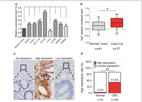
Fig. 1 The expression of GEP in CRC. a GEP mRNA expression was significantly higher in CRC cell lines (6/8) than normal colon. b GEP mRNA expression was significantly higher in colon cancer tissues than the normal colon in the Ki colon dataset of ONCOMINE. c Representative IHC images showing no expression in normal colon (H-score = 0), high expression in tumor cells (H-score > 150), and low expression in tumor cells (H-score ≤ 150) of GEP in human CRC samples. d GEP protein expression was higher in primary CRCs (51.6%), compared to normal colonic mucosa (4.3%). (*P < 0.05; **P < 0.01)
protein was predominantly localized in the cytoplasm and most of the CRC cases demonstrated abundant GEP protein compared with non-tumorous tissues (Fig. 1c). High expression (H-score ≥ 150) of GEP protein was detected in 51.6% (98/190) of tumor tissues. Whereas, only 4.3% (3/70) of normal tissues were found high GEP expression (P < 0.01, Fig. 1d).
High GEP expression correlates with poor survival in CRC patients
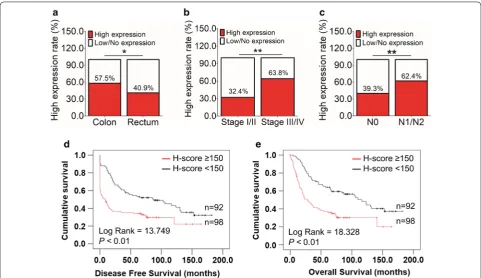
The correlation of GEP with clinicopathologic param-eters in CRC patients was summarized in Table 1. GEP expression varied significantly among CRC samples with different tumor locations (P= 0.032), AJCC stages
(P < 0.01), lymph node involvement (P < 0.01) and distant metastasis status (P < 0.01). Specifically, a high GEP pro-tein level more frequently occurred in tumors located on colon than that on rectum (P < 0.05, Fig. 2a). High GEP expression was also more commonly happened in patients with advanced AJCC stage (Stage III/IV, P < 0.01, Fig. 2b), including the presence of lymphatic (P < 0.01, Fig. 2c) and distant metastasis (P < 0.01, Table 1).
[image:5.595.61.539.86.431.2]high CEA level before operation (> 10 ng/ml) also pre-dicted worse disease-free survival and overall survival (Additional file 2). By multivariable Cox regression, com-pared with those classic prognostic parameters, high GEP expression was not an independent prognosticator for patients’ disease-free (Additional file 3) and overall survival (Additional file 4).
GEP exerts an oncogenic function on CRC cells
In DLD-1 and HCT 116 cell lines with endogenous high GEP expression, siRNA-mediated knockdown reduced GEP expression at mRNA (P < 0.01, Fig. 3a). siGEP treat-ments significantly decreased cell proliferation (Fig. 3b), anchorage-dependent growth (Fig. 3c), and abilities of
the invasion (Fig. 3d) and migration (Fig. 3e) in both DLD-1 and HCT 116 cells compared to the control group. These observations suggested that GEP had onco-genic properties.
To determine the anti-tumor effect of shGEP in vivo, DLD-1 cells stably expressing GEP shRNA was estab-lished using retrovirus system. shControl and shGEP were subcutaneously injected into the left and right flanks of nude mice respectively. The shGEP group formed smaller tumor within 27 days (Fig. 3f). In con-trast to shControl injected mice, the mice injected with shGEP exhibited greatly reduced mean tumor size (P < 0.05) and mean tumor weight (P < 0.01) in xenograft model (Fig. 3f).
Fig. 2 Clinicopathological and prognostic features of CRC patients with high GEP expression. a High GEP protein level was more frequently occurred in tumor located on colon than that on rectum (P < 0.05). b Higher GEP expression was also more commonly happened in patients with advanced AJCC stage (Stage III/IV, P < 0.01). c GEP upregulation was more involved in patients with lymphatic metastasis (P < 0.01). d, e GEP high expression correlated with both shorter disease-free survival (P < 0.01, d) and overall survival (P < 0.01, e). (*P < 0.05; **P < 0.01)
Fig. 3 GEP exerts oncogenic function in CRC cells. a GEP showed decreased expression at the mRNA level by qRT-PCR in DLD-1 and HCT 116 cells. b A significantly decreased proliferation was observed in the siGEP treated group compared with siControl group in all 2 cell lines examined (P < 0.01). c GEP knockdown significantly reduced anchorage-dependent growth in CRC cell lines by Foci-Formation assay (P < 0.01). d, e
[image:6.595.57.539.87.365.2]GEP knockdown results in G1 arrest and increased apoptosis in CRC
Since a growth inhibitory effect was involved in siGEP transfected cells, we further explored the molecular basis involved in siGEP-suppressed tumor cell growth. We analyzed the transfectants for cell cycle parameters and apoptosis using flow cytometry. 24 h after transfection, accumulation of G1 cells increased in siGEP transfect-ant compared with siControls (57.8% vs. 44.8% in DLD-1; 52.6% vs. 36.2% in HCT 116 cells), while S-phase cell percentage decreased after siGEP transfection (11.6% vs. 16.8% in DLD-1; 23.8% vs. 27.3% in HCT 116 cells) in these 2 cell lines (Fig. 4a). For apoptosis analysis, the per-centage of early apoptotic cells in siGEP treated cells was significantly increased compared to the siControl cells in both DLD-1 (0.8% ± 0.1% vs. 4.2% ± 0.2%, P < 0.01, Fig. 4b) and HCT 116 cells (0.8% ± 0.1% vs. 5.5% ± 0.2%, P < 0.01, Fig. 4b).
As proliferation-inhibition phenotypes were observed in siGEP groups, the associated cell cycle regulators and apoptosis markers were analyzed by western blot. Cyclin D1 was decreased in GEP knockdown cells, sup-porting the G0/G1-phase cell cycle arrest (Fig. 4c). Acti-vation of cleaved Caspase 3, 8 and cleaved PARP were observed after silencing GEP expression (Fig. 4c), indicat-ing that GEP inhibited apoptosis via a caspase-dependent pathway. Furthermore, luciferase reporter activity also showed GEP knockdown significantly suppressed cell cycle pathway in DLD-1and HCT 116 cells (Fig. 4d), con-firming the flow cytometry and western blot result for cell cycle analysis. Besides, GEP expression was positively associated with CyclinD1 expression from RNA level in TCGA database (Fig. 4e).
GEP promotes carcinogenesis via MAPK/ERK pathway in CRCs
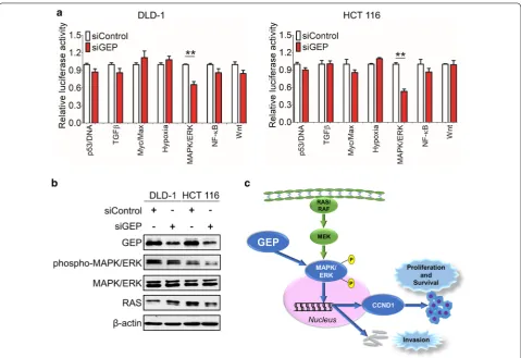
To further gain insights into the downstream signal-ing pathways modulated by GEP in CRC tumorigene-sis, we examined the functional effect of GEP in several important cancer pathways including p53, TGFβ, Myc, Hypoxia, MAPK/ERK, NF-κB and Wnt by luciferase reporter activity assay. GEP knockdown significantly suppressed MAPK/ERK luciferase reporter activity in DLD-1 and HCT 116 cells (Fig. 5a). Western blot results
showed significant suppression of phosphop-MAPK/ERK was observed in the siGEP treated CRC tumors (Fig. 5b), indicating that the MAPK/ERK signaling pathway was essential for anti-GEP-mediated growth inhibition in CRC cells. GEP led to MAPK/ERK phosphorylation and translocate into the nucleus, which stimulated cell prolif-eration, cell survival and metastasis of CRC (Fig. 5c).
Discussion
In this study, we characterized clinicopathological fea-tures of GEP expression and delineated its oncogenic function in colorectal carcinogenesis.
GEP was overexpressed in CRC cell lines and patients’ tumor samples. Overexpression of GEP in CRC was associated with nodal and distant metastasis and poorer clinical outcome. These findings concurred with previ-ous observations showing GEP overexpression in differ-ent human cancers including gliomas, renal, prostatic and hepatocellular carcinomas [25–28], and the correla-tion with poor survival [29]. Moreover, GEP expression was also positively associated with the MSI/CIMP sub-types of CRC in the TCGA cohort and patients with this kind of subtype had a very poor survival rate after relapse [30, 31] (Additional file 5). More importantly, increased DNA copy number of GEP could be detected in tumor samples in TCGA and Kurashina colon statistics from ONCOMINE (Additional file 6), indicating a strong pos-sibility that overexpression of GEP was caused by copy number change from DNA level.
We further investigated the underlying mechanism of upregulating GEP on CRC. A series of in vitro and in vivo experiments confirmed GEP as an oncogenic factor in CRC. Notably, a decreased GEP level by RNA interference decreased the cell proliferation rate by MTT and colony-forming ability in the anchorage-dependent environment and decelerating xenograft formation in nude mice by dis-turbing cell cycle and apoptosis process and targeting the MAPK/ERK pathway. Thus, GEP played a crucial role in carcinogenesis of CRC. GEP expression has been reported in other aggressive tumors [26, 29] with a promoting role in cell growth regulation [32, 33], wound-healing process [34, 35], and murine development [36]. It has been previ-ously shown that GEP binds with TNFR1/2 and diminish TNF-dependent activation of MAPK/ERK by altering the
(See figure on next page.)
TNF/TNFR interaction [12]. Then it triggered activation of the MAPK/ERK and PI3K/AKT/PKB signaling cascades as well as focal adhesion kinase in the adhesion/motility path-way in cell lines derived from adrenal, breast, cervical, and bladder cancer cells, as well as in modified mouse embryo fibroblasts [16, 37–39]. Therefore, control of CRC growth by GEP could be also mediated through these pathways. A decreased level of MAPK/ERK phosphorylated form was observed in the GEP knockdown group from our study. Here, we also provided evidence that GEP promoted cell migration and invasion in CRC cells in vitro. A clinical association evaluation showed that overexpression of GEP was associated significantly with advanced AJCC stage, suggesting that overexpression of GEP in CRC may facili-tate an invasive and metastatic phenotype.
GEP enhanced tumor cell survival, which was partly attributable to its anti-apoptotic ability. Defective
apoptosis is one of the major causative factors in tumo-rigenesis and caspase activation has been considered a hallmark of apoptosis [40]. In this study, we demon-strated that knockdown of GEP induced cell apoptosis with caspase activation, suggesting that GEP modulated cell apoptosis via a caspase-dependent pathway. On the other hand, p53 was well-documented to play an impor-tant role in inducing cell apoptosis [41], and the inhibition of p53 may contribute to the anti-apoptotic effect of GEP. We found that GEP also exhibited the effect of apoptosis both in p53 wild type and mutated CRC cells, indicating that GEP promoted tumorigenesis regardless of p53 muta-tion status. In keeping with this, the p53 pathway was not significantly affected when GEP knockdown by luciferase reporter activity assay for p53 pathway analysis. Thus, GEP knockdown activated cell apoptosis in a p53 independent manner.
Fig. 5 GEP promotes carcinogenesis via MAPK/ERK pathway in CRC. a A serial of promoter-luciferase assays (p53, TGFβ, Cell cycle, Myc, Hypoxia, MAPK/ERK, NF-κB and Wnt) were performed to screen for GEP target signaling pathways in DLD-1 and HCT 116 cells with GEP knockdown cells.
[image:10.595.59.539.86.415.2]Conclusions
Collectively, our findings demonstrated the oncogenic role of GEP in promoting tumorigenesis and metastasis in CRC. These findings suggest that GEP is a crucial fac-tor in carcinogenesis of colon and rectum, and that GEP has the potential to serve as a prognostic marker and therapeutic target for CRC.
Abbreviations
CRC : colorectal cancer; GEP: granulin epithelin precursor; pTNM: pathological tumor-node-metastasis; ATCC : American Type Culture Collection; qRT-PCR: quantitative real-time polymerase chain reaction; TCGA : the cancer genome atlas; TMA: tissue microarray; IHC: immunohistochemistry; H-score: histoscore; MTT: 3-(4,5-dimethylthiazol-2-yl)-2,5-diphenylte-trazolium bromide; PI: pro-pidium iodide.
Authors’ contributions
STC and AWHC conceived the project idea and designed the experiments; YP and HL performed the experiments; YP and FW analyzed the data; YP, WK and AWHC wrote and/or reviewed and/or revised the manuscript; STC, JHMT, EKYT, RWML, SSMN and TWCM provided technical or material support; KFT and AWHC supervised this study. All authors read and approved the final manuscript.
Author details
1 Department of Anatomical and Cellular Pathology, State Key Laboratory
in Oncology in South China, Prince of Wales Hospital, The Chinese University of Hong Kong, 30-32 Ngan Shing Street, Shatin, NT, Hong Kong, SAR, China.
2 Li Ka Shing Institute of Health Science, Sir Y.K. Pao Cancer Center, The Chinese
University of Hong Kong, Hong Kong, SAR, China. 3 Institute of Digestive
Dis-ease, Partner State Key Laboratory of Digestive DisDis-ease, The Chinese University of Hong Kong, Hong Kong, SAR, China. 4 Division of Hepatobiliary and
Pan-creatic Surgery, Department of Surgery, Prince of Wales Hospital, The Chinese University of Hong Kong, Hong Kong, SAR, China. 5 Division of Colorectal
Sur-gery, Department of SurSur-gery, Prince of Wales Hospital, The Chinese University of Hong Kong, Hong Kong, SAR, China.
Acknowledgements
We acknowledge the TCGA research Network (http://cance rgeno me.nih. gov/), ONCOMINE (https ://www.oncom ine.org/) and the cBioPortal for Cancer Genomics (http://www.cbiop ortal .org/) for providing and analyzing the CRC database. This study is supported by General Research Fund (RGC Reference No. CUHK 14104415, and 14138016) from The Research Grants Council of Hong Kong. We thank the Core Utilities of Cancer Genomics and Pathobiology (CUHK) for providing the facilities and assistance in support of this project. Additional files
Additional file 1. SuperArray SureSilencing shRNA plasmids mediated GEP knockdown.
Additional file 2. Clinicopathological factors predicted prognosis for 190 CRC patients.
Additional file 3. Univariable and multivariable Cox regression of prog-nostic parameters for disease free survival in 190 patients with colorectal cancer.
Additional file 4. Univariable and multivariable Cox regression of prognostic parameters for overall survival in 190 patients with colorectal cancer.
Additional file 5. GEP mRNA expression was higher in MSI/CIMP subtype than other subtypes in TCGA cohort. (*P < 0.05).
Additional file 6. Copy number change of GEP in ONCOMINE colon tis-sue datasets. (*P < 0.05; P < 0.01).
Competing interests
The authors declare that they have no competing interests.
Availability of data and materials
The datasets during and/or analyzed during the current study available from the corresponding author on reasonable request.
Consent for publication Not applicable.
Ethics approval and consent to participate
The use of human samples was approved by Joint Chinese University of Hong Kong-New Territories East Cluster Clinical Research Ethics Committee, Hong Kong.
Publisher’s Note
Springer Nature remains neutral with regard to jurisdictional claims in pub-lished maps and institutional affiliations.
Received: 5 March 2018 Accepted: 28 May 2018
References
1. Torre LA, Bray F, Siegel RL, Ferlay J, Lortet-Tieulent J, Jemal A. Global cancer statistics, 2012. CA Cancer J Clin. 2015;65:87–108.
2. Punt CJA, Tol J. OPINION More is less-combining targeted therapies in metastatic colorectal cancer. Nat Rev Clin Oncol. 2009;6:731–3. 3. Hanahan D, Weinberg RA. Hallmarks of cancer: the next generation. Cell.
2011;144:646–74.
4. Ebos JM, Kerbel RS. Antiangiogenic therapy: impact on invasion, disease progression, and metastasis. Nat Rev Clin Oncol. 2011;8:210–21. 5. Dimova I, Popivanov G, Djonov V. Angiogenesis in cancer—general
pathways and their therapeutic implications. J BUON. 2014;19:15–21. 6. Bateman A, Bennett HPJ. The granulin gene family: from cancer to
dementia. BioEssays. 2009;31:1245–54.
7. Liu CJ, Bosch X. Progranulin: a growth factor, a novel TNFR ligand and a drug target. Pharmacol Ther. 2012;133:124–32.
8. Jian J, Konopka J, Liu C. Insights into the role of progranulin in immunity, infection, and inflammation. J Leukoc Biol. 2013;93:199–208.
9. Arechavaleta-Velasco F, Perez-Juarez CE, Gerton GL, Diaz-Cueto L. Pro-granulin and its biological effects in cancer. Med Oncol. 2017;34:194. 10. Suzuki M, Nishiahara M. Granulin precursor gene: a sex steroid-inducible
gene involved in sexual differentiation of the rat brain. Mol Genet Metab. 2002;75:31–7.
11. Zhu J, Nathan C, Jin WW, Sim D, Ashcroft GS, Wahl SM, Lacomis L, Erdjument-Bromage H, Tempst P, Wright CD, Ding AH. Conversion of proepithelin to epithelins: roles of SLPI and elastase in host defense and wound repair. Cell. 2002;111:867–78.
12. Tang W, Lu Y, Tian QY, Zhang Y, Guo FJ, Liu GY, Syed NM, Lai YJ, Lin EA, Kong L, et al. The growth factor progranulin binds to tnf receptors and is therapeutic against inflammatory arthritis in mice. Science. 2011;332:478–84.
13. Han JJ, Yu MS, Houston N, Steinberg SM, Kohn EC. Progranulin is a poten-tial prognostic biomarker in advanced epithelial ovarian cancers (vol 120, pg 5, 2011). Gynecol Oncol. 2011;121:427.
14. Selmy MA, Ibrahim GH, El Serafi TI, Ghobeish AA. Evaluation of urinary proepithelin as a potential biomarker for bladder cancer detection and prognosis in Egyptian patients. Cancer Biomarkers. 2010;7:163–70. 15. Wang MQ, Li G, Yin JY, Lin T, Zhang J. Progranulin overexpression
predicts overall survival in patients with glioblastoma. Med Oncol. 2012;29:2423–31.
16. Swamydas M, Nguyen D, Allen LD, Eddy J, Dreau D. Progranulin stimu-lated by LPA promotes the migration of aggressive breast cancer cells. Cell Commun Adhes. 2011;18:119–30.
•fast, convenient online submission •
thorough peer review by experienced researchers in your field • rapid publication on acceptance
• support for research data, including large and complex data types •
gold Open Access which fosters wider collaboration and increased citations maximum visibility for your research: over 100M website views per year •
At BMC, research is always in progress.
Learn more biomedcentral.com/submissions
Ready to submit your research? Choose BMC and benefit from:
confers letrozole resistance to aromatase overexpressing breast cancer cells. Bmc Cancer. 2011;11:231.
18. Diaz-Cueto L, Arechavaleta-Velasco F, Diaz-Arizaga A, Dominguez-Lopez P, Robles-Flores M. PKC signaling is involved in the regulation of pro-granulin (acrogranin/PC-Cell-derived growth factor/pro-granulin–epithelin precursor) protein expression in human ovarian cancer cell lines. Int J Gynecol Cancer. 2012;22:945–50.
19. Carlson AM, Maurer MJ, Goergen KM, Kalli KR, Erskine CL, Behrens MD, Knutson KL, Block MS. Utility of progranulin and serum leukocyte pro-tease inhibitor as diagnostic and prognostic biomarkers in ovarian cancer. Cancer Epidemiol Biomark Prev. 2013;22:1730–5.
20. Cheung ST, Cheung PFY, Cheng CKC, Wong NCL, Fan ST. Granulin–epithe-lin precursor and ATP-dependent binding cassette (ABC) B5 regulate liver cancer cell chemoresistance. Gastroenterology. 2011;140:344.
21. Cheung PFY, Cheng CKC, Wong NCL, Ho JCY, Yip CW, Lui VCH, Cheung ANY, Fan ST, Cheung ST. Granulin–epithelin precursor is an oncofetal protein defining hepatic cancer stem cells. PLoS ONE. 2011;6:e28246. 22. Frampton G, Invernizzi P, Bernuzzi F, Pae HY, Quinn M, Horvat D, Galindo
C, Huang L, McMillin M, Cooper B, et al. Interleukin-6-driven progranulin expression increases cholangiocarcinoma growth by an Akt-dependent mechanism. Gut. 2012;61:268–77.
23. Ho JC, Ip YC, Cheung ST, Lee YT, Chan KF, Wong SY, Fan ST. Granulin–epi-thelin precursor as a therapeutic target for hepatocellular carcinoma. Hepatology. 2008;47:1524–32.
24. Choudhury KR, Yagle KJ, Swanson PE, Krohn KA, Rajendran JG. A robust automated measure of average antibody staining in immunohistochem-istry images. J Histochem Cytochem. 2010;58:95–107.
25. Liau LM, Lallone RL, Seitz RS, Buznikov A, Gregg JP, Kornblum HI, Nelson SF, Bronstein JM. Identification of a human glioma-associated growth factor gene, granulin, using differential immuno-absorption. Cancer Res. 2000;60:1353–60.
26. Donald CD, Laddu A, Chandham P, Lim SD, Cohen C, Amin M, Gerton GL, Marshall FF, Petros JA. Expression of progranulin and the epithelin/ granulin precursor acrogranin correlates with neoplastic state in renal epithelium. Anticancer Res. 2001;21:3739–42.
27. Pan CX, Kinch MS, Kiener PA, Langermann S, Serrero G, Sun L, Corvera J, Sweeney CJ, Li L, Zhang S, et al. PC cell-derived growth factor expression in prostatic intraepithelial neoplasia and prostatic adenocarcinoma. Clin Cancer Res. 2004;10:1333–7.
28. Cheung ST, Wong SY, Leung KL, Chen X, So S, Ng IO, Fan ST. Granulin– epithelin precursor overexpression promotes growth and invasion of hepatocellular carcinoma. Clin Cancer Res. 2004;10:7629–36.
29. Jones MB, Michener CM, Blanchette JO, Kuznetsov VA, Raffeld M, Serrero G, Emmert-Buck MR, Petricoin EF, Krizman DB, Liotta LA, Kohn EC. The granulin–epithelin precursor/PC-cell-derived growth factor is a growth factor for epithelial ovarian cancer. Clin Cancer Res. 2003;9:44–51. 30. Gao JJ, Aksoy BA, Dogrusoz U, Dresdner G, Gross B, Sumer SO, Sun YC,
Jacobsen A, Sinha R, Larsson E, et al. Integrative analysis of complex cancer genomics and clinical profiles using the cBioPortal. Sci Signaling. 2013;6:1.
31. Cerami E, Gao JJ, Dogrusoz U, Gross BE, Sumer SO, Aksoy BA, Jacobsen A, Byrne CJ, Heuer ML, Larsson E, et al. The cBio cancer genomics portal: an open platform for exploring multidimensional cancer genomics data. Cancer Discov. 2012;2:401–4.
32. Elkabets M, Gifford AM, Scheel C, Nilsson B, Reinhardt F, Bray MA, Carpen-ter AE, Jirström K, Magnusson K, Ebert BL, et al. Human tumors instigate granulin-expressing hematopoietic cells that promote malignancy by activating stromal fibroblasts in mice. J Clin Invest. 2011;121:784–99. 33. Lu Y, Zheng L, Zhang W, Feng T, Liu J, Wang X, Yu Y, Qi M, Zhao W, Yu X,
Tang W. Growth factor progranulin contributes to cervical cancer cell proliferation and transformation in vivo and in vitro. Gynecol Oncol. 2014;134:364–71.
34. He ZH, Ong CHP, Halper J, Bateman A. Progranulin is a mediator of the wound response. Nat Med. 2003;9:225–9.
35. Wang H, Sun Y, Liu S, Yu H, Li W, Zeng J, Chen C, Jia J. Upregulation of progranulin by Helicobacter pylori in human gastric epithelial cells via p38MAPK and MEK1/2 signaling pathway: role in epithelial cell prolifera-tion and migraprolifera-tion. FEMS Immunol Med Microbiol. 2011;63:82–92. 36. Kuse Y, Tsuruma K, Sugitani S, Izawa H, Ohno Y, Shimazawa M, Hara H.
Progranulin promotes the retinal precursor cell proliferation and the photoreceptor differentiation in the mouse retina. Sci Rep. 2016;6:23811. 37. Monami G, Gonzalez EM, Hellman M, Gomella LG, Baffa R, Iozzo RV,
Mor-rione A. Proepithelin promotes migration and invasion of 5637 bladder cancer cells through the activation of ERK1/2 and the formation of a paxillin/FAK/ERK complex. Can Res. 2006;66:7103–10.
38. Zhang XY, Pan ZX, Liu H, Yu JL, Li GX, Wang HY, Liu MM. Effect of pro-granulin (PGRN) on the proliferation and senescence of cervical cancer cells. Genet Mol Res. 2015;14:14331–8.
39. Tanimoto R, Lu KG, Xu SQ, Buraschi S, Belfiore A, Iozzo RV, Morrione A. Mechanisms of progranulin action and regulation in genitourinary can-cers. Front Endocrinol (Lausanne). 2016;7:100.
40. Johnstone RW, Ruefli AA, Lowe SW. Apoptosis: a link between cancer genetics and chemotherapy. Cell. 2002;108:153–64.