S H O R T R E P O R T
Open Access
The presence of
Giardia intestinalis
in
donkeys,
Equus asinus
, in China
Xiao-Xuan Zhang
1,2, Fu-Kai Zhang
1, Fa-Cai Li
1, Jun-Ling Hou
1, Wen-Bin Zheng
1, Shuai-Zhi Du
3, Quan Zhao
2and Xing-Quan Zhu
1*Abstract
Background:Giardia intestinalisis one of the most important zoonotic enteric parasites. As no information regarding prevalence and genotype ofG. intestinalisin donkeys (Equus asinus) in China is available, 181 faecal samples from 48 donkeys from Jilin Province, from 104 from Shandong Province and from 29 from Liaoning Province were examined between May and December 2015.
Findings:Twenty-eight (15.47%) out of 181 donkey samples were testedG. intestinalis-positive by nested amplification of the triosephosphate isomerase (tpi) gene. The prevalence in different regional groups varied from 10.42 to 18.27%. The prevalence in adult and young donkeys was 14.29 and 22.92%, respectively. Otherwise, the prevalence was 11.69% in summer and 18.27% in winter. However, no statistically significant differences were found in relation to region or age group. Sequence analysis of thetpi, glutamate dehydrogenase (gdh) and beta giardin (bg) loci identified 4, 1 and 3 subtypes of assemblage B, respectively. Moreover, four novel multilocus genotypes (MLGs novel-1 to novel-4) were identified in assemblage B.
Conclusions:This first report ofG. intestinalisin donkeys in China indicates that further studies of nation-wide molecular epidemiology and geographical distribution ofGiardiain donkeys are warranted. Effective strategies should be implemented to controlG. intestinalisinfection in donkeys, other animals and humans.
Keywords:Giardia intestinalis, Prevalence, Genotyping, Donkey, China
Background
Giardia intestinalis is the only species ofGiardia which
is found in human beings [1–4]. It is not only distributed worldwide, but also can infect many vertebrates [5, 6]. Eight assemblages (A-H) have been identified within G.
intestinalis [6–8]. Of these, assemblages C-H seem to be
animal-specific [9], but assemblages A and B can infect humans and a wide range of non-human hosts [6, 10]. Diarrhea is the main symptom of giardiasis [11] and trans-mission is mainly through ingestion of Giardia cysts in contaminated food or water [12]; approximately 2.8 × 10⁸ cases of human giardiasis are reported world-wide per year, and the majority of them are reported in developing countries [6]. In view of such a serious situation, giardiasis
has attracted considerable attention around the world. AlthoughG. intestinalis infections have been reported in humans and a variety of animal species [13–16], there is little information in donkeys (Equus asinus).
Giardia intestinalis infection in horses has been
reported in many countries around the world including China [17–21]. The donkey belongs to the genusEquus. It is an important edible animal species and used in Chinese traditional medicine, and is closely related to horses. Because they are maintained in a close association with their owners and veterinary personnel, donkeys are the important reservoirs for transmission of pathogens (such
as Cryptosporidium hominis and Toxoplasma gondii) to
humans and other animals [22, 23]. To determine whether donkeys are hosts ofG. intestinalis, we conducted a study on the prevalence and genotypes of G. intestinalis in donkeys in Jilin, Liaoning and Shandong Provinces, China. * Correspondence:xingquanzhu1@hotmail.com
1
State Key Laboratory of Veterinary Etiological Biology, Key Laboratory of Veterinary Parasitology of Gansu Province, Lanzhou Veterinary Research Institute, Chinese Academy of Agricultural Sciences, Lanzhou, Gansu Province 730046, People’s Republic of China
Full list of author information is available at the end of the article
Methods
Collection and preparation of faecal samples
A total of 181 donkey faecal samples (48 from Jilin, 27 from Liaoning and 104 from Shandong) were collected from three provinces, in northeastern and eastern China, be-tween March and December 2015. Each of the fresh faecal samples was collected into sterile gloves separately after its defecation onto the ground, placed into box with ice and transported to the laboratory immediately. Genomic DNA was extracted directly from each faecal sample using the Stool DNA kit (OMEGA, Norcross, Georgia, USA) accord-ing to the manufacturer’s instructions. Genomic DNA was stored at -20 °C until PCR amplification.
PCR amplification
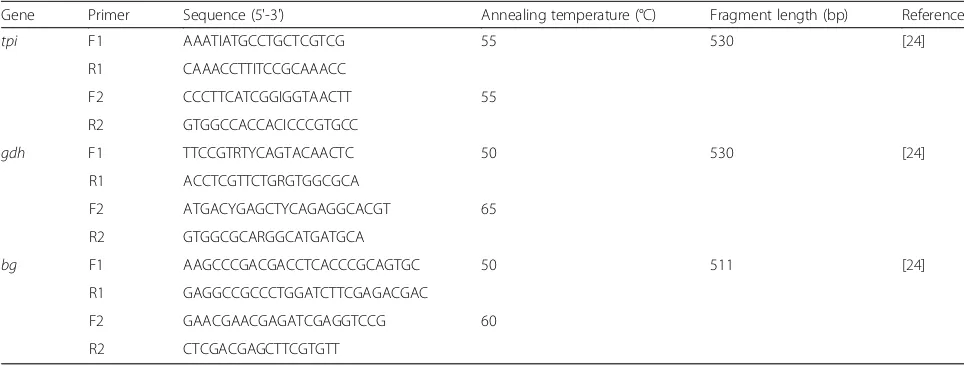
The prevalence and genotypes ofG. intestinaliswere deter-mined by the nested PCR amplification of the triosepho-sphate isomerase (tpi) gene, beta giardin (bg) gene and glutamate dehydrogenase (gdh) genes as described by Zhao et al. [24]. The primers and their annealing temperatures are listed in Table 1. Positive and negative controls were included in each amplification. Amplification products were observed under UV light after electrophoresis in 1.5% agar-ose gels containing GoldView™(Solarbio, Beijing, China).
Sequence and phylogenetic analyses
Positive secondary PCR products were sequenced by Genscript Company (Nanjing, China). Bidirectional sequen-cing was used to confirm the accuracy of the sequences. Se-quences with mutations were considered as novel genotypes when confirmed from independent two PCR reactions on the same sample. To identify the assemblages and subtypes, nucleotide sequences were aligned with known reference tpi,gdhandbggene sequences ofG. intestinalisavailable in GenBank using the BLAST (http://www.ncbi.nlm.nih.gov/ BLAST/) and computer program Clustal X 1.83.
Statistical analysis
The relationship between prevalence of G. intestinalis-in-fected donkeys and different variables including age, geo-graphic origin and seasons were analyzed by Chi-square test using SPSS version 17.0 (SPSS Inc., Chicago, IL, USA) [25]. The results were considered significant statistically if P< 0.05. Odds ratios (ORs) and their 95% confidence inter-vals (CI) are also given.
Results and discussion
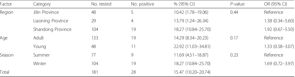
In this study, a total of 28 (15.47%, 95% CI: 10.20–20.74) out of 181 donkey samples were PCR-positive forG. intesti-nalis(Table 2). The prevalence was 22.92% (95% CI: 11.03– 34.81) in young donkeys and 14.29% (95% CI: 8.34–20.23) in adults; no significant difference was observed (χ2= 1.90, df= 1,P= 0.17) (Table 2). The prevalence in summer and winter was 11.69% (95% CI: 4.51–18.87) and 18.27% (95% CI: 10.84–25.70), respectively (χ2
= 1.47, df= 1, P= 0.23) (Table 2). Donkeys from Jilin Province (5/48, 10.42%, 95% CI: 1.78–19.06) had a lower prevalence than those from Shandong Province (19/104, 18.27%, 95% CI: 10.84–25.70) and Liaoning Province (4/29, 13.79%, 95% CI: 1.24–26.34); however, these differences were not significant (χ2= 1.62, df= 2,P= 0.44) (Table 2). Moreover, G. intestinalis preva-lence in different farms ranged from 6.12 to 29.09% (Table 3). Sequences analysis of thetpi,gdhandbgloci in-dicated only assemblage B was found in the present study (Additional file 1: Figure S1).
[image:2.595.56.538.549.732.2]The overall prevalence of G. intestinalis infection in donkeys was 15.47% (28/181, 95% CI: 10.20–20.74), which was higher than that in horses in Xinjiang (1.5%) [17], Brazil (0.5%) [26], foals in Belgium (14.2%) [20], Germany (10%) [20], Greece (11.6%) [20], the Netherlands (11.4%) [20] and Italy (8.6%) [18], but lower than that in horses in Colombia (17.4%) [19]. Previous studies demonstrated that survival ofG. intestinalisis more likely to be affected
Table 1Primers used in the study, annealing temperatures used in the PCRs and expected sizes of the PCR products
Gene Primer Sequence (5'-3') Annealing temperature (°C) Fragment length (bp) Reference
tpi F1 AAATIATGCCTGCTCGTCG 55 530 [24]
R1 CAAACCTTITCCGCAAACC
F2 CCCTTCATCGGIGGTAACTT 55
R2 GTGGCCACCACICCCGTGCC
gdh F1 TTCCGTRTYCAGTACAACTC 50 530 [24]
R1 ACCTCGTTCTGRGTGGCGCA
F2 ATGACYGAGCTYCAGAGGCACGT 65
R2 GTGGCGCARGGCATGATGCA
bg F1 AAGCCCGACGACCTCACCCGCAGTGC 50 511 [24]
R1 GAGGCCGCCCTGGATCTTCGAGACGAC
F2 GAACGAACGAGATCGAGGTCCG 60
by climate (temperature and relative humidity) [6, 27], so the difference inG. intestinalisprevalence in different re-gions may be due to different local climatic conditions, as well as the detection methods, sampling time and sample sizes. Moreover, probably because of the smaller sample sizes, seasonal and age-related correlates previously found in cattle [6] and horses [20] were not found in this study.
Assemblages A and B, responsible for the vast majority of human giardiasis [6, 28], and assemblage E, a common assemblage ofG. intestinalisin cattle [9], have also been re-ported in horses [20]. However, perhaps due to the smaller sample sizes, only assemblage B was identified in donkeys in the present study. Assemblage B has a broad host range worldwide [20, 28]. In China, isolates of assemblage B have also been found in non-human primates [2], rabbits [3], horses [17], cattle [29], golden takins (Budorcas taxicolor bedfordi) [24], pet chinchillas (Chinchilla lanigera) [30], captive wildlife [13], sheep [31] and goats [31], suggesting interspecies transmission of G. intestinalis may be commonly occurring in China, and we should pay enough attention to. More importantly, assemblage B was also identified in raw urban wastewater in northern China [32]. Therefore, our results also suggest that donkeys could be a source of giardiasis outbreaks.
Mixed infections ofG. intestinalisgenotypes have been recorded from a wide range of hosts worldwide [29, 33]. In cases of co-infections, some assemblages may be de-tected preferentially using a single locus primers [34]; thus PCR amplification of a single locus may not reflect the
accurate information toG. intestinalis infection [34, 35]. A multilocus genotype (MLG) method (tpi, gdh and bg loci) has been developed and widely used for detection of G. intestinalisinfection [6, 33, 34]. In the present study,
28G. intestinalis-positive samples were also genotyped
[image:3.595.57.540.99.225.2]based onbgand gdhloci. A total of 28tpi, 22bgand 16 gdhgene sequences were obtained, and analysis of these genes revealed only one assemblage (B); however, high genetic polymorphism was observed at these loci within this assemblage (Table 4), implying high genetic diversity of G. intestinalis in donkeys in the investigation regions. At thetpilocus, seven polymorphic sites were found com-pared with the GenBank reference sequence AY368169 (Table 4), and four different assemblage B subtypes were identified (Table 4, Additional file 1: Figure S1c). These sequences all represented new subtypes (KU892519– KU892522), and showed a 99% similarity with the refer-ence sequrefer-ence (accession no. AY368169, from wastewater in USA [36]). Only one subtype was found at thegdhlocus (Additional file 1: Figure S1b), and the sequence (KU892523) had 99% similarity with the reference se-quence available in GenBank with accession number of KR048463 (from a takin in China [24]). Moreover, a total of five SNPs were observed at thebg locus (Table 4), and these sequences (KU892516–KU892518) represented three subtypes (Additional file 1: Figure S1a). Moreover, these subtypes were closely clustered with sub-assemblage BIV (Additional file 1: Figure S1), suggesting that sub-assemblage BIV was the most common subtype in donkeys
Table 2Factors associated with prevalence ofGiardia intestinalisin donkeys in northern China
Factor Category No. tested No. positive % (95% CI) P-value OR (95% CI)
Region Jilin Province 48 5 10.42 (1.78–19.06) 0.44 Reference
Liaoning Province 29 4 13.79 (1.24–26.34) 1.38 (0.34–5.60)
Shandong Province 104 19 18.27 (10.84–25.70) 1.92 (0.67–5.50)
Age Adult 133 19 14.29 (8.34–20.23) 0.17 Reference
Young 48 11 22.92 (11.03–34.81) 1.33 (0.58–3.07)
Season Summer 77 9 11.69 (4.51–18.87) 0.23 Reference
Winter 104 19 18.27 (10.84–25.70) 1.69 (0.72–3.97)
[image:3.595.58.536.638.732.2]Total 181 28 15.47 (10.20–20.74)
Table 3Giardia intestinalisgenotypes identified in donkeys in different farms
Region Farm ID Age category (n) No. positive/No. tested (%) Genotype (n)
Jilin Province Farm 1 Young (10); Adult (38) 5/48 (10.42) BIV-1 (n= 2); BIV-novel-2 (n= 2); BIV-novel-3 (n= 1) Liaoning Province Farm 2 Young (6); Adult (23) 4/29 (13.79) BIV-1 (n= 2); BIV-novel-3 (n= 1); BIV-novel-4 (n= 1) Shandong Province Farm 3 Young (14); Adult (35) 3/49 (6.12) BIV-1 (n= 1); BIV-novel-4 (n= 2)
Farm 4 Young (18); Adult (37) 16/55 (29.09) BIV-1 (n= 12); BIV-novel-3 (n= 4)
in the investigated regions. Furthermore, phylogenetic ana-lysis of these isolates showed that these isolates exhibit close relationship with isolates from horses, humans and chinchillas, suggesting that transmission ofG. intestinalis may be occurring among these hosts.
Furthermore, 10 out of 28 positive isolates were successfully amplified at all three loci. These samples pro-vided four novel MLGs in the assemblage B, namely MLGs novel-1 to novel-4 (Table 5, Fig. 1). Of these, MLG novel-1 (n= 5) was the most prevalent MLG, and respon-sible for 50% of all MLGs in the present study. These find-ings suggest a high genetic diversity of this prevalent genotype in donkeys in China, in agreement with previous conclusions thatG. intestinalisisolates of the same assem-blage may be grouped into distinct MLGs [6, 24].
Conclusions
The present study demonstrated the occurrence of G.
intestinalis infection in donkeys in China. Sequences
[image:4.595.57.538.99.308.2]analysis suggested that all the G. intestinalis isolates represented assemblage B, with four, one and three subtypes of assemblage B at the tpi gdh and bgloci, re-spectively. Moreover, four novel MLGs (MLGs novel-1 to novel-4) were identified within assemblage B. The re-sults of the present study not only improve the informa-tion of the distribuinforma-tion of G. intestinalis genotypes in China, but also provide the foundation data for prevent-ing and controllprevent-ing G. intestinalis infection in donkeys, other animals and humans.
Table 4Variations intpi,gdhandbgnucleotide sequences among the subtypes ofGiardia intestinalisassemblage B
Locus Subtype (n) Nucleotide at position GenBank ID
tpi 10 11 16 182 197 384 525
Ref. sequence C G – G A G G AY368169
BIV-1a(n= 17) C G – A G A G KU892520
BIV-novel-2 (n= 2) T C G A G A G KU892519
BIV-novel-3 (n= 6) T C – A G A G KU892521
BIV-novel-4 (n= 3) T G – A G A T KU892522
gdh 219
Reference sequence G KR048463
BIV-novel-1 (n= 16) C KU892523
bg 14 179 248 446 447
Reference sequence G C C A G KM926514
BIV-1b
(n= 12) G C C G A KU892517
BIV-2b(n= 4) G T C G A KU892518
Bb-7c(n= 6) A C T G A KU892516
a
Identified by Qi et al. [17]
b
Identified by Coronato Nunes et al. [37]
c
[image:4.595.303.540.555.673.2]Identified by Karim et al. [2]
Table 5Multilocus characterization ofGiardia intestinalis
assemblage B isolates from donkeys attpi,gdhandbgloci
Isolate (n) Genotype GenBank ID MLGs L7 (5) BIV-1,
BIV-novel-1, BIV-1
KU892520, KU892523, KU892516
novel-1
L13 (1) BIV-novel-2, BIV-novel-1, BIV-2
KU892519, KU892523, KU892517
novel-2
L64 (2) BIV-novel-3, BIV-novel-1, Bb-7
KU892521, KU892523, KU892518
novel-3
L93 (2) BIV-1, BIV-novel-1, Bb-7
KU892520, KU892523, KU892518
novel-4
[image:4.595.56.293.590.731.2]Additional file
Additional file 1: Figure S1.Phylogenetic tree ofGiardia intestinalis
based on nucleotide sequences of theβ-giardin(a),gdhgene (b) andtpi
gene (c). Trees were constructed using using the neighbor-joining (NJ) method (Kimura 2-parameter model). Bootstrapping was performed using 1000 replicates.G. intestinalisisolates identified in the present study are indicated by solid circles. (TIF 4193 kb)
Abbreviations
bg:Beta giardin gene; CI: Confidence interval;gdh: Glutamate dehydrogenase gene; MLG: Multilocus genotype; OR: Odds ratio;
tpi: Triosephosphate isomerase (tpi) gene
Acknowledgements
The authors thank the staff and workers in the donkey farms who helped in the collection of faecal samples.
Funding
This work was supported by the Agricultural Science and Technology Innovation Program (ASTIP) (Grant No. CAAS-ASTIP-2014-LVRI-03).
Availability of data and material
Representative nucleotide sequences were deposited in GenBank with the following accession numbers: KU892516–KU892518 for thebggene, and KU892519–KU892522 for thetpigene, and KU892523 for thegdhgene.
Authors’contributions
XQZ and QZ conceived and designed the study, and critically revised the manuscript. XXZ, FKZ and WBZ performed the experiments. XXZ and FKZ analyzed the data. XXZ drafted the manuscript. FCL, JLH and SZD helped in study design, study implementation and manuscript preparation. All authors read and approved the final manuscript.
Competing interests
The authors declare that they have no competing interests.
Consent for publication
Not applicable.
Ethics approval
This study was approved by the Animal Ethics Committee of Lanzhou Veterinary Research Institute, Chinese Academy of Agricultural Sciences (Approval No. LVRIAEC2014-011). Donkeys used for the study were handled in accordance with good animal practices required by the Animal Ethics Procedures and Guidelines of the People's Republic of China.
Author details
1
State Key Laboratory of Veterinary Etiological Biology, Key Laboratory of Veterinary Parasitology of Gansu Province, Lanzhou Veterinary Research Institute, Chinese Academy of Agricultural Sciences, Lanzhou, Gansu Province 730046, People’s Republic of China.2College of Animal Science and Technology, Jilin Agricultural University, Changchun, Jilin Province 130118, People’s Republic of China.3Qilu Animal Health Products Co., Ltd., Jinan, Shandong Province 250100, People’s Republic of China.
Received: 26 August 2016 Accepted: 13 December 2016
References
1. Minetti C, Lamden K, Durband C, Cheesbrough J, Fox A, Wastling JM. Determination ofGiardia duodenalisassemblages and multi-locus genotypes in patients with sporadic giardiasis from England. Parasit Vectors. 2015;8:444.
2. Karim MR, Wang R, Yu F, Li T, Dong H, Li D, et al. Multi-locus analysis of
Giardia duodenalisfrom nonhuman primates kept in zoos in China:
geographical segregation and host-adaptation of assemblage B isolates. Infect Genet Evol. 2015;30:82–8.
3. Qi M, Xi J, Li J, Wang H, Ning C, Zhang L. Prevalence of zoonoticGiardia
duodenalisassemblage B and first identification of assemblage E in rabbit fecal
samples isolates from Central China. J Eukaryot Microbiol. 2015;62:810–4. 4. De Liberato C, Berrilli F, Marangi M, Santoro M, Trogu T, Putignani L, et al.
Giardia duodenalisin Alpine (Rupicapra rupicapra rupicapra) and Apennine
(Rupicapra pyrenaica ornata) chamois. Parasit Vectors. 2015;8:650.
5. Feng Y, Xiao L. Zoonotic potential and molecular epidemiology ofGiardia
species and giardiasis. Clin Microbiol Rev. 2011;24:110–40.
6. Zhang XX, Tan QD, Zhao GH, Ma JG, Zheng WB, Ni XT, et al. Prevalence, risk factors and multilocus genotyping ofGiardia intestinalisin dairy cattle, northwest China. J Eukaryot Microbiol. 2016;63:498–504.
7. Ramírez JD, Heredia RD, Hernández C, León CM, Moncada LI, Reyes P, et al. Molecular diagnosis and genotype analysis ofGiardia duodenalisin asymptomatic children from a rural area in central Colombia. Infect Genet Evol. 2015;32:208–13.
8. Santin M, Fayer R.Enterocytozoon bieneusi,Giardia, andCryptosporidium
infecting white-tailed deer. J Eukaryot Microbiol. 2015;62:34–43.
9. Sprong H, Cacciò SM, van der Giessen JW, ZOOPNET network and partners. Identification of zoonotic genotypes ofGiardia duodenalis. PLoS Negl Trop Dis. 2009;3, e558.
10. Mohamed AS, Levine M, Camp JW, Jr Lund E, Yoder JS, Glickman LT, et al. Temporal patterns of human and canineGiardiainfection in the United States: 2003–2009. Prev Vet Med. 2014;113:249–56.
11. Shin JC, Reyes AW, Kim SH, Kim S, Park HJ, Seo KW, et al. Molecular detection ofGiardia intestinalisfrom stray dogs in animal shelters of Gyeongsangbuk-do (Province) and Daejeon, Korea. Korean J Parasitol. 2015; 53:477–81.
12. Karanis P, Kourenti C, Smith H. Waterborne transmission of protozoan parasites: a worldwide review of outbreaks and lessons learnt. J Water Health. 2007;5:1–38.
13. Li J, Qi M, Chang Y, Wang R, Li T, Dong H, et al. Molecular characterization of
Cryptosporidiumspp.,Giardia duodenalis, andEnterocytozoon bieneusiin captive
wildlife at Zhengzhou Zoo, China. J Eukaryot Microbiol. 2015;62:833–9. 14. Ehsan AM, Geurden T, Casaert S, Parvin SM, Islam TM, Ahmed UM, et al.
Assessment of zoonotic transmission ofGiardiaandCryptosporidium
between cattle and humans in rural villages in Bangladesh. PLoS One. 2015; 10, e0118239.
15. Cacciò SM, Ryan U. Molecular epidemiology of giardiasis. Mol Biochem Parasitol. 2008;160:75–80.
16. Bouzid M, Halai K, Jeffreys D, Hunter PR. The prevalence ofGiardiainfection in dogs and cats, a systematic review and meta-analysis of prevalence studies from stool samples. Vet Parasitol. 2015;207:181–202. 17. Qi M, Zhou H, Wang H, Wang R, Xiao L, Arrowood MJ, et al. Molecular
identification ofCryptosporidiumspp. andGiardia duodenalisin grazing horses from Xinjiang, China. Vet Parasitol. 2015;209:169–72.
18. Traversa D, Otranto D, Milillo P, Latrofa MS, Giangaspero A, Di Cesare A, et
al.Giardia duodenalissub-assemblage of animal and human origin in
horses. Infect Genet Evol. 2012;12:1642–6.
19. Santín M, Cortés Vecino JA, Fayer R. A large scale molecular study ofGiardia
duodenalisin horses from Colombia. Vet Parasitol. 2013;196:31–6.
20. Kostopoulou D, Casaert S, Tzanidakis N, van Doorn D, Demeler J, von Samson-Himmelstjerna G, et al. The occurrence and genetic characterization
ofCryptosporidiumandGiardiaspecies in foals in Belgium, the Netherlands,
Germany and Greece. Vet Parasitol. 2015;211:170–4.
21. Veronesi F, Passamonti F, Cacciò S, Diaferia M, Piergili FD. Epidemiological survey on equineCryptosporidiumandGiardiainfections in Italy and molecular characterization of isolates. Zoonoses Public Health. 2010;57:510–7. 22. Jian F, Liu A, Wang R, Zhang S, Qi M, Zhao W, et al. Common occurrence of
Cryptosporidium hominisin horses and donkeys. Infect Genet Evol. 2016;43:261–6. 23. Alvarado-Esquivel C, Alvarado-Esquivel D, Dubey JP. Prevalence ofToxoplasma
gondiiantibodies in domestic donkeys (Equus asinus) in Durango, Mexico
slaughtered for human consumption. BMC Vet Res. 2015;11:6.
24. Zhao GH, Du SZ, Wang HB, Hu XF, Deng MJ, Yu SK, et al. First report of zoonotic
Cryptosporidiumspp.,Giardia intestinalisandEnterocytozoon bieneusiin golden takins (Budorcas taxicolor bedfordi). Infect Genet Evol. 2015;34:394–401. 25. Xu H, Jin Y, Wu W, Li P, Wang L, Li N, et al. Genotypes ofCryptosporidium
spp.,Enterocytozoon bieneusiandGiardia duodenalisin dogs and cats in Shanghai, China. Parasit Vectors. 2016;9:121.
26. De Souza PN, Bomfim TC, Huber F, Abboud LC, Gomes RS. Natural infection by
Cryptosporidiumsp.,Giardiasp. andEimeria leuckartiin three groups of equines
27. Alum A, Absar IM, Asaad H, Rubino JR, Ijaz MK. Impact of environmental conditions on the survival ofCryptosporidiumandGiardiaon environmental surfaces. Interdiscip Perspect Infect Dis. 2014;2014:210385.
28. Ballweber LR, Xiao L, Bowman DD, Kahn G, Cama VA. Giardiasis in dogs and cats: update on epidemiology and public health significance. Trends Parasitol. 2010;26:180–9.
29. Huang J, Yue D, Qi M, Wang R, Zhao J, Li J, et al. Prevalence and molecular characterization ofCryptosporidiumspp. andGiardia duodenalisin dairy cattle in Ningxia, northwestern China. BMC Vet Res. 2014;10:292. 30. Qi M, Yu F, Li S, Wang H, Luo N, Huang J, et al. Multilocus genotyping of
potentially zoonoticGiardia duodenalisin pet chinchillas (Chinchilla lanigera) in China. Vet Parasitol. 2015;208:113–7.
31. Zhang W, Zhang X, Wang R, Liu A, Shen Y, Ling H, et al. Genetic characterizations ofGiardia duodenalisin sheep and goats in Heilongjiang Province, China and possibility of zoonotic transmission. PLoS Negl Trop Dis. 2012;6, e1826.
32. Liu A, Ji H, Wang E, Liu J, Xiao L, Shen Y, et al. Molecular identification and distribution ofCryptosporidiumandGiardia duodenalisin raw urban wastewater in Harbin. China Parasitol Res. 2011;109:913–8.
33. Ankarklev J, Hestvik E, Lebbad M, Lindh J, Kaddu-Mulindwa DH, Andersson JO, et al. Common coinfections ofGiardia intestinalisandHelicobacter pylori
in non-symptomatic Ugandan children. PLoS Negl Trop Dis. 2012;6, e1780. 34. Cacciò SM, Beck R, Lalle M, Marinculic A, Pozio E. Multilocus genotyping of
Giardia duodenalisreveals striking differences between assemblages A and
B. Int J Parasitol. 2008;38:1523–31.
35. Wielinga CM, Thompson RC. Comparative evaluation ofGiardia duodenalis
sequence data. Parasitology. 2007;134:1795–821.
36. Sulaiman IM, Jiang J, Singh A, Xiao L. Distribution ofGiardia duodenalis
genotypes and subgenotypes in raw urban wastewater in Milwaukee, Wisconsin. Appl Environ Microbiol. 2004;70:3776–80.
37. Coronato Nunes B, Pavan MG, Jaeger LH, Monteiro KJ, Xavier SC, Monteiro FA, et al. Spatial and molecular epidemiology ofGiardia intestinalisdeep in the Amazon, Brazil. PLoS One. 2016;11, e0158805.
38. Lebbad M, Mattsson JG, Christensson B, Ljungström B, Backhans A, Andersson JO, et al. From mouse to moose: multilocus genotyping of
Giardiaisolates from various animal species. Vet Parasitol. 2010;168:231–9.
• We accept pre-submission inquiries
• Our selector tool helps you to find the most relevant journal
• We provide round the clock customer support
• Convenient online submission
• Thorough peer review
• Inclusion in PubMed and all major indexing services
• Maximum visibility for your research
Submit your manuscript at www.biomedcentral.com/submit