R E S E A R C H
Open Access
Within-host temporal fluctuations of
Trypanosoma cruzi
discrete typing units: the
case of the wild reservoir rodent
Octodon
degus
Gemma Rojo
1†, Alejandra Sandoval-Rodríguez
1†, Angélica López
1, Sylvia Ortiz
1, Juana P. Correa
4, Miguel Saavedra
2,
Carezza Botto-Mahan
3, Pedro E. Cattan
4and Aldo Solari
1*Abstract
Background:Chagas disease caused byTrypanosoma cruziis considered a major public health problem in America. After an acute phase the disease changes to a chronic phase with very low parasitemia. The parasite presents high genetic variability with seven discrete typing units (DTUs): TcI-TcVI and Tc bat. The aim of this work is to evaluate fluctuation of parasitemia andT. cruziDTUs in naturally infectedOctodon degus.
Methods:After animal capture parasitemia was obtained by qPCR and later the animals were evaluated by three serial xenodiagnoses using two insect vector species,Mepraia spinolaiandTriatoma infestans. The parasites amplified over time by insect xenodiagnosis were analyzed by conventional PCR and after that the infectiveT. cruziwere characterized by means of hybridization tests.
Results:The determination ofO. degusparasitemia before serial xenodiagnosis by qPCR reveals a great heterogeneity from 1 to 812 parasite equivalents/ml in the blood stream. TheT. cruziDTU composition in 23 analyzed animals by xenodiagnosis oscillated from mixed infections with different DTUs to infections without DTU identification or vice versa, this is equivalent to 50% of the studied animals. Detection of triatomine infection and composition ofT. cruzi DTUs was achieved more efficiently 40 days post-infection rather than after 80 or 120 days.
Conclusion:Trypanosoma cruziDTUs composition fluctuates over time in naturally infectedO. degus.Three replicates of serial xenodiagnosis confirmed that living parasites have been studied. Our results allow us to confirm thatM. spinolai andT. infestansare equally competent to maintainT. cruziDTUs since similar results of infection were obtained after xenodiagnosis procedure.
Keywords:Trypanosoma cruzi,Octodon degus, Xenodiagnosis, Triatomine species, qPCR
Background
Chagas disease is a vector-borne disease caused by the
flagellated parasiteTrypanosoma cruzi, and transmitted
via triatomine insects (Hemiptera: Reduviidae) to several
mammal species [1]. The infection caused by T. cruzi
presents two phases, an acute and a chronic one. For both domestic and wild transmission cycles the infection
of mammal hosts occurs by contamination of mucous membranes/skin abrasions with infected triatomine feces or by ingestion of infective material [2]. Although the contaminative vectorial transmission also occurs, the most common way of infection for mammalian species, as for any other predator in nature, seems to be the oral route through ingestion of infected mammals or triato-mines since several carnivore species are avid insect
con-sumers [3].Trypanosoma cruziexhibits a heterogeneous
structure composed of six different discrete typing units (DTUs hereafter) known as TcI-TcVI, which circulate in nature involving all kinds of mammals, including man, * Correspondence:asolari@med.uchile.cl
†Equal contributors
1Programa de Biología Celular y Molecular, ICBM, Facultad de Medicina,
Universidad de Chile, Santiago, Chile
Full list of author information is available at the end of the article
and vectors [4]. However, a new entity known as Tcbat has been described. Despite being genetically related to TcI, multiple analyses strongly support the definitive classification of Tcbat as a new DTU [5]. Additionally, this new DTU, initially believed to be strictly associated with bats, has been found to also infect humans [6].
Trypanosoma cruzikinetoplast DNA (kDNA) contains two components (minicircles and maxicircles). The
minicircles (1.4 kb long) are abundant (10–20 thousand/
cell) and contain different classes present in different ratios in the differentT. cruzi DTUs [7]. This
character-istic of theT. cruzi DTUs has been used to genotypeT.
cruzi directly with high sensitivity from biological samples by means of hybridization tests [8–11].
Several species of sylvatic mammals have been found naturally infected withT. cruzi in different geographical areas [12–16]. Some of these serve as reservoir hosts ofT. cruzias described by Begon [17]. By definition, a parasite species feature in assessing reservoir competence is host infection prevalence, their relative abundance and host in-fectivity or infectiousness, i.e. infection transmission to a feeding vector [18, 19]. This occurs with the endemic
Chilean caviomorph rodent Octodon degus[20–22]. This
wild rodent has a high relative abundance and has been found highly infected, from 18 up to 70%, only affected by some temporal variations, presumably explained by large-scale global climatic fluctuations [21–23].
By definition, a parasite species that persists in a reser-voir host population must have a basic reproduction
number (R0) equal to or greater than 1. A competent
reservoir host is one capable of sustaining a pathogen, a host capable of passing on the infection may not be
competent because of the dependence of R0 on the
abundance of susceptible hosts and on the average life expectancy of infectious hosts (determined by death or recovery) [17, 24].
Octodon degushas been found with single or mixed
in-fections with more than twoT. cruziDTUs [13, 21, 25].
Trypanosoma cruzi DTUs fluctuation was observed in
one highly infected O. degus followed-up in captivity
[26]. The wild vector species in Chile, characterized by
mitochondrial sequences, are the endemicMepraiaspp.,
Mepraia gajardoi in the north, Mepraia spinolai in the
central endemic area, and Mepraia parapatrica in the
intermediate area [27]. The only domestic vector of
Chagas disease in Chile isTriatoma infestans, which has
already been controlled [28]. Mepraia spinolai exists in
rocky ecotopes associated with small rodents such as O.
degus. Both, the vectorM. spinolaiand the hostO. degus, exhibit gregarious behavior, share the same habitats and form large aggregated colonies [22, 29]. Parasitemia in the chronic phase of Chagas disease can be very low, with a detection sensitivity of 1 parasite equivalent/0.2 ml in peripheral blood of a small mammal detected by
xenodiagnosis (XD), a sophisticaded method which uti-lizes the vector acting as a biological culture medium for the detection ofT. cruziinfection in any kind of host [30]. In the XD non-infected unfed laboratory reared nymphs of triatomines are allowed to feed, during 20–30 min, on the host to be examined. After this time the nymphs
become engorged and later, about 30–40 days a pool of
feces of the insects is microscopically examined for mov-ingT. cruzi. Meanwhile a higher detection sensitivity (0.01 parasite equivalents/ml) is obtained by conventional PCR or qPCR amplifying repeated DNA sequences in blood
samples [31, 32]. Previous studies on T. cruzi DNA
detected in blood samples ofO. degusvia XD, revealed bet-ter results whenM. spinolaiis used instead ofT. infestans
to amplifyT. cruzi minicircle DNA by means of
conven-tional PCR, using triatomine’s intestinal contents as tem-plate [33, 34]. The aim of this study is to look for evidence of fluctuations over time in theT. cruziDTUs composition in a group of naturally infectedO. degus, which may result from natural oscillations in the parasite-vector-reservoir system. The following questions will be addressed using
three serial XD followed by PCR assays andT. cruzi
geno-typing: (i) DoesT. cruzi DTU composition infecting each
O. deguschange over time? (ii) DoesT. cruziDTU compos-ition infectingO. degusvary over time in XD triatomines?
(iii) Does T. cruzi DTU composition in O. degus change
depending on which XD triatomine species is used?
Methods
Population under study:Octodon degus
Fifty-seven O. degus were used for this study (91.6:100
male:female sex ratio), of which only four were juvenile individuals. The animals were captured using traps (H.B. Sherman Trap Company, Tallahasee, FL, USA) in diurnal and nocturnal periods. Each animal was weighed and anesthetized with isofurene at a dose of 13 mg/kg body weight. Once anesthetized, a blood sample of 0.2 ml from the saphenous vein was obtained in the field from each animal to determine infection status. This sample was mixed with a similar volume of solution 6 M Guan-idine and 0.2 EDTA pH 8.0 and boiled for 10 min. Animals were ear-tagged and separated into groups of
2–4 rodents of the same sex and kept in acrylic cages.
The animals were maintained in an animal room with controlled temperature (25 °C) and relative humidity (45–65%). They received a mixture of three pellets and a complement of fresh vegetables, fruits and water ad
libi-tum. The animals were inspected every 24–48 h. Three
animals died during the follow-up study and were necropsied immediately after death.
Xenodiagnosis
Medicine, University of Chile. About 4–8 nymphs of each species were fed each time for 30 min and later inspected visually to check they had engorged. A pool of insects of the same species were weighed before and after feeding to record the amount of blood ingested. Three XD, separated by a period of 6 months each were performed. The first XD was performed four months after animals were collected in the field.
Triatomine feeding withMus musculusand fecal/urine sample collection
Mepraia spinolai and T. infestansnymphs used for XD
were fed using theO. degusindividuals under study and
maintained separately inside a climate chamber at 27 °C with relative humidity of 70% and 14:10 h light:dark photoperiod. Forty, eighty and one hundred and twenty days after feeding with O. degus, a pool of feces from all the insects of each vector species was obtained spontan-eously after feeding the triatomines using non-infected Mus musculus(2% thiopental). Each pool of fecal/urine
contents was mixed with 200μl of double-distilled water,
boiled for 10 min, submitted to DNA extraction using a commercial kit (EZNA Blood DNA Mini Kit OMEGA
biotek, Nercross, GA) eluted in 100 μl of
double-distilled water, and later frozen at −20 °C ready to per-form a PCR assay.
Blood and xenodiagnosis samples analyses
Both blood samples collected from each O. degusat the
beginning of the study and fecal/urine samples from triatomine of the three XD, each one with analyses after
40, 80 and 120 days of feeding with M. musculus, were
used to perform the PCR assays.
PCR assay test
Conventional PCR was performed as previously reported using primers 121 and 122 directed to amplify the vari-able region of minicircle DNA [35]. Each assay included positive and negative controls. Samples were tested at least three times, and to improve the method’s sensitivity when resulted negative, the DNA extract was concen-trated up to four times by evaporation and PCR assayed again. A sample was considered positive when at least one of these assays handed a positive result (presence of amplicon). The qPCR was performed using primers cruzi 1 and cruzi 2 directed to a DNA satellite under the conditions described in [36].
Genotyping test:T. cruziDTUs
For genotyping, PCR-DNA for blot analyses was performed
by using 10μl of each PCR product. FourT. cruzi clones
(sp 104 cl 1, CBB cl 3, NR cl 3, and V195 cl 1), correspond-ing to TcI, TcII, TcV and TcVI, respectively, were used to
generate DTU specific probes. Construction of minicircle probes and radiolabeling was performed as described [10]. The PCR products were subjected to electrophoresis, trans-ferred onto Hybond N + nylon membranes (Amersham, Piscataway, NJ, USA), and cross-linked by ultraviolet light for DNA fixation. After transferring PCR products, four membranes were pre-hybridized for at least 4 h at 55 °C. Each membrane was then hybridized overnight with a
DTU-specific DNA probe labeled with 32P (1 × 10⁶cpm/
membrane). After hybridization, membranes were washed under high stringency conditions and then exposed using the Molecular Imager FX (Bio-Rad Laboratories, Hercules,
CA, USA). DNA amplicons (30–300 ng DNA) were
elec-trophoresed onto an agarose gel 2% after which the DNA was denaturized and later transferred to nylon membranes. Four copies of identical membranes containing DNA blots were hybridized against a panel of the DNA probes specific to recognize specifics lineages of Tcl, TcII, TcV and TcVI,
which are the representativeT. cruzi DTUs circulating in
Chile as previously described [9, 13, 37].
Statistical analyses
We determined whether differences could be detected between the amount of detected DTUs and the weight and sex of the individuals, and the number of parasites detected by means of qPCR. To perform these analyses the program GraphPadPrism 7.0 for Windows was used.
One-way ANOVA, Friedman and/or χ2 tests were
per-formed depending on the nature of data.
Results
Determination of parasitemia by qPCR andT. cruziDTUs composition
Immediately after capture, twenty-nine out of the forty infected animals were evaluated for quantification of parasite-equivalents/ml of blood. Figure 1 shows parasite loads of animals immediately after their captures. One sample gave a minimum of 1 parasite equivalent/ml, an-other gave a maximum of 812 parasite equivalents/ml. The median was 6.2 parasite equivalents/ml and the
average was 43 parasite equivalents/ml. The T. cruzi
DTU composition in nineteen animals revealed single and mixed infections with a preponderance of TcV, and TcVI as the less represented (Table 1).
Serial xenodiagnosis onOctodon degus
Octodon degus (23 in total) were followed-up after the
three XD to gather information on the infectiveT. cruzi
DTUs composition. In the case of other four O. degus,
even though the infective status was demonstrated, no
information aboutT. cruziDTUs was obtained since the
PCR amplicons had insufficient DNA (under 30 ng) to genotype or the infective genotype belonged to an
during the study (one after the first XD and two after the second XD). The overall information after the
follow-ups on the animals regardingT. cruziDTU
com-position rates TcV as the most prevalent (56%, with 70 total detections), followed by TcII (20%, with 25 total de-tections) and TcI (18.4%, with 23 total dede-tections), and finally TcVI (5.6%, with 7 total detections) in the circu-lating blood and amplified in the guts of the XD triato-mines (Table 1). The most frequent infections after the whole follow-up are mixed infections of DTUs I, II and VI (one case), DTUs I, II and V (four cases), DTUs II, V and VI (three cases), DTUs I and V (four cases), DTUs II and V, and DTUs V and VI (one case each). The only single infections were TcV in three cases and, TcI in one
case. Fluctuations onT. cruziDTUs are observed in
sev-eralO. degus followed in more than one XD (changes in DTU composition) as observed in animals number 2, 3, 13, 22, 29, 34, 43, 50, 51, 52, 53 and 57. These animals represent over 50% of the total infective 23 infective ani-mals studied.
The fluctuations of DTU composition were mainly of mixed infections with two DTUs to single infections, or vice versa. However, in few cases fluctuations of mixed infections with three DTUs were observed in the first and third XD (animals numbers 13, 29, 43 and 50), and fluctuations with four DTUs were observed in the first and second XD (animals numbers 2 and 3). Despite obtaining confirmation of their infective status, it was
not possible to determineT. cruzi DTU composition for
four animals (animals numbers 25, 31, 32 and 33). Three animals died during the follow-up as indicated in Table 1. It was not possible to determine the cause of death of animals numbers 2 and 10. Meantime the animal number 8 presented a dilated heart and mega colon with
copro-lites. The serial XD in several O. degus evidenced
fluctuations in parasitemia over time. There were cases in which at one time due to a high level of parasitemia both
triatomine species became infected with more than oneT.
cruzi DTU, and later no infection or no T. cruzi DTUs
was detected, or vice versa. Octodon degus numbers 22,
29, 34, 40, 52, 53 and 57 represented typical cases with maximal parasitemia fluctuations. However, there were cases with high parasitemia over time with fluctuations in theT. cruziDTU composition (animals numbers 2, 3, 50).
Other cases presentedO. deguswith very low parasitemia
over time (animals numbers 25, 31, 33, 41, 42 and 54). In
these cases, it was possible to detect the presence of T.
cruzi DNA but not to determine the infective T. cruzi DTU composition due to insufficient DNA to perform genotyping or the presence of unknown DTU. The
deter-mination of O. degus parasitemia before serial XD by
qPCR revealed a great heterogeneity, i.e. between 1 and 812 parasite equivalents/ml of blood. The results on qPCR and serial XD indicate that parasitemia on the studied ani-mals fluctuated over time between qPCRs and XDs. Quantification by qPCR determines the amount of para-site equivalents/ml making the detection of high and low levels of parasitemia possible, high levels of parasitemia allow for the determination of DTU composition while low levels only serve to determine the infective status of the animal. Additional parasitemia in the animals was esti-mated by the serial XD followed by conventional PCR on the infected triatomine samples. These estimates are the minimal figures based on the total amount of blood ingested from both triatomine species pool and the
num-ber of differentT. cruziDTUs detected by them. Both
in-sect species ingest an average of 150 μl of blood. The
minimal theoretical parasitemia is 7 parasite equivalents/ ml when one single parasite infects only one insect. When both insect species became infected that minimal parasit-emia would fringe to 14 parasite equivalents/ml. However, when both insect species became infected with two differ-ent DTUs that minimal parasitemia would be over 28 parasite equivalents/ml and much higher when three DTUs are detected by both species.
Serial evaluations of infectivity andT. cruziDTU detection over time with triatomine xenodiagnosis
Both triatomine species exhibited almost the same results for infectivity,M. spinolaiwith 90 andT. infestanswith 85
positive cases and/or DTU composition (M. spinolaiwith
10 TcI, 12 TcII, 34 TcV and 4 TcVIvs T. infestanswith 12 TcI, 12 TcII, 34 TcV and 2 TcVI) even though the average
of blood ingested byT. infestansnymphs was greater than
the blood ingested byM. spinolainymphs, 93μlvs55μl,
respectively (Table 1). Most of the gathered information
on infectivity and T. cruzi DTUs composition was
ob-tained in the first (40 days) rather than at the second and
third time (80–120 days) post-infection or triatomine
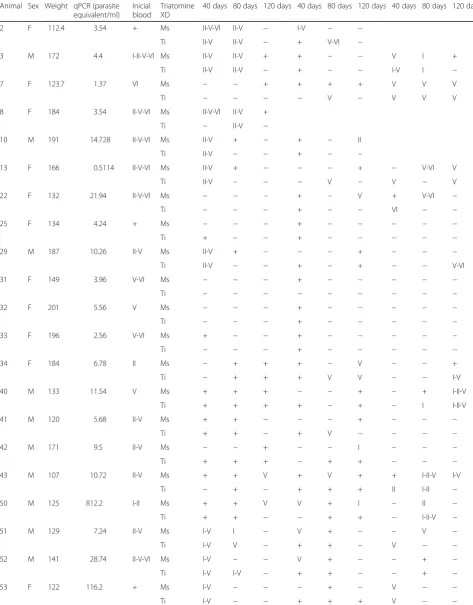
[image:4.595.57.291.87.275.2]Table 1DTUs detected by PCR from blood samples, and from XD coupled with PCR (1XD, 2XD and 3XD), per individualOctodon degusanalyzed
1XD 2XD 3XD
Animal Sex Weight qPCR (parasite equivalent/ml)
Inicial blood
Triatomine XD
40 days 80 days 120 days 40 days 80 days 120 days 40 days 80 days 120 days
2 F 112.4 3.54 + Ms II-V-VI II-V − I-V − −
Ti II-V II-V − + V-VI −
3 M 172 4.4 I-II-V-VI Ms II-V II-V + + − − V I +
Ti II-V II-V − + − − I-V I −
7 F 123.7 1.37 VI Ms − − + + + + V V V
Ti − − − − V − V V V
8 F 184 3.54 II-V-VI Ms II-V-VI II-V +
Ti − II-V −
10 M 191 14.728 II-V-VI Ms II-V + − + − II
Ti II-V − − + − −
13 F 166 0.5114 II-V-VI Ms II-V + − − − + − V-VI V
Ti II-V − − − V − V − V
22 F 132 21.94 II-V-VI Ms − − − + − V + V-VI −
Ti − − − + − − VI − −
25 F 134 4.24 + Ms − − − + − − − − −
Ti + − − + − − − − −
29 M 187 10.26 II-V Ms II-V + − − − + − − −
Ti II-V − − + − + − − V-VI
31 F 149 3.96 V-VI Ms − − − + − − − − −
Ti − − − − − − − − −
32 F 201 5.56 V Ms − − − + − − − − −
Ti − − − + − − − − −
33 F 196 2.56 V-VI Ms + − − + − − − − −
Ti − − − + − − − − −
34 F 184 6.78 II Ms − + + + − V − − +
Ti − + + + V V − − I-V
40 M 133 11.54 V Ms + + + − − + − + I-II-V
Ti + + + + − + − I I-II-V
41 M 120 5.68 II-V Ms + + − − − + − − −
Ti + + − + V − − − −
42 M 171 9.5 II-V Ms − − + − − I − − −
Ti + + + − + + − − −
43 M 107 10.72 II-V Ms + + V + V + + I-II-V I-V
Ti − + − + + + II I-II −
50 M 125 812.2 I-II Ms + + V V + I − II −
Ti + + − − + + − I-II-V −
51 M 129 7.24 II-V Ms I-V I − V + − − V −
Ti I-V V − + + − V − −
52 M 141 28.74 II-V-VI Ms I-V − − V + − − + −
Ti I-V I-V − + + − − + −
53 F 122 116.2 + Ms I-V − − − + − V − −
feeding on O. degus (infectivity detection: 75, 56 and 44 cases over the first, second and third insect evaluations, respectively) (χ2= 16.118,df= 2,P= 0.0003). The amount of cases in which DTU detection was possible was of: 56, 57 and 44 cases over the first, second and third insect evaluations, respectively. Some results of genotyping were obtained at the third analysis (120 days post-infection) and not in the previous ones, this being the case for XD1 O. degusnumbers 43 and 50; XD2O. degusnumbers 22,
34 and 42 and XD3O. degus numbers 29, 34 and 40. In
these cases, the parasites required more time to flourish in the midgut. One special and singular case was observed
with animal number 50 and M. spinolai XD, which
har-boured different DTUs at the first and the third evalu-ation. Figure 2 shows representative results on infectivity
(presence of amplicon), and T. cruzi DTUs composition
(hybridization tests with the four T. cruzi DTU specific
probes). This figure also shows that the amplicon signal can exhibit differential intensities.
Trypanosoma cruzidetection at capture and after serial xenodiagnosis
The number of T. cruzi DTUs present in the initial
blood sample and after the third XD-PCR (Table 1) were analyzed using the Friedman test. We found a decrease
in the number of detectedT. cruzi DTUs when
compar-ing blood samples and XD samples (Friedman statistic:
16.19, P = 0.0010). However, we did not find that
decrease for T. cruzi DTUs between the different XD
[image:6.595.54.542.113.170.2](Friedman statistic: 2.413,P= 0.2993).
Table 1DTUs detected by PCR from blood samples, and from XD coupled with PCR (1XD, 2XD and 3XD), per individualOctodon degusanalyzed(Continued)
54 F 163 80.28 + Ms − − − + − − V − −
Ti − − − − − + V − −
57 M 117 5.14 II-V Ms − − − + + − V − +
Ti I-V − − + − + − + +
Abbreviations: Ffemale,Mmale,Ms Mepraia spinolai,Ti Triatoma infestans
The“–”sign indicates a negative conventional PCR result. The“+”sign indicates a positive PCR result, but these samples did not hybridize with any of the probes used. The dark lockers indicate that the individual perished before performing the XD. Analyses 1, 2 and 3 correspond to post-XD feeding withMus musculusat 40, 80 and 120 days, respectively
[image:6.595.59.539.427.653.2]Statistical analyses on qPCR, body size, sex and DTUs We did not find statistically significant differences between the number of detected DTUs and the number
of parasites detected by means of qPCR (P= 0.8868). In
addition, we did not find statistically significant differ-ences between the number of detected DTUs and the
weight of O. degus individuals (P = 0.5170). The low
number of juveniles (n= 4) did not allow us to perform
comparisons with adults of O. degus. We did not find
statistical differences between sexes in the detected
DTUs (P= 0.0623).
Discussion
Parasitemia in mammals can be very low in the case of chronic Chagas disease patients, where fluctuations of
circulating T. cruzi are expected. Therefore, there is a
need to perform serial determinations to obtain more sensitive parasitological diagnosis [38, 39]. In a previous report, temporal fluctuations of parasitemia with differ-entT. cruziDTUs was observed in one out of two
natur-ally infected O. degus studied by means of serial
XD-PCR [26]. The longitudinal study performed in this study
with 23 naturally infected O. degus submitted to three
serial XD, using two vector species, analyzed three times by means of serial PCR assays confirms and extends the previous study now with a larger sample size. We have modified the classical XD of microscopic observation and replaced it with XD-PCR that includes permanent triatomine artificial feedings after infection to provide enough nutrients and allow cases with minimal trypano-some burden to proliferate. We found temporal
fluctua-tions ofT. cruziDTUs composition in the animals, with
DTU detection (higher parasitemia) to cases of infec-tions without DTU detection (lower parasitemia) or vice versa. Other cases involved fluctuations of DTU com-position within the same animal. The gathered
informa-tion collected from all the XD is that O. degus were
infected with four, three and two different DTUs at the same time. Only few animals resulted infected with only one T. cruzi DTU. However, the circulating parasite composition was less complex when XD-PCR was per-formed only once or when the triatomines were analyzed at a single time. Most of the results were obtained with insect analysis performed 40 or more days post-infection. However, there are XD triatomines from which
informa-tion about T. cruzi infections and/or DTU composition
was obtained 120 days post-infection suggesting cases of low parasitemia at the time XD was performed. These results indicate thatO. degushas different host infectivity at different times, and the reasons deserve to be studied.
In this study, a prevalence of 75% ofT. cruzi infection in O. degus was found. A previous study of T. cruzi
prevalence in O. degus during four consecutive years in
the same endemic area of this study by means ofT. cruzi
detection using one single determination in peripheral
blood ofO. degus, varied over the years (between 18 and
70%) [21]. The resultingT. cruziDTU composition
con-sisted of equivalent rates of single and mixed infections; however, the relative importance of each DTU changed among years, TcV being the most represented. Most of
mixed infections consisted of a combination of two T.
cruzi DTUs [21]. The results of the present follow-up study show a higher rate of mixed infections, which were composed of two, three and four DTUs (15 animals), with TcV as the most represented. Single infections were detected in only four animals. These results demonstrate that a longitudinal study increases the sensitivity ofT. cruzi DTU detection in animals. Even though in the present study with serial determinations we detected mostly mixed
infections with three of the four T. cruzi DTUs studied,
probably other unknown DTUs are also infecting these animals. It is worth noting that the temporal fluctuations
of the observedT. cruziDTUs can be explained either by
elimination of one specificT. cruziDTU, or by the possibil-ity of aT. cruziDTU not being accounted due low
parasit-emia and the method’s sensitivity limit which makes
parasite detection impossible. The temporal fluctuations of
the four T. cruziDTUs reported here could be explained
by permanent colonization of different tissues and release ofT. cruziinto the vascular system. However, it is also ex-pected that parasitemia could be permanently controlled by the immune system. This equilibrium can be changed in immunocompetent hosts submitted to several kinds of stress. This could be the case of the rodents used in this study, which were submitted to a stress condition after be-ing captured from the wild environment, transported to the laboratory, and later submitted to an acclimatization process under controlled laboratory conditions. Evidence to the general assumption that when an animal is under stress, its resistance to infection decreases, since inflamma-tory responses would diminish due to increase in gluco-corticoid levels [40, 41]. For example, animals infected with Trichomonassp. showed an increase in the number of par-asites for about two weeks after being submitted to stress factors. After this period the number of parasites decreased possibly due to the stress adjustment [41]. We suggest that
the tendency for a decrease in the number of T. cruzi
and has high infectiousness to triatomines [44]. In this study, we support the idea that the two analyzed vector species are equally competent to maintainT. cruzi.
Our results confirm that circulatingT. cruziDTUs inO. deguscan be quite different in a longitudinal study [26]. In another study using the same two triatomine species used
here, M. spinolai showed a better susceptibility than T.
infestans to amplify T. cruzi from naturally infected O. degus [33]. The reason for this inconsistency remains
unknown butT. infestans specimens used in [33] and the
present study pertained to the same insect colony
estab-lished since 1950. Meantime theM. spinolaicolonies used
varied between the two studies; probably some of them have a higher susceptibility to become infected. Regarding
the parasitemia level of naturally infected O. degus, we
estimate that such figure should be much higher than 1 parasite/0.15 ml, which is the minimal theoretical amount
to be detected in the 150 μl of blood intake (7 parasite
equivalents/ml). We estimate that parasitemia in animals
with one single T. cruzi DTU is probably 5–10 times
higher than the minimal theoretical amount for each XD that is 35–70 parasite equivalents/ml.
Higher parasitemia can be expected when both insect species are infected with mixedT. cruziDTUs. Parasitemia of theO. degusstudied by qPCR varied from less than 1 to 812 equivalents/ml with a median of 6.16 parasite-equivalents/ml of blood. Parasitemia in human chronic cases has been estimated in different countries by means of qPCR [45, 46]. The obtained medians fluctuated between
1.27–2.38 parasite equivalents/ml. In the meantime, the
established medians for dogs and cats were 8.1 and 9.7 parasite equivalents/ml, respectively [47]. To overcome these limitations of parasite detection sensitivity, and to study the dynamics of circulatingT. cruziDTUs in a small rodent such asO. degusit is necessary to amplify first the
infecting T. cruzi by means of XD, to further amplify T.
cruziDNA with PCR assays and later characterize them. In this way, using XD we are also confident that living parasites have been studied and results are not due to lysed ones. This result resembles the finding of infectiousness of T. infestans by infected dogs and cats which increased steeply with parasitemia. Dogs and cats with 80% of
infec-tiousness on T. infestans displayed median parasite load
determined by qPCR of 21.3 or 96.1 parasite equivalents/ ml, respectively [48]. In summary, using a combination of XD and PCR assays, this study reports for the first time the
occurrence of temporal fluctuations of T. cruzi DTU
composition in a large sample of naturally infectedO. degus reservoirs. We showed that it is possible to analyze complex
mixtures of circulating T. cruzi DTUs using two vector
species in the XD over time. The time necessary to detect easily infected triatomines ranges from 40 to 120 days. We also conclude thatM. spinolaiandT. infestansare equally competent to maintain severalT. cruziDTUs.
Conclusion
Serial XD coupled withT. cruzi DTU identification (TcI,
TcII, TcV, TcVI) by means of conventional PCR and hybridization tests, provided evidence for different DTUs
composition of T. cruzi circulating in peripheral blood
over time. The XD performed with the vectorsM. spinolai
and T. infestans reveals that O. degus infects both insect
species, confirming that M. spinolaiand T. infestansare
equally competent to maintainT. cruziDTUs, since
simi-lar results of infection were obtained after xenodiagnosis procedure. XD triatomines analyzed 40 days post - infec-tion resulted the optimum to evaluate infectiveness rather
than insects analyzed at 80 or 120 days. Trypanosoma
cruzi parasitemia is estimated based on XD triatomines
able to detect infection and/or T. cruzi DTUs
compos-ition. In addition, an evaluation of parasitemia was per-formed before the serial XD study by means of qPCR.
Abbreviations
DNA:Deoxyribonucleic acid; DTUs: Discrete typing units; kDNA: Kinetoplastidic DNA; PCR: Polymerase chain reaction; PCR-XD: Conventional PCR performed with fecal samples of XD; qPCR: quantitative PCR; XD: Xenodiagnosis
Acknowledgments
The authors thank the collaboration of Dr. Lafayette Eaton for manuscript language revision.
Funding
This study was financially supported by FONDECYT-Chile 1120122 to A. Solari, CONICYT Doctoral Fellowship 21120685 to G. Rojo, FONDECYT-Chile 1140521 to C. Botto-Mahan and FONDECYT-Chile 1140650 to P. E. Cattan.
Availability of data and materials
All data used for this manuscript has been uploaded to a public repository and can be accessed via the following link: https://figshare.com/projects/ Within host_temporal_fluctuations_of_Trypanosoma_cruzi_discrete_typing _units_the_case_of_the_wild_reservoir_rodent_Octodon_degus_/18029
Authors’contributions
Conceived and designed the experiments: AS, GR, PEC, CB-M and AS-R. Per-formed the experiments: GR, AS-R, AL, JPC, MS and AS. Analyzed the data: GR, AS-R, PEC, CB-M and AS. Contributed reagents/materials/analysis tools: PEC, SO and AS. Wrote the paper: GR, AS-R, AS and SO. All authors read and approved the final manuscript.
Ethics approval
This study was approved by Ethics Committee of the Faculty of Medicine of University of Chile (CBA≠0443 FMUCH).Biosafety and animal processing methods were performed following the protocols approved by the Bioethical Committee (CBA≠0443 FMUCH) dated August 16, 2011. The captures ofO. deguswere performed under the permission of the Servicio Agrícola y Ganadero (resolution N° 1792 of April 2, 2012).
Consent for publication
Not applicable.
Competing interests
The authors declare that they have no competing interests.
Publisher’s Note
Author details
1Programa de Biología Celular y Molecular, ICBM, Facultad de Medicina,
Universidad de Chile, Santiago, Chile.2Laboratorio de Parasitología
Básico-Clínica, ICBM, Facultad de Medicina, Universidad de Chile, Santiago, Chile.3Departamento de Ciencias Ecológicas, Facultad de Ciencias,
Universidad de Chile, Santiago, Chile.4Laboratorio de Ecología,
Departamento de Ciencias Biológicas Animales, Facultad de Ciencias Veterinarias y Pecuarias, Universidad de Chile, Santiago, Chile.
Received: 4 January 2017 Accepted: 27 July 2017
References
1. Coura JR, Viñas PA. Chagas disease: 402 a new worldwide challenge. Nature. 2010;465:56–7.
2. Shikanai-Yasuda MA, Carvalho NB. Oral transmission of Chagas disease. Clin Infect Dis. 2012;54:845–52.
3. Telleria J, Tibayrenc M. American Trypanosomiasis Chagas Disease: One Hundred Years of Research. 2nd Edition. Amsterdam: Elsevier Academic Press; 2017.
4. Zingales B, Miles MA, Campbell DA, Tibayrenc M, Macedo AM, Teixeira MM, et al. The revisedTrypanosoma cruzi: subspecific nomeclature: rationale, epidemological relevance and research applications. Infect Genet Evol. 2012; 12:240–53.
5. Lima L, Espinosa-Álvarez O, Ortiz PA, Trejo-Varón JA, Carranza JC, Pinto CM, et al. Genetic diversity ofTrypanosoma cruziin bats, and multilocus phylogenetic and phylogeographical analyses supporting Tcbat as an independent DTU (discrete typing unit). Acta Trop. 2015;151:166–77. 6. Ramírez JD, Hernández C, Montilla M, Zambrano P, Flórez AC, Parra E,
Cucunubá ZM. First report of humanTrypanosoma cruziinfection attributed to TcBat genotype. Zoonoses Public Health. 2014;61:477–9.
7. Telleria J, Lafay B, Virreira M, Barnabe C, Tibayrenc M, Svdoba M. Trypanosoma cruzi: sequence analysis of the variable región of kinetoplast minicircles. Exp Parasitol. 2006;144:279–88.
8. Brenière SF, Bosseno MF, Telleria J, Bastrenta B, Yacsik N, Noireau F, et al. Different behavior of twoTrypanosoma cruzimajor clones: transmission and circulation in young Bolivian patients. Exp Parasitol. 1998;89:285–95. 9. Coronado X, Rozas M, Botto-Mahan C, Ortiz S, Cattan PE, Solari A. Molecular
epidemiology of Chagas disease in the wild transmission cycle: the evaluation in the sylvatic vectorMepraia spinolaifrom and endemic area of Chile. Am J Trop Med Hyg. 2009;81:656–9.
10. Arenas M, Campos R, Coronado X, Ortiz S, Solari A.Trypanosoma cruzi genotypes of insect vectors and patients with chagas of Chile studied by means of citochromebgene seguencing, minicircle hybridization, and nuclear gene polymorphisms. Vector-Borne Zoon Dis. 2012;12:196–205. 11. Egaña C, Pinto R, Vergara F, Ortiz S, Campos-Soto R, Solari A. Fluctuations in
Trypanosoma cruzidiscrete typing unit composition in two naturally infected triatomines:Mepraia gajardoiandM. spinolaiafter laboratory feeding. Acta Trop. 2016;160:9–14.
12. Rozas M, Botto-Mahan C, Coronado X, Ortiz S, Cattan PE, Solari A. Trypanosoma cruziinfection in wild mammals from a chagasic area of Chile. Am J Trop Med Hyg. 2005;73:517–9.
13. Rozas M, Botto-Mahan C, Coronado X, Ortiz S, Cattan PE, Solari A. Coexistence ofTrypanosoma cruzigenotypes in wild and periodomestic mammals in Chile. Am J Trop Med Hyg. 2007;77:647–53.
14. Charles RA, Kjos A, Ellis AE, Barnes JC, Yabsley MJ. Southern Plains woodrats (Neotoma micropus) from southern Texas are important reservoirs of two genotypes ofTrypanosoma cruziand host of a putative novelTrypanosoma species. Vector-Borne Zoon Dis. 2013;13:22–30.
15. Lopes F, Rodrigues AL, Saab de Lima J, Carvalho C, Gemesio F, Cavalcanti de Azevedo F, et al.Trypanosoma cruziinfection in Neotropical wild carnivores (Mammalia: Carnivora): at the top of theT.cruzitransmission chain. PLoS Negl Trop Dis. 2013;8:1–12.
16. Buitrago R, Bosseno M-F, Depickere S, Waleckx E, Salas R, Aliaga C, et al. Blood meal sources of wild and domesticTriatoma infestans(Hemiptera: Reduviidae) in Bolivia: connectivity between cycles of transmission of Trypanosoma cruzi. Parasit Vectors. 2016;9:214.
17. Begon M. Effects of host diversity on disease dynamics. In: Ostfeld RS, Keesing F, Eviner VT, editors. Infectious disease ecology: effects of ecosystems on disease and of disease on ecosystems. Princeton: Princeton University Press; 2008. p. 12–29.
18. Gürtler RE, Cardinal MV. Reservoir host competence and the role of domestic and commensal hosts in the transmission ofTrypanosoma cruzi. Acta Trop. 2015;151:32–50.
19. Orozco MM, Piccinali RV, Mora MS, Cardinal MV, Enriquez GF, Gürtler RE. The role of sigmodontine rodents as sylvatic hosts ofTrypanosoma cruziin the Argentinian Chaco. Inf Gen Evol. 2014;22:12–22.
20. Botto-Mahan C, Bacigalupo A, Correa JP, Oda E, Solari A. Field assessment of Trypanosoma cruziinfection and host survival in the native rodentOctodon degus. Acta Trop. 2012;122:164–7.
21. Botto-Mahan C, Rojo G, Sandoval-Rodriguez A, Peña F, Ortiz S, Solari A. Temporal variation inTrypanosoma cruzilineages from the native rodent Octodon degusin semiarid Chile. Acta Trop. 2015;151:178–81. 22. Oda E, Solari A, Botto-Mahan C. Effects of mammal host diversity and
density on the infection level ofTrypanosoma cruziin sylvatic kissing bugs. Med Vet Entomol. 2014;28:384–90.
23. Botto-Mahan C, Campos R, Acuña-Retamar M, Coronado X, Cattan PE, Solari A. Temporal variation ofTrypanosoma cruziinfection in native mammals in Chile. Vector-Borne Zoon Dis. 2010;10:317–9.
24. Fabrizio MC, Schweigmann NJ, Bartoloni NJ. Analysis of the transmission of Trypanosoma cruziinfection through hosts and vectors. Parasitology. 2016; 143:1168–78.
25. Gallupo S, Bacigalupo A, García A, Ortiz S, Coronado X, Cattan PE, et al. Predominance ofTrypanosoma cruzigenotypes in two reservoirs infected by sylvaticTriatoma infestansof an endemic area of Chile. Acta Trop. 2009; 111:90–3.
26. Campos R, Botto-Mahan C, Ortiz S, Coronado X, Solari A. Temporal fluctuation of infection with differentTrypanosoma cruzigenotypes in the wild rodentOctodon degus. Am J Trop Med Hyg. 2010;83:380–1. 27. Campos R, Torres-Perez F, Botto-Mahan C, Coronado X, Solari A. High
phylogenetic structure in sylvatic vectors of Chagas disease of the genus Mepraia(Hemiptera: Reduviidae). Infec Genet Evol. 2013;19:280–6. 28. Lorca M, García A, Contreras M, Schenone H, Rojas A. Evaluation of a
Triatoma infestanselimination program by the decrease of aTriatoma infestanselimination program by the decrease ofTrypanosoma cruzi infection frequency in children younger than 10 years, Chile 1991–1998. Am J Trop Med Hyg. 2001;65:861–4.
29. Botto-Mahan C, Ortiz S, Rozas M, Cattan PE, Solari A. DNA evidence of Trypanosoma cruziin the Chilean wild vectorMepraia spinolai(Hemiptera: Reduviidae). Mem I Oswaldo Cruz. 2005;100:237–9.
30. Brumpt E. Le xenodiagnostic. Application au diagnostic de quelques infections parasitaires et en particulier à la trypanosomose de Chagas. Bull Soc Pat Exot. 1914;7:706–10.
31. Schijman AG, Altcheh J, Burgos JM, Biancardi M, Bisio M, Levin MJ, et al. Aetiological treatment of congenital Chagas disease diagnosed and monitored by the polymerase chain reaction. J Antimicrob Chemother. 2003;52:441–9.
32. Duffy T, Bisio M, Altcheh J, Burgos JM, Diez M, Levin MJ, et al. Accurate real time PCR strategy for monitoring bloodstream parasitic loads in Chagas disease patients. PLoS Negl Trop Dis. 2009;3:e419.
33. Campos R, Botto-Mahan C, Ortiz S, Acuña M, Cattan PE, Solari A. Trypanosoma cruzidetection in blood by xenodiagnosis and polymerase chain reaction in the wild rodentOctodon degus. Am J Trop Med Hyg. 2007; 76:324–6.
34. Campos R, Acuña-Retamar M, Botto-Mahan C, Ortiz S, Cattan PE, Solari A. Susceptibility ofMepraia spinolaiandTriatoma infestansto different Trypanosoma cruzistrains from naturally infected rodent hosts. Acta Trop. 2007;104:25–9.
35. Wincker P, Britto C, Pereira JB, Cardoso MA, Oeleman O, Morel CM. Use of a simplified polymerase chain reaction procedure to detectTrypanosoma cruziin blood samples from chronic chagasic patients in a rural endemic area. Am J Trop Med Hyg. 1994;51:771–7.
36. Piron M, Fisa R, Casamitjana N, López-Chejade P, Puig L, Vergés M, et al. Development of a real-time PCR assay forTrypanosoma cruzidetection in blood samples. Acta Trop. 2007;103:195–200.
37. Barnabé C, Neubauer K, Solari A, Tibayrenc M.Trypanosoma cruzi: presence of the two major phylogenetic lineages and of several lesser discrete typing units (DTUs) in Chile and Paraguay. Acta Trop. 2001;78:127–37.
38. Dias C. Xenodiagnóstico seriados em caes infectados como amostras venezuelanas de“Schizotrypanum cruzi”. Bras Med. 1940;52:859–61. 39. Schenone H, Contreras MC, Rojas A. Rendimiento del xenodiagnóstico,
chagásica crónica diagnósticada mediante la reacción de hemaglutinación indirecta. Bol Chil Parasitol. 1991;46:58–61.
40. Brivio F, Grignolio S, Sica N, Cerise S, Bassano B. Assessing the impact of capture on wild animals: the case study of chemical immobilisation on alpine ibex. PLoS One. 2015;10:1–18.
41. Noble G. Stress as a factor in parasitism. Jackson Hole Research Station Annual Report. 1959;1959:14–6.
42. Petersen RM, Gürtler RE, Cecere MC, Rubel DN, Hansen D, Lauricella MA, Carlomagno M. Association between nutritional indicators of dogs seroactive forTrypanosoma cruziin a rural area of northwestern Argentina. Parasitol Res. 2001;87:208–14.
43. Orozco MM, Enriquez GF, Alvarado-Otegui JA, Cardinal MV, Schijman AG, Kitron U, Gürtler RE. New sylvatic hosts ofTrypanosoma cruziand their reservoir competence in the humid Chaco of Argentina. Am J Trop Med Hyg. 2013;88:872–82.
44. Cacere MC, Cardinal MV, Arrabal JP, Moreno C, Gurtler R.Microcavia australis (Cavidae, Rodentia),a new highly competent reservoir host ofTrypanosoma cruziin Northernwestern Argentina. Acta Trop. 2014;142:34–40.
45. Moreira O, Ramirez JD, Velasquez E, Diaz Melo FA, Lima-Ferreira C, Guhl F, et al. Towards the establishment of a consensus real-time qPCR to monitor Trypanosoma cruziparasitemia in patiens with chronic Chagas disease cardiomyopathy: a substudy from the BENEFIT trial. Acta Trop. 2013;125:23–31. 46. Apt W, Arribada A, Zulantay I, Saavedra M, Araya E, Solari A, et al.
Trypanosoma cruziburden, genotypes and clinical evaluation of Chilean patients with chronic Chagas cardiopathy. Parasitol Res. 2015;114:3007–18. 47. Enriquez GF, Cardinal MV, Orozco MM, Schijman AG, Gürtler RE. Detection of
Trypanosoma cruziinfection in naturally infected dogs and cats using serological, parasitological and molecular methods. Acta Trop. 2013;126:211–7. 48. Enriquez GF, Bua J, Orozco MM, Wirth S, Schijman AG, Gürtler RE, et al. High levels ofTrypanosoma cruziDNA determined by qPCR and infectiousness to Triatoma infestanssupport dogs and cats are major sources of parasites for domestic transmission. Inf Genet Evol. 2014;25:36–43.
• We accept pre-submission inquiries
• Our selector tool helps you to find the most relevant journal
• We provide round the clock customer support
• Convenient online submission
• Thorough peer review
• Inclusion in PubMed and all major indexing services
• Maximum visibility for your research
Submit your manuscript at www.biomedcentral.com/submit