R E S E A R C H
Open Access
Plasmodium yoelii
blood-stage primes
macrophage-mediated innate immune response
through modulation of toll-like receptor
signalling
Yong Fu
1, Yan Ding
1, Taoli Zhou
1, Xiaolan Fu
2and Wenyue Xu
1*Abstract
Background:Toll-like receptors (TLRs) signalling is reported to be primed by the infection of human malaria parasite,Plasmodium falciparum. However, little is known about the regulation of macrophages TLR signalling by the infection of lethal or non-lethal strain of rodent malaria parasites.
Methods:BALB/c mice were infected with non-lethal strainPlasmodium yoelii 17XNL or lethal strainP. yoelii17XL. Peritoneal macrophages were isolated to study its immune response to pRBC lysate, and TLRs (TLR2, TLR4, and TLR9) agonists, and the expression of TLRs and intracellular signalling molecules were also investigated by flow cytometry and semi-quantitive RT-PCR.
Results:The reactivity of peritoneal macrophages from the mice infected with lethal strainP. y17XL or non-lethal strainP. y17XNL were enhanced to pRBC lysate, and TLR2, TLR4, and TLR9 agonists at one, three and five days post-infection. Of all the tested TLRs, only TLR2 was up-regulated on peritoneal macrophages of mice infected with either strain. However, transcription of intracellular signalling molecules MyD88, IRAK-1, and TRAF-6 was significantly up-regulated in peritoneal macrophages from mice infected either withP. yoelii17XL or P. yoelii17XNL at one, three and five days post-infection. However, the enhanced TLRs response of macrophage fromP. yoelii 17XNL-infected mice persisted for a much longer time than that fromP. yoelii17XL-infected mice.
Conclusion:BothP. yoelii17XL and 17XNL strains could enhance the response of peritoneal macrophages to pRBC lysate and TLR agonists, through up-regulating the expression of TLR2 and intracellular signalling molecules MyD88, IRAK-1, and TRAF-6. In addition, prolonged high response of macrophage fromP. yoelii17XNL-infected mice might be associated with the more efficiently controlling ofP. yoelii17XNL growth in mice at early stage.
Keywords:Plasmodium yoelii, Macrophage, Toll-like receptors
Background
Malaria remains one of the most devastating diseases worldwide, with ~40% of the population at risk, and 200-300 million new cases each year, resulting in about one million deaths annually [1]. The causative agents of malaria are parasitic protozoa belonging to the genus Plasmodium. Except for the virulence of infected
malaria parasite, presentation of clinical malaria is mainly dependent on the balance between pro- and anti-inflammatory responses against these parasites. Individuals who exhibit particularly weak immune responses often lead to uncontrolled parasitaemia. Thus, understanding the regulation mechanism of the immune
response brought on by infection with Plasmodium
parasites will provide us with potential therapeutic approaches for treating infected individuals.
Although adaptive immune effectors, such as
Plasmo-dium-specific CD4+ ab T cells and antibody [2], are mandatory for effective clearance of parasitized red
* Correspondence: xuwenyue@gmail.com
1Department of Pathogenic Biology, Third Military Medical University, 30
Gaotanyan Zhengjie, Shapingba District, Chongqing 400038, People’s Republic of China
Full list of author information is available at the end of the article
blood cells (pRBCs) after infection, the control of para-site growth during the early stage of infection is largely dependent on the innate immune response. Previous study has shown that the primary peak of parasitaemia in T-cell-deficient mice tends to be comparable to that of wild-type mice [3]. It has been reported that NK cell-derived IFN-gthat contributes to the early control of Plasmodium chabaudiandPlasmodium yoeliiinfections [4,5]. However, a recent study showed that the macro-phage-mediated innate immune response, but not IFN-g, has a significant role in controlling the primary wave of P. yoelii infection [6]. Another study recently reported that a population of CD11bhighLy6C+monocyte migra-tion from bone marrow to the spleen was important for killing ofP. chabaudiblood stage at early stage [7].
It is well established that macrophages are activated by malaria parasites to release pro-inflammatory cytokines, mainly through toll-like receptors (TLRs). For example, Plasmodium falciparum-derived glycosylphosphatidyli-nositols (GPI) moieties are known to induce potent TNF responses in macrophages by TLR2, and to a lesser extent TLR4[8,9]. It has also been reported thatP. falci-parumhaemozoin, a crystalline by-product of haemoglo-bin metabolism by malaria parasites, is recognized by TLR9 on macrophages[10], although more recently it has been suggested that instead haemozoin-bound nucleic acids are the true ligand for this receptor [11,12].
TLR signalling is subject to modulation by microor-ganisms. For instance, it is well known that lipopolysac-charide (LPS), the toxic wall component of Gram-negative bacteria, induces macrophages into a tolerance status by down-regulating TLR4 receptor expression [13], or its association with MyD88, and by IRAK-1
acti-vation [14]. In contrast, infection of P. falciparum
primes peripheral blood mononuclear cells (PBMC) for TLR signalling by enhancing mitogen-activated protein kinase (MAPK) activation during the early stage [15-17].
However, the infection of P. yoelii non-lethal strain
17XNL was reported to induce dendritic cell (DC) TLR tolerance during the late stage [18].
Little is known about the regulation of macrophages TLR signalling by the infection of lethal or non-lethal strain of rodent malaria parasites at the early stage. In
the present study, both P. yoelii 17XL and 17XNL
strains were found to be able to enhance the response of peritoneal macrophages to pRBC lysate and TLR ago-nists, through up-regulating the expression of TLR2 and intracellular signalling molecules MyD88, IRAK-1, and TRAF-6.
Methods
Mice andplasmodium
BALB/c mice (specific pathogen free, ~6-8 week old females) were purchased from the Animal Institute of
Third Military Medical University (Chongqing, China). These studies have been reviewed and approved by the Third Military Medical University Institute of Medical Research Animal Ethics Committee.P. yoelii17XNL is a non-lethal strain, originally isolated in 1965 from the
blood of a wild thicket rat, Thamnomys rutilans [19],
and P. yoelii17XL is a lethal strain cloned fromP. yoelii 17XNL, which was suddenly virulent in the laboratory of J. Finerty [20]. Cohorts of 10 mice were infected intra-peritoneally with 2 × 10⁵ non-lethal strain P. yoelii 17XNL-infected pRBCs or 2 × 10⁵ lethal strain P. yoelii 17XL. For control purposes, 10 mice were also infected
with 2 × 10⁵RBCs taken from normal mice (nRBCs).
Reagents and antibody
Biotin anti-mouse TLR4 (MTS510), TLR2 (6 C2 clone), TLR9 (M9.D6 clone), Biotin rat IgG2a and IgG2b iso-type control antibodies, anti-mouse CD16/32 (93 clone), FITC-F4/80 and PE-streptavidin were all purchased from eBioscience (San Diego, CA). TLR2 agonist
Pam3CSK4, TLR4 agonist LPS (Escherichia.coli 0111:
B4), TLR9 agonist CpG(ODN 1826), and its control CpG were purchased from InvivoGen (San Diego, CA).
Isolation of peritoneal macrophage
All the mice infected withP. yoelii17XL or 17XNL, or injected with nRBCs, were administrated with 1 mg/ml thioglycollate (Sigma, St. Louis, MO, USA) via intraperito-neally three days before killing. Peritoneal exudate cells (PECs) were then extracted and allowed to adhere on tis-sue culture dishes for two hours, and non-adherent cells were removed. The adherent cells were collected as perito-neal macrophages, and F4/80 expression was analysed using a FACSCalibur flow cytometer with Cell Quest soft-ware (Becton Dickinson, Lincoln Park, NJ).
Cytokine detection by ELISA
Peritoneal macrophage from mice infected withP. yoelii 17XL, 17XNL, or nRBCs were cultured in the presence
of 1 × 10⁷ nRBC or pRBC lysate, or LPS (10μg/ml),
Pam3CSK4 (10μg/ml), or CpG (10μg/ml). Lysates from 1 × 10⁷ red blood cells (RBCs) or pRBCs were prepared by twice freeze-thaw. After 24 hours, supernatants were collected and analysed by ELISA (eBioscience) according to manufacturer’s instructions to detect IL-6 and TNF.
Flow cytometry assay
min. To examine TLR9 expression, peritoneal macro-phages were permeabilized with fixation/permeabilization (eBioscience) prior to labelling with biotin-conjugated anti-mouse TLR9 and PE-streptavidin. Cells were finally analysed using a FACSCalibur flow cytometre using Cell Quest software (Becton Dickinson).
Semi-quantitive reverse-transcriptase PCR
Total RNA was isolated from 1 × 10⁶peritoneal macro-phages from mice injected withP. yoelii17XL or 17XNL strain, or with nRBCs, one, three and five days after infec-tion using Trizol (Boehringer Mannheim, Germany), and reversely transcribed with MMLV (Promega, Madison, WI, USA). Initial cDNAs amount for PCR amplification were normalized with GAPDH signal, which was opti-mized to be visible, but not saturated. Changes in the expression of intracellular signalling molecules TRAF-6, IRAK-1, and MyD88 were investigated in peritoneal macrophages from mice infected with or without malaria parasites. Specific primers used were as follows:
IRAK-1781: 5’- GAACAGCTATCAAGGTTTCGTCA-3’;
IRAK-11260: 5’-ACCAGCAAGGGTCTCCAGTA-3’; MyD2341: 5’
-CCCACTCGCAGTTTGTTG-3’; MyD2569: 5’-CTCC
CAGTTCCTTTGTTTG-3’; TRAF162: 5’
-GGGCTAC-GATGTGGAGTT-3’; TRAF396: 5’
-TACCGTCAGG-GAAAGAAT-3’.
Statistic analysis
Statistical significance was determined with SPSS software (version 13.0). Differences between two experimental
groups (P.yoelii 17XNL-infection vs naive or P.yoelii
17XL-infection vs naive), or two time points were analysed for statistical significance by means of a nonparametric Mann-Whitney U test, and for multiple groups (TLR2 expression amongP.yoelii17XNL-infected,P.yoelii 17XL-infected and naive mice) using Kruskal-Wallis test. *P <0.05 or **P <0.01 were considered statistically significant.
Results
Plasmodium yoelii17XNL and 17XL infection enhance the response of peritoneal macrophage to pRBCs
Plasmodium yoelii17XNL is a non-lethal Plasmodium strain, which grows slowly and would be cleared by
mice at 20 days after infection. In contrast, P. yoelii 17XL is a lethal strain, which grows quickly and infected mice usually die at six to nine days after injection (Fig-ure 1).
Since the macrophage-mediated innate immune response plays an important role in controlling the pri-mary wave of P. yoeliiinfection [6], the responsiveness of macrophages collected on one, three and five days after infection with either theP. yoeliilethal 17XL strain or non-lethal 17XNL strain was investigated. Pooled adherent PECs were used as peritoneal macrophages in the following experiments without additional indication, as 85% of pooled adherent PECs were positive for the macrophage-specific marker F4/80(data not shown). Compared to the peritoneal macrophages from mice injected with nRBCs, the reactivity of macrophages from
mice infected either with P. yoelii17XL or 17XNL was
enhanced at one, three and five days post infection, as both inflammatory cytokines TNF and IL-6 released by macrophages stimulated with pRBC lysate were much higher in either strain infected mice (Figure 2). How-ever, the response of macrophages reached a peak at three days, and it was then tend to return to the base-line level at five days postP. yoelii 17XL-infection (Fig-ure 2B,D). In contrast, the response of macrophages from P. yoelii 17XNL-infected mice even increased at five days post infection (Figure 2A,C). Thus, these data suggested that the response of peritoneal macrophage
was enhanced after mice were infected either with P.
yoelii 17XL or 17XNL strain, but the duration time was
much longer for the macrophages from P. yoelii
17XNL-infected mice.
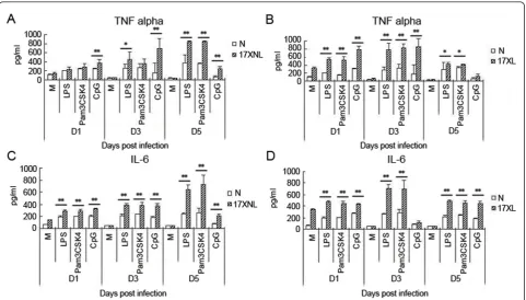
Plasmodium yoelii17XNL and 17XL infection prime the response of peritoneal macrophages to TLR agonists
[image:3.595.60.540.618.710.2]It is well known that malaria parasite components, including GPI and haemozoin, could induce macro-phages to release inflammatory cytokines through TLR2/4 and TLR9[8-10]. To investigate the mechanism of the enhanced response of macrophages to pRBC lysate primed by infection ofP. yoelii17XL or 17XNL, the levels of inflammatory cytokines of TNF and IL-6 released by macrophages stimulated with TLR2 agonist
Figure 1In vivoinfection course after inoculation withP. y17XL or 17XNL. BALB/c were intraperitoneally injected with 2 × 10⁵P. yoelii
Pam3CSK4, TLR4 agonist LPS, or TLR9 agonist CpG, were measured. As shown in Figure 3, peritoneal macro-phages stimulated with any of the three TLR agonists exhibited dramatically increased TNF and IL-6 levels at one, three and five days after infection with P. yoelii 17XNL(Figure 3A,C) or 17XL(Figure 3B,D), which was consistent with the response of macrophages to pRBC lysate. Take together, these data supported that the
response of macrophages primed by P. yoelii 17XL or
17XNL infection was due to enhanced TLR response of macrophages.
Effect ofP. Yoelii17XL and 17XNL infection on TLR expression of peritoneal macrophage
It was previously reported that TLR4 and MD-2 over-expression was attributed to TLR4 signal priming
induced by Propionibacterium acnes[21]. Furthermore,
P. falciparum-induced priming of monocyte TLR response is also associated with increased expression of both TLR2 and TLR4 [15]. Hence, the expression of TLR2, TLR4, and TLR9 on peritoneal macrophages
from mice infected with P. yoelii17XL or 17XNL was
investigated to explore the mechanism of TLR response
priming induced by P. yoelii. As shown in Figure 4,
macrophages from mice infected with 17XL or 17XNL had significantly increased expression of TLR2, but not TLR4 or TLR9, compared to the control mice at all time points measured. The strong response of
macro-phages fromP. yoelii 17XL- or 17XNL-infected mice to
TLR2 agonist Pam3CSK4 might be correlated with the relatively high level of TLR2 on PECs.
Infection ofP. Yoelii17XL and 17XNL up-regulate TLR-MyD88 dependent pathway intracellular molecules
These data so far does not explain the underlying mechanism of the primed TLR4 and TLR9 responses of
macrophages induced by P. yoelii 17XL and 17XNL
infection[15]. Since p38 activity enhancement are related to TLR response priming in malaria parasite infection [16], then the transcription of MyD88, IRAK-1, and TRAF6, which are shared by the TLR2-, TLR4-, and TLR9- mediated MyD88-dependent pathway, were
investigated in macrophages from P. yoelii 17XL- or
[image:4.595.57.540.88.355.2]17XNL-infected mice. As shown in Figure 5, the tran-scriptional levels of MyD88, IRAK-1, and TRAF-6 were significantly increased in mice infected with either P.
Figure 2Peritoneal macrophage response from mice infected withP. y17XL or 17XNL to pRBC lysate. Three days after mice were treated with thioglycollate, peritoneal macrophages were isolated from mice on one, three and five days after intraperitoneally injected with 2 × 10⁵nRBCs (N), 2 × 10⁵P. yoelii17XNL- or 17XL-infected RBCs. Each 1 × 10⁶macrophages from mice infected withP. yoelii17XNL were stimulated with medium (M), 1 × 10⁷P. yoelii17XNL-infected RBCs lysate for 24 h, and supernatants were collected to detect TNF-a(A) and IL-6
(C). Each 1 × 10⁶macrophages from mice infected withP. yoelii17XL were also stimulated with medium (M), 1 × 10⁷P. yoelii17XL-infected RBCs
yoelii17XL or 17XNL compared to control mice at one, three and five days post-infection. The change pattern of transcription levels of MyD88, IRAK-1, and TRAF-6
of macrophage was consistent with its response to pRBC lysate after infection with either strain. These data strongly suggested an important role for the
[image:5.595.58.540.88.362.2]up-Figure 3The response of macrophages from mice infected withP. y17XL or 17XNL to TLR agonists. Peritoneal macrophages were collected from mice at one, three and five days after being intraperitoneally injected with 2 × 10⁵nRBCs (N), 2 × 10⁵P. yoelii17XL- or 17XNL-infected RBCs. 1 × 10⁶macrophages were then stimulated with medium (M), LPS (10μg/ml), Pam3CSK4 (10μg/ml), or CpG (10μg/ml) for 24 h, and cell supernatants were collected to detect TNF-a(A) and IL-6 levels (B). Three individual experiments were performed, and pooled data are presented as the mean ± SD (*P <0.05, **P <0.01).
[image:5.595.57.539.490.683.2]regulation of MyD88, IRAK-1, and TRAF-6 in the TLR
response priming induced by either P. yoelii strain
infection.
Discussion
Macrophage-mediated innate immune response is criti-cal for controlling theP. yoeliiparasitaemia at its early stage, so it tends to be modulated by exposure to malaria parasites. It was previously reported that Plas-modium bergheiinfection inhibits IL-12 p40 production by peritoneal macrophages at the transcriptional level [22]. However, infection with lethal strainP. yoelii 17XL
or non-lethal strain P. yoelii 17XNL was found to be
able to prime the response of macrophages through up-regulating TLR2 expression and signalling intracellular molecules, although infection of the two strains resulted in dramatically different disease outcome.
Pre-exposure to a variety of TLR agonists, including LPS, Pam3CSK4, and CpG, often induces macrophages into a tolerant status to prevent over-activation [23-25]. It is well known that pre-administration of a low dose (sub-lethal dose) of LPS induces macrophages into endotoxin tolerance to protect the host from the chal-lenge of lethal-dose of LPS. However, tissue injury[26] andPropionibacteriuminfection [21] have been demon-strated to prime the TLR response. In this study, the lethal P. yoelii17XL strain was also found to enhance
the response of murine peritoneal macrophages to pRBC at one, there and five days post-infection (Figure 3B-D), which is consistent with recent reports of
prim-ing the TLR response with P. f at the early stage
[15-17]. Interestingly, infection with the non-lethal P. yoelii 17XNL strain could also prime the response of macrophages to pRBC lysate (Figure 3A-C). Like the response of macrophages to pRBC lysate, macrophages fromP. yoelii17XL- or 17XNL-infected mice responded to TLR2, TLR4, and TLR9 agonists much more strongly than macrophages from nRBCs-injected mice at one and there days post-infection (Figure 4). Thus, the increased response of macrophages to TLR2, TLR4, and TLR9 agonists resulted in their hypersensitivity to pRBC lysate. In a previous study, McCallet al attempted to corre-late TLR2/4 expression with the priming response, how-ever the investigators did not observe enhanced expression of TLR2/4 on PBMCs [17]. In contrast,
Flanklinet alfound that TLR2, TLR4 and TLR9
expres-sion was significantly augmented in PBMCs from patients with relatively high parasitaemia [15]. Here, the
infection with P. yoelii 17XL or 17XNL induced the
expression of TLR2, but not TLR4 and TLR9, on mur-ine macrophages in the present study (Figure 5). The disparity could be interpreted as P. yoelii used in this study, butP.f was used in their research. Interestingly, the transcription levels of intracellular molecules of the MyD88-dependent pathway were also found to be
aug-mented in macrophages fromP. yoelii17XL- or
17XNL-infected mice (Figure 5). Therefore, hypersensitivity of macrophages to TLR agonists was contributed to up-regulation of intracellular signalling molecules by malaria parasite infection. However, it remains to be determined whether up-regulation of MyD88, IRAK-1, and TRAF-6 would result in enhancement of MAPK activation, which was previously contributed to priming
the TLR response on PBMCs fromP. f-infected patients
[16].
It was recently reported that malaria-induced priming
of the TLR response was TLR9-, MyD88-, and IFN-g
-dependent[15]. Hence, it is reasonable to find that lethal and non-lethal strains can prime the macrophage response in this study, as a relative high level of IFN-gis induced in the spleen of either strain-infected mice dur-ing the early stage [27]. It is well known that IFN-gwas mainly secreted by NK and T cells after infection with lethal strainP. yoelii17XNL and nonlethal strainP. yoe-lii 17XL [4], but the production of IFN-g by NK cells required the help of IL-12 of DC activated by rodent malaria parasite [28].
[image:6.595.57.290.88.291.2]Single amino acid substitution of erythrocytic binding ligand (EBL) was reported to determine the erythrocyte invasion preference and virulence of P. yoelii 17XL and P. yoelii17XNL [29], but the early induction of TGF-b
[30] and activation of CD4+CD25+T cells could suppress the host immune response, and result in the overgrowth ofP. yoelii17XL in mice. Although the level of macro-phage response between the two strains could not be compared in this study, as their parasitaemia were sig-nificantly different at one, there and five days post infec-tion (Figure 1), the durainfec-tion time of primed TLRs
response of macrophage fromP. yoelii17XNL-infected
mice was much longer than that from P. yoelii
17XL-infected mice. This is consistent with a relative higher level of IFN-gin the spleen ofP. yoelii 17XNL-infected mice than that ofP. yoelii17XL-infected mice [27], and might be associated with more efficiently controlling of P. yoelii 17XNL growth than P. yoelii 17XL in mice at the early stage.
Conclusion
It was observed that P. y, either its lethal 17XL strain or non-lethal 17XNL strain, primes the TLR response on macrophages, mainly through modulating the transcrip-tion of intracellular signalling molecules. However, the
enhanced macrophage response induced by P. yoelii
17XNL maintained longer than that induced by lethalP. yoelii17XL. This finding provides us with a novel aspect of TLR response modulated by malaria parasites with different virulence, and clues to understand the control
mechanism of the primary wave ofPlasmodium yoelii.
Abbreviations
DC: Dendritic cell; EBL: Erythrocytic binding ligand; GPI: Glycosylphosphatidylinositols; LPS: Lipopolysaccharide; MFI: Mean fluorescence intensity; PBMC: Peripheral blood mononuclear cells; MAPK: Mitogen-activated protein kinase; RBCs: Red blood cells; nRBCs: Normal red blood cells; PECs: Peritoneal exudate cells; pRBCs: Parasitized red blood cells;
P. chabaudi:Plasmodium chabaudi; P. yoelii:Plasmodium yoelii; P. falciparum:
Plasmodium falciparum; TLRs: Toll-like receptors.
Acknowledgements
We thank W Peters and B L Robinson for providingPlasmodium yoelii17XL to the Malaria Research and Reference Reagent Resource Center, and thank Professor Kaifa Wang for his assistance for stastistical analysis. This work is supported by National Basic Research Program of China, 973 Program 2007CB513105, National Natural Science Foundation of China (30972773), and Natural Science Foundation Project of CQ CSTC (2008BA5010).
Author details
1Department of Pathogenic Biology, Third Military Medical University, 30
Gaotanyan Zhengjie, Shapingba District, Chongqing 400038, People’s Republic of China.2Institute of Immunology, PLA, Third Military Medical
University, 30 Gaotanyan Zhengjie, Shapingba District, Chongqing 400038, People’s Republic of China.
Authors’contributions
WYX designed research; YF, YD, TLZ, XLF performed research; WYX analysed data and wrote paper. All authors have read and approved the final manuscript.
Competing interests
The authors declare that they have no competing interests.
Received: 4 January 2012 Accepted: 1 April 2012 Published: 1 April 2012
References
1. WHO:World Malaria Report2009 [http://www.who.int/malaria/ world_malaria_report_2009/en/index.html].
2. Good MF, Xu H, Wykes M, Engwerda CR:Development and regulation of cell-mediated immune responses to the blood stages of malaria: implications for vaccine research.Annu Rev Immunol2005,23:69-99. 3. Mannoor MK, Halder RC, Morshed SR, Ariyasinghe A, Bakir HY, Kawamura H,
Watanabe H, Sekikawa H, Abo T:Essential role of extrathymic T cells in protection against malaria.J Immunol2002,169:301-306.
4. Choudhury HR, Sheikh NA, Bancroft GJ, Katz DR, De Souza JB:Early nonspecific immune responses and immunity to blood-stage nonlethal Plasmodium yoeliimalaria.Infect Immun2000,68:6127-6132.
5. Mohan K, Moulin P, Stevenson MM:Natural killer cell cytokine production, not cytotoxicity, contributes to resistance against blood-stage Plasmodium chabaudiAS infection.J Immunol1997,159:4990-4998. 6. Couper KN, Blount DG, Hafalla JC, van Rooijen N, de Souza JB, Riley EM:
Macrophage-mediated but gamma interferon-independent innate immune responses control the primary wave ofPlasmodium yoelii parasitemia.Infect Immun2007,75:5806-5818.
7. Freitas do Rosario AP, Voisine C, Mastelic B, Thompson J, Koernig S, Jarra W, Renia L, Mauduit M, Potocnik AJ, Langhorne J, Sponaas AM:Migrating monocytes recruited to the spleen play an important role in control of blood stage malaria.Blood2009,114:5522-5531.
8. Zhu J, Krishnegowda G, Gowda DC:Induction of proinflammatory responses in macrophages by the glycosylphosphatidylinositols of Plasmodium falciparum: the requirement of extracellular signal-regulated kinase, p38, c-Jun N-terminal kinase and NF-kappaB pathways for the expression of proinflammatory cytokines and nitric oxide.J Biol Chem
2005,280:8617-8627.
9. Krishnegowda G, Hajjar AM, Zhu J, Douglass EJ, Uematsu S, Akira S, Woods AS, Gowda DC:Induction of proinflammatory responses in macrophages by the glycosylphosphatidylinositols ofPlasmodium falciparum: cell signaling receptors, glycosylphosphatidylinositol (GPI) structural requirement, and regulation of GPI activity.J Biol Chem2005,
280:8606-8616.
10. Coban C, Ishii KJ, Kawai T, Hemmi H, Sato S, Uematsu S, Yamamoto M, Takeuchi O, Itagaki S, Kumar N, Horii T, Akira S:Toll-like receptor 9 mediates innate immune activation by the malaria pigment hemozoin.J Exp Med2005,201:19-25.
11. Parroche P, Lauw FN, Goutagny N, Latz E, Monks BG, Visintin A, Halmen KA, Lamphier M, Olivier M, Bartholomeu DC, Gazzinelli RT, Golenbock DT:
Malaria hemozoin is immunologically inert but radically enhances innate responses by presenting malaria DNA to Toll-like receptor 9.Proc Natl Acad Sci USA2007,104:1919-1924.
12. Wu X, Gowda NM, Kumar S, Gowda DC:Protein-DNA complex is the exclusive malaria parasite component that activates dendritic cells and triggers innate immune responses.J Immunol2010,184:4338-4348. 13. Nomura F, Akashi S, Sakao Y, Sato S, Kawai T, Matsumoto M, Nakanishi K,
Kimoto M, Miyake K, Takeda K, Akira S:Cutting edge: endotoxin tolerance in mouse peritoneal macrophages correlates with down-regulation of surface toll-like receptor 4 expression.J Immunol2000,164:3476-3479. 14. Medvedev AE, Lentschat A, Wahl LM, Golenbock DT, Vogel SN:
Dysregulation of LPS-induced Toll-like receptor 4-MyD88 complex formation and IL-1 receptor-associated kinase 1 activation in endotoxin-tolerant cells.J Immunol2002,169:5209-5216.
15. Franklin BS, Parroche P, Ataíde MA, Lauw F, Ropert C, de Oliveira RB, Pereira D, Tada MS, Nogueira P, da Silva LH, Bjorkbacka H, Golenbock DT, Gazzinelli RT:Malaria primes the innate immune response due to interferon-gamma induced enhancement of toll-like receptor expression and function.Proc Natl Acad Sci USA2009,106:5789-5794.
16. Hartgers FC, Obeng BB, Voskamp A, Larbi IA, Amoah AS, Luty AJ, Boakye D, Yazdanbakhsh M:Enhanced Toll-like receptor responsiveness associated with mitogen-activated protein kinase activation inPlasmodium falciparum-infected children.Infect Immun2008,76:5149-5157. 17. McCall MB, Netea MG, Hermsen CC, Jansen T, Jacobs L, Golenbock D, van
der Ven AJ, Sauerwein RW:Plasmodium falciparuminfection causes proinflammatory priming of human TLR responses.J Immunol2007,
179:162-171.
19. Landau I, Chabaud AG:Natural infection by 2plasmodiaof the rodent Thamnomys rutilans in the Central African Republic.Comptes Rendus Hebdomadaires des Seances de l’Academie des Sciences. D: Sciences Naturelles
1965,261:230-232.
20. Yoeli M, Hargreaves B, Carter R, Walliker D:Sudden increase in virulence in a strain of Plasmodium berghei yoelii.Ann Trop Med Parasitol1975,
69:173-178.
21. Romics L Jr, Dolganiuc A, Kodys K, Drechsler Y, Oak S, Velayudham A, Mandrekar P, Szabo G:Selective priming to Toll-like receptor 4 (TLR4), not TLR2, ligands byP. acnesinvolves up-regulation of MD-2 in mice. Hepatology2004,40:555-564.
22. Xu X, Sumita K, Feng C, Xiong X, Shen H, Maruyama S, Kanoh M, Asano Y:
Down-regulation of IL-12 p40 gene inPlasmodium berghei-infected mice.J Immunol2001,167:235-241.
23. Medvedev AE, Kopydlowski KM, Vogel SN:Inhibition of lipopolysaccharide-induced signal transduction in endotoxin-tolerized mouse macrophages: dysregulation of cytokine, chemokine, and toll-like receptor 2 and 4 gene expression.J Immunol2000,164:5564-5574.
24. Siedlar M, Frankenberger M, Benkhart E, Espevik T, Quirling M, Brand K, Zembala M, Ziegler-Heitbrock L:Tolerance induced by the lipopeptide Pam3Cys is due to ablation of IL-1R-associated kinase-1.J Immunol2004,
173:2736-2745.
25. Yeo SJ, Yoon JG, Hong SC, Yi AK:CpG DNA induces self and cross-hyporesponsiveness of RAW264.7 cells in response to CpG DNA and lipopolysaccharide: alterations in IL-1 receptor-associated kinase expression.J Immunol2003,170:1052-1061.
26. Paterson HM, Murphy TJ, Purcell EJ, Shelley O, Kriynovich SJ, Lien E, Mannick JA, Lederer JA:Injury primes the innate immune system for enhanced Toll-like receptor reactivity.J Immunol2003,171:1473-1483. 27. De Souza JB, Williamson KH, Otani T, Playfair JH:Early gamma interferon
responses in lethal and nonlethal murine blood-stage malaria.Infect Immun1997,65:1593-1598.
28. Ing R, Stevenson MM:Dendritic cell and NK cell reciprocal cross talk promotes gamma interferon-dependent immunity to blood-stage Plasmodium chabaudiAS infection in mice.Infect Immun2009,
77:770-782.
29. Otsuki H, Kaneko O, Thongkukiatkul A, Tachibana M, Iriko H, Takeo S, Tsuboi T, Torii M:Single amino acid substitution inPlasmodium yoelii erythrocyte ligand determines its localization and controls parasite virulence.Proc Natl Acad Sci USA2009,106:7167-7172.
30. Omer FM, de Souza JB, Riley EM:Differential induction of TGF-beta regulates proinflammatory cytokine production and determines the outcome of lethal and nonlethalPlasmodium yoeliiinfections.J Immunol
2003,171:5430-5436.
doi:10.1186/1475-2875-11-104
Cite this article as:Fuet al.:Plasmodium yoeliiblood-stage primes macrophage-mediated innate immune response through modulation of toll-like receptor signalling.Malaria Journal201211:104.
Submit your next manuscript to BioMed Central and take full advantage of:
• Convenient online submission
• Thorough peer review
• No space constraints or color figure charges
• Immediate publication on acceptance
• Inclusion in PubMed, CAS, Scopus and Google Scholar
• Research which is freely available for redistribution