R E S E A R C H
Open Access
Diagnostic performance of ELISA,
IFAT and Western blot for the detection of
anti-
Leishmania infantum
antibodies in cats
using a Bayesian analysis without a gold
standard
Maria Flaminia Persichetti
1, Laia Solano-Gallego
2, Angela Vullo
1, Marisa Masucci
3, Pierre Marty
4, Pascal Delaunay
5,
Fabrizio Vitale
6and Maria Grazia Pennisi
3*Abstract
Background:Anti-Leishmaniaantibodies are increasingly investigated in cats for epidemiological studies or for the diagnosis of clinical feline leishmaniosis. The immunofluorescent antibody test (IFAT), the enzyme-linked immunosorbent assay (ELISA) and western blot (WB) are the serological tests more frequently used. The aim of the present study was to assess diagnostic performance of IFAT, ELISA and WB to detect anti-L. infantumantibodies in feline serum samples obtained from endemic (n= 76) and non-endemic (n= 64) areas and from cats affected by feline leishmaniosis (n= 21) by a Bayesian approach without a gold standard.
Methods:Cut-offs were set at 80 titre for IFAT and 40 ELISA units for ELISA. WB was considered positive in presence of at least a 18 KDa band. Statistical analysis was performed through a written routine with MATLAB software in the Bayesian framework. The latent data and observations from the joint posterior were simulated in the Bayesian approach by an iterative Markov Chain Monte Carlo technique using the Gibbs sampler for estimating sensitivity and specificity of the three tests.
Results:The median seroprevalence in the sample used for evaluating the performance of tests was estimated at 0.27 [credible interval (CI) = 0.20–0.34]. The median sensitivity of the three different methods was 0.97 (CI: 0.86–1.00), 0.75 (CI: 0.61–0.87) and 0.70 (CI: 0.56–0.83) for WB, IFAT and ELISA, respectively. Median specificity reached 0.99 (CI: 0.96–1.00) with WB, 0.97 (CI: 0.93–0.99) with IFAT and 0.98 (CI: 0.94–1.00) with ELISA. IFAT was more sensitive than ELISA (75vs70%) for the detection of subclinical infection while ELISA was better for diagnosing clinical leishmaniosis when compared with IFAT (98vs97%).
Conclusions:The overall performance of all serological techniques was good and the most accurate test for anti-Leishmania antibody detection in feline serum samples was WB.
Keywords: Bayesian analysis, Cat, Diagnostic performance, ELISA, Gold standard, IFAT,Leishmania, Leishmaniosis, Serological diagnosis, Western blot
* Correspondence:mariagrazia.pennisi@unime.it
3Dipartimento di Scienze Veterinarie, Università degli Studi di Messina , Polo Universitario Annunziata, Messina 98168, Italy
Full list of author information is available at the end of the article
Background
Leishmaniosis due toLeishmania infantumis a zoonotic vector-borne disease of worldwide importance, transmit-ted by phlebotominae sand flies. Dogs are the primary reservoir host; however other animal species can be in-fected, including cats [1, 2]. The role of cats as reservoirs ofL. infantum is strongly suspected as infected cats are able to transmit the parasite to vector sand flies [3]. Moreover, clinical cases of feline leishmaniosis and sub-clinical infections due toL. infantumare increasingly be-ing reported in Europe [1, 2].
IFAT and ELISA are amongst the most common sero-logical techniques used for the diagnosis and for clinical and research studies on canine and feline L. infantum
infection [1, 4–6]. For both IFAT and ELISA, quantifica-tion using antibody titer or optical density allows classi-fication of antibody levels against L. infantum antigens. IFAT method is considered the reference technique by the World Organization for Animal Health (OIE) [7]. However, this technique depends on the operator’s skills and experience for the microscopical reading of IFAT antigen slides [4, 8]. Moreover, appropriate setting of cut-off level is crucial in determining sensitivity (Se) and specificity (Sp) of this test. Conversely, reading of ELISA plates is rapidly operated in a plate reader at the re-quired absorbance and, in addition to the selected cut-off, Sp and Se strongly depend on the kind of antigen used [9, 10]. Western blot (WB) analysis, mainly a quali-tative serological method, distinguishes the molecular weight of theL. infantum antigens stimulating antibody production, but is less frequently used in veterinary practice for the diagnosis of leishmaniosis [11]. One po-tential field of application of WB method is the discrim-ination between subclinical infections and disease [12].
Numerous epidemiological studies demonstrated the presence of anti-Leishmania antibodies in feline sera by means of different techniques such as IFAT, ELISA or WB as previously reviewed elsewhere [1, 2]. It is import-ant to highlight that sensitivity and specificity estimates of these serological methods in cats unfortunately were rarely evaluated [4, 11]. However, ELISA and WB tests were reported to be more sensitive than IFAT [10, 13–15]. Variation in sensitivity and specificity is mainly attribut-able to differences among the reference population stud-ied and sampling strategies that are used for the validation procedure [16]. In addition, the serological diagnostic techniques used may have considerable influence on the estimate obtained for the true seroprevalence; however, comparative studies on serological techniques used in cats are limited and scarce [4, 11, 17].
True differences of test accuracy among studies are not directly observable because studies are not free of random and systematic errors such as technical variation of test characteristics (among laboratories; by time),
laboratory proficiency, choice of gold standard or cut-off value for interpretation, and handling of intermediate or uninterpretable results [16].
A common practice in many diagnostic accuracy stud-ies is to evaluate a novel test by using another test as a gold standard. This approach yields strongly biased test accuracy estimates if the test considered gold standard have Se and Sp not approaching 100%. This may occur with leishmaniosis caused by L. infantum as a gold standard technique does not exist for diagnosis of in-fection or disease [18]. In order to avoid imperfect standard bias, we used the Bayesian method which has been proposed to estimate accuracy parameters of the tests [19, 20] by an iterative Markov Chain Monte Carlo (MCMC) technique using the Gibbs sampler for estimating Se and Sp.
The aim of the present study was to assess diagnostic performance of IFAT, ELISA and WB to detect anti-L. infantum antibodies in feline serum samples obtained from endemic (n= 76) and non-endemic (n= 64) areas and from cats affected by feline leishmaniosis (n= 21) by a Bayesian approach without a gold standard.
Methods
Feline serum samples
Overall, 161 residual feline sera samples were obtained in 2013 as described below and stored at−20 °C until analyzed.
Feline sera from a non-endemic area of leishmaniosis Sixty-four feline serum samples of cats seen for medical reasons at Beamount and Queen Mother Hospitals from Royal Veterinary College University of London (UK), where leishmaniosis is not endemic, were obtained. No travel history, clinical or clinicopathological information was available for these cats.
Feline sera of cats affected by feline leishmaniosis
Twenty-one sera were of cats from South of Italy with clin-ical and clinicopathologclin-ical findings compatible with feline leishmaniosis and diagnosis confirmed by at least two dif-ferent parasitological methods among cytology, immuno-histochemical staining, PCR, culture and xenodiagnosis [1]. Clinical findings included lymph node enlargement, skin and mucosal lesions (nodules, ulcers, crusts), weight loss, chronic stomatitis, ocular lesions; clinicopathological abnor-malities found were normocytic normochromic anemia, leukopenia, thrombocytopenia, pancytopenia, hyperprotei-nemia, hypoalbumihyperprotei-nemia, hypergammaglobulihyperprotei-nemia, azote-mia and increased urinary protein/creatinine ratio.
Serological techniques IFAT
Immunoglobulin G antibodies were detected using L. infantum (strain MHOM/IT/80/IPT1) antigen slides produced by C.Re.Na.L. (Centro di Referenza Nazionale per la Leishmaniosi, Palermo, Italy). Fluoresceinated goat anti-cat immunoglobulin G (IgG) antibody (Anti-cat IgG-FITC conjugate, SIGMA, Saint Louis, Missouri, USA) diluted in PBS (from 1:180 to 1:200 according to the batch) was used. The IFAT was performed according to the manufacturer’s instructions and the end-point titer of positive samples was determined preparing serial two-fold dilutions of serum starting from 1:20. The cut-off value for positivity was established at 1:80 [5].
ELISA
An ELISA previously described was performed with slight modifications [11]. Briefly, each plate was coated with 100μl/well of 20μg/ml antigen extracted from son-icated L. infantum promastigote culture in 0.1 M car-bonate/bicarbonate buffer (pH 9.6 at 25 °C) and incubated overnight at 4 °C. Plates were then frozen and stored at -20 °C.
One hundred microliters of cat sera, diluted 1:800 in PBS-0.05% Tween 20 (PBST)-1% dried skimmed milk (PBST-M), were added to each well and the plate was in-cubated for 1 h at 37 °C in moist chamber. After three washes with PBST for 3 min and one wash with PBS for 1 min, 100μl per well of anti-cat IgG (Serotec, Bangkok, Thailand) 1:10000 in PBST-M were added and incubated for 1 h at 37 °C in moist chamber. The substrate solu-tion (orthophenylenediamine, 0.5 mg/ml; Thermo Fisher, Waltham, Massachusetts, USA) plus H2O2(0.4μl/ml) in
0.1 M phosphate/citrate buffer at pH 5.0, was added at 100μl per well and developed for 20 ± 5 min at 24 °C in the dark. The reaction was stopped with 100μl of 2.5 M H2SO4. The optical density (OD) was measured using an
automatic micro-ELISA (Anthos 2020, Cambridge, UK) at a wavelength of 492 nm.
All plates included pooled serum from three sick cats with a confirmed infection as a positive control (calibra-tor) and serum of a cat from an area where leishmanio-sis was not endemic as a negative control and all samples were analyzed in duplicate. The reaction was quantified as ELISA units (EU) related to positive cat sera used as calibrators and arbitrarily set at 100 EU. The cut-off was established at 40 ELISA units [mean ± 4 standard deviations (SD), of sera from 87 cats from non-endemic area] [11].
Western blot
WB analysis was performed as described previously for the diagnosis of clinical leishmaniosis due toL. infantum
and cutaneous leishmaniosis due toLeishmania majorin humans [21–23]. A nitrocellulose sheet sensitized with 2 mg/ml of antigen extract from L. infantum promasti-gote culture (zymodeme, MON-1) was carried out as described [21]. The homemade nitrocellulose paper was rehydrated with 500 μl of non-fat dried milk and incu-bated for 30 min in slow agitation. The liquid of each gutter was removed and more 500μl of milk were added with 40 μl of feline serum samples and only 10 μl of serum for the positive control. The bowl was left in slow agitation overnight with a lid.
After 3 washes of 5 min with solution buffer (1/10 di-lution of buffer + surfactant + NaN3), 1.2 ml of conjugate anti-human [buffer + polyclonal rabbit anti-human IgG conjugated with alkaline phosphatase + NaN3 (below 0.1%) + stabilizers, LDBIO] was distributed on each gut-ter, the bowl was covered with a lid and incubated 1 h 30 min in slow agitation. After repeating the washes, 1.2 ml of substrate (buffer + NBT+ BCIP+ stabilizers, LDBIO) was put in each gutter and incubated with a lid in slow agitation for 20–30 min. The reaction was stopped with distilled water when the characteristic bars appeared on the positive control sample.
In WB analysis for the diagnosis of feline leishmaniosis only bands with low molecular weight (14, 18, 21, 23 and 31 kDa) were considered diagnostic [11, 24]. In par-ticular, only the presence of the 18 kDa band was sug-gestive of L. infantum infection as described previously in cats [17, 25] and humans [22, 23].
Statistical analysis
infection status of the cat and let Yi, i = 1, 2, 3, be di-chotomous test variables assuming yi= {0, 1} respect-ively, for negative and positive results. The Se and Sp of the i-th test areSei=P(yi= 1|D= 1) and Spi=P(yi= 0|D = 0), respectively. We assume that the test outcomes for a given cat are independent, conditional on infection sta-tus of the cat. With three tests, for each one of the 23 possible realizations is computed the joint probability:
P yð 1;y2;y3Þ ¼πY
3
i¼1
Pse yð Þ þi ð1−πÞY
3
i¼1
Psp yð Þi ;
ð1Þ
withPse yð Þ ¼i
1−Seiif yi¼0 Seiif yi¼1
andPsp yð Þ ¼i
Spiif yi¼0
1−Spiif yi¼1
The observed number of test results in each of the eight cells in the 2 × 2 × 2 contingency table can be thought as the sum of those that are truly infected and those that are truly non-infected. Let us indicate as
d111|y111 the unknown frequency of truly infected cats
given the test response pattern (y1= 1,y2= 1,y3= 1). It is
binomially distributed (y111,p111) where p111is the
posi-tive predicposi-tive value of test pattern y111. Using Bayes’
theorem:
p111¼ Prob Dð ¼1jy1¼1;y2¼1;y3¼1Þ
¼ π
Q
i¼1 3
Sei
πQ
i¼1 3
Seiþð1−πÞ
Q
i¼1 3
1−Sei
ð Þ
ð2Þ
and d111is the expectation ofd111|y111. The other
prob-abilities are similarly computed.
The latent data and observations from the joint poster-ior are simulated in the Bayesian approach by an itera-tive MCMC technique using the Gibbs sampler.
The Gibbs sampling proceeds iteratively with two steps as follows: (i) arbitrary starting values for the pa-rameters (one prevalence, three sensitivities and three specificities) are chosen; and (ii) parameter values are substituted within binomial distributions and di1i2i3 are
sampled from the respective binomial distributions. For each one of the seven parameters, the augmented beta posterior is computed. For example, for the first test,Se1it isbeta αse1þd1…;βse1þd0…
whereαSe1 and βSe1 are the parameters of the beta prior of the Se and
for Sp1 it is beta αsp1þy0…−d0…;βse1þy1…−d1…
. The joint posterior distribution of the parameters is obtained as the product of these seven independent beta poste-riors. Once the posterior distributions of all parameters approach equilibrium condition the following qth quan-tiles as parameters for the diagnostic test are considered:
q = 0.50 for calculating the median and q = 0.025 and 0.975 for calculating the 95% credibility intervals of the sample generated from the posterior distribution. The non-informative beta prior distribution for parameters (i.e.α= 1β= 1) of diagnostic test and prevalence sample is employed.
In this study, we used J cycles of the Gibbs sampler and the last N generations of the chain forms the sample of the equilibrium distribution [20]. The first J-N itera-tions ensures the convergence of all the full conditional distributions. Thus we used a vector of size N to esti-mate the posterior median and 95% credibility intervals of the prevalence and the Se and Sp of each test. We fix
N= 2,000 whereas the value of J (< 3,300) depends on the rate of the convergence. We use starting values for the prevalence 0.1, for Se and Sp for each of three test respectively 0.50 and 0.80.
The routine was applied to each group of cats to evaluate the performance of the three tests in each group and in the total sample composed by 161 cats. In details, the statistical analysis at first compared simultan-eously the three tests in the three groups studied and gained the true prevalence in each group. In this way it was possible to understand which test was best accord-ing to the purpose of diagnosis. Furthermore, this ana-lysis revealed the critical points related to the estimates when sample size is small and they are not representa-tive of the population under investigation. Then, in order to improve estimates and find the most accurate test we considered the total sample composed by the three groups of cats.
Results
Table 1 shows the combination of results obtained by IFAT, ELISA and WB in each group of cats studied. The results of the performed simulation of Se, Sp and preva-lence in the three groups is shown in Table 2.
In cats from non-endemic area, Se of the tests was not high with best value (41%) offered by WB but with a high statistical variability (low precision) as represented by 95% credible intervals (CI) (CI: 2–96%). Obviously, the high uncertainty is determined by 95% of negative results obtained by diagnostic tests in this group and by sample size. Conversely, Sp of tests is high and with high precision and IFAT offered the best value (99%) as well as high precision (CI: 94–100%). The true prevalence calculated simultaneously by the three tests in the non-endemic area group was 2%.
In the group of cats affected by leishmaniosis, ELISA and WB had the same estimate of Se (94%) but ELISA (CI: 13–100%) had a smaller uncertainty than WB (CI: 9–100%). As expected, in the group of sick indi-viduals, Sp values were low and had a low precision with all tests. However, the best Sp (21%; CI: 1–95%) was obtained by IFAT. The prevalence sample in this group was 89% but with a low precision (CI: 1–100%).
One hundred and thirty-five serum sample units out of 161 (83.8%) produced results in agreement. Table 3
shows the test results for the number of cats which have one of the 8 different test patterns.
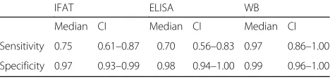
The results of simulation of Se and Sp of IFAT, ELISA and WB and sample prevalence in the overall 161 sam-ples are shown in Table 4. As the sample size is larger, test accuracy was higher and provided a greater preci-sion. Western blot was the most accurate test: Se = 97% (CI: 86–100%); Sp = 99% (CI: 96–100%). Sensitivity of IFAT (75%) was higher and with a higher precision (CI: 61–87%) compared to that of ELISA (70%; CI: 56–83%).
Conversely ELISA Sp (98%) and precision (CI: 94–100%) slightly exceeded that of IFAT (97%; CI: 93–100%).
Discussion
This is the first study that assesses the diagnostic per-formance of IFAT, ELISA and WB for the detection of anti-L. infantum antibodies in sera of cats from non-endemic and non-endemic areas (included cats with con-firmed clinical leishmaniosis) using a Bayesian approach in the absence of a gold standard. Parasite specific anti-body detection is a fundamental diagnostic tool for con-firming a clinical suspicion of leishmaniosis in dogs [6] but the possibility of discrepancy between different sero-logical techniques is well known in both dogs and cats [4, 9, 11, 13, 26]. Serological methods are however less useful for assessing the infection seroprevalence of dogs living in endemic areas as subclinical infections are usu-ally associated with negative or border-line results [6]. In this study, cats with clinical leishmanosis were 100% positive with WB or ELISA and 95% with IFAT, support-ing that antibody detection can be used for confirmsupport-ing feline leishmaniosis in clinical practice as for canine leishmaniosis. However, some caution is needed before excluding leishmaniosis in IFAT negative cats. Sick cats with clinical picture compatible with leishmaniosis but negative by IFAT should perform other serological tests or complementary diagnostic tools such as cytology, histology or PCR [1].
In endemic areas for Trypanosoma spp. or other
[image:5.595.304.539.100.226.2]Leishmania spp. cross reactions with L. infantum must be taken into account for interpretation of serological Table 1Combination of results of the three serological tests
detected in each group of cats Serological technique Non-endemic area
(n= 64)
Endemic area (n= 76)
Affected by leishmaniosis (n= 21)
IFAT ELISA WB Number of
observations
– – – 61 53 0
– – + 2 9 0
– + – 1 0 0
– + + 0 1 1
+ – – 0 9 0
+ – + 0 3 0
+ + – 0 0 0
+ + + 0 1 20
[image:5.595.55.292.110.277.2]+, positive test result;−, negative test result
Table 2Output parameters of the accuracy of tests for each group of cats studied
IFAT ELISA WB
Median CI Median CI Median CI
Non-endemic area: sample prevalence 0.02% (CI: 0.00–0.16)
Sensitivity 0.33 0.01–0.96 0.38 0.02–0.97 0.41 0.02–0.96
Specificity 0.99 0.94–1.00 0.98 0.92–1.00 0.96 0.90–0.99
Endemic area: sample prevalence 0.10% (CI: 0.01–0.96)
Sensitivity 0.43 0.07–0.93 0.26 0.00–0.94 0.64 0.07–0.99
Specificity 0.84 0.18–0.95 0.98 0.30–1.00 0.84 0.06–0.96
Affected by leishmaniosis: sample prevalence 0.89% (CI: 0.01–1.00)
Sensitivity 0.89 0.08–0.99 0.94 0.13–1.00 0.94 0.09–1.00
[image:5.595.56.290.581.723.2]Specificity 0.21 0.01–0.95 0.18 0.00–0.93 0.16 0.00–0.94 Abbreviation:CI0.95 credible interval
Table 3Results of three serological tests applied to all 161 cats
IFAT ELISA WB No. of observations
– – – 114
– – + 11
– + – 1
– + + 2
+ – – 9
+ – + 3
+ + - 0
+ + + 21
+, positive test result;−, negative test result
Table 4Output parameters of the accuracy of the tests with all cats studied. Sample prevalence 0.27; CI = 0.20–0.34
IFAT ELISA WB
Median CI Median CI Median CI
Sensitivity 0.75 0.61–0.87 0.70 0.56–0.83 0.97 0.86–1.00
Specificity 0.97 0.93–0.99 0.98 0.94–1.00 0.99 0.96–1.00
[image:5.595.305.539.669.725.2]tests but this is not the case of the geographical area of investigation of this study [27–29].
The percentage of antibody positive samples to at least one technique in the group of 76 cats living in South Italy reached 30% confirming that feline Leishmaniainfection is frequent in endemic areas (2). This feline group is rep-resentative of an adult feline population admitted for het-erogeneous reasons to clinical practice in endemic area. These cats are potentially exposed to the parasite and can stand in one of the following conditions: not come into contact withLeishmania, come into contact but not be-come infected, otherwise they are infected. Moreover, in-fected individuals can stand at the time of sampling in a different point of the wide spectrum of clinical situations going from a subclinical infection to overt disease. ELISA testing appears to have a low Se for detecting antibodies compared to WB and IFAT in subclinical infections. This finding is in contrast with other studies and is of clinical relevance when there is a need of testing cats in endemic areas because they are blood donors, before their export-ation to a non-endemic area or because an immune-suppressive therapy has to be given [1, 30]. The very strict calculation that we used (mean value ± 4 SD of sera from non-endemic area) for ELISA cut-off setting obviously contributed to this result.
Despite the large number of published serological inves-tigations, very few studies validated serological techniques testing for anti-Leishmaniaantibodies a consistent num-ber of serum samples obtained from feline patients of non-endemic area [5, 11, 31, 32]. It is important to have as much extensive as possible data by testing fe-line populations of non-endemic regions in order to exclude the possibility of cross-reactions with other microbial agents or even with endogenous feline pro-teins. The current state of the art clearly rule out cross-reactivity with Toxoplasma gondii only, there-fore we cannot exclude cross-reactions for some posi-tive results we obtained [10, 14, 15, 32, 33]. It is important to remark that the travel and clinical his-tory were unknown in all cats studied from non-endemic area and this can be considered a limitation of this study. However, the overall good discrimin-ation that we obtained between the three different categories of cats confirms that we set appropriate methodologies for IFAT, ELISA and WB techniques. In particular, we confirmed that 80 is appropriate IFAT cut-off and the 18 kDa band is marker for posi-tive WB when testing feline sera likewise it occurs in dogs and humans [5, 21–23].
Various problems may arise when comparing diagnos-tic performance of different tests given the absence of a gold standard diagnostic test for Leishmania infection. One problem concerns the influence of spectrum (stage of the disease) and selection (inclusion criteria) bias. For
example, it is more difficult to suspect a disease at an early stage and the criteria for inclusion of patients to be tested in a study will be crucial. Ideally, each unit in the sample should be randomly selected so that the sample is representative of the target population. This is difficult to do in field studies and this type of error yields to misleading statistics. Therefore, it is important to be aware of the influence of spectrum and selection bias on the accuracy of diagnostic tests for Leishmania
infection, but this must be kept in perspective. This means that to carefully assess the Se and Sp of diagnos-tic tests, samples of cats with confirmed leishmaniosis as well as of animals from endemic and non-endemic areas should be selected. Other important factors influ-encing the diagnostic performance of tests include tech-nical variations. In this study, cut-off values used for IFAT and ELISA and the criteria for interpretation of positive immunoblots in feline sera were confirmed to be appropriate for discriminating samples from endemic and non-endemic areas and, among those from endemic area, between cats with confirmed leishmaniosis and cats with other clinical problems. The aim of this study was to evaluate the diagnostic performance of tests fre-quently used for the detection of anti-Leishmania anti-bodies in feline sera in the absence of a gold standard. For the first time, this analytical problem was solved by writing a routine with MATLAB software in the Bayes-ian framework. This approach has the main advantage of drawing inference from diagnostic tests in the ab-sence of a gold standard. The non-informative beta prior for parameter provided a minimal effect on the final inference of three diagnostic tests and we obtained the maximum of information from the experimental data. One weak point of this study is, however, given by the sample size of cats with confirmed leishmaniosis compared to those non-infected that we could test, as this can be one possible source for uncertainty of Se of ELISA (27%) and IFAT (26%) compared to that of WB (14%). The analysis of a more extensive and better bal-anced specimen of feline serum samples can confirm or reject this hypothesis.
Conclusion
perfect diagnostic test, the best choice for each specific purpose of diagnosis (infectionversusdisease) theoretic-ally is offered by WB because of the highest Se and Sp, however the analysis of more samples is necessary for confirming our results.
Abbreviations
ELISA:Enzyme-linked immunosorbent assay; IFAT: Immunofluorescence antibody test; MCMC: Markov Chain Monte Carlo; WB: Western blot
Acknowledgements
Publication of this paper has been sponsored by Bayer Animal Health in the framework of the 12thCVBD World Forum Symposium. The authors are grateful
to Sergio Villanueva-Saz, Karine Le Plus, Rachid Mahmoud and Angela Burrascano for technical collaboration for serological techniques. The authors also thank Dr. Shazia Hosein for the collection of serum samples from a non-endemic area.
Funding Not applicable.
Availability of data and materials
The datasets used or analyzed during the current study are available from the corresponding author on reasonable request.
Authors’contributions
MGP and LSG conceived the research study. MFP worked in the field, contributed with data analysis and interpretation, wrote the first draft and revised the manuscript. MFP performed all laboratory techniques with the collaboration of MM and PD. PM, MGP, LSG and FV supervised the technical procedures. MGP contributed with data analysis, interpretation, wrote and revised the manuscript. LSG contributed with data analysis, interpretation and revised the manuscript. AV performed statistical analysis, interpretation and wrote and revised the manuscript. All authors read and approved the final manuscript.
Competing interests
The authors declare that they have no competing interests.
Consent for publication Not applicable.
Ethics approval and consent to participate
This study was approved by the Royal Veterinary College (University of London) Ethics and Welfare Committee (Project number 1235, 2013). Consent to use residual serum samples was preliminarily obtained from owners or animal trust legal representatives and they received complimentary tests for retroviral infections of their cats.
Author details
1Istituto Zooprofilattico Sperimentale della Sicilia, A. Mirri, Via G. Marinuzzi 3, Palermo 90129, Italy.2Departament de Medicina i Cirurgia Animals. Facultat de Veterinària, Universitat Autònoma de Barcelona, Bellaterra, Cerdanyola 08193, Barcelona, Spain.3Dipartimento di Scienze Veterinarie, Università degli Studi di Messina , Polo Universitario Annunziata, Messina 98168, Italy.4Centre Hospitalier Universitaire de Nice, Faculté de Médecine, Université de Nice-Sophia Antipolis, Inserm U 1065, Hôpital de l’Archet, 151, route de Saint Antoine de Ginestière, CS 23079 06202 Nice Cedex 3, France.
5Parasitologie-Mycologie, Hôpital de l’Archet, Centre Hospitalier Universitaire de Nice, France-MIVEGEC, UMR IRD224 - CNRS 5290 - Université de Montpellier, Montpellier Cedex 5, France.6Centro di Referenza Nazionale per le Leishmaniosi (C.Re.Na.L), Istituto Zooprofilattico Sperimentale della Sicilia, A. Mirri, Via G. Marinuzzi 3, Palermo 90129, Italy.
Received: 20 January 2017 Accepted: 17 February 2017
References
1. Pennisi MG, Cardoso L, Baneth G, Bourdeau P, Koutinas A, Miró G, et al. LeishVet update and recommendations on feline leishmaniosis. Parasit Vectors. 2015;8:302.
2. Pennisi MG. Leishmaniosis of companion animals in Europe: an update. Vet Parasitol. 2015;208:35–47.
3. Maroli M, Pennisi MG, Di Muccio T, Khoury C, Gradoni L, Gramiccia M. Infection of sand flies by a cat naturally infected withLeishmania infantum. Vet Parasitol. 2007;145:357–60.
4. Chatzis MK, Leontides L, Athanasiou LV, Papadopoulos E, Kasabalis D, Mylonakis M, et al. Evaluation of indirect immunofluorescence antibody test and enzyme-linked immunosorbent assay for the diagnosis of infection by
Leishmania infantumin clinically normal and sick cats. Exp Parasitol. 2014; 147:54–9.
5. Pennisi MG, Lupo T, Malara D, Masucci M, Migliazzo A, Lombardo G. Serological and molecular prevalence ofLeishmania infantuminfection in cats from Southern Italy. J Feline Med Surg. 2012;14:656–7.
6. Solano-Gallego L, Miró G, Koutinas A, Cardoso L, Pennisi MG, Ferrer L, et al. LeishVet guidelines for the practical management of canine leishmaniosis. Parasit Vectors. 2011;4:86.
7. O.I.E. Leishmaniosis. In: Manual of diagnostic tests and vaccines for terrestrial animals (chap.2.1.11) 6th edition. World Organisation for Animal Health, France. 2016. p.1–12. http://www.oie.int/fileadmin/Home/eng/Health_ standards/tahm/2.01.11_LEISHMANIOSIS.pdf. Accessed 18 Jan 2017. 8. Swets JA. Measuring the accuracy of diagnostic systems. Science. 1988;240:
1285–93.
9. Solano-Gallego L, Villanueva-Saz S, Carbonell M, Trotta M, Furlanello T, Natale A. Serological diagnosis of canine leishmaniosis: comparison of three commercial ELISA tests (Leiscan, ID Screen and Leishmania 96), a rapid test (Speed Leish K) and an in-house IFAT. Parasit Vectors. 2014;7:111. 10. da Silveira NL, Sobrinho LSV, Martins CO, Machado RZ, Marcondes M, de
Lima VMF. Use of crude, FML and rK39 antigens in ELISA to detect
anti-Leishmaniaspp. antibodies inFelis catus. Vet Parasitol. 2011;177:374–7. 11. Solano-Gallego L, Rodríguez-Cortés A, Iniesta L, Quintana J, Pastor J, Espada
Y, et al. Cross-sectional serosurvey of feline leishmaniasis in ecoregions around the northwestern Mediterranean. Am J Trop Med Hyg. 2007;76:676–80. 12. Iniesta L, Gállego M, Portús M. Idiotype expression of IgG1 and IgG2 in dogs
naturally infected withLeishmania infantum. Vet Immunol Immunopathol. 2007;119:189–97.
13. Figueiredo FB, Bonna ICF, Nascimento LD, Costa T, Baptista C, Pacheco TMV, et al. Serological evaluation for detection of anti-Leishmaniaantibodies in dogs and cats in the district of Santa Rita de Cássia, municipality of Barra Mansa, State of Rio de Janeiro. Rev Soc Bras Med Trop. 2009;42:141–5. In Portuguese. 14. Coelho WMD. do Amarante AFT, Apolinário J de C, Coelho NMD, de Lima
VMF, Perri SHV, et al. Seroepidemiology ofToxoplasma gondii, Neospora caninumandLeishmaniaspp. infections and risk factors for cats from Brazil. Parasitol Res. 2011;109:1009–13.
15. Sobrinho LSV, Rossi CN, Vides JP, Braga ET, Gomes AAD, de Lima VMF, et al. Coinfection ofLeishmania chagasiwithToxoplasma gondii, Feline Immunodeficiency Virus (FIV) and Feline Leukemia Virus (FeLV) in cats from an endemic area of zoonotic visceral leishmaniasis. Vet Parasitol. 2012;187:302–6. 16. Greiner M, Gardner IA. Epidemiologic issues in the validation of veterinary
diagnostic tests. Prev Vet Med. 2000;45:3–22.
17. Grevot A, Jaussaud Hugues P, Marty P, Pratlong F, Ozon C, Haas P, et al. Leishmaniosis due toLeishmania infantumin a FIV and FELV positive cat with a squamous cell carcinoma diagnosed with histological, serological and isoenzymatic methods. Parasite. 2005;12:271–5.
18. Rodríguez-Cortés A, Ojeda A, Francino O, López-Fuertes L, Timón M, Alberola J.Leishmaniainfection: laboratory diagnosing in the absence of a
“gold standard.”. Am J Trop Med Hyg. 2010;82:251–6.
19. Joseph L, Gyorkos TW, Coupal L. Bayesian estimation of disease prevalence and the parameters of diagnostic tests in the absence of a gold standard. Am J Epidemiol. 1995;141:263–72.
20. Principato F, Vullo A, Matranga D. On implementation of the Gibbs sampler for estimating the accuracy of multiple diagnostic tests. J Appl Stat. 2010;37: 1335–54.
21. Marty P, Lelievre A, Quaranta JF, Rahal A, Gari-Toussaint M, Le Fichoux Y. Use of the leishmanin skin test and western blot analysis for
epidemiological studies in visceral leishmaniasis areas: experience in a highly endemic focus in Alpes-Maritimes (France). Trans R Soc Trop Med Hyg. 1994;88:658–9.
23. Pomares C, Despierres L, del Giudice P, Delaunay P, Michel G, Ferrua B, et al. Western blot analysis as an aid for the diagnosis of cutaneous leishmaniasis due toLeishmania major. Trans R Soc Trop Med Hyg. 2012;106:452–4. 24. Laruelle-Magalon C, Toga I. Un cas de leishmaniose féline. Prat Méd Chir
Anim Comp. 1996;31:255–61.
25. Pocholle E, Reyes-Gomez E, Giacomo A, Delaunay P, Hasseine L, Marty P. Un cas de leishmaniose féline disséminée dans le sud de la France. Parasite. 2012;19:77–80.
26. Ozon C, Marty P, Pratlong F, Breton C, Blein M, Lelièvre A, et al. Disseminated feline leishmaniosis due toLeishmania infantumin Southern France. Vet Parasitol. 1998;75:273–7.
27. Longoni SS, López-Cespedes A, Sánchez-Moreno M, Bolio-Gonzalez ME, Sauri-Arceo CH, Rodríguez-Vivas RI, et al. Detection of differentLeishmania
spp. andTrypanosoma cruziantibodies in cats from the Yucatan Peninsula (Mexico) using an iron superoxide dismutase excreted as antigen. Comp Immunol Microbiol Infect Dis. 2012;35:469–76.
28. Can H, Döşkaya M, Özdemir HG,Şahar EA, Karakavuk M, PektaşB, et al. Seroprevalence ofLeishmaniainfection and molecular detection of
Leishmania tropicaandLeishmania infantumin stray cats ofİzmir. Turkey Exp Parasitol. 2016;167:109–14.
29. Sartor PA, Cardinal MV, Orozco MM, Gürtler RE, Leguizamón MS. Trans-Sialidase neutralizing antibody detection inTrypanosoma cruzi-infected domestic reservoirs. Clin Vaccine Immunol. 2011;18:984–9.
30. Pennisi MG, Hartmann K, Addie DD, Lutz H, Gruffydd-Jones T, Boucraut-Baralon C, et al. Blood transfusion in cats: ABCD guidelines for minimising risks of infectious iatrogenic complications. J Feline Med Surg. 2015;17:588–93. 31. Martín-Sánchez J, Acedo C, Muñoz-Pérez M, Pesson B, Marchal O,
Morillas-Márquez F. Infection byLeishmania infantumin cats: epidemiological study in Spain. Vet Parasitol. 2007;145:267–73.
32. Sherry K, Miró G, Trotta M, Miranda C, Montoya A, Espinosa C, et al. A serological and molecular study ofLeishmania infantuminfection in cats from the Island of Ibiza (Spain). Vector Borne Zoonotic Dis. 2011;11:239–45. 33. Nasereddin A, Salant H, Abdeen Z. Feline leishmaniasis in Jerusalem:
serological investigation. Vet Parasitol. 2008;158:364–9.
• We accept pre-submission inquiries
• Our selector tool helps you to find the most relevant journal • We provide round the clock customer support
• Convenient online submission • Thorough peer review
• Inclusion in PubMed and all major indexing services • Maximum visibility for your research
Submit your manuscript at www.biomedcentral.com/submit