RESEARCH
Study of the microRNA expression
profile of foreskin derived mesenchymal stromal
cells following inflammation priming
Hussein Fayyad‑Kazan
1*†, Mohammad Fayyad‑Kazan
2†, Bassam Badran
1, Dominique Bron
3,
Laurence Lagneaux
3and Mehdi Najar
3Abstract
Background: Due to their self‑renewal capacity, multi‑lineage potential, and immunomodulatory properties, mes‑ enchymal stromal cells (MSCs) are an attractive tool for different therapeutic strategies. Foreskin (FSK), considered as a biological waste material, has already been shown to be a valuable source of MSCs. Besides their typical fibroblast like morphology and International Society for cellular Therapy compliant phenotype, foreskin‑MSCs (FSK–MSCs) are clo‑ nogenic, and highly proliferative cells with multi‑lineage and strong immunomodulatory capacities. Of importance, FSK–MSCs properly adjust their fate following exposure to inflammatory signals. Being potent regulators of gene expression, miRNAs are involved in modulating nearly all cellular processes and in orchestrating the roles of different immune cells. In this study, we characterized the miRNome of FSK–MSCs by determining the expression profile of 380 different miRNAs in inflammation primed vs. control non‑primed cells.
Methods: TaqMan low density array (TLDA) was performed to identify dysregulated miRNAs after exposing FSK– MSCs to inflammatory signals. Quantitative real‑time RT‑PCR was carried out to validate the observations. DIANA‑miR‑ Path analysis web server was used to identify potential pathways that could be targeted by the dysregulated miRNAs.
Results: Sixteen miRNAs were differentially expressed in inflammation‑primed vs. non‑primed FSK–MSCs. The expres‑ sion level of miR‑27a, ‑145, ‑149, ‑194, ‑199a, ‑221, ‑328, ‑345, ‑423‑5p, ‑485‑3p, ‑485‑5p, ‑615‑5p and ‑758 was down‑ regulated whilst that of miR‑155, ‑363 and ‑886‑3p was upregulated. Target pathway prediction of those differentially expressed miRNAs identified different inflammation linked pathways.
Conclusions: After determining their miRNome, we identified a striking effect of inflammatory signals on the miR‑ NAs’ expression levels in FSK–MSCs. Our results highlight a potential role of miRNAs in modulating the transcription programs of FSK–MSCs in response to inflammatory signals. Further, we propose that specific miRNAs could serve as interesting targets to manipulate some functions of FSK–MSCs, thus ameliorating their therapeutic potential.
Keywords: MSCs, Foreskin, Inflammation, miRNA
© The Author(s) 2017. This article is distributed under the terms of the Creative Commons Attribution 4.0 International License (http://creativecommons.org/licenses/by/4.0/), which permits unrestricted use, distribution, and reproduction in any medium, provided you give appropriate credit to the original author(s) and the source, provide a link to the Creative Commons license, and indicate if changes were made. The Creative Commons Public Domain Dedication waiver (http://creativecommons.org/ publicdomain/zero/1.0/) applies to the data made available in this article, unless otherwise stated.
Background
Mesenchymal stromal cells (MSCs) are multipotent fibro-blast-like cells found in almost all tissues [1]. This multi-lineage potential, along with their capacity of supporting
hematopoiesis and modulating immune responses are the main properties of MSCs. The simple and easy iso-lation procedures together with the great expansion potential, render MSCs as ideal candidates for different cellular therapies [2]. Despite some common character-istics and properties shared by MSCs of different origin [3], significant differences in their immunobiological fea-tures exist [4, 5]. Although it has long been considered as a biological waste material, foreskin (FSK) is, nowadays, considered as an important reservoir of therapeutic cells
Open Access
*Correspondence: hfayyadk@gmail.com
†Hussein Fayyad‑Kazan and Mohammad Fayyad‑Kazan contributed equally to this work
1 Laboratory of Cancer Biology and Molecular Immunology, Faculty of Sciences I, Lebanese University, Hadath, Lebanon
having potential value for several clinical applications [6, 7]. Recently, we have demonstrated that the foreskin is a new source of MSCs, designated as FSK–MSCs [8]. Being compliant with ISCT criteria, non-immunogenic and showing powerful immunomodulatory capacities [8], FSK–MSCs are highlighted as a valuable tolerogenic tool for cell-based immunotherapy. As known, MSCs are particularly sensitive to the local inflammatory microen-vironment that modulate their functions and responses. Indeed, inflammation-priming leads to significant changes in the immunobiology of FSK–MSCs [8].
MicroRNAs (miRNAs) are small (19-22-nt) single-stranded noncoding RNA molecules that regulate the transcription or translation of the target gene mRNAs. Thus, they play crucial roles in many different cellular processes including cell differentiation, proliferation and immune homeostasis [9–12]. Accumulating data show that abnormal miRNA expression is a common feature of various diseases [9–12]. Nowadays, many evidences dem-onstrate a role for miRNAs in regulating distinct aspects of the immune system including cell proliferation, dif-ferentiation, fate determination and function mediated by different cell types [13–15]. The understanding of the miRNome of FSK–MSCs under inflammatory settings will help to identify the pattern of miRs that are modu-lated and that could serve as targets to enhance MSCs therapeutic functions. In this study, comparative analysis showed that the miRNA expression profile of FSK–MSCs is critically different in inflammation primed vs. control non-primed cells. Our results reveal specific miRNA expression differences following inflammation priming and identified 16 differentially expressed miRNAs. Those altered miRNAs might be involved in the molecular mechanisms regulating FSK–MSCs immunobiology dur-ing inflammatory conditions and could be further used as targets to manipulate FSK–MSCs functions.
Methods Ethical guidelines
This study was conducted in accordance with the Decla-ration of Helsinki (1964) and approved by the local eth-ics committee of the “Institut Jules Bordet” (Belgium). All donors and/or their parents or legal guardian were volun-tary for giving the foreskin (FSK) samples as obtained fol-lowing a circumcision procedure from healthy subjects. They gave written informed consent before specimen col-lection for using the samples and for publishing any asso-ciated scientific data. However, all personal information are kept confidential.
Isolation, culture and characterization of FSK–MSCs Briefly, after circumcision, foreskin was aseptically col-lected into a sterile specimen container containing sterile
phosphate-buffered saline (PBS) buffer supplemented with penicillin/streptomycin. The samples were processed as previously published [8]. After a sterile wash with PBS, the sample was transferred into a petri dish where epidermis was manually removed from the skin and the dermis was cut into small pieces. By using Liberase Research Grade solution (Roche Diagnostics, Belgium), the tissue was digested and the resulting cell suspension was washed by centrifugation (800g, 5 min) in Dulbecco’s modified Eagle’s medium with low glucose (DMEM-LG; Lonza, Belguim) and containing 10% fetal bovine serum (FBS; Sigma-Aldrich, Belgium). The subsequent cell pellet was seeded in culture flasks with DMEM-LG (Lonza) supplemented with 10% FBS (Sigma-Aldrich), 2 mM l-glutamine and 50 U/ ml penicillin (both from Lonza) and the cultures were incubated at 37 °C in a 5% CO2 humidified atmosphere. Non-adherent cells were removed when the medium was changed (once a week). When sub-confluence (80–90%) was achieved, adherent cells were harvested by TrypLE Select (Lonza) and expanded until the desired passage.
The cells were characterized according to the ISCT criteria. Thus, the immunophenotype was analysed by flow cytometry (MacsQuant analyzer (Miltenyi Biotec, Netherlands)) using fluorochrome labelled monoclo-nal antibodies: anti-CD45-FITC and anti-HLA-Dr-PE (Exalpha Biologicals, Maynard, MA), anti-CD34-PE and anti-CD73-PE (BD Biosciences, San Diego, CA, USA), anti-CD14-PE, anti-CD19-PE, anti-CD105-FITC and anti-CD90-PE (R&D systems, Minneapolis, MN, USA). Their multilineage potential was confirmed by induc-ing specific lineage commitment (adipogenic, osteogenic and chondrogenic differentiation) using appropriate induction culture medium (NH media, Miltenyi Biotec). Specific lineage commitment was highlighted by using lineage-specific cell staining techniques and microscopic examination.
Inflammation priming of FSK–MSCs
Foreskin–mesenchymal stromal cells were analyzed under both constitutive and inflammatory conditions. Inflammation priming of FSK–MSCs was performed as previously described [16]. Briefly, cells were treated (overnight) with a pro-inflammatory cytokine cock-tail containing IL-1β (Peprotech, Rocky Hill, NJ, USA) (25 ng/ml), TNF-α (50 ng/ml), IFN-α (3000 U/ml or 10 ng/ml) and IFN-γ (1000 U/ml or 50 ng/ml) (all from Prospec Inc., Rehovot, Israel). After priming, the medium was removed, and the cells were washed and became available for analysis.
MiRNA expression profile
concentration was quantified using a NanoDrop spec-trophotometer. A three-step procedure was performed to profile the miRNAs. First, for cDNA synthesis from the miRNAs, 30 ng of total RNA was subjected to RT (reverse transcription) using a TaqMan® microRNA Reverse Transcription Kit (#4366596; Applied Bio-systems) and Megaplex RT primers (Human Pool A, #4399966; Applied Biosystems) following the manufac-turer’s protocol, allowing simultaneous reverse transcrip-tion of 380 mature human miRNAs to generate a miRNA cDNA library corresponding to each plasma sample. RT was performed on a Mastercycler Epgradient thermocy-cler (Eppendorf) with the following cycling conditions: 40 cycles at 16 °C for 2 min, 42 °C for 1 min and 50 °C for 1 s followed by a final step of 80 °C for 5 min to inacti-vate reverse transcriptase. Thereafter, to generate enough miRNA cDNA template for the following real-time PCR, the cDNA libraries were pre-amplified using Megaplex PreAmp primer (Humam Pool A, #4399233; Applied Bio-systems) and PreAmp Master Mix (#4384266; Applied-Biosystems) following the manufacturer’s instructions. The PreAmp primer pool used here consisted of forward primers specific for each of the 380 human miRNAs and a universal reverse primer. The pre-amplification cycling conditions were as follows: 95 °C for 10 min, 55 °C for 2 min, 72 °C for 2 min followed by 12 cycles at 95 °C for 30 s and 60 °C for 4 min; the samples were then held at 99.9 °C for 10 min. After the pre-amplification step, the products were diluted with RNase-free water, com-bined with TaqMan gene expression Master Mix and then loaded into TaqMan Human MicroRNA Array A (#4398965; Applied Biosystems), which is a 384-well for-matted plate and real-time PCR-based microfluidic card with embedded TaqMan primers and probes in each well for the 380 different mature human miRNAs. Real-time PCR was performed on an ABI PRISM 7900HT sequence detection system (Applied Biosystems) with the following cycling conditions: 50 °C for 2 min, 94.5 °C for 10 min fol-lowed by 40 cycles at 95 °C for 30 s and 59.7 °C for 1 min. The Ct (cycle threshold) was automatically given by SDS 2.4 software (Applied Biosystems) and is defined as the fractional cycle number at which the fluorescence passes the fixed threshold of 0.2. RNU48 embedded in the TaqMan human microRNA arrays was used as an endog-enous control. The relative expression levels of miRNAs were calculated using the comparative ΔΔCt method as described previously [17, 18]. The fold changes in miR-NAs were calculated by the equation 2−ΔΔCt.
Taqman miRNA assay for individual miRNAs
Gene-specific reverse transcription was performed for each miR using 10 ng of purified total RNA, 100 mM dNTPs, 50 U MultiScribe reverse transcriptase, 20 U
RNase inhibitor, and 50 nM of gene-specific RT primer samples using the TaqMan MicroRNA Reverse Transcrip-tion kit (Applied Biosystems, Gent, Belgium). 15 μl reac-tions were incubated for 30 min at 16 °C, 30 min at 42 °C, and 5 min at 85 °C to inactivate the reverse transcriptase. Real time RT-PCR reactions (5 μl of RT product, 10 μl TaqMan 2 × Universal PCR master Mix, (Applied Biosys-tems, Gent, Belgium), and 1 μl TaqMan MicroRNA Assay Mix containing PCR primers and TaqMan probes) were carried out on ABI Prism 7900HT Sequence Detection System (Applied Biosystems, Gent, Belgium) at 95 °C for 10 min followed by 40 cycles at 95 °C for 15 s and 60 °C for 1 min. The qRT-PCR reactions were performed in tripli-cate, and the signal was collected at the end of every cycle. The Ct (cycle threshold) was automatically given by SDS 2.4 software (Applied Biosystems) and is defined as the fractional cycle number at which the fluorescence passes the fixed threshold of 0.2.
RNU48 was used as an endogenous control. The rela-tive expression levels of miRNAs were calculated using the comparative ΔΔCt method as described previously [31, 32]. The fold changes in miRNAs were calculated by the equation 2−ΔΔCt.
Pathway enrichment analysis
Pathway enrichment analysis was employed to investigate the regulatory mechanisms of significantly differentially expressed miRNAs. DIANA-miRPath v.3.0 analysis web server was used for pathway enrichment analyses. The enriched pathways were defined by their enrichment of significantly differentially expressed miRNA target genes.
Statistical analysis
Data analyses were performed using the SDS RQ Man-ager 1.2 software and DataAssist v2.0 software (Applied Biosystems). Statistical significance of miRNA expres-sion between control and treated cells was determined according to unpaired Mann–Whitney U test. p Values <0.05 (*), <0.01 (**) were considered significant.
Results
Obtaining and characterizing FSK–MSCs
These adherent cells were then characterized accord-ing to the ISCT criteria. Flow cytometry analysis demon-strated that those cells are positive (>95%) for the MSC markers CD73, CD90 and CD105 whilst negative (<5%) for CD45, CD34, CD14, CD19 and HLA-DR markers. It is noteworthy that inflammation priming had no sig-nificant impact on the expression level of those markers (data not shown).
To comply with the ISCT criteria, we further con-firmed that the obtained cells exhibited a multi-lineage potential since they were able to differentiate into cells of adipogenic, osteogenic or chondrogenic lineage after being cultured with appropriate induction media (data not shown). After 21 days of osteogenic differentiation, a mineralization matrix following calcium deposits was observed by Alizarin red staining demonstrating the gen-eration of osteoblasts. After 10 days of adipogenic differ-entiation, cells with cytoplasm containing several lipid vacuoles were revealed by Oil Red O staining indicating the formation of adipocytes. After 21 days of chondro-genic differentiation, the production of proteoglycans-based extracellular matrix was shown by Alcian blue staining illustrating the generation of chondrocytes.
Impact of inflammation priming on miRNA‑expression profile in FSK–MSCs cells
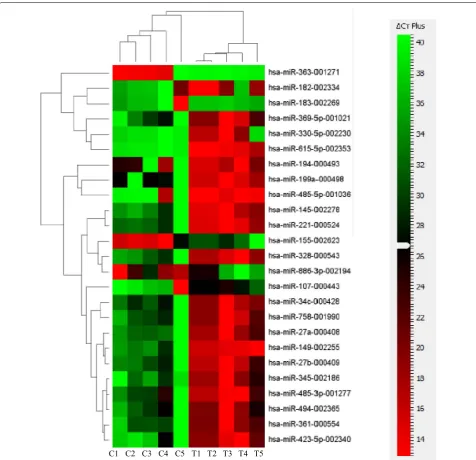
Aware that MSCs can serve as sensors and switchers of inflammation, and given the importance of miRNAs in regulating inflammatory responses, we aimed at char-acterizing the expression level of different miRNAs in FSK–MSCs and investigating the impact of inflammation on this expression. FSK–MSCs were derived from 5 inde-pendent donors and total RNA was prepared from those cells being either untreated (control cells) or treated with a pro-inflammatory cytokine cocktail (treated cells). TaqMan low density array (TLDA) was then carried out to assess miRNA expression level in those cells. Before measuring miRNAs’ levels and in order to identify suit-able endogenous controls, three different candidate miRNAs (RNU44, RN48, and U6 snRNA) were analyzed for variance in gene expression using Data Assist soft-ware v2.0. The statistical method ranked the candidate endogenous control genes with an excellent correlation of raw stability values (data not shown). The most stably expressed RNU48 was chosen as the endogenous control, and relative miRNA expression was normalized against RNU48. Our TLDA analysis revealed that the miRNA expression profile in treated cells is not totally similar to that of untreated cells. In fact, we show the expres-sion level of 25 miRNAs that were modulated in treated vs. control cells (Fig. 1). Among these, 20 miRNAs (miR-145, -149, -182, -194, -199a, -221, -27a/b, -328, -330-5p, 345, -34c, -361, -369-5p, -423-5p, 485-3p, 485-5p, -494,
-615-5p and-758) were downregulated whilst 5 miRNAs (miR-107, -155, -183, -363 and -886-3p) were upregu-lated. Details of the results are shown in Table 1. A clustergram of the samples as well as the significantly dif-ferentially expressed miRNAs in control and treated cells are illustrated in Fig. 1 in the form of a heat map gener-ated using ΔCt values. Heat map is a typical method used in gene-expression analysis where the expression level of many genes across a number of tested samples can be displayed in a two dimensional image with a color code representing the expression intensity of each gene.
The numbers corresponding to these colors are the ΔCt values. The dendrogram on the left side of the heat map classifies miRNAs into groups based on the divergence of miRNA expression values among the different samples. The dendrogram presented at the top indicates the relat-edness of the samples based on overall miRNA expres-sion values and separates the control from the treated group of samples.
In a second step, and in order to validate their differ-ential expression, miRNAs that appeared to be upregu-lated or downreguupregu-lated in treated vs. control cells were further examined using individual quantitative Real Time PCR (qRT-PCR). Interestingly, out of the 25 miRNAs that showed altered expression (Table 1), 16 miRNAs were confirmed to exhibit such differential expression in treated vs. control cells (Fig. 2). Those 16 miRNAs fall in two groups. Group 1 contains 13 miRs that were down-regulated (ratio between 0.1and 0.005) in treated cells in comparison to control cells and includes miR-27a, -145, -149, -194, -199a, -221, -328, -345, -423-5p, -485-3p, -485-5p, -615-5p and -758 (Fig. 2). Observing that those 16 miRs are not equal in terms of their downregulation rate led us to further classify them into subgroups. Group 1A corresponds to miRNAs that were most strikingly downregulated and consists of miR-27a, -145 and -221 that decreased 10, 13.7 and 15 folds, respectively. Group 1B consists of miRNAs that were less strikingly down-regulated and includes miR-149, -194, -615-5p and -758 that exhibited decreased rates of 7, 8.4, 5 and 5.3 folds, respectively. Group 1C contains the least strongly down-regulated miRNAs and includes miR-199a, -328, -345, -423-5p, 485-3p and -485-5p that showed downregula-tion rates of 3.8, 2, 4.8, 2.5, 3.4 and 3.7 folds, respectively.
cells suggesting a potential role for miRNAs in modulat-ing FSK–MSCs’ transcriptional programs in response to inflammatory conditions.
Analysis of inflammation primed MSCs–associated miRNA pathways
Since each miRNA can regulate the expression of many target genes but multiple miRNAs can modulate specific
pathways, we explored the pathways that were poten-tially regulated by the miRNAs observed to be altered in inflammation primed FSK–MSCs in comparison to untreated control MSCs.
For the identification of these pathways, DIANA-miRPath v.3.0 analysis web server was used and each of the statistically differentially expressed miRNAs was used as a query. The output Kyoto encyclopedia of genes
C4 C2 C3
C1 C5 T1 T2 T3 T4 T5
Fig. 1 Differentially expressed miRNAs in inflammation‑primed FSK–MSCs. Heat map presenting the differentially expressed miRNAs in inflamma‑ tion‑primed FSK–MSCs compared to their respective untreated partners (n = 5 in each group). MiRNA expression data was generated by perform‑ ing TaqMan low‑density arrays (TLDA). Columns correspond to inflammation‑cocktail treated samples (T) or untreated control samples (C). Each row
[image:5.595.59.536.86.546.2]and genomes (KEGG) pathways obtained for each miR included many results and for reasons of simplicity and specificity we focused only on pathways linked to inflam-mation. These pathways were previously summarized in [19] and included Adhesion-Extravasation-Migration, apoptosis signaling, calcium signaling, cytokine signaling, leukocyte signaling, innate pathogen detection, MAPK signaling, NF-κB signaling, PI3 K-AKT signaling, TNF superfamily signaling, NK cell signaling, GPCR signal-ing, ROS/Glutathione/cytotoxic granules, phagocytosis-antigen presentation, complement cascade, Eicosanoid signaling and Glucocorticoid signaling/PPAR signaling. The pathways targeted by upregulated and downregu-lated miRs are shown in Table 2. It is noteworthy that no KEGG pathways linked to inflammation were identified
for miR-149, -328, -345, -363, -423-5p, -615-5p, -758 and -886-3p.
Discussion
In addition to their self-renewing and multi-lineage dif-ferentiation properties, MSCs exhibit immunoregulatory abilities through influencing both immune and inflamma-tory responses [20]. In fact, MSCs, can actively sense the surrounding microenvironment or inflammatory milieu and adopt their phenotype and responses, accordingly [20]. For instance, and depending on the environmental signals, MSCs can play both pro- and anti-inflammatory roles [20]. This marked functional plasticity of MSCs ren-ders them as clinically relevant cell types with important therapeutic potential for tissue repair and regeneration as well as for the treatment of different inflammatory diseases and malignancies [20, 21]. Nowadays, different popula-tions of MSCs, derived from distinct human tissues, are available and exhibit dissimilar immunological profiles and functional capacities [5, 22, 23]. FSK–MSCs are emerg-ing as a highly interestemerg-ing MSC-type due to their recently characterized properties [8]. Besides, FSK–MSCs, when exposed to inflammatory conditions, have been shown to exhibit induced expression of different immunoregulatory genes and factors [8]. Their high sensitivity to inflamma-tion increased the interest in understanding the molecular mechanisms involved in coupling FSK–MSCs’ responses to the surrounding inflammatory microenvironment.
Among the different regulatory elements that controls gene expression and modulates cellular fate and behav-ior are miRNAs. For instance, they can significantly alter the responses of different innate and adaptive immune cells where any disturbance of their expression profiles can lead to several diseases [24–27]. Therefore, miRNAs are, nowadays, considered as attractive therapeutic tar-gets [24–27]. Besides their ability to modulate immune responses, miRNAs play a key role in MSCs’ biology. For example, miRNAs can strikingly affect MSCs’ decision to either proliferate or differentiate into either cell lineage including adipogenic, chondrogenic, myogenic, neuro-genic or other lineages [28]. The aim of this work was to determine the miRNome of FSK–MSCs by investigating the expression profile of 380 different miRNAs in inflam-mation primed vs. control non-primed cells. The major outcome of this study is our demonstration of a signifi-cantly altered miRNA expression profile in inflamma-tion-primed FSK–MSCs where 16 miRNAs were found
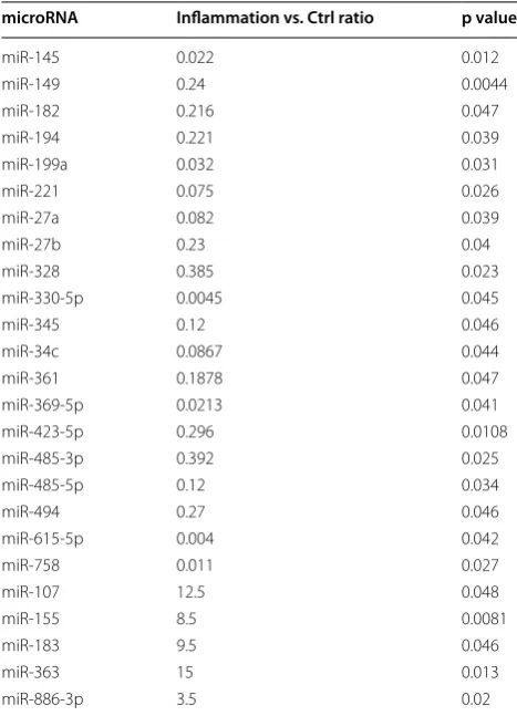
Table 1 MiRNA signature identified by TLDA Technique
Our TLDA analysis identified 25 miRNAs to be differentially expressed in treated vs. untreated control cells with a p value <0.05
microRNA Inflammation vs. Ctrl ratio p value
miR‑145 0.022 0.012
miR‑149 0.24 0.0044
miR‑182 0.216 0.047
miR‑194 0.221 0.039
miR‑199a 0.032 0.031
miR‑221 0.075 0.026
miR‑27a 0.082 0.039
miR‑27b 0.23 0.04
miR‑328 0.385 0.023
miR‑330‑5p 0.0045 0.045
miR‑345 0.12 0.046
miR‑34c 0.0867 0.044
miR‑361 0.1878 0.047
miR‑369‑5p 0.0213 0.041
miR‑423‑5p 0.296 0.0108
miR‑485‑3p 0.392 0.025
miR‑485‑5p 0.12 0.034
miR‑494 0.27 0.046
miR‑615‑5p 0.004 0.042
miR‑758 0.011 0.027
miR‑107 12.5 0.048
miR‑155 8.5 0.0081
miR‑183 9.5 0.046
miR‑363 15 0.013
miR‑886‑3p 3.5 0.02
(See figure on next page.)
[image:6.595.56.290.101.425.2]Contr ol Trea ted 0.0 0.1 0.2 0.3 0.4 0.5
miR-27a p= 0.039
noi ss er px e evi tal e R a7 2-Ri m Contr ol Trea ted 0.0 0.2 0.4 0.6 0.8
miR-145 p= 0.023
noi ss er px e evi tal e R 54 1-Ri m Contr ol Trea ted 0.0 0.2 0.4 0.6 0.8 1.0
miR-149 p= 0.0036
noi ss er px e evi tal e R 94 1-Ri m Contr ol Trea ted 0.000 0.001 0.002 0.003 0.004 0.005
miR-194 p= 0.032
noi ss er px e evi tal e R 49 1-Ri m Contr ol
Treated
0.0000 0.0005 0.0010 0.0015 0.0020 0.0025
miR-199a p= 0.028
noi ss er px e evi tal e R a9 91-Ri m
13.7 fold decrease
10 fold decrease 7 fold decrease
8.4 fold decrease
3.8 fold decrease
Contr ol Trea ted 0.0 0.1 0.2 0.3 0.4 0.5
miR-221 p= 0.016
noi ss er px e evi tal e R 12 2-Ri m
15 fold decrease
* * ** * * * Contr ol Trea ted 0.000 0.005 0.010 0.015 0.020
miR-328 p= 0.012
noi ss er px e evi tal e R 82 3-Ri m Contr ol
Treated
0.000 0.001 0.002 0.003 0.004
miR-345 p= 0.038
noi ss er px e evi tal e R 54 3-Ri m Contr ol Trea ted 0.0000 0.0002 0.0004 0.0006 0.0008 0.0010
miR-423-5p p= 0.035
noi ss er px e evi tal e R p5-32 4-Ri m Contr ol
Treated
0.000 0.001 0.002 0.003
miR-485-3p p= 0.017
noi ss er px e evi tal e R p3-58 4-Ri m
2 fold decrease
4.8 fold decrease
2.5 fold decrease
3.4 fold decrease
Contr ol Trea ted 0.0000 0.0005 0.0010 0.0015 0.0020 0.0025
miR-485-5p p= 0.024
noi ss er px e evi tal e R p5-58 4-Ri m
3.7 fold decrease
Contr ol
Treated
0.00 0.02 0.04 0.06 0.08 0.10
miR-615-5p p= 0.043
noi ss er px e evi tal e R p5-51 6-Ri m
5 fold decrease * * * * * * Contr ol Trea ted 0.0000 0.0001 0.0002 0.0003 0.0004 0.0005
miR-758 p= 0.012
noi ss er px e evi tal e R 85 7-Ri m
5.3 fold decrease
Contr ol
Treated 0.00
0.01 0.02 0.03
miR-155 p= 0.0075
noi ss er px e evi tal e R 55 1-Ri m 9.4 increase Contr ol
Treated 0.00
0.05 0.10 0.15
miR-363 p= 0.022
noi ss er px e evi tal e R 36 3-Ri m 4.7 increase Contr ol Trea ted 0.00 0.01 0.02 0.03 0.04
miR-886-3p p= 0.013
to be differentially expressed. This observed inflamma-tion-triggered alteration of miRNA expression pattern is not surprising especially that different miRNAs have been reported to show altered expression in response to inflammatory signals. For example, miR-146 expression level is described to be upregulated in response to inflam-matory stimuli [29], whilst other miRNAs including miR-9, -101, -125b, -155, -146, -192 and -203 are described to exhibit altered expression profiles in some inflammatory diseases [30].
We classified our differentially expressed miRNAs in two major groups: (1) a first one containing the miRNAs being downregulated in inflammation-primed FSK– MSCs and includes miR-27a, -145, -149, -194, -199a, -221, -328, -345, -423-5p, -485-3p, -485-5p, -615-5p and -758; (2) and a second one bearing the upregulated miR-NAs and consists of miR-155, -363 and -886-3p. Inter-estingly, among those identified miRNAs, there are ones that have already been demonstrated to regulate MSCs’ behavior. For instance, miR-27a has been reported to regulate osteogenesis of MSCs [31] whilst miR-145, -194, and -199a have been described to regulate their chon-drogenesis [32–34]. Moreover, miR-221 has been shown to be involved in the adipogenesis process of MSCs [35]. Besides its ability to regulate adipogenesis [35], miR-155 has also been described to modulate the responses of MSCs to inflammatory signals [36]. Furthermore, miR-886-3p overexpression has been shown to inhibit MSCs migration [37].
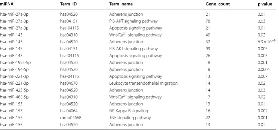
Given that a variety of genes, encoding cell adhesion molecules, co-stimulatory elements as well as immu-noregulatory cytokines and factors, are modulated in inflammation-primed FSK–MSCs, it is strongly pos-sible that the differentially expressed miRNAs could be involved in the regulatory processes accounting for such altered transcription programs in inflammation-primed vs. non-primed FSK–MSCs. This proposal is strongly supported by the observation that miR-155 expres-sion can be modulated upon inflammation-priming of mouse MSCs and that miR-155 expression levels are tightly related to its ability to modulate the immunosup-pressive capacity of those MSCs [36], thus providing a striking evidence for the importance of miRNAs in reg-ulating the immunoregulatory capacity of MSCs. To get more insight into the potential mechanisms and signal-ing events that could be targeted by our differentially expressed miRNAs, we searched for the predicted target pathways of those miRNAs. In fact, given that multiple miRNAs may cooperate to regulate related target genes in a certain signaling route [38, 39], analysis of target pathways instead of individual genes was carried out. The miRNAs, being downregulated in inflammation-primed FSK–MSCs, are predicted to target pathways enriched in genes involved in Adherens junction, PI3-AKT pathway, apoptosis pathway, Wnt/Ca2+ pathway and leukocyte trans-endothelial migration. On the other hand, adherens junction, TNF and NF-KB signaling pathways are pre-dicted to be the targets of miR-155 which is upregulated
Table 2 Significantly enriched KEGG pathways (p < 0.05) targeted by miRNAs
KEGG pathways, number of target genes, and p values were identified by DIANA miRPath v.3.0 for statistically significant differentially expressed miRNAs in inflammation primed FSK–MSCs vs. untreated control cells
miRNA Term_ID Term_name Gene_count p value
hsa‑miR‑27a‑3p hsa04520 Adherens junction 21 0.01
hsa‑miR‑27a‑3p hsa04151 PI3‑AKT signaling pathway 78 0.03
hsa‑miR‑27a‑3p hsa‑04115 Apoptosis signaling pathway 21 0.01
hsa‑miR‑145 hsa04310 Wnt/Ca2+ signaling pathway 40 0.02
hsa‑miR‑145 hsa04520 Adherens junction 32 6.9 x 10⁻⁸
hsa‑miR‑145 hsa04151 PI3‑AKT signaling pathway 99 0.005
hsa‑miR‑145 hsa‑04115 Apoptosis signaling pathway 26 0.005
hsa‑miR‑199a‑5p hsa04520 Adherens junction 8 0.001
hsa‑miR‑194‑5p hsa04520 Adherens junction 8 0.0004
hsa‑miR‑221‑3p hsa‑04115 Apoptosis signaling pathway 13 0.007
hsa‑miR‑221‑3p hsa04670 Leukocyte transendothelial migration 14 0.02
hsa‑miR‑423‑5p hsa04520 Adherens junction 14 0.03
hsa‑miR‑485‑5p hsa04310 Wnt/Ca2+ signaling pathway 7 0.02
hsa‑miR‑155 hsa04520 Adherens junction 13 0.01
hsa‑miR‑155 hsa04064 NF‑Kappa B signaling 16 0.002
hsa‑miR‑155 mmu04668 TNF signaling pathway 22 0.001
[image:8.595.57.544.102.330.2]in inflammation-primed FSK–MSCs. Indeed, it was previously reported that miR-155, being upregulated in inflammation primed mouse MSCs, modulates MSCs immunosuppressive capacity upon modulating NF-KB activity [36].
The observed altered miRNA expression profile in inflammation-primed vs. non-primed FSK–MSCs imposes a detailed mechanistic characterization of the involvement of each of the identified miRNAs as well as their predicted pathway(s) on FSK–MSCs inflammatory responses in future studies. However, our observations might provide an early step toward developing a novel strategy to improve the immunosuppressive activity of FSK–MSCs. For instance, identifying the miRNAs that either dampen or trigger the expression of genes encod-ing immunosuppressive cytokines, factors and receptors would enable us to trigger, in vitro, the immunomodula-tory capacity of FSK–MSCs by downregulating or upreg-ulating the expression of certain miRNAs. This, in vitro, pre-activation of FSK–MSCs would greatly improve their therapeutic potential and medical value.
Conclusions
In this report, we show that inflammation-priming of FSK–MSCs substantially alters their miRNA expression profile where 13 miRNAs are downregulated and 3 oth-ers are upregulated. Those differentially expressed miR-NAs are predicted to target several candidate signaling pathways being involved in regulating cellular behavior in response to inflammatory signals.
Aware that the in vivo communication between grafted MSCs and the host inflammatory microenvironment orchestrates the plasticity of the immunological proper-ties of the grafted MSCs, understanding the molecular events that transplanted MSCs undergo upon exposure to an inflammatory signal is of great importance. In this report, we highlight a potential role for miRNAs in regu-lating FSK–MSCs’ responses to inflammatory signals. Further, we suggest that miRNAs could serve as poten-tial targets to improve, in vitro, the therapeutic value of MSCs before being grafted into the patient.
Abbreviations
FSK: foreskin; GPCR: G protein‑coupled receptor; IL: interleukin; IFN: interferon; ISCT: International Society for cellular Therapy; KEGG: Kyoto encyclopedia of genes and genomes; miR: microRNA; MSCs : mesenchymal stromal cells; PBS: phosphate‑buffered saline; PPAR: peroxisome proliferator‑activated receptor; qRT‑PCR: quantitative Real‑Time PCR; TNF: tumor necrosis factor; TLDA: TaqMan low density array.
Authors’ contributions
HFK and BB conceived and designed the study, performed experiments, acquired data, analyzed the data, interpreted data, drafted the manuscript and revised the manuscript. MFK collected and provided samples, performed experiments, acquired data, analyzed the data, interpreted data, drafted the manuscript and revised the manuscript. DB, LL and MN conceived and
designed the study, collected and provided samples, interpreted data, drafted the manuscript and revised the manuscript. All authors read and approved the final manuscript.
Author details
1 Laboratory of Cancer Biology and Molecular Immunology, Faculty of Sci‑ ences I, Lebanese University, Hadath, Lebanon. 2 Institut de Biologie et de Médecine Moléculaires, Université Libre de Bruxelles, 6041 Gosselies, Belgium. 3 Laboratory of Clinical Cell Therapy, Institut Jules Bordet, Université Libre de Bruxelles (ULB), Campus Erasme, Brussels, Belgium.
Competing interests
The authors declare that they have no competing interests.
Availability of data and supporting materials
Authors do not wish to share their data as they are confidential.
Received: 28 February 2016 Accepted: 6 December 2016
References
1. Clark EA, Kalomoiris S, Nolta JA, Fierro FA. Concise review: Micro‑ RNA function in multipotent mesenchymal stromal cells. Stem Cells. 2014;32:1074–82.
2. da Silva Meirelles L, Fontes AM, Covas DT, Caplan AI. Mechanisms involved in the therapeutic properties of mesenchymal stem cells. Cytokine Growth Factor Rev. 2009;20:419–27.
3. Via AG, Frizziero A, Oliva F. Biological properties of mesenchymal stem cells from different sources. Muscles Ligaments Tendons J. 2012;2:154–62. 4. De Kock J, Najar M, Bolleyn J, Al Battah F, Rodrigues RM, Buyl K, et al.
Mesoderm‑derived stem cells: the link between the transcriptome and their differentiation potential. Stem Cells Dev. 2012;21:3309–23. 5. Najar M, Raicevic G, Fayyad‑Kazan H, Kazan HF, De Bruyn C, Bron D, et al.
Immune‑related antigens, surface molecules and regulatory factors in human‑derived mesenchymal stromal cells: the expression and impact of inflammatory priming. Stem Cell Rev. 2012;8:1188–98.
6. Ashley F. Foreskins as skin grafts. Ann Surg. 1937;106:252–6. 7. Zaroo MI, Sheikh BA, Wani AH, Darzi MA, Mir M, Dar H, et al. Use of pre‑
putial skin for coverage of post‑burn contractures of fingers in children. Indian J Plast Surg. 2011;44:68–71.
8. Najar M, Raicevic G, André T, Fayyad‑Kazan H, Pieters K, Bron D, et al. Mes‑ enchymal stromal cells from the foreskin: tissue isolation, cell characteri‑ zation and immunobiological properties. Cytotherapy. 2016;18:320–35. 9. Pallante P, Visone R, Croce CM, Fusco A. Deregulation of microRNA
expression in follicular‑cell‑derived human thyroid carcinomas. Endocr Relat Cancer. 2010;17:F91–104.
10. Schetter AJ, Heegaard NHH, Harris CC. Inflammation and cancer: inter‑ weaving microRNA, free radical, cytokine and p53 pathways. Carcinogen‑ esis. 2010;31:37–49.
11. Miller BH, Wahlestedt C. MicroRNA dysregulation in psychiatric disease. Brain Res. 2010;1338:89–99.
12. Haramati S, Chapnik E, Sztainberg Y, Eilam R, Zwang R, Gershoni N, et al. miRNA malfunction causes spinal motor neuron disease. Proc Natl Acad Sci US A. 2010;107:13111–6.
13. Lindsay MA. microRNAs and the immune response. Trends Immunol. 2008;29:343–51.
14. Chan CWY, Kay LS, Khadaroo RG, Chan MWC, Lakatoo S, Young KJ, et al. Soluble fibrinogen‑like protein 2/fibroleukin exhibits immunosuppressive properties: suppressing T cell proliferation and inhibiting maturation of bone marrow‑derived dendritic cells. J Immunol. 2003;170:4036–44. 15. Lodish HF, Zhou B, Liu G, Chen C‑Z. Micromanagement of the immune
system by microRNAs. Nat Rev Immunol. 2008;8:120–30. 16. Raicevic G, Rouas R, Najar M, Stordeur P, Boufker HI, Bron D, et al.
Inflammation modifies the pattern and the function of toll‑like recep‑ tors expressed by human mesenchymal stromal cells. Hum Immunol. 2010;71:235–44.
• We accept pre-submission inquiries
• Our selector tool helps you to find the most relevant journal
• We provide round the clock customer support
• Convenient online submission
• Thorough peer review
• Inclusion in PubMed and all major indexing services
• Maximum visibility for your research
Submit your manuscript at www.biomedcentral.com/submit
Submit your next manuscript to BioMed Central
and we will help you at every step:
18. Livak KJ, Schmittgen TD. Analysis of relative gene expression data using real‑time quantitative PCR and the 2 (‑delta delta C(T)) method. Methods. 2001;25:402–8.
19. Loza MJ, McCall CE, Li L, Isaacs WB, Xu J, Chang B‑L. Assembly of inflammation‑related genes for pathway‑focused genetic analysis. PLoS ONE. 2007;2:e1035.
20. Bernardo ME, Fibbe WE. Mesenchymal stromal cells: sensors and switch‑ ers of inflammation. Cell Stem Cell. 2013;13:392–402.
21. Yagi H, Kitagawa Y. The role of mesenchymal stem cells in cancer devel‑ opment. Front Genet. 2013;4:261.
22. Campagnoli C, Roberts IA, Kumar S, Bennett PR, Bellantuono I, Fisk NM. Identification of mesenchymal stem/progenitor cells in human first‑ trimester fetal blood, liver, and bone marrow. Blood. 2001;98:2396–402. 23. El Omar R, Beroud J, Stoltz J‑F, Menu P, Velot E, Decot V. Umbilical cord
mesenchymal stem cells: the new gold standard for mesenchymal stem cell‑based therapies? Tissue Eng Part B Rev. 2014;20:523–44.
24. Jeker LT, Bluestone JA. MicroRNA regulation of T‑cell differentiation and function. Immunol Rev. 2013;253:65–81.
25. He H, Jazdzewski K, Li W, Liyanarachchi S, Nagy R, Volinia S, et al. The role of microRNA genes in papillary thyroid carcinoma. Proc Natl Acad Sci US A. 2005;102:19075–80.
26. Xiao C, Srinivasan L, Calado DP, Patterson HC, Zhang B, Wang J, et al. Lymphoproliferative disease and autoimmunity in mice with increased miR‑17‑92 expression in lymphocytes. Nat Immunol. 2008;9:405–14. 27. Raisch J, Darfeuille‑Michaud A, Nguyen HTT. Role of microRNAs in
the immune system, inflammation and cancer. World J Gastroenterol. 2013;19:2985–96.
28. Collino F, Bruno S, Deregibus MC, Tetta C, Camussi G. MicroRNAs and mesenchymal stem cells. Vitam Horm. 2011;87:291–320.
29. Taganov KD, Boldin MP, Chang K‑J, Baltimore D. NF‑kappaB‑dependent induction of microRNA miR‑146, an inhibitor targeted to signal‑ ing proteins of innate immune responses. Proc Natl Acad Sci USA. 2006;103:12481–6.
30. Sonkoly E, Pivarcsi A. Advances in microRNAs: implications for immunity and inflammatory diseases. J Cell Mol Med. 2009;13:24–38.
31. Hassan MQ, Gordon JAR, Beloti MM, Croce CM, van Wijnen AJ, Stein JL, et al. A network connecting Runx2, SATB2, and the miR‑23a ~ 27a ~ 24‑2 cluster regulates the osteoblast differentiation program. Proc Natl Acad Sci USA. 2010;107:19879–84.
32. Martinez‑Sanchez A, Dudek KA, Murphy CL. Regulation of human chon‑ drocyte function through direct inhibition of cartilage master regulator SOX9 by microRNA‑145 (miRNA‑145). J Biol Chem. 2012;287:916–24. 33. Xu J, Kang Y, Liao W, Yu L. MiR‑194 regulates chondrogenic differentia‑
tion of human adipose‑derived stem cells by targeting Sox5. PLoS ONE. 2012;7:e31861.
34. Lin EA, Kong L, Bai X‑H, Luan Y, Liu C‑J. miR‑199a, a bone morphogenic protein 2‑responsive MicroRNA, regulates chondrogenesis via direct targeting to Smad1. J Biol Chem. 2009;284:11326–35.
35. Skårn M, Namløs HM, Noordhuis P, Wang M‑Y, Meza‑Zepeda LA, Myklebost O. Adipocyte differentiation of human bone marrow‑derived stromal cells is modulated by microRNA‑155, microRNA‑221, and micro‑ RNA‑222. Stem Cells Dev. 2012;21:873–83.
36. Xu C, Ren G, Cao G, Chen Q, Shou P, Zheng C, et al. miR‑155 regulates immune modulatory properties of mesenchymal stem cells by targeting TAK1‑binding protein 2. J Biol Chem. 2013;288:11074–9.
37. Pillai MM, Yang X, Balakrishnan I, Bemis L, Torok‑Storb B. MiR‑886‑3p down regulates CXCL12 (SDF1) expression in human marrow stromal cells. PLoS ONE. 2010;5:e14304.
38. Cloonan N, Brown MK, Steptoe AL, Wani S, Chan WL, Forrest ARR, et al. The miR‑17‑5p microRNA is a key regulator of the G1/S phase cell cycle transition. Genome Biol. 2008;9:R127.