STUDIES ON THE SPARGANUM OF S~IROMETRA ERINACEI
by B. H. "ICTvA
A thesis submitted for the degree of Master
The work reported in this thesis, except where specif'ically mentioned, was performed
entirely by me.
ACKNOWLEDGEMENTS
I should like to express deep and sincere appreciation to my supervisor, Dr. M.J. Howell of ~ha Department of Zoology in the Australian National University.
I
am indebted to him for his guidance, encouragement and patience, and for his generosity in giving me so much of his time.I am also grateful for the assistance given by members of the staff and fellow students of the Departmant of Zoology. In particular I should like to thank
Mr.
N. Call who assisted me in my histochemical experiments; Mrs. J.M. Shield who assisted me in some aspects of my work; Ivlr. I. Fox who rendered photographic assistance; and Professor J.D. Smyth, to whom I am profoundly grateful for making all thispossible.
To Miss K.F. Tze, I am deeply indebted for all the typing done in this thesis.
TABLE
OF CONTENTS
ACKNOWLEDGENENTS
GENERAL INTRODUCTION
CHAPTER I
THE SPARGANUM
IntroductionClassification Life cycle Sparganosis
CHAPTER 2
IDENTIFICATION OF
THE
SPARGANUM
OF SPIRO:METRA
ERINACEI
AND ITS
PENETRATIO~
INTO
THE INTERMEDIATE
ii i 1 5 5 6 8 10
ROST
12Introduction 12
Methods and Materials 13
(a) Identification of the sparganum 13
(b) Penetration experiments 15
Results 16
(a) Identification of the sparganum 16
(b) Penetration experiments 21
CHAPTER
3
THE MORPHOLOGY AND HISTOCHEMISTRY OF THE SPARGANUM SCOLEX OFiii
SPIROMETRA ERINACEI 29
Introduction 29
Methods and Materials 30
(a) Morphological study of the sparganum
scolex 30
(b) Histological study of the sparganum
scolex 31
(c) Histochemical study of the sparganum
scolex 32
Result~
33
(a) The sparganum scolex 33
(b) Histology of the sparganum scolex 34 (c) Histochemical study of the sparganum
scolex 36
(i) Distribution of non-enzymatic
Discussion CHAPTER 4
Introduction
substances 36
(ii) Distribution of enzymatic substances
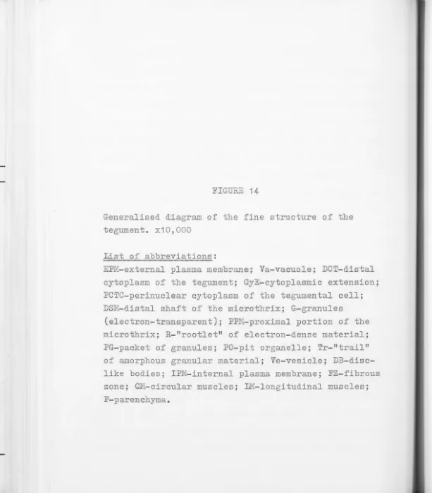
THE FINE STRUCTURE OF THE TEGUMENT OF THE SPARG.ANUM SCOLEX OF SPIROI'1ETRA ERINACEI
38 39
Methods and Materials
Results
(a) The tegument - General (b) The tegument - Organelles Discussion
CHAPTER 5 PROTEOLYTIC ACTIVITY IN THE SPARGANUM OF S:PIROMETRA ERINACEI
In-troduction
Methods and Materials
(a) Riochemical demonstration of proteolytic
-
enzymes by colorimetric assay(b) Histochemical demonstration of
proteolytic enzymes by gelatin-silvex film substrate
Results
(a) Biochemical demonstration of proteolytic enzymes in spargana
(b) Histochemical demonstration of protease
Discussion
1
GENERAL INTRODUCTION
Penetration by a parasite through or into the intestinal wall of its host is a common phenomenon; various examples are seen in all major groups of parasites. In the case of the plerocercoids, or spargana, of pseudophyllidean cestodes (genera
Diphyllobothrium and Spirometra), considerable interest has been aroused because these organisms are extremely paratenic and exhibit low host-specificity; they are able to penetrate through the gut of a wide variety of intermediate hosts (see Chapter I for review of life cycle). The remarkable behaviour of this larval cestode implies that it possesses an efficient mechanism for penetration of the intestinal wall.
Wardle and McLeod (1952) noted of the pseudophyllidean sparganum that "The method by which such a soft-bodied
organism, lacking anything in the nature of a penetrant mechanism and relying mainly upon its own turgidity, can
travel from the alimentary canal of its host to an inter-muscular position, or to a position within the layers of the gut wall, is far from clearly understood." By
however, that some mechanism must exist to account for the sparganum's efficacy in penetration.
2
There would appear to be three main methods by which parasites are able to penetrate the intestine of their hosts. Firstly, by the use of hooks or teeth a parasite may be able to physically tear through the tissues and
thus eventually penetrate the intestine by mechanical destruction of the intestinal wall. Secondly, even with-out hooks or teeth, a parasite with powerful suckers may be able to cause sufficient initial damage to intestinal tissues so as to permit autolysis by intracellular enzymes and enzymes of the intestinal lumen which would facilitate penetration. Thirdly, a parasite may secrete histolytic enzymes for digesting its way through the intestinal wall of the host. These methods may not necessarily be mutually exclusive, and varying combinations of the three may be utilised by different parasites.
In
the case of a sparganum, the absence of hooks orteeth rules out the possibility of the first method. The sparganum is also unlikely to make use of the second
method for penetration since it does not possess strong muscular suckers, but weakly developed bothria. Since
3
the intestinal wall, the second method may be thought of
as even less likely as an adequate mechanism for
penetra-tion. That leaves the third possibility as the most
likely mechanism involved; however, it should be stressed
that once initial damage is done by the parasite, autolytic
effects could play a significant role as well.
Previous authors have suggested that histolytic
enzymes may be involved in the mechanism of sparganum
penetration (Smyth and Reath, 1970). Although, as far as
can be determined, such enzymes have not been previously
described in any Spirometrid sparganum, proteolytic enzymes
have been identified in preparations (plerocercoids or
adults not specified) of Diphyllobothrium latum (Read and
Simmons, 1963). In addition, a study by Fischer and Freeman
(1969), on Proteocephalus ambloplitis plerocercoids,
demon-strated that a large gland in the scolex of the parasite
secreted a substance which apparently aided penetration.
Furthermore, glands have been described in the scolex of
other species of cestodes (see Chapter 3) as well as in
the procercoid of Spirometra erinacei (Li, 1929). This
present study was therefore concerned with an investigation
to determine whether glandular secretions might be the
4
CHAPTER I
THE SPARGANUM
INTRODUCTION
The term "sparganum" is used to describe the
plerocercoid larva of pseudophyllidean cestodes of the
5
genera Diphyllobothrium and Spirometra (Smyth and Heath, 1970).
Infections due to these larval parasites are common and
widespread, and earlier workers described them as a
separate group of cestodes. This gave rise to the use of
the name Sparganum in a generic sense whenever the
specific identity of the adult was not known; this usage
persisted even after it was evident that the term was an
artificial one. More recently, the term has become a
vernacular one.
An astonishing number of "species" of spargana have
been described. This has led to an extremely complicated
problem in classification, yet to be satisfactorily
resolved (see below). The wide geographical distribution
of spargana and the large range of animals which can act
as intermediate hosts for these parasites make them
impoFtant parasites for study. Human infection by
spargana, known as human sparganosis, also has a wide
geographical distribution, although i t is especially
6
This chapter is not a detailed review of literature
since several excellent reviews have already been written
on the subject (Huang and Kirk, 1962; Mueller, 1966;
Smyth and Heath, 1970). This account merely serves as
an outline of sparganosis and as an introduction to the
following study of the sparganum of Spirometra erinacei.
Classification
Diesing 1854, originally proposed that the term
Sparganum be used "as a generic name for any unidentified
pseudophyllidean larva" (Wardle and McLeod, 1952). This
usage is now considered artificial (Smyth and Heath, 1970).
The classification of this group of parasites is,
however, still extremely complicated and unresolved.
Vik (1964) has pointed out that the classification of even
the genera Diphyllobothrium and Spirometra is still
largely uncertain since "the validity of the generic as
well as the specific features is still an unsolved
problem."
In
two recent comprehensive works on cestodes(Wardle and McLeod, 1952; Yamaguti, 1959) the striking
differences in the classification of the family
Diphyllobothriidae by the authors give a good indication
of the difficulties of classification confronting
7
Luhe, 1902. According to Wardle and McLeod (1952), Luhe (1910) changed the name to Diphyllobothriidae when he accepted that the tapeworm he named Dibothriocephalu.s
latus belonged to the same genus as Cobboldts (1858) Diphyllobothrium stemmacephalum; Luhe (1910) thus renamed his tapeworm Diphyllobothrium latum.
Wardle and McLeod (1952) did not accept this change and retained the family name Dibothriocephalidae Luhe, 1902, and named 17 genera within that family. Yamaguti (1959), however, accepted the change and described the family
-Diphyllobothriidae Luhe, 1910, which contained 8 genera. Both Wardle and McLeod (1952) and Yamaguti (1959) accepted the genus Spirometra,which was first proposed by Mueller (1937). That Spirometra and Diphyllobothrium are distinct genera was also accepted by Vik (1964) on morphological and biological grounds.
Wardle and McLeod (1952) named a total of 17 species within the genus Spirometra, including S. erinacei
8
controversial (Wardle and McLeod, 1952).
S. erinacei has been described in a number of
intermediate hosts in Australia (Bearup, 1948, 1953;
Sandars, 1953, 1954; Gordon, Forsyth and Robinson, 1954)
and the life cycle has been studied by Bearup (1953).
Bearup (1948, 1953) used Iwata's ·(1933) description of
s.
erinacei for identification of the species inAustralia, and the same authority has been followed for
the identification of S. erinacei in the present study.
Iwata (1933) synonymised most species described up to
that time withs. erinacei and it would appear that
this was a valid decision.
Life cycle
For a detailed review of the life cycle of
sparganum-type cestodes, see Huang and Kirk (1 962). A brief
outline of the life cycle is as follows:
--Eggs are liberated in large number from the distal
segments of the adult worm as they are passed out in the
faeces of the definitive host. These eggs hatch in water
and a free-swimming ciliated larva, the coracidium, is
released. On ingestion by copepods, the coracidium
9
de~elops into the next stage known as the procercoid.
When the first intermediate host is ingested by a
second intermediate host e.g. a frog, the procercoid
penetrates the gut and develops into a plerocercoid,
usually in an intramuscular site, within this host. The
plerocercoid larva is known as the sparganum; if it is
ingested by a suitable host e.g. a dog, it attaches
itself to the intestinal wall and matures into the
typically segmented adult. If, however, the sparganum
is ingested by another intermediate host it will penetrate
-through. the intestine and reestablish itself as a
plerocercoid in the muscles and other tissue.
In
thisway it can pass from one intermediate host to another as
long as a definitive host does not intervene. The
sparganum can infect a wide range of intermediate hosts
including several species of amphibians, reptiles, birds
and mammals (Huang and Kirk, 1962).
According to ffuang and Kirk (1962) a number of workers
have failed to establish spargana in frogs by feeding
them copepods infected with procercoids, and passage of
procercoids through tadpoles has been assumed by many
to be an obligatory phase in the life cycle. The results
to this view. H.e found that he could not infect pigs
and rats by feeding procercoids, and only one of 12
10
mice became infected following this procedure. Rowever,
spargana readily became reestablished in all these hosts,
as well as frogs, following ingestion of tadpoles
infected by ingesting copepods with procercoids.
On the other hand, Mueller (1938a) has been able
to infect mice with spargana of
s.
mansonoides by feedingprocercoids from infected copepods. Furthermore, Mueller
(1938b) could also infect monkeys withs. mansonoides by
the same route.
Thus i t would appear that there are species differences with regard to the necessity for passage through tadpoles
before spargana are infective to other intermediate hosts.
For a comprehensive review of the natural hosts of
spargana in Australia, see Sandars (1953). Mueller (1966) has given a detailed review of the biology of the North
American species, s. mansonoides.
Sparganosis
Although a number of species have been implicated
in causing human sparganosis (Smyth and Reath, 1970),
in most cases, the actual species involved has not been
1 1
The geographical distribution of human sparganosis is extremely wide. Faust and Russell (1964) listed China, Japan, Formosa, Korea, and Vietnam as countries with high incidence of sparganosis, with a lower incidence in Africa, Australia, Indonesia, Holland, British Guiana, Puertro Rico, Honduras, Uruguay, Colombia and 12 states in the U.S. Huang and Kirk (1962) also listed Italy besides the ones mentioned, and Belding (1965) added
Malaya to the list. Reviews on the pathology of sparganosis have been published by Ruang and Kirk (1962) and Smyth and
CHAPTER 2
IDENTIFICATION OF THE SPARGANUM OF SPIROMETRA ERINACEI
.AND ITS PENETRATION INTO THE INTERMEDIATE HOST
INTRODUCTION
12
The sparganum or plerocercoid of species of the
genus Spirometra, re~resents the terminal larval stage
in the life history of the parasite. If it is ingested
by a host in which it cannot mature into an adult, the sparganum will often penetrate the gut and reestablish
itself in another part of the body. It appears that
provided a definitive host does not intervene, ~argana
may be passaged successively through intermediate hosts
which may follow one another in a particular food chain.
The present chapter reports studies made to
deter-mine the identity of spargana recovered from black snakes
and Queensland cane toads, and to investigate penetration
of the gut of the mouse which was found to act as a
13
METHODS AND MATERIALS
(a) Identification of the sparganum
Spargana were collected from two different hosts: Queensland cane toads, Bufo marinus, from Innisfail,
Queensland, and the black snake, Demansia textilis textilis,
from the Lake George area, N.S.W. It was necessary to establish the species of spargana from cane toads since these animals have not been previously recorded as hosts
of spargana in Australia (see Sandars, 1953). Accordingly, two dogs were fed with spargana to obtain adult worms
for identification purposes.
The first dog was fed 15 spargana collected from Bufo marinus. On the 10th day following infection, eggs began to appear in the faeces of the dog, and these were washed in distilled water and examined under the microscope.
On the 23rd day several pieces of strobila consisting of gravid proglottids were found in the faeces. These were
washed, stained with Fast Red Salt B (Johri and Smyth, 1956), dehydrated in alcohol and mounted in Canada balsam. Two
14
A second dog was fed 2 spargana collected from the
black snake Demansia textilis textilis, and 2 spargana
from Bufo marinus. The dog was killed 28 days later
and immediately dissected. About 2 feet of the small
intestine immediately distal to the pylorus was rapidly
removed, tied at both ends, and injected with warm Bouin1s
fixative. The entire piece of gut was then opened
longitudinally and immersed in Bouin's fixative for 48 hrs.
Four adult cestodes were found attached tro the duodenum,
about 15 cm from the pyloric sphincter and separated from
each other by a few mm. No attempt was made to remove
the scoleces of the worms as they were firmly attached.
Instead, pieces of gut with the attached scoleces were
~xcised, dehydrated and embedded in paraffin in the usual
manner. Sections of the scolex were then cut atr 6,,.«-8/4
and stained with either haematoxylin and eosin or by the
Maximow technique for morphological details.
Pieces of the strobila were also excised and some
were stained with Fast Red Salt B (Johri and Smyth, 1956)
and mounted in balsam. The remainder were embedded in
paraffin in the usual manner and sections were cut at
6.p-8_µ , and stained with haematoxylin and eosin for
15
(b) Penetration experiments
Nearly all the spargana of
s.
erinacei used for theexperiments were obtained from Queensland cane toads,
Bufo marinus, which were collected by the Apex Club of
Innisfail, and sent to Canberra. On two occasions, some
spargana were dissected from the black snake, Demansia
textilis textilis collected from the Lake George area,
N.s
.w
.
The spargana were found to be either free or
encapsulated. The free specimens were found lying either just below the skin in the superficial connective tissues
or entwined among the muscles. The encapsulated specimens
were firmly enclosed in tough yellowish capsules which
varied from 3 mm to 10 mm in diameter (see Fig. 4A). The
free spargana seemed to be much more vigorous and active
than those released mechanically from their capsules.
Initially spargana were fed to laboratory mice (strain
Quakenbush) in different forms, i.e. either intact capsules,
or spargana dissected from capsules, or whole
non-encap-sulated sparganao In later infections approximately 10 mm
of the scolex and anterior end of free spargana were used.
In all cases the mice were forced fed while lightly
.
.-G.
iFIGURE 1
T.S. of scolex of adult Spirometra erinacei in duodenum of dog.
A - Bothria clamped around villi for attachment. (H&E) x50
B -
As
above. (PAS) x50A series of mice were fed one to three spargana each and killed after 10 min, 20 min, 30 min, 40 min,
16
50 min, 1 hr, 24 hr, 48 hr, 72 hr, and 2 mth, respective-ly. They were dissected rapidly after being killed
and the locations of spargana were recorded. These infections were carried out on different occasions due to the unavailability of large numbers of spargana at any one time. Some of the infections were, however, repeated several times. When it was found necessary to hold some spargana until sufficient had been collected for experi-ments, the spargana were washed once in Ringer's solution after removal from the host and maintained in 20 ml of
0
Ringer's in closed Petri dishes at 4
c.
The spargana were never used later than 48 hr after removal from the host.RESULTS
(a) Identification of the sparganum
In the first dog infected, only gravid proglottids and eggs were recovered. The adult(s), presumably
derived from spargana of cane toad origin, were not recovered following dosing.
G.
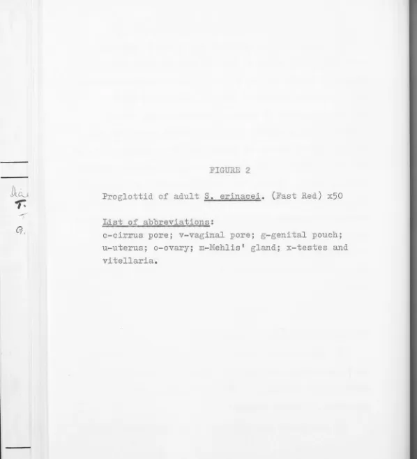
FIGURE 2
Proglottid of adults. erinacei. (Fast Red) x50
List of abbreviations:
[image:25.657.10.608.16.674.2]developed from the
4
spargana fed -- 2 each from thecane toad and black snake respectively. Although in
this case it was impossible to establish the
interme-diate host origin of the respective adults, there did
not appear to be any major differences between them
with regard to their morphology and accordingly, all 17
4 are regarded here as the same species. The morphology
of gravid material recovered from the first dog agreed
with material from second dog.
The following description is based on the material
recovered from the two dogs infected. The colour of
the adult is grayish or milky white. The scolex lacks
suckers and hooks but bears shallow bothria on the
dorsal and ventral surfaces. In cross-section the
scolex has a shape as seen in Fig. 1,A, and is
approxi-mately 0.5 mm wide.
Sections of the adult scolex illustrate the mode of
attachment of adult S. erinacei in the intestine of the
dog (see Fig. 1,A and B). The bothria are firmly clamped
around villi; the tip of the scolex may lie between
villi, but sometimes penetrates into the crypts of
Lieberkuhn (see Fig. 1,C and D). However, no pronounced
18
tissue damage was evident. Although the epithelial cells
of the villi sometimes appear to be firmly compressed
by the bothria, no necrosis, or destruction of cell
membranes was apparent. The tegument of the adult scolex
is AB positive (Fig. 3,B) but PAS negative (Fig. 1,B) which is the converse of the situation in the sparganum
(see Chapter 3).
In
the following description, the terminology ofIwata (1933) is followed.
The total length of the strobila is about 50 cm
long, abou~ 8 mm wide and 1 mm thick. The mature
proglottids are usually near the last quarter of the
stxobila, but this is variable since groups of gravid
proglottids are constantly shed by the worm. The shape
of the proglottids is also variable; the more anterior
ones are usually broader than long, whereas the most
posterior ones are longer than broad (see Fig. 2).
The testes are found in the medullary layer and are
oval or lobed in shape, about 110px 160,>Lin size (Fig. 3,A). The testes (as well as the vitellaria) are absent from
G.
FIGURE
3
A - T.S. of adult S. erinacei proglottid. (H&E) x100 V-vitellaria; T-testes.
B - T.S. of adult scolex. (AB) x100
C - L.S. of sparganum scolexy protruded. (R&E) x30 D - L.S. of sparganu.m scolex, invaginated.
,
,-•
'
"
"'',,''fl I, ,,
.
•
•
I, I,
•
,
,
4
JJ
'
,
.
I
-. '.JI. .
·.:,&
I..•
·,
;.
'
..
•
••\"''.
-
((
.
•
If•
,-~
-
r...
~
•
. ;
,
•
.
•
•
I •
..
'
.
,
I
19
entire axis so that the testes are confined to the
lateral regions of the proglottid. In the longer
posterior proglottids, however, ~he testes are situated
laterally as well as across the anterior end of the
proglottid, meeting in front of the genital pouch. The
cirrus sac is oval in shape and is situated below the
genital pouch. It communicates to the exterior via a
cirrus pore which is situated at the apex of the genital
pouch.
Three ventrally situated pores are present in each
mature proglottid; these are the cirrus pore and vaginal
pore, which are located more anteriorly than the uterine
pore (see Fig. 2). The cirrus pore and vaginal pore are
united to form the genital pouch in the median axis.
In
long proglottids, the genital pouch is situated some
distance anteriorly from the uterine pore.
The uterus lies in the median axis and extends from
the posterior end of the proglottid as far as the anterior
third. It is spiral in form, consisting of about three
to six coils; in the vicinity of the uterine pore it
forms a hemispherical mass. The width of the coils
usually increases posteriorly. The ovary tends to be
it lies ventral to and on either side of the proximal
portion of the uterus. The vitellaria are in the
cortical layer of the proglottid and each follicle is
oval in shape. The vitellaria sometimes obscure the
ovary in long proglottids. The eggs measure about
57 ,#'-to 66_.P x 32,µ to 36~. They are oval in shape but
tend to be slightly assymetrical with one side rounder
than the other; both ends are slightly pointed. An
operculum is present at the sharper end of the egg.
20
The above description agrees substantially with
that given by Iwata (1933) for D. erinacei. Significant
and consistent features which characterise this species
are egg size, egg shape, structure of the scolex with
bothria on dorsal and ventral sides, reproductive organs
one pair in each proglottid, uterus forming spiral coils,
and the uterine pore and cirrus pore opening on the
ventral side in the median axis. D. erinacei, however,
has been transferred to the genus Spirometra (Wardle and
McLeod, 1952) and this change in generic status has
been accepted by later workers (Yamaguti, 1959).
Thus, the spargana from Bufo marinus, the Queensland
cane toad from Innisfail, Queensland, and Demansia textilis
G
FIGURE
4
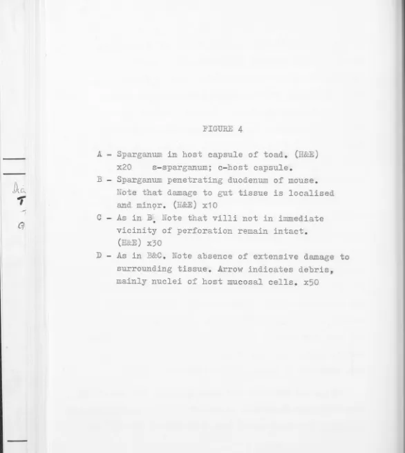
A - Sparganum in host capsule of toad. (H&E) x20 s-sparganum; c-host capsule.
B - Sparganum penetrating duodenum of mouse.
Note that damage to gut tissue is localised and min9r.
(H&E)
x10C - As in B • Note that villi not in immediate vicinity of perforation remain intact.
(H
&E
)
x3021
be confidently referred to as S. erinacei, as characterised
by Iwata (1933).
s.
erinacei spargana have been recoveredfrom a variety of Australian host species previously
(Bearup, 1948, 1953; Sandars, 1953; Gordon, Forsyth
and Robinson, 1954).
(b) Penetration experiments
Mice fed with intact capsules were dissected 24 hr
later but no spargana were found.
Both free spa~gana and those liberated mechanically
from the capsules readily penetrated the gut of the mouse
after oral administration. The point of penetration
was always in the duodenum, about 2-4 cm below the
pyloric sphincter.
In
one mouse, 3 spargana penetratedthe duodenum within 1 mm of each other at different
points around the intestinal wall. As shown in Fig.
5,
penetration of the gut commences approximately 30 min
after ingestion. This time lapse between ingestion and
penetration was reasonably consistent on the 7 occasions
this experiment was carried out. The actual process of
gut penetration was observed to take only 5-10 min on
most occasions, if no great disarrangement of the gut
22
freed from remaining viscera to facilitate observation, penetration was delayed by up to 15 min. Only the
scolex (2-5 mm of the anterior portion of the sparganum) emerged into the abdominal cavity from the gut. The rest of the strobila became detached posterior to the scolex and remained in the intestinal lumen. Fig. 4,B, shows a section through the penetrating sparganum. The size of the actual perforation caused by the penetrating sparganum is about 0.5 mm in diameter and constitutes a direct connection between the gut lumen and the coelom. In spite of the relatively large size of the perforation, however, damage to gut tissues appeared minor and
localised, suggesting that the mechanism of penetration was direct and efficient (see Fig. 4,C and D). It is
evident that the scolex breaks through the peritoneal lining of the gut into the abdominal cavity and does not remain attached to the gut just below the peritoneal lining.
Ten minutes after feeding, one sparganum was found in the stomach with the strobila still attached to the scolex. After 20 min a sparganum was found in the
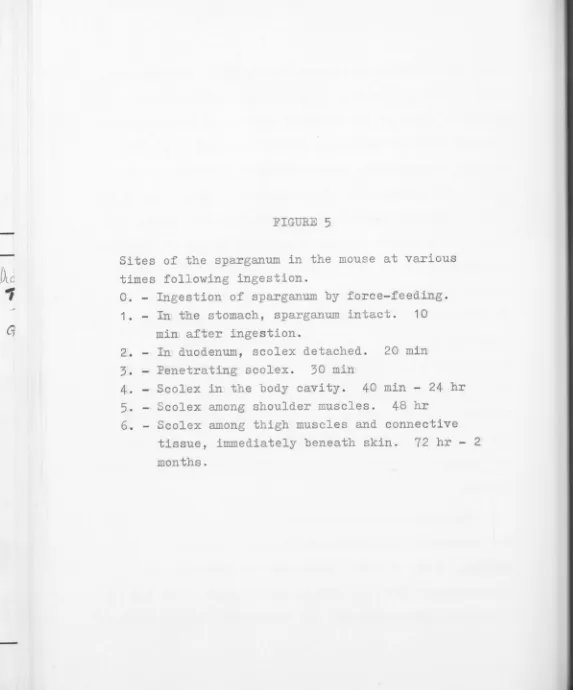
[image:36.646.58.631.24.709.2]FIGURE 5
Sites of the sparganum in the mouse at various times following ingestion.
0. - Ingestion of sparga.num by force-feeding.
1. - In the stomach, sparganum intact. 10 min after ingestion.
2. In duodenum, scolex detached. 20 min 3. Penetrating scolex. 30 min
4
.
Scolex in the body cavity. 40 min-
24 hr5
.
- Scolex among shoulder muscles. 48 hr6. - Scolex among thigh muscles and connective tissue, immediately beneath skin. 72 hr
-months.
0
/
If
/
r
.
,.-,
...
\
(,
.
~
:\
,,, . . ··,
6 1
2
3
Hence, it appears the bulk of the strobila becomes
detached before penetration.
23
Between 40 min and 24 hr the sparganum remains in
the viscera, moving between the organs in the abdominal
cavity and over the mesenteriaes. In one mouse two
spargana were found among the shoulder muscles 48 hr
after ingestion. In another, 72 hr after ingestion,
one sparganum was found among the thigh muscles of the
hind leg and another, lying below the skin in the
abdominal region. _In the mouse dissected after two
months, the two spargana found averaged 8-10 cm long,
and were located beneath the skin in the abdominal and
thoracic regions respectively.
In all infected mice examined between 40 min and
24 hr, there were no occasions on which the spargana
were found either within any visceral organs or
penetra-ting through mesenteries.
All infected mice examined appeared healthy prior
to dissection and there was no evidence of peritonitis
which might be considered possible as a result of the ,r
fairly large perforation made by the sparganum in the
gut wall. No inflammation at or around the site of
24
significant host reaction to spargana in tissue sites. However, the latter point was not checked histologically.
DISCUSSION
The identification of the spargana from Bufo marinus (Innisfail, Qld.) as belonging to the species Spirometra e.rinacei (Rudolphi, 1819) has established a new host for this parasite. Sandars (1953) has already noted
spargana of this species in Demansia textilis in Australia. From the results of penetration experiments described above, the sparganum of
s.
erinacei was shown to complete penetration of the duodenum of a mouse within 40 minafter ingestion. The actual time taken for penetration was, however, much less than this since 10 min after ingestion the sparganum was still present in the stomach, and even after 20 min penetration had not commenced. It was, in fact, observed that penetration commenced only after 30 min and the entire process of gut penetration took only 5 to 10 min to complete. The times recorded for the penetration of mouse intestine by S. erinacei
25
1 to 1! hr after ingestion and penetration itself took 20 to 30 min to complete. The results of Takahashi
(1959a), which showed that the sparganum of D. mansoni (= s. erinacei ?) penetrated the mouse gut within 40 min after ingestion, is however consistent with the observa-tions of the present study.
Any significant disarrangement of the gut during penetration was seen to delay penetration bys. erinacei spargana by up to 15 min. This occurred in cases when the duodenal loop WJiS pulled away from the rest of the
viscera to facilitate observation. When this was done before penetration had begun, the sparganum failed to penetrate the gut or even initiate penetration. Similar-ly, a sparganum failed to penetrate when a section of the duodenum with an enclosed sparganum was removed, tied
0
at both ends and immersed in Ringer's solution at 37 C. These results were not surprising, since a great deal of physiological damage was probably done to the gut as a result of such drastic treatment, which probably inter-fered with sparganum penetration.
sparganum is about 0.5 mm in diameter and is thus a
relatively large opening. Contents of the intestinal
lumen (including bacteria and other organisms) would
presumably be released into the peritoneal cavity as
the sparganum penetrates. These views receive support
from studies by Feng and Hoeppli (1936) with spargana 26
0£ S. erinacei in Chinese hamsters. In this host, the
spargana perforated the gut wall and thus establish a
connection between the intestinal lumen and peritoneal
cavity. Feng and Hoeppli found that bacteria and other
organisms were released into the peritoneal cavity
through the perforation. Moreover, they established
beyond doubt that the absence of fatal peritonitis was
due to bacteriacidal properties possessed by the hamster.
They repeated experiments earlier made by Joyeux and
Baer (1929) and found no evidence that the spargana
possessed bacteriacidal properties. However, Joyeux and
Baer (1929) may have been dealing with a different
species. It is not unlikely that mice may possess a
similar mechanism to hamsters in preventing fatal
peritonitis due to penetrating spargana, since a mouse
infected with two spargana was still in an apparently
27
In
contrast to the spargana of S. erinacei, the spargana of S. mansonoides, after penetrating through the mucosa of the mouse intestine, do not pass directly into the body cavity but migrated tangentially beneath the serosa and finally escaped where the mesenteryjoins the gut (Mueller, 1938b). Sometimes the sparganum of
s
.
mansonoides travelled upwards between the twocoats of the mesentery and reach the muscles via that route. Thus Mueller (1938b) explained that no infection can occur in the mouse since "no direct communication is formed between the cavity of the intestine and the outside and hence the infective contents of the gut do not escape into the coelom11o
Another feature of the penetrating sparganum of
s.
erinacei noted in this study was that only about 2-5 mm of the scolex reached the peritoneal cavity. The major part of the strobila was discarded before penetration and remained in the duodenum of the mouse. It appears possible that one of the factors which activate thesparganum to detach its scolex from the rest of the larval
0
body is an increase in temperature ~o 37 C. When a sparganum kept in Ringer's solution at room temperature
0
of the sparganum is significantly increased and within
15 to 25 min the scolex separates from the remainder
of the strobila. This time interval coincides with
that in which an ingested sparganum is found in the
duodenum of the host with the scolex detached from
the strobila.
CHAPTER
3
THE MORPHOLOGY AND HISTOCHEMISTRY OF THE
SPARGANUM SCOLEX OF SPIROMETRA ERINACEI
INTRODUCTION
The sparganum of
s
.
erinacei is able to rapidlypenetrate the gut of an intermediate host following ingestion and become established as a tissue parasite
29
(see Chapter 2). A number of studies (Li, 1929; Mueller, 1938a, 1938b; Bearu~, 1953; Sandars, 1953; Takahashi, 1959, of the spargana of S. erinacei and related species have
been made but the mechanism of penetration has not yet
been established. The remarkable migration of the
sparganum is accomplished despite the fact that it lacks
any striking modifications of the scolex which migh~ facilitate mechanical penetration through the relatively
thick muscular and collagenous layers of the host
intestine. Thus, it would seem that any mechanical effects
exerted by the scolex might have to be augmented by some
other mechanism to effect penetration.
Smyth and Heath (1970) have suggested that "cephalic
30
of penetration. However, evidence for the existence of such glands in Spirometrid pseudophyllidean cestodes is lacking. Glands have been described in the plerocercoid scolex of several other species of pseudophyllidean
cestodes, including Diphyllobothrium latum, D. vogeli, D. osmeri, D. dendriticum (Kuhlow, 1953) D. dalliae,
D. norvegicum
(Vik,
1964), as well as in the adult scolex of Dibothriocephalus wilsoni, Adenocephalus pacificus and Glandicephalus antarticus (Wardle and McLeod, 1952), and Abothrium gadi (Williams, 1960). "Histolytic glands"
-have also been described in the procercoid of Spirometra erinacei by Li (1929).
A s~udy of the morphology and histochemistry of the scolex of the sparganum of S. erinacei, at the light microscope level, was therefore undertaken initially to
establish whether recognisable gland cells are present in this region which might be linked with penetration. This chapter describes the results of this investigation.
METHODS AND MATERIALS
(a) Morphological study of the sparganum scolex
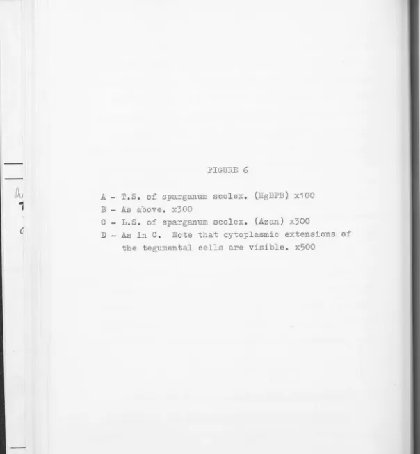
FIGURE 6
A - T.S. of sparganum scolex. (HgBPB) x100 B - As above. x300
C - L.S. of sparganum scolex. (Azan) x300
[image:47.657.5.609.16.669.2]A
,
• J ,,, ~ .. '
.•
.
),
l'
' _,
•
•
~
..
-.;
·
...r
31
(B.D.H. tablets) and studied under a low power binocular
microscope.
For the study of living worms at higher
magnifica-tions, about 2 cm of the anterior portion of the
sparganum was cut off and flattened with gentle pressure
between a slide and cover slip. Care was taken to
prevent preparations from drying out during examination.
Neutral Red (1% aqueous) was used on occasions as a vital stain.
-(b) Histological study of the sparganum scolex
The spargana used for histological studies were
either collected directly from the natural host (e.g.
B. marinus), or were first fed to laboratory mice
(Quakenbush strain) and then collected after penetration
of the gut (see Chapter 2).
The spargana were washed in Ringer's and
3
-
5
mm ofthe scolex was cut off. They were then fixed in Bouints
and Carney's fixatives overnight, dehydrated in alcohol,
and embedded in paraffin in the usual manner. Sections
were cut at 6p-- 8,?-and stained with haematoxylin and
eosin. Some sections were stained with Heidenhain's
•
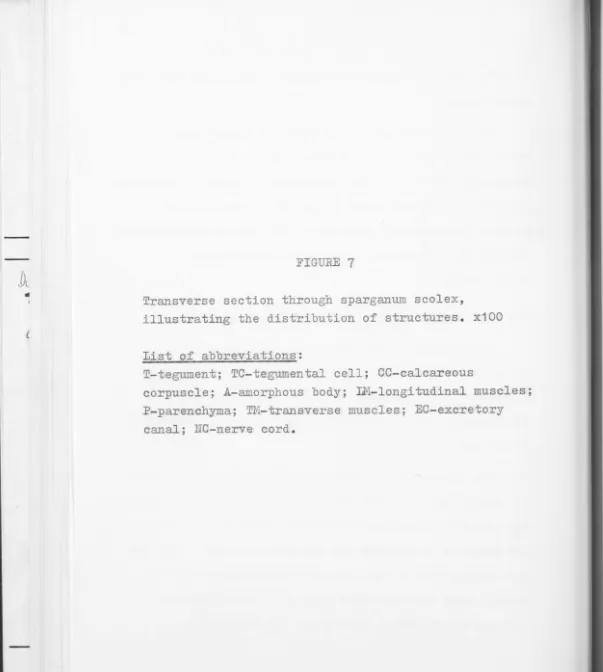
FIGURE
7Transverse section through sparganum scolex,
illustrating the distribution of structures. x100
List of abbreviations:
T-tegument; TC-tegumental cell; CC-calcareous
corpuscle; A-amorphous body; LM-longitudinal muscles; P-parenchyma; TM-transverse muscles; EC-excretory
~---TC
~~~*+----~cc
~ 7 ~ 1 - - - A
~':-:tf'---LM
~~---P
TM
..-7~~---A
EC
32
(c) Histochemical study of the sparganum scolex
The spargana used we~e as above, either collected
directly from the natural hosts or recovered from
laboratory mice (Quakenbush strain) soon after
experi-mental infection.
Material was fixed in either Bouin's fixative,
0
70% alcohol, 4% formal-saline at 4 C, or Carney's
fixative. For enzyme tests, the scoleces of spargana
were fixed in cold formal-saline, embedded in 10%
gelatine, frozen on ~o the chuck of a International
model CTD cryostat and sections cut at 20?. For other
histochemical tests, the variously fixed scoleces were
embedded in paraffin and sections cut at 6_µ- 8_,µ .
The following histochemical tests were carried out
using procedures given by Pearse (1 961)
:-( i ) for carbohydrates, the periodic acid Schiff (PAS),
alcian blue (AB) and toluidine blue metachromasia
tests;
( ii) for lipids, the Sudan black B test;
(iii) for proteins, the mercury-bromo-phenol blue (HgBPB)
test;
( iv) for RNA and DNA, the methyl green pyronin (MGP) test;
•
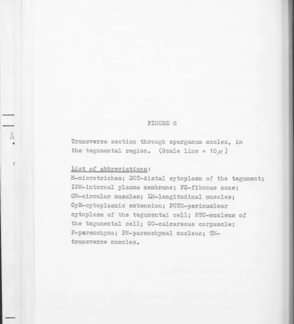
FIGURE 8
Transverse section through sparganum scolex, in the tegumental region. (Scale line= 10f1 )
List of abbreviations:
M-microtriches; DCT-distal cytoplasm of the tegument; IPM-internal plasma membrane; FZ-fibrous zone;
CM-circular muscles; LN-longitudinal muscles; CyE-cytoplasmic extension; PCTC-perinuclear
[image:53.646.6.598.18.671.2]M
(
.. .
. ·, .
. , . ' ..
,• .
phosphatase, the Nachlas, Crawford and Seligman method for leucine amino-peptidase, the Holt and
Withers method for esterase.
In
addition, the acridine orange method for RNA33
and DNA as detailed by ,Culling( 1963), and the Karnovsky
and Roots (1964) techniques for esterases were carried
out.
Appropriate controls were used for all histochemical
tests. These included, for the FAS test, treatment
with diastase (Sigma) to remove glycogen; for the MGP
and acridine orange method, the examination of sections
treated with 1% RNA-ase in P0
4
buffer, pH7.3
(Sigma)before application of the test; for the enzyme vests,
heat treatment and incubations in media lacking substrates were carried out. Where appropriate, mammalian tissues
known to contain specific substances were processed
simultaneously.
RESULTS
(a) The sparganum scolex
Spargana from D. textilis textilis measure up to
1 cm wide and 45 cm long, and are generally much larger
FIGURE
9Transverse section through sparganum scolex, in medullary region. (Scale line= 10.f"')
List of abbreviations:
. .
.
...
, - - - ~ PN
' . · . .... ·. · .. ·.
:·· ..
,":.
~
:··:•
.
· .· .· ...·
.
TM
LM
. . . : .
. . . . A
. ;_. ~ · ..
. ~
--
.. '...
...
·~. . :'. .. · .. :.,..
. ...
...
. . . . . . . . . . ~ . . .·· ..
.
.... . · .
34
· 8-10 cm long) .
In live specimens, the scolex of the sparganum is
extremely vigorous and muscular, and closely resembles
that of the adult. Ovoid calcareous corpuscles appear
as transparent, ovoid, lamellated granules scattered
randomly throughout the scolex, apart from the tip. No
gland cells could be detected within the scolex.
In
the presence of neutral red, the scolex granuallybecame uniformly stained; again, no gland cells were
delineated. No secretion of material from within the
scolex to the exterior was detected.
(b) Histology of the sparganum scolex
See Figs. 7,8 and 9
The external surface of the sparganum scolex is covered
by microtriches 2.3.J,<long. These microtriches arise from
the tegument which is about 10)..lthick. Under the light
microscope the tegument appears fairly homogenous and
clear (Fig. 6,D) . Immediately below the tegument are
the tegumental cells which appear as a single layer of
closely packed cells with darkly staining nuclei. Their
cytoplasmic connections with the distal cytoplasm are
just faintly discernible under the light microscope
35
Below the tegument and comprising most of the scolex
is the parenchyma. The cells of the parenchyma are
irregular in shape and size and cell membranes are not
distinct; the parenchyma thus appears syncytial with
nuclei randomly scattered within it. Calcareous
corpuscles about 1~µ -15;,t in diameter, appear irregularly throughout the parenchyma (see Fig. 6,A,B and C)o
A conspicuous ring of longitudinal muscles lies
about within the parenchyma (see Fig. 6,A and B).
En
-closed by these muscles and distributed randomly, are
irregularly shaped masses of material, referred to here
as amorphous bodies. These range in size from 10,M-20.,.u . The amorphous bodies appear to be extracellular deposits
since nuclei could not be detected within them.
Some-times these bodies are not distinctly demarcated and
they merge gradually with the parenchyma. They are com-posed of masses of granules which stain a reddish-orange
with Heidenhain's Azan technique.
Two lateral excretory canals, about 10)-'in diameter
each, may be observed in cross-sections of the middle and posterior parts of the scolex. In the more anterior
regions the excretory canals anastomose. Conspicuous nerve
1
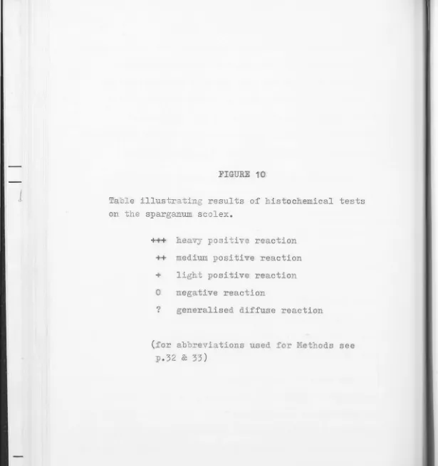
FIGURE 1;0
Table illustrating results of histochemical tests on the sparganum scolex.
+++ heavy positive reaction
++ medium positive reaction
+ light positive reaction
0 negative reaction
? generalised diffuse reaction
[image:60.657.0.615.15.670.2]Method
PAS
Glycogen
AB
Toluidine Blue
..
Sudan Black B
HgBPB MGP
Acridine Orange
Esterase
LAP
Acid Phosphatase
Alk. Phosphatase
* Metachromasia
D=distal tegument
T=tegumental cells
P=paranchyma M=muscles D +++ 0 0 0 +++ ++ 0 0 - ? 0 0 +++
Sites in the scolex
T p M E
+ + ++ 0
0 ++ ++ 0
0 0 0 0
0 0 0 0
0 0 + +
+ 0 +++ ++
+++ + 0 0
+++ + 0 0
? ? ? ?
0 0 0 0
0 0 0 0
++ 0 0 0
E=excretory canals
N=nerves N 0 0 0 0 0 + 0 0 ? 0 0 0
C=calcareous corpuscles
A=amorphous bodies
C A
++ +++
0 0
0 0
0 0
++ +
++ +++
0 0
0 0
? ?
0 0
0 0
36
No gland cells, which might be involved in secreting material to the exterior, could be detected in the scolex
(see Fig. 3,C and D).
(c) Histochemical study of the sparganum scolex
Fig. 10 presents the results of the histochemical tests on the sparganum scolex of
s
.
erinacei. No differ-ences could be detected between scoleces of sparganataken from infected cane toads and those which had recent-ly penetrated the gut of mice, and the following remarks apply equally to spargana recovered from both sources.
(i) Distribution of non-enzymatic substances.
The Sudan black B method for lipids gave a strong reaction in the tegument. Small amounts of lipid were
present in the muscles, excretory canals, and the amorphous bodies mentioned earlier. Slightly larger amounts were
associated with the calcareous corpuscles (see Fig. 12,A and
B)
.
For carbohydrates, the PAS reaction was strongly positive in the tegument (Fig. 11 ,A) and smaller amounts of carbohydrates were evident in the muscles, amorphous bodies and calcareous corpuscles. A very light and diffuse reaction was also observed in the parenchyma and
J
FIGURE 11
A - Test for carbohydrates (PAS). Note strong reaction in distal tegument. x500
B - Test for proteins (HgBPB) . Strongest reaction in amorphous bodies and tegument. x50
C - Test for nucleic acids (MGP). Strong reaction for fu~A in perinuclear cytoplasm of tegumental cells. x50
D - Control section; after action by RNAase (MGP).
C
,
..
'
'
:. ... -.• ·-~ • .,..:,.Y,
37
was clearly due to the presence of glycogen since it was abolished following diastase treatment. PAS reactions in other locations however persisted after
diastase and thus showed that other types of polysaccharide were present in the sparganum scolex. (see Fig. 12,C and D). The AB test was negative and there was no evidence of metachromasia with the toluidine blue test, thus
showing that acid mucopolysaccharides were absent. Thus, the mucopolysaccharides were neutral mucopolysaccharides.
The HgBPB test ~tained almost the whole section of the scolex except for negligible amounts in the parenchyma. The muscles and amorphous bodies gave especially strong reactions indicating higher concentrations of protein in these regions (Fig. 6,A and B; 11,B).
Tests for DNA and RNA were carried out using the MGP method and the acridine orange techniques. DNA was demon-strated in the nuclei in the scolex. RNA was seen to be almost totally concentrated in the cytoplasm of the
tegumental cells with only small scattered amounts in the parenchymal cells. Ribonuclease destroyed RNA activity
(Fig. 11 ,C and D). It was noted, with the acridine orange technique, that the normal green fluorescence seen in
J
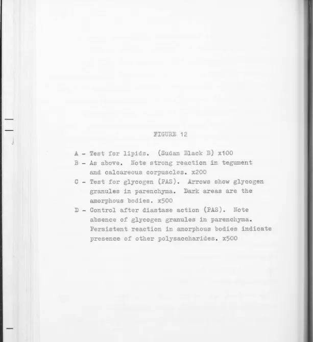
FIGURE 12
A - Test for lipids. (Sudan Black B) x100
B - As above. Note strong reaction in tegu.ment
and calcareous corpuscles. x200
C - Test for glycogen (PAS). Arrows show glycogen
granules in parenchyma. Dark areas are the
amorphous bodies. x500
D - Control after diastase action (PAS). Note
absence of glycogen granules in parenchyma.
Persistent reaction in amorphous bodies indicate
[image:66.666.4.617.14.685.2]38
of RNA. The green fluorescence became visible, however,
following ribonuclease treatment.
(ii) Distribution of enzymatic substances.
The tegument and the tegumental cells of the sparganum
scolex showed a very distinct reaction for alkaline
phosphatase (see Fig. 13,A and B) . There was no apparent
reaction for the enzyme in any other regions. False
positive reactions (i.e. persistence of reaction product
in controls) were seen in the calcareous corpuscles and
these were probably due to the presence of insoluble
inorganic phosphate known to occur in these structures
(von Brand, 1966).
Acid phosphatase was absent in the scolex of the
sparganum (Fig. 13,C and D). Again, a false positive
reaction was given by the calcareous corposcles. The
test for leucine amino-peptidase was negative in the
sparganum scolex. The limited number of tests for
esterase which were carried out gave inconclusive results.
A weak and diffuse reaction was given throughout entire
sections of the scolex, and no localised concentrations
FIGURE
13
A - Test for alkaline phosphatase (Gomori). Strong
reaction integument; reaction in calcareous corpuscles is a false positive probably due to
reaction with inorganic phosphate deposits. x100
B - Control for above; note persistence of reaction
in calcareous corpuscles. x200
C - Acid phosphatase test (Gomori) . Negative
except again for false positive in calcareous
corpuscles. x100
D - Control for C; false positive again persistent.
39
DISCUSSION
From the results in this chapter, the structure of
the sparganum scolex appears to be relatively simple.
Apart from the muscles, nerves and excretory canals, the
scolex is filled with parenchymatous tissue. Calcareous
corpuscles and other deposits in the form of amorphous
bodies are also present in the parenchyma but no special
-ised gland cells were detected. No secretion from the
scolex was detected in living spargana.
The rationale for this examination of the scolex of
the sparganum of S. erinacei for glands, as mentioned in
the introduction, was to find if glands and glandular
secretions were present which might facilitate the process
of penetration by the sparganum through the gut wall of
the intermediate host. That glands might be involved was
suggested by Smyth and Heath (1970) who stated that
"Presumably, the cephalic glands present in the plerocercoid
are histolytic in nature, although their nature does not
appear t o have been investigated." However, as far as
can be determined, glands have not been previously
described in the plerocercoid of S. erinacei or any other
species of Spirometra. The "cephalic glands" mentioned
40
glands of the plerocercoids of Diphyllobothrium osmeri,
D. l atum, D. vogeli and D. dendriticum described by
Kuhlow
(
1
953).
These glands predominate in the medullary layer and
vary in distribution and number in the different species
he described. Furthermore, Kuhlow
(1953)
noted that theglands had a crystalline content and discharge to the
exterior by means of tortuous canals. The function of
these glands, Kuhlow suggested, was to enable the
plerocercoid to diges; host intestinal tissue during
penetration of the fish host. He cited as evidence that
they were involved in this process the fact that adult
individuals of D. osmeri, D. vogeli, D. dendriticum and
D. latum do not possess the glands; if they were involved
only in penetration they would presumably be unnecessary
in the adult.
From this study of the anatomy of the scolex of the
S. erinacei sparganum, the only structures which resemble
the glands described by Kuhlow
(
1953
)
are the amorphousbodies described earlier. These bodies appear in cross
-sections to be roughly in the same position in the
plerocercoid scolex and to also have the same irregular
41
is especially true in the case of D. latum in Fig.
9
,
p. 201 . It was at first thought that these bodies in the sparganum scolex of S. erinacei were in fact glands
analogous to those described by Kuhlow (1953) . Results of studies described in this chapter, however, suggest strongly that they are not glands but rather extracellular
deposits of organic material.
In the first place, the bodies do not have a discrete
cellular structure and no nuclei have been seen within
them. The bodies appear amorphous and irregular and frequently blend into the surrounding parenchyma. On staining with the Heidenhain Azan technique, these bodies
appear reddish- orange in colour. The glands described by Kuhlow appeared bright blue in colour when stained by
the same technique. Secondly, no canals, or connections of any kind, could be detected which connected these bodies to the exterior. Thirdly, these bodies also appear further down the length of the sparganum and are
not limited to the scolex. Fourthly, no reduction in size, or change in their histochemical properties, could be
detected in these bodies after the sparganum had penetrated
42
involved in protein synthesis. Sixthly, from the results of Chapter
5
,
it is clear that although proteolyticenzyme(s) could be demonstrated around the tegument of the scolex, no proteolytic activity was found within the rnedullary region of the scolex where the amorphous bodies
occur.
The function of these amorphous bodies is not known.
They contain substantial amounts of neutral rnucopolysaccharide
and protein but relatively little lipid. One possible
explanation is that they are deposits of excretory produc~s
possibly inactivated by association with protein. The sparganum lives within the tissue of the host and is there -fore in direct and intimate contact with the hosto The
parasite might be expected, therefore, to minimise the
amount of antigenic material being released into the host
tissue so as not to provoke a strong host reaction against
it. From the results of the penetration experiments in Chapter 2, it was evident that the sparganum can remain in the mouse host up to two months without causing any
43
are established in an "external" environment such as the
intestine, the amorphous bodies are no l onger present in
the scolex.
Thus it seems certain that glands similar to those
described by Kuhlow (1953) are not present in the sparganum
of S. erinacei. Takahashi (1959b) in a fairly detailed
study of the histochemistry of the plerocercoid of
s
.
mansoni also failed to mention glands in the scolex ordeposits comparable to those described here. Li (1929),
in a study of the life history of S. erinacei, described
"histolytic glands" in the procercoid stage but did not
describe any glands or amorphous bodies in the plerocercoid
stage.
The absence of glands visible with the light microscope
could mean that there are no histolytic glands of any kind
present in the sparganum, thus implying that the mechanism
of penetration is by some means other than with glandular
enzymes. It could also mean that enzyme-producing cells
may be structurally organised in such a way that they are
not visibly apparent as glands under the light microscope.
The results in the following chapters indicate that the
latter case is more likely, since proteolytic enzyme(s)
44
At this stage it is uncertain why distinct glands
are present in the procercoid but not in the plerocercoid
of S. erinacei. It is certain, however, that there is great variability among species of Pseudophyllidea since
in D. osmeri, D. vogeli, D. dendriticum and D. latum, glands are present in the plerocercoid but not in the
adults (Kuhlow, 1953), whereas glands are present in the
adults of Dibothriocephalus wilsoni, Adenocephalus pacificus,
Glandicephalus antarticus (Wardle and McLeod, 1952) and
Abothrium gadi (WilliclJils, 1960).
Takahashi (1959b) detected both acid and alkaline
phosphatase in the "cuticle and subcuticle cells" of
s.
mansoni spargana. In this study of S. erinacei spargana,only alkaline phosphatase was detected. All the other
histochemical results obtained for
s.
mansoni by Takahashi(1959b) agree substant ially with those for S. erinacei,
however.
Findings described by Saraki (1961 ) for the
distribu-tion of DNA and RNA in the plerocercoids of S. mansoni also agree with those for S. erinacei spargana as found
in this study.
and the PAS positive material in the distal tegument,
may be related to the proteolytic enzyme(s) found in
the same region (see Chapter
5).
Further discussionon the importance of this will be described later in
Chapter 6.