by
BRIAN RONALD CREESE
A t h e s i s s u b m i t t e d f o r t h e d e g r e e o f Doctor of Ph il o so ph y o f t h e A u s t r a l i a n N a t i o n a l U n i v e r s i t y
The investigations described in this thesis are my own original work.
Brian Creese
A C K N O W L E D G E M E N T S
I am greatly indebted to my supervisor, Dr. Michael Denborough, for his help and encouragement over the last three years. For his many helpful suggestions and criticisms I would also like to thank Dr.
Howard Mitchell. I am grateful to my fellow students, John Sullivan and Joe Jakubowski, for their help and friendship, and to my wife Helen, for her encouragement and assistance in preparing the thesis. Thanks are also due to Mrs. Margaret Bacon for typing this thesis.
A B S T R A C T
1. Regulation of airways smooth muscle contraction and relaxation was studied in vitro by using a strip preparation of guinea- pig trachea. Changes in the cyclic AMP content of the guinea- pig tracheal muscle were also determined.
2. Drug-induced contractions of the tracheal muscle were diminished, but not abolished, in the absence of extracellular calcium ions, indicating that contractile responses involve both the influx of Ca2+ from the extracellular space and release of Ca2+ from
o + intracellular sites. The relative contributions of these two Ca sources depended on both the contractile agent and its concentra tion. Repeated additions of histamine or acetylcholine in the absence of extracellular Ca resulted in progressively smaller
2 *4" contractile responses, reflecting depletion of intracellular Ca stores.
3. Tracheal muscle depolarized with high concentrations of KC1 still contracted in response to histamine and acetylcholine, indicating the involvement of pharmacomechanical coupling mechanisms. In calcium-depleted tissues, KC1 potentiated contractile responses to histamine and prostaglandin F2CU
4. Calcium metabolism of the tracheal muscle was also investigated using the calcium ionophore A23187, which contracted the muscle,
and the Ca2+ antagonist TMB-8, which caused relaxation and inhibited drug-induced contractures.
5. The basal level of cyclic AMP in the tracheal muscle was 2.7 pmoles/mg protein. Cyclic AMP levels increased in response to isoprenaline and prostaglandin Ei supporting the concept of
a bronchoconstrictor agent, also caused a dose-dependent increase of cyclic AMP levels. 100 yM histamine caused a 3-fold increase of cyclic AMP content after 2 minutes. The increase was not affected by propranolol or atropine, but partially inhibited by mepyramine.
6. Imidazole (0.1 to 30 mM) contracted the tracheal muscle, and this response was partially inhibited by atropine and mepyramine. Imidazole did not affect the cyclic AMP content of the muscle, indicating that its contractile action is unrelated to stimulation of cyclic nucleotide phosphodiesterase.
7. Low concentrations of imidazole (0.05 to 2 mM) potentiated contractile responses to histamine, but not to other agents. The histamine concentration-response curve was shifted to the left in the presence of imidazole, but the maximum response was not affected. Imidazole (0.5 mM) did not affect the accumulation
of cyclic AMP induced by histamine, prostaglandin Ei or isopre- naline. Imidazole-induced hyperreactivity was not affected by atropine or indomethacin, and was more pronounced in the absence of extracellular calcium ions. The results suggest that-the potentiating effect of imidazole on histamine contractions is unrelated to activation of phosphodiesterase, enhancement of acetylcholine release or inhibition of prostaglandin or throm boxane synthesis. The phenomenon may involve enhanced release of Ca2+ from intracellular sites, or, more likely, inhibition of the enzyme histaminase.
8. Indomethacin relaxed the guinea-pig tracheal muscle and also potentiated contractile responses to histamine and acetylcholine.
9. The results suggest that the contractile response of guinea-pig tracheal smooth muscle to histamine is modulated by a negative feedback regulatory mechanism involving the release of prosta glandins and elevation of cyclic AMP levels.
CONTENTS
P a g e
STATEMENT
ACKNOWLEDGEMENTS ABSTRACT
LIST OF FIGURES LIST OF TABLES ABBREVIATIONS
i i i i v x x i x i i CHAPTER 1 - INTRODUCTION
1 . 1 B r o n c h i a l h y p e r r e a c t i v i t y i n a s t h m a 1 . 2 I m m u n o l o g i c a l m e c h a n i s m s i n a s t h m a
1 . 2 . 1 M e c h a n i s m o f m e d i a t o r r e l e a s e
1 . 2 . 2 The m e d i a t o r s o f a l l e r g i c r e a c t i o n s 1 . 2 . 2 . 1 H i s t a m i n e
1 . 2 . 2 . 2 S l o w r e a c t i n g s u b s t a n c e o f a n a p h y l a x i s
1 . 2 . 2 . 3 O t h e r m e d i a t o r s
1 . 2 . 3 R e g u l a t i o n o f m e d i a t o r r e l e a s e 1 . 2 . 4 M e d i a t o r r e l e a s e i n a s t h m a
8 10 1 . 3 A d r e n e r g i c m e c h a n i s m s a n d a s t h m a
1 . 3 . 1 A d r e n e r g i c r e g u l a t i o n o f a i r w a y s s m o o t h m u s c l e
1 . 3 . 2 B e t a - a d r e n e r g i c b l o c k a d e i n a s t h m a 1 . 3 . 3 A l p h a - a d r e n e r g i c r e c e p t o r s i n a s t h m a 1 . 4 C h o l i n e r g i c r e g u l a t i o n o f a i r w a y s s m o o t h m u s c l e
1 . 4 . 1 Nor ma l a i r w a y s
1 . 4 . 2 C h o l i n e r g i c m e c h a n i s m s i n a s t h m a
13 13 17 20 22 23 1 . 5 P r o s t a g l a n d i n s
1 . 5 . 1 P r o s t a g l a n d i n r e l e a s e i n a l l e r g i c r e a c t i o n s 27 1 . 5 . 2 E f f e c t s o f p r o s t a g l a n d i n s on a i r w a y s s m o o t h 29
m u s c l e
1 . 5 . 3 The r o l e o f p r o s t a g l a n d i n s i n a s t h m a 34 1 . 5 . 4 A s p i r i n - s e n s i t i v e a s t h m a 37 1 . 6 C a l c i u m m e t a b o l i s m o f s m o o t h m u s c l e 39 1 . 6 . 1 C a l c i u m m e t a b o l i s m i n a i r w a y s s m o o t h m u s c l e 41 1 . 7 C y c l i c n u c l e o t i d e m e t a b o l i s m o f s m o o t h m u s c l e 42
1 . 7 . 1 C y c l i c AMP 42
1 . 7 . 2 C y c l i c GMP 45
Page
1.7.3.1 Adrenergic agents 46
1.7.3.2 Phosphodiesterase inhibitors 49
1.7.3.3 Prostaglandins 50
1.7.3.4 Other bronchodilators 51
1.7.3.5 Bronchoconstrictors 52
1.7.4 Cyclic GMP metabolism in airways smooth 53
muscle
1.8 Summary 55
1.9 Aims of the project 55
CHAPTER 2 - MATERIALS AND METHODS 58
2.1 Preparation of guinea-pig tracheal strips 58
2.2 Experimental procedures 58
2.3 Determination of intracellular cyclic AMP levels 60
2.4 Determination of protein 62
2.5 Active sensitization of guinea-pigs 62
2.6 Analysis of data 62
2.7 Materials 63
CHAPTER 3 - CHARACTERIZATION OF GUINEA-PIG TRACHEAL STRIPS 64
3.1 Introduction 64
3.2 Contractile responses 67
3.3 Anaphylactic contractures , 71
3.4 Cyclic AMP levels 71
3.5 Discussion 75
3.6 Summary 77
CHAPTER 4 - CALCIUM METABOLISM OF AIRWAYS SMOOTH MUSCLE 78
4.1 Introduction 78
4.2 Calcium dependence of contractile responses 81 4.3 Effect of depolarization on contractile responses 86
4.4 Effect of a calcium ionophore and a calcium 88
antagonist
4.5 Discussion 93
4.6 Summary 99
CHAPTER 5 - THE EFFECTS OF IMIDAZOLE ON AIRWAYS SMOOTH MUSCLE 100
5.1 Introduction 100
5.2 Contractile responses to imidazole 102
5.3 Potentiating effects of imidazole 108
5.4 Discussion 118
CHAPTER 6 - MECHANISM OF IMIDAZOLE-INDUCED HYPERREACTIVITY 119
6.1 Introduction 119
6.2 Effects of imidazole and histamine on cyclic 124 AMP metabolism
6.3 Potentiation by imidazole in the presence of 126 atropine or indomethacin
6.4 Potentiation by imidazole in zero-calcium medium 132
6.5 Discussion 135
6.6 Summary 140
CHAPTER 7 - REGULATION OF AIRWAYS SMOOTH MUSCLE BY PROSTAGLANDINS 141
7.1 Introduction 141
7.2 The relaxant effect of indomethacin 143
7.3 Potentiation of contractile responses by indomethacin 145 7.4 Effect of indomethacin on cyclic AMP metabolism 148
7.5 Discussion 148
7.6 Summary 153
CHAPTER 8 - GENERAL DISCUSSION AND CONCLUSIONS 155
8.1 Regulation of guinea-pig tracheal smooth muscle 155
8.2 Models of bronchial hyperreactivity 160
8.3 Possible mechanisms of bronchial hyperreactivity in asthma
163
8.4 Summary 166
LIST OF FIG U R E S
Page 1.1 Metabolism of dienoic prostaglandins and thromboxanes 28
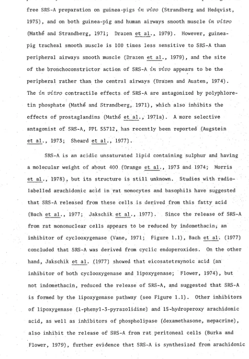
1.2 Regulation of airways smooth muscle 47
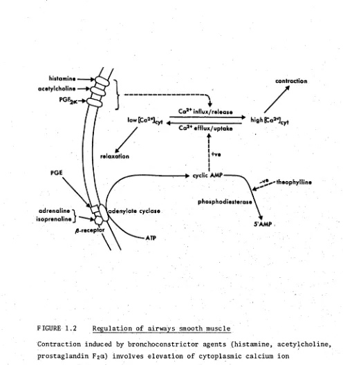
2.1 Method of preparation of guinea-pig tracheal smooth muscle 59 strip
2.2 Experimental procedure used in the determination of cyclic 61 AMP levels in guinea-pig tracheal smooth muscle
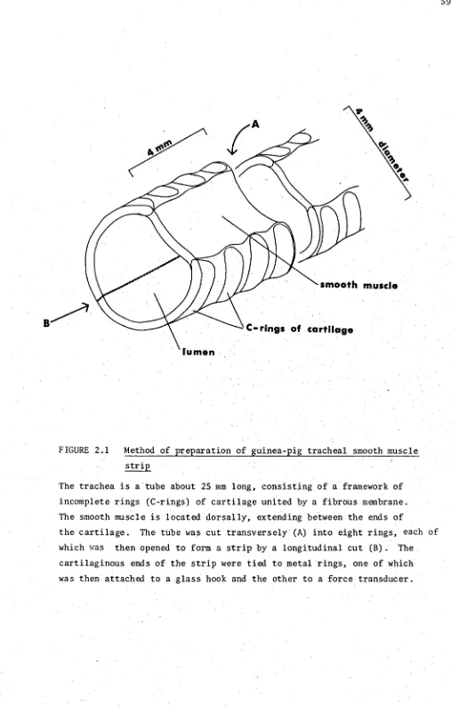
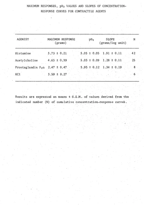
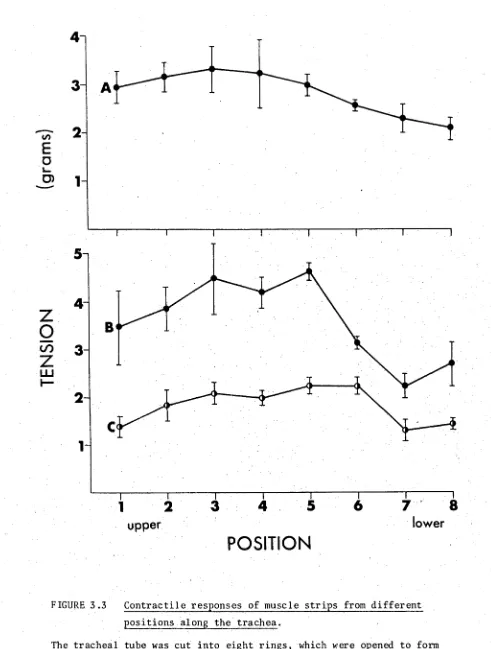
3.1 Contractile responses of the guinea-pig tracheal strip 68 3.2 Concentration-response curves for contractile agents 69 3.3 Contractile responses of muscle strips from different 72
positions along the trachea
3.4 Anaphylactic contracture of tracheal muscle from sensitized 23 guinea-pig
4.1 Effect of zero-calcium medium on concentration-response 82 curves of KC1, histamine and acetylcholine
4.2 Repeated doses of contractile agents in zero-calcium medium 83 4.3 Readdition of calcium ions after contractile responses in 85
zero-calcium medium
4.4 Potentiation by KC1 of histamine-induced contractures after 89 repeated additions in zero-calcium medium
4.5 Concentration dependence of A23187-induced contractures 91
5.1 Contractile responses to imidazole 103
5.2 Concentration dependence of imidazole-induced contractures • 105 5.3 Effect of atropine on the contractile response to imidazole 107 5.4 The effect of calcium depletion on the contractile response 109
to imidazole
5.5 Potentiation of histamine contractures by imidazole 110 5.6 Concentration dependence of the potentiating action of 111
imidazole
5.7 Effect of imidazole on histamine concentration-response curve 112 6.1 Concentration dependence of cyclic AMP accumulation and 127
contraction induced by histamine
6.2 Time courses of cyclic AMP accumulation and contraction 128 induced by histamine
6.3 Imidazole-induced hyperreactivity to histamine in zero- 134 calcium medium
7.1 Effects of indomethacin on resting tension and histamine- 144 induced contractures
7.2 Effects of NSAID on histamine concentration-response curve 146 7.3 Effects of indomethacin on concentration-response curves of 147
contractile agents
3.1 Maximum responses, pE>2 values and slopes of concentration- 7 0 response curves for contractile agents.
3.2 Anaphylactic contractures: dependence on antigen 74 concentration and sensitivity to mepyramine.
4.1 Effects of zero-calcium and EGTA treatment on contractile 87 responses.
4.2 Potentiation of contractile responses by KC1 after calcium 90 depletion.
5.1 Modification of contractile responses to imidazole, 106 acetylcholine and histamine.
5.2 Effect of imidazole (0.5 mM) on contractile responses to 114 various agents.
6.1 Effect of imidazole on cyclic AMP accumulation in response 125 to various agents.
6.2 Effect of atropine on imidazole-induced hyperreactivity to 129 histamine.
6.3 Effect of indomethacin on imidazole-induced hyperreactivity 131 to histamine.
6.4 Effect of calcium ion concentration in imidazole-induced 133 hyperreactivity to histamine.
6.5 Effect of imidazole on contractile responses in zero- 136 calcium medium.
ATPase adenosine triphosphatase 5 ’-AMP adenosine-5’-monophosphate
BSA bovine serum albumin
cAMP, cyclic AMP cyclic adenosine-3', 5'-monophosphate
df degrees of freedom
ECF-A eosinophil chemotactic factor of anaphylaxis EDTA ethylenediamine tetraacetate
EGTA ethylene glycol-bis-(3-aminoethyl ether)-N,N'- tetraacetate
FEVj forced expiratory volume in one second
HA histamine
HSA human serum albumin
IgE immunoglobulin E
NSAID non-steroidal anti-inflammatory drugs
OA ovalbumin
PAF platelet activating factor PG (E2, F2a etc.) prostaglandin (E2> F 2a etc.)
RCS rabbit aorta contracting substance
S .D. standard deviation
S .E . M . standard error of the mean SG
aw specific airways conductance
SRS-A slow reacting substance of anaphylaxis
TCA trichloroacetic acid
C H A P T E R 1
INTRODUCTION
Asthma is a disease of the respiratory tract in which wheezing and dyspnoea are caused by widespread narrowing of the airways. This narrow ing is characteristically reversible, either spontaneously or by treat ment, and is due to abnormal contraction of smooth muscle in the airways.
Other factors, such as oedema of bronchial mucosa and hypersecretion of mucus may also contribute to airways obstruction in asthma (Hayes, 1976).
The basic abnormality in asthma involves hyperreactivity of smooth muscle in the airways: bronchoconstriction is induced by various
stimuli that are without effect on the airways of normal subjects.
These stimuli include allergens, exercise, emotional upsets, and exposure to dusts, cold and chemical irritants. Bronchial hyperreactivity is discussed further in Section 1.1.
Several explanations of bronchial hyperreactivity in asthma have been proposed, and almost all of them involve a defect in the normal regulatory mechanisms controlling airways smooth muscle tone. These mechanisms range from intracellular biochemical processes to hormonal or neural control involving the whole organism. Nervous regulation of airways smooth muscle involves both the sympathetic and parasympathetic systems (Sections 1.3 and 1.4 respectively). Smooth muscle tone may also be modulated by various biologically active substances, either in the circulation or released locally. These include catecholamines
(Section 1.3), histamine and other mediators (Section 1.2), and
1.1 Bronchial hyperreactivity in asthma
Bronchial hyperreactivity to histamine and cholinergic agents has been recognized in asthmatic patients for many years (Weiss, 1928; Curry, 1946, 1947; Tiffeneau, 1958), and has been demonstrated in many studies since (reviewed by Benson, 1975). However, it is only recently that bronchial hyperreactivity has been acknowledged as the underlying common abnormality in all forms of asthma (Cade and Pain,
1971) , and demonstration of bronchial hyperractivity by bronchial provocation testing with methacholine has been proposed as a means of diagnosing asthma (Parker et a l ., 1965; Chai et a l ., 1975).
Some studies of bronchial hyperreactivity measure the changes in pulmonary function parameters such as forced expiratory volume in one second (FEVi) or specific airways conductance (SG ) in response to a standard dose of the bronchoconstrictor (usually aerosols of histamine or methacholine). Others determine the threshold dose of the broncho constrictor agent required to produce significant airways obstruction
(20% decrease in FEVi or 25% increase in SG ). In the former type of study, the response of asthmatic patients is greater than that of normal subjects (Parker et a l ., 1965), and in the latter experiments, 10- to
It is possible that the enhanced bronchoconstrictor responses to
histamine observed in asthmatic patients are due to an increased initial
bronchomotor tone or some degree of airways obstruction existing before
the bronchial provocation test (Benson and Graf, 1977; Brown et a l .,
1977) rather than an inherent hypersensitivity of the smooth muscle to
histamine. This suggestion, however, is not consistent with other
results (Rubinfeld and Pain, 1977) which show that bronchial reactivity
to methacholine is not related to initial level of airways obstruction.
Furthermore, in dogs, bronchial reactivity to histamine is not related
to baseline pulmonary function (Snapper et a l ., 1978).
Although methacholine and histamine are most commonly used in studies
of bronchial hyperreactivity, asthmatic patients also show abnormal sensi
tivity to other bronchoconstricting agents and stimuli. Most notable
amongst these is prostaglandin F201 to which asthmatic patients are about
8000-fold more sensitive than normal controls, whereas the difference in
sensitivity to histamine is only about 10-fold (Mathe et a l ., 1972).
Bronchial hyperreactivity to bradykinin and to SRS-A has also been reported
in asthmatics (Herxheimer and Stresemann,1961 and 1963), but it is
probable that the SRS-A preparation used in the latter study contained
prostaglandins, and that the observed responses resulted from PGF20t-
induced bronchoconstriction.
Asthmatic patients also respond with bronchoconstriction to
3-adrenergic blockers (McNeill, 1964; MacDonald et a l ., 1967), including
the cardioselective 31-antagonists (Bernecker and Roetscher, 1970;
Singh et a l ., 1976). Furthermore, asthmatic subjects are hypersensitive
to a-adrenergic stimulants in the presence of 3-blockade (Kerr and Patel,
1975 ; Snashall, 1978). These observations are pertinent to the 3-
adrenergic blockade theory of asthma, and will be further discussed in Sec
tion 1.3.2. Further examples of stimuli to which the airways of asthmatic
patients are hyperreactive include exercise (McNeill et a l ., 1976), cold air
(Sterling and Batten, 1969), and citric acid (Simonsson et al., 1967).
The diversity of agents and stimuli which cause bronchoconstriction in susceptible patients suggests a fundamental abnormality in the airways of these people. However, one factor which may contribute to the heigh tened bronchial sensitivity in asthma is the increased amount of bronchial smooth muscle which exists in asthmatic airways (Hossain, 1973). It
was suggested that this increased volume of bronchial smooth muscle
is the result of the disease process, rather than a genetically determined fundamental abnormality (Hossain, 1973).
Other possible explanations of bronchical hyperreactivity in asthma are increased sensitivity of irritant receptors in the airways (Section 1.4.2) and a defective ß-adrenergic receptor system (Section 1.3.2).
1.2 Immunological mechanisms in asthma
Perhaps the most common precipitating factor in asthma is an allergic reaction, known as an immediate hypersensitivity or Typ.e I immunological reaction. It involves the interaction of an allergen
(antigen) with specific reaginic antibodies fixed to target cells, the release of various chemical mediators from these cells, and their actions on secondary target tissues to produce the characteristic symptoms of asthma.
1.2.1 Mechanism of mediator release
Mediator release has been studied in a variety of cell types and tissues including rat peritoneal mast cells, human basophilic leucocytes, sensitized guinea-pig lung and passively sensitized human lung. The following discussion concentrates on studies of mediator release from passively sensitized human lung, which have been reviewed by Kaliner and Austen (1975 and 1976).
allergic patients, after which challenge with specific antigen induces the release of histamine and slow reacting substance of anaphylaxis
(SRS-A) (Sheard et al., 1967). Sensitization occurs by transfer of antibodies of the immunoglobulin E class (IgE; reaginic antibody) from the sensitizing serum to target cells in the lung (Ishizaka et al., 1966 a, b ) . The target cells which bind IgE and which are responsible for mediator release have been identified as mast cells (Parish, 1967), large cells containing electron-dense granules. Histamine is stored within these granules, and its release is effected by exocytosis of the granules (Orr, 1977).
When sensitized human lung is challenged with specific antigen, the antigen is bound by two molecules of IgE on the mast cell surface. Bridging of IgE molecules in this manner leads to a complex sequence of biochemical events in the mast cell which eventually leads to mediator release (Kaliner and Austen, 1975). The mechanism involves activation of a serine esterase, and is dependent on extracellular calcium ions and glycolytic activity. The microfilament system of the mast cell also appears to have a major role in the process of mediator release (Orange,
1973; Kaliner, 1977).
1.2.2 The mediators of allergic reactions
The chemical mediators released during antigenic challenge of passively sensitized human lung are histamine, SRS-A, eosinophil chemo-
1.2.2.1 Histamine
The major pathophysiological effects of histamine in the lung are increased vascular permeability and bronchoconstriction (Beaven, 1976). Histamine contracts the isolated smooth muscle of human bronchus (Hawkins and Schild, 1951) and guinea-pig trachea (Ak^asu, 1959). Atropine reduces the severity of histamine-induced bronchoconstriction in vivo (Simonsson et a l ., 1967), suggesting that vagal reflexes initiated by stimulation of irritant receptors in the airways may contribute to the response
(Section 1.4). The in vitro contractile actions of histamine on airways smooth muscle are inhibited by classic antihistaminic drugs (mepyramine, diphenhydramine), indicating the involvement of histamine Hi-receptors. However, histamine H.2-receptors are also present in guinea-pig trachea
and mediate a relaxant effect on the smooth muscle (Okpako et al., 1978). Treatment of guinea-pig tracheal smooth muscle with the H2-antagonist metiamide enhances the contractile response to histamine (Okpako et al.,
1978), suggesting that these receptors constitute an important regulatory mechanism in the control of airways calibre.
1.2.2.2 Slow reacting substance of anaphylaxis
SRS-A was discovered in 1940 in the perfusate of antigenically challenged guinea-pig lung (Kellaway and Trethewie, 1940), and its release from human lung was later shown by Brocklehurst (1960). SRS-A has been reported to cause bronchoconstriction in vivo in guinea-pigs
(Berry and Collier, 1964) and asthmatic humans (Herxheimer and Stresemann, 1963) , and to contract isolated smooth muscle of guinea-pig trachea
(Berry and Collier, 1964) and human bronchus (Brocklehurst, 1962). However, the preparations of SRS-A used in these studies may have con
possibility and showed the bronchonconstrictor effects of a prostaglandin- free SRS-A preparation on guinea-pigs in vivo (Strandberg and Hedqvist, 1975) , and on both guinea-pig and human airways smooth muscle in vitro
(Mathe and Strandberg, 1971; Drazen et al., 1979). However, guinea- pig tracheal smooth muscle is 100 times less sensitive to SRS-A than peripheral airways smooth muscle (Drazen et al., 1979), and the site of the bronchoconstrictor action of SRS-A in vivo appears to be the peripheral rather than the central airways (Drazen and Austen, 1974) .
The in vitro contractile effects of SRS-A are antagonized by polyphlore-
tin phosphate (Mathe and Strandberg, 1971), which also inhibits the effects of prostaglandins (Mathe et al., 1971a). A more selective antagonist of SRS-A, FPL 55712, has recently been reported (Augstein et a l ., 1973; Sheard et a l ., 1977).
SRS-A is an acidic unsaturated lipid containing sulphur and having a molecular weight of about 400 (Orange et al., 1973 and 1974; Morris et a l ., 1978), but its structure is still unknown. Studies with radio-
labelled arachidonic acid in rat monocytes and basophils have suggested that SRS-A released from these cells is derived from this fatty acid
(Bach et a l ., 1977; Jakschik et al., 1977). Since the release of SRS-A from rat mononuclear cells appears to be reduced by indomethacin, an inhibitor of cyclooxygenase (Vane, 1971; Figure 1.1), Bach et al. (1977) concluded that SRS-A was derived from cyclic endoperoxides. On the other hand, Jakschik et a l . (1977) showed that eicosatetraynoic acid (an
inhibitor of both cyclooxygenase and lipoxygenase; Flower, 1974), but not indomethacin, reduced the release of SRS-A, and suggested that SRS-A
[image:19.534.15.525.67.802.2]acid by lipoxygenase,at least in some cell types. SRS-A release from guinea-pig lung is not inhibited by either indomethacin or eicosatetray- noic acid (Dawson and Tomlinson, 1974; Engineer et al., 1978b),
indicating that it is unlikely that SRS-A is derived from arachidonic acid in this tissue. Thus, the biosynthetic pathway of SRS-A may vary between species or cell types, and its structure remains unknown.
1.2.2.3 Other mediators
ECF-A and PAF are the other two primary mediators released during antigenic challenge of sensitized human lung (Austen and Orange, 1975) . Unlike histamine and SRS-A, these agents do not affect bronchial smooth muscle. Rather, their roles in allergic reactions are attraction and deactivation of eosinophils, and stimulation of platelet aggregation and release reaction respectively (Austen and Orange, 1975) . ECF-A is a tetrapeptide (Goetzl and Austen, 1975), whilst PAF is a lipid-like substance of unknown structure (Kater et a l ., 1976).
In spite of the potent effects of prostaglandins on airways smooth muscle (Section 1.5.2), they are not considered as primary mediators
(Austen and Orange, 1975). They may however, play an important role in the modulation of the release of other mediators (Section 1.2.3). Their involvement in allergic reactions is discussed in Section 1.5.1.
1.2.3 Regulation of mediator release
AMP levels (Bourne et a l ., 1972). Lung cyclic AMP levels also rise in response to catecholamines (Vaughan, 1976), but the contribution of the mast cell subpopulation to this effect is uncertain. On the other hand, a-adrenergic stimulation with phenylephrine decreases cyclic AMP
levels and enhances mediator release (Kaliner et al., 1972). Thus, mast cells possess both a- and 3-adrenergic receptors which mediate opposing effects on intracellular cyclic AMP levels and mediator release.
Cholinergic stimulation of sensitized lung also enhances mediator release in response to antigen, but this effect is not associated with any change in cyclic AMP levels (Kaliner et al., 1972). Rather,
acetylcholine increases cyclic GMP levels in lung (Stoner et al., 1973, Kaliner, 1977). Both effects of acetylcholine are blocked by atropine
(Kaliner, 1977).
Prostaglandins Ei and E2 inhibit mediator release from human lung, and these effects are accompanied by increased levels of cyclic AMP
(Tauber et al., 1973). Both responses are independent of the 3-adrenergic receptor. PGF201, on the other hand, reduces cyclic AMP levels and
augments histamine release in response to antigen (Tauber et a l ., 1973; Hitchcock, 1978). PGF201 also increases cyclic GMP levels in human
lung (Kaliner, 1977). Since prostaglandins are released during antigen challenge of sensitized lung (Section 1.6.1), these observations suggest that prostaglandins may have an important role in regulating mediator release. Indeed, treatment of guinea-pig lungs with indomethacin (an inhibitor of prostaglandin synthesis) before anaphylactic challenge increases the amounts of histamine and SRS-A released (Engineer et al., 1978b; Hitchcock, 1978), and sodium meclofenamate (another inhibitor of prostaglandin synthesis) potentiates anaphylactic bronchonconstriction of guinea-pigs
in vivo
(Miller and Robson, 1976).mediators is the negative feedback inhibition of histamine release by histamine itself. In basophilic leucocytes, this effect is accompanied by an increase of cyclic AMP levels (Bourne et a l ., 1971), and is medi
ated by histamine H2-receptors (Lichtenstein and Gillespie, 1973 and 1975).
Thus modulation of mediator release by adrenergic or cholinergic stimulation, by prostaglandins or by histamine, is associated with changes in the levels of cyclic nucleotides, with cyclic AMP and cyclic GMP having opposing effects. Further evidence for this hypothesis is the effect of imidazole, an activator of cyclic nucleotide phosphodie sterase (Section 5.1) which simultaneously reduces lung cyclic AMP levels and enhances the release of mediators (Kaliner and Austen, 1974; Austen and Orange, 1976).
Antigen-induced mediator release from human lung in vitro is also inhibited by disodium cromoglycate (Sheard and Blair 1970; Orange and Austen, 1971). The action of disodium cromoglycate is thought to be independent of cyclic nucleotide metabolism but rather the result of mast cell membrane stabilization (Cox, 1976). However, disodium cromogly cate does have an inhibitory effect on cyclic nucleotide phosphodiesterase from another tissue (Roy and Warren, 1974; Lavin et al., 1976). The importance of the inhibitory effect of disodium cromoglycate on mediator release is seen in the widespread use of this drug in the treatment of asthma (Cox, 1976) .
1.2.4 Mediator release in asthma
and aspirin-sensitive asthma (Section 1.5.4).
The involvement of histamine in allergic asthma was suggested many years ago (Epstein, 1932), but the importance of its contribution
to allergic bronchoconstriction is still unclear. Histamine is released upon antigen challenge of isolated lung and bronchial tissue of asthmatic patients (Schild et al., 1951). Plasma histamine levels in asthmatic patients are increased during naturally occurring asthmatic attacks
(Simon et al., 1977), and after experimental challenge with specific antigen (Bhat et al., 1976), methacholine (Rosenblum et a l ., 1978), or exercise (Anderson et a l ., 1978) . Histamine levels are also elevated in sputum of patients with allergic asthma (Turnbull et al., 1977).
On the other hand, antihistaminic drugs are of limited benefit in most asthmatic patients (Karlin, 1972), mainly because of their dessicant action on the airways which increases bronchial obstruction
(Bodes, 1976) . Also, antihistamines themselves have bronchoconstrictor activity which may limit their therapeutic effectiveness in asthma
(Hawkins, 1955). However, these drugs do prevent allergen-induced bronchoconstriction in some asthmatic patients (Itkin and Anand, 1970), and some have significant bronchodilator activity when administered as aerosols (Nogrady et al., 1978).
Thus, histamine appears to play some role in allergic bronchocon striction in asthma, but the relative importance of this involvement is unclear, and other mediators may be more significant. SRS-A, as well as histamine, is detectable in sputum of patients with allergic asthma and bronchitis, but not other lung diseases (Turnbull et al., 1977). Brocklehurst (1970) compared the bronchoconstricting activity attribut able to histamine and SRS-A released during antigen challenge of
this hypothesis, which has gained widespread acceptance. However, more direct evidence is lacking because of the unavailability of clinically acceptable selective inhibitors of SRS-A release and action.
The release of histamine and SRS-A in response to antigen exposure and their effects on airways smooth muscle may therefore explain bron- choconstriction in allergic asthma. However, this explanation does not account for the bronchial hyperreactivity characteristic of bronchial asthma (Section 1.1). The release of histamine in allergic asthma does not explain why exogenous histamine causes severe bronchoconstriction in asthmatic patients in doses which have no effect on normal airways.
Indeed the bronchoconstrictor response to allergen exposure is influenced by the degree of underlying bronchial hyperreactivity determined by
histamine inhalation challenge, so that allergen-induced asthma is
triggered more easily as the level of hyperreactivity increases (Killian et al., 1976) .
One explanation of this discrepancy may be that the defect in asthma involves an impaired degradative metabolism of histamine. An in vitro model of this possibility is the potentiation of the contractile responses of guinea-pig tracheal smooth muscle by inhibitors of diamine oxidase (histaminase), the enzyme responsible for histamine degradation in this species (Arunlakshana et a l ., 1954). However, in man the principal route of histamine catabolism is methylation by histamine methyltransferase (Schayer, 1956), and this activity is normal in asth matic patients (Thom et al., 1973). The possibility remains, however, that impaired degradation of SRS-A may contribute to bronchial hyperreac tivity.
does n o t e x p l a i n t h e n a t u r e o f t h e d e f e c t r e s p o n s i b l e f o r b r o n c h i a l h y p e r r e a c t i v i t y .
T hu s, a l l e r g i c m e d i a t o r r e l e a s e i s a common s t i m u l u s f o r a s t h m a t i c b r o n c h o c o n s t r i c t i o n , b u t t h e c o n c e p t o f a s thma as a p u r e l y i m m un ol og i ca l
d i s e a s e i s u n t e n a b l e , s i n c e i t f a i l s t o e x p l a i n t h e f u n d a m e n t a l a b n o r m a l i t y i n as th ma . U n d e r l y i n g a l l e r g i c b r o n c h o c o n s t r i c t i o n , t h e r e i s a n o n
s p e c i f i c h y p e r r e a c t i v i t y o f a i r w a y s smooth m u s c l e t o t h e m e d i a t o r s
r e l e a s e d d u r i n g a n t i g e n e x p o s u r e .
1 . 3 A d r e n e r g i c mechanisms and asthma
1 . 3 . 1 A d r e n e r g i c r e g u l a t i o n o f a i r w a y s smooth mus cl e
A d r e n e r g i c r e g u l a t i o n o f t h e a i r w a y s i n v o l v e s b o t h t h e s y m p a t h e t i c
n e r v o u s s y st e m and c i r c u l a t i n g c a t e c h o l a m i n e s from t h e a d r e n a l g l a n d s . N o r a d r e n a l i n e r e l e a s e d from s y m p a t h e t i c n e r v e t e r m i n a l s and c i r c u l a t i n g c a t e c h o l a m i n e s a c t on a d r e n e r g i c r e c e p t o r s i n a i r w a y s smooth mu scl e t o p r o d u c e b r o n c h o d i l a t i o n i n many a ni m a l s p e c i e s . The i m p o r t a n c e o f a d r e n e r g i c a l l y m e d i a t e d b r o n c h o d i l a t i o n i s shown by t h e w i d e s p r e a d u s e
o f sy mp at homi met i c a e r o s o l s i n t h e t r e a t m e n t o f a s th m a .
The p r e s e n c e o f a d r e n e r g i c n e r v e s i n t h e a i r w a y s o f v a r i o u s s p e c i e s
h a s been d e m o n s t r a t e d by f l u o r e s c e n c e h i s t o c h e m i s t r y (Mann, 1971;
O’D o n n e l l and S a a r , 1973) . A d r e n e r g i c i n n e r v a t i o n i s most d e n s e a t t h e
l a r y n g e a l end o f t h e t r a c h e a , d e c r e a s e s down t h e t r a c h e a and b r o n c h i , and i s s c a r c e i n p e r i p h e r a l a i r w a y s smooth mu s c l e ( O ' D o n n e l l e t a l . ,
1 9 78) . I t has b e e n s u g g e s t e d t h a t a d r e n e r g i c n e r v e s r e g u l a t e o n l y t h e c e n t r a l a i r w a y s , and t h a t o t h e r f a c t o r s s uch as n o n - a d r e n e r g i c n e r v e s
o r c i r c u l a t i n g c a t e c h o l a m i n e s c o n t r o l r e l a x a t i o n o f t h e s m a l l e r a i r w a y s ( O 'D o n n e l l e t a l . , 1 97 8) . A n o n - a d r e n e r g i c i n h i b i t o r y n e r v o u s s yst em
h a s b ee n d e m o n s t r a t e d i n g u i n e a - p i g t r a c h e a (Coburn and T o m i t a , 1973;
Coleman and Levy, 1974; R i c h a r d s o n and Bou ch ar d , 1 9 75 ) , and i n human
The order of potency of catecholamines in relaxing isolated airways smooth muscle (isoprenaline > adrenaline > noradrenaline > phenylephrine), and inhibition of the response by the ß-adrenergic
antagonist propranolol, indicate that bronchodilation is mediated by ß-adrenergic receptors (Ahlquist, 1948; Foster, 1966). However, after ß-adrenergic blockade, adrenaline and noradrenaline contract isolated smooth muscle of guinea-pig trachea (Everitt and Cairncross, 1969; Fleisch et a l ., 1970) and human bronchus (Adolphson et a l ., 1971; Mathe et al., 1971b), and cause bronchoconstriction in guinea-pigs in vivo (Persson and Johnson, 1970). These responses are blocked by oi- adrenergic receptor antagonists, indicating the presence of a-adrenergic receptors in airways smooth muscle.
ß-adrenergic receptors have been further classified into ßi- and ß2-subtypes (Lands et al., 1967), and bronchial smooth muscle has pre dominantly ß2-adrenoceptors (Lands et a l ., 1967; Daly et a l ., 1971). This observation has led to the development of specific ß2-adrenergic agents such as salbutamol (Cullum et a l ., 1969) and terbutaline
(Carlström, 1970), which have minimal cardiac stimulant effect (mediated by ß i-adrenoceptors).
The relaxant effects of adrenergic agents on airways smooth muscle are associated with an increase of intracellular cyclic AMP levels
(Section 1.7.3) and it is widely accepted that cyclic AMP is the "second messenger" for the bronchodilator actions of these compounds. Indeed,
it was suggested that the ß-adrenergic receptor is identical to the
adenylate cyclase enzyme (Robison et al., 1967). However, more recently it has been shown that adenylate cyclase and the ß-adrenoceptor are
distinct proteins (Insel et al., 1976; Schramm et al., 1977), but that there is a close association between them in the smooth muscle cell membrane.
be assessed by the administration of 3-adrenergic antagonists. Although propranolol contracts isolated human bronchial smooth muscle (Mathe et al.,
1971), it has little or no effect on the airways of normal subjects (McNeill and Ingram, 1966; Turner et al., 1971; Tattersfield et a l ., 1973; Bradley et al., 1976), indicating the absence of any significant bronchodilating influences mediated by 3-adrenergic mechanisms. However, administration of propranolol to asthmatic patients causes severe bron- choconstriction (McNeill, 1964; MacDonald et a l ., 1967; Richardson and Sterling, 1969). The slight bronchoconstriction sometimes observed in normal subjects and the greater response in asthmatic patients are both inhibited by atropine (Langer, 1967; MacDonald et al., 1967; Grieco and Pierson, 1971). This observation could indicate that cholinergically mediated reflex mechanisms are involved (Section 1.4.1), but a more
like.ly explanation is that after 3-blockade with propranolol, bronchocon striction is caused by unopposed vagal activity, which would be blocked by atropine. Stimulation of a-adrenergic receptors may also contribute to the bronchoconstriction observed after propranolol (MacDonald et al.,
1967).
Propranolol also causes bronchoconstriction
in vivo
in dogs(Woolcock et al., 1969b) and guinea-pigs (Herxheimer, 1967; MacLagan and Ney, 1977) . Although prior administration of atropine delays the onset of dyspnoea (Herxheimer, 1967), it does not reduce the severity of the bronchoconstriction (MacLagan and Ney, 1977), indicating that parasympa thetic mechanisms do not mediate the airways response. Practolol, a selective 3i-adrenoceptor antagonist, and (+)-propranolol, the stereoiso mer devoid of 3-blocking activity, also causes bronchoconstriction in guinea-pigs (MacLagan and Ney, 1977), suggesting that the bronchoconstric- tor response to propranolol may not be related to blockade of 32-
1976; Formgren, 1977), although the effect is not as common or as
severe as propranolol-induced bronchoconstriction (MacDonald and McNeill, 1968).
Apart from mediating resting bronchodilator activity in guinea- pigs and asthmatic humans, adrenergic mechanisms are also important in regulating airways smooth muscle during exposure to bronchoconstrictor
stimuli. Bronchoconstriction induced by antigen challenge of sensitized guinea-pigs, or by the administration of histamine, SRS-A or bradykinin, is accompanied by the release of catecholamines from the adrenal medulla into the circulation (Staszewska-Barczak and Vane, 1965; Piper et al., 1967; Bernauer et al., 1971). Furthermore, prior administration of ß-adrenergic blocking drugs enhances bronchoconstriction induced in
guinea-pigs by bradykinin (Collier et al., 1965) and histamine (McCulloch et a l ., 1967; Diamond, 1972; Douglas et al., 1973). Adrenalectomized guinea-pigs also display increased reactivity to histamine (Diamond,
1972), and this effect is additive to that of propranolol administration (McCulloch et al., 1967). This suggests that histamine-induced-broncho- constriction is modulated by both adrenaline released from the adrenal glands and by noradrenaline released from adrenergic neurones in the airways. Non-neuronal catecholamine stores in the lung may also be involved (Drazen, 1978) .
Similar mechanisms have been demonstrated in other species: adrenalectomy enhances histamine-induced bronchoconstriction in cats
sensitivity to histamine (Ploy-Song-Sang et al., 1978), acetylcholine (Orehek et al., 1975) , and cigarette smoke (Zuskin et a l ., 1974) .
Studies demonstrating bronchial hyperreactivity induced by
3-blockade in experimental animals have all been in vivo, and some have shown that in vitro, propranolol does not enhance histamine-induced contractions (McCulloch et al., 1967). On the other hand, another 3-adrenergic antagonist, practolol, does potentiate the contractile responses of isolated guinea-pig trachea to histamine, acetylcholine and serotonin (Chang et a l ., 1978). However, it seems unlikely that this effect is related to 3-adrenergic blockade, since practolol is a
cardioselective 3u-adrenoceptor antagonist, and would be expected to have much less of an effect than propranolol on the predominantly 3 2-adreno ceptors of airways smooth muscle. The mechanism of action of practolol on guinea-pig trachea remains obscure.
Thus, 3-adrenergic mechanisms may constitute an important negative feedback regulatory system modulating the bronchoconstrictor effects of various agents in vivo, and serve as a protective mechanism against excessive bronchoconstriction in response to allergic or other stimuli. Although the evidence for such a mechanism in healthy humans is not as
strong as for guinea-pigs and other experimental animals, it is possible that a defect in the normal 3-adrenergic regulatory system of airways smooth muscle underlies the bronchial hyperreactivity characteristic of asthma.
1.3.2 Beta-adrenergic blockade in asthma
underlying abnormality is a diminished responsiveness of B-adrenergic receptors in the airways. B-adrenergic regulation of airways smooth muscle is thus impaired, and exposure to contractile stimuli results in unopposed bronchoconstriction.
Some indirect evidence for the theory of B-adrenergic blockade comes from the studies described in the preceding section, in which
administration of propranolol to experimental animals enhances the airways responses to bronchoconstrictor stimuli. However, similar studies in humans are few, and the results conflicting (Section 1.3.1).
Further indirect evidence is derived from studies of B-adrenergic function in various tissues and metabolic processes in asthmatic patients. For example, the hyperglycaemic and cardiac stimulant responses to
infused adrenaline or isoprenaline are less in asthmatic patients than in normal controls (Cookson and Reed, 1963; Lockey et a l ., 1967;
Middleton and Finke, 1968) , and the impairment of these responses
correlates with the degree of bronchial hyperreactivity. Also the normal increases in plasma and urinary cyclic AMP levels after adrenaline are diminished in patients with asthma (Bernstein et al., 1972; Schwartz et al., 1973), and an inverse correlation exists between the plasma cyclic AMP response to adrenaline and bronchial sensitivity to histamine
(Apold and Aksnes, 1976). Inhalation of salbutamol aerosol also increases urinary and plasma cyclic AMP levels in normal subjects, but not in
asthmatic patients (Raij et a l ., 1976).
The effect of B-adrenergic stimulation on cyclic AMP metabolism has also been studied in lymphocytes and polymorphonuclear leucocytes. Cyclic AMP in lymphocytes from normal patients increases three-to four fold after in vitro exposure to isoprenaline, but this response is diminished in cells from patients with asthma (Smith and Parker, 1970;
Logsdon et a l ., 1972; Parker and Smith, 1973). The decreased
asthmatic condition, being most marked during severe active asthma and returning towards normal during periods of remission. On the other hand, increases in cyclic AMP levels induced by PGEi are no different in lymphocytes from asthmatic and normal patients (Parker et al., 1973), indicating that the defect in catecholamine responsiveness is associated with the B-adrenergic receptor rather than with the adenylate cyclase enzyme or some other component of the cyclic AMP metabolic pathway. Similar results have also been obtained in polymorphonuclear leucocytes
(Busse, 1977) . Furthermore, the cyclic AMP response of lymphocytes
from asthmatic patients to noradrenaline, predominantly a
Bi
-adrenostimu- lant, was not as impaired as the response to salbutamol (predominantly a B2-adrenostimulant), suggesting that the abnormality underlying asthma is associated with B2-adrenoceptors (Makino et a l ., 1977). Further investigation of B-adrenergic receptors of asthmatic patients by measure ment of adrenaline binding to leucocytes has shown that there is no difference in either the number or binding affinity of adrenergicreceptors between cells from normal and asthmatic subjects, in spite of decreased cyclic AMP production in the latter (Sokol and Beall, 1975).
One criticism of all of the above studies is that many asthmatic patients are treated with B-adrenostimulant drugs, and the diminished
B-adrenergic responsiveness may result from this treatment, rather than being the underlying cause of asthma. For example, administration of ephedrine or terbutaline to normal subjects for one week depresses the metabolic and cardiovascular responses to adrenaline (Nelson et a l .,
with ephedrine for two weeks show no changes in the lymphocyte cyclic AMP response to isoprenaline (Parker and Smith, 1973). On the other hand, cyclic AMP responses to adrenergic agents are normal in leucocytes
from asthmatic children who have never received sympathomimetic bron- chodilators (Kalisker et a l ., 1977), or who have not received sympathomi- metics for one week (Morris et a l ., 1977) . In the latter study,
leucocyte cyclic AMP responses decreased after one week of 3-adrenostimu- lant therapy. (3-adrenoceptor desensitization may also occur in asthma as a result of exposure to increased amounts of catecholamines released during asthmatic attacks, and this could explain why asthmatic patients in remission show less impairment of (3-adrenergic function than patients with active asthma (Parker and Smith, 1973; Alston et a l ., 1974).
Since asymptomatic asthmatic patients still display bronchial hyperreacti vity (Cade and Pain, 1971), these observations may indicate that impaired 3-adrenergic function is a result rather than the cause of asthma.
In spite of the wealth of information showing impaired 3-adrenergic function in a variety of cell types and metabolic processes, there has > been no report of a similar defect in the airways smooth muscle of
asthmatic patients. On the contrary, Svedmyr et al. (1976) found normal responses to isoprenaline in bronchial smooth muscle removed from three asthmatic patients undergoing surgery for bronchial carcinoma.
Thus asthma is associated with impaired 3-adrenergic function in various cell types and tissues, but it is not certain if this defect underlies bronchial hyperreactivity or results from the symptoms and/or treatment of the disease, or if it extends to airways smooth muscle.
1.3.3 Alpha-adrenergic receptors in asthma
Another proposed mechanism which could explain bronchial
(phentolamine, phenoxybenzamine) inhibit histamine-induced bronchocon- striction in asthmatic patients (Kerr et al., 1970). Although these drugs have other actions, such as anti-histaminic activity and inhibition of catecholamine uptake (Nickerson, 1970), these results were taken to indicate the participation of a-adrenergic receptors in the constriction of asthmatic airways in response to histamine. The existence of
a-adrenergic receptors in human bronchial smooth muscle has been confirmed in vitro (Mathe et a l ., 1971), and Patel and Kerr (1973) suggested that increased a-receptor activity may lead to bronchial hyperreactivity in asthma. Simonsson et al., (1972) found that bacterial endotoxins poten tiate contraction of airways smooth muscle in response to a-adrenergic stimulation in vitro and proposed that a-adrenoceptors were involved in bronchoconstriction occurring during bronchial infections.
Inhalation of phenylephrine (an a-adrenergic stimulant) by asthmatic patients after (3-blockade induces bronchoconstriction, but has no effect
on normal airways (Kerr and Patel, 1975). In vitro, platelets of asthmatic children have an ATPase activity (an a-adrenergic function) twice as great as normal platelets (Coffey and Middleton, 1975). Also, the enhancement by a-adrenergic blockade of the cyclic AMP response to isoprenaline is greater in leucocytes from asthmatic patients than in normal cells (Alston et a l ., 1974) indicating that the impaired cyclic AMP response of asthmatic leucocytes is due to overactivity of
a-adrenoceptors rather than reduced (3-adrenergic function. All these observations lend support to the concept that asthma is associated with increased a-adrenergic activity. Furthermore, a-adrenergic hypersensiti vity, determined by pupil dilation in response to phenylephrine, is found
1.4 Cholinergic regulation of airways smooth muscle 1.4.1 Normal airways
The parasympathetic nervous system plays an important role in the normal regulation of airways smooth muscle (reviewed by Nadel, 1976), and provides one site where a defect may contribute to bronchial hyperreacti vity in asthma.
Parasympathetic (cholinergic) innervation has been demonstrated by acetylcholinesterase staining in the bronchial smooth muscle of all species studied (Spencer and Leof, 1964; Nadel, 1975), and this innervation is derived from the pulmonary branches of the vagus nerve. Cutting the vagus nerve of experimental animals or blocking cholinergic transmission with atropine in humans causes bronchodila- tion (Severinghaus and Stupfel, 1955; Dautebrande et a l ., 1962;
Nadel and Widdicombe, 1963), indicating that the airways are subject to a resting cholinergic tonic constriction. In humans, the maximum
bronchodilator effect of atropine is similar to that of a ß-adrenostimu- lant such as isoprenaline (Altounyan, 1974) . Electrical stimulation of the vagus in animals causes acetylcholine release from nerve terminals within airways smooth muscle and constriction of the larger airways
(Olsen et al., 1965; Woolcock et al., 1969a).
Parasympathetic regulation of airway calibre depends on various receptors in the tracheobronchial tree, which are of three main types:
1) cough receptors, located mainly in the tracheal and larger bronchi,
2) irritant receptors, located in the more peripheral bronchi, and
et a l ., 1962; Sellick and Widdicombe, 1971). Stimuli capable of
inducing reflex bronchoconstriction include chemicals (sulphur dioxide, citric acid) (Nadel et al., 1965; Simonsson et al., 1967), chemically inert dusts (charcoal, cigarette smoke) (Widdicombe et al., 1962;
Sterling 1967), cold air (Simonsson et al., 1967), mechanical stimulation (Nadel and Widdicombe, 1962b), hypoxaemia and hypercapnia (Nadel and Widdicombe, 1962a), and hyperinflation (Simonsson et al., 1967).
Inhibition of the bronchoconstrictor responses to these stimuli by atropine, vagotomy or vagal cooling demonstrates their parasympathetic reflex nature. Furthermore, increased activity of single vagal nerve fibres has been recorded in response to some of these stimuli, indicating stimulation of irritant receptors in the lung (Mills et al., 1969;
Sellick and Widdicombe, 1969 and 1971).
1.4.2 Cholinergic mechanisms in asthma
Asthmatic patients display bronchial hyperreactivity to a variety of stimuli (Section 1.2), and it is noteworthy that many of these
stimuli cause reflex bronchoconstriction, even in healthy subjects. On this basis, Simonsson et a l . (1967) suggested that increased sensiti vity of irritant receptors in the airways could be a cause of bronchial hyperreactivity in asthma. That is, a lower threshold of stimulation of sensory nerve endings in asthmatic airways results in reflex broncho constriction at stimulus levels which are without effect in normal subjects. The involvement of cholinergic pathways is indicated by inhibition of the abnormal bronchoconstrictor responses by atropine.
Some evidence for this hypothesis comes from animal experiments in which sensitization of airway receptors is achieved by lung infections or by inhalation of ammonia or ozone, resulting in increased responsive ness to bronchoconstrictor stimuli (Widdicombe, 1961; Easton and Murphy,
s e n s o r y r e c e p t o r s by e p i t h e l i a l damage may e x p l a i n t h e b r o n c h i a l h y p e r r e a c t i v i t y t o h i s t a m i n e and c i t r i c a c i d o b s e r v e d d u r i n g r e c o v e r y from a c u t e v i r a l r e s p i r a t o r y i n f e c t i o n s (Empey e t a l . , 1976) .
However, a c c e p t a n c e o f any h y p o t h e s i s i m p l i c a t i n g t h e p a r a s y m p a t h e
t i c n e r v o u s s yst em i n b r o n c h i a l h y p e r r e a c t i v i t y must depend on a demon s t r a t i o n t h a t a l l s t i m u l i t o which a s t h m a t i c p a t i e n t s a r e h y p e r r e a c t i v e
do o p e r a t e by a c h o l i n e r g i c r e f l e x mechanism. The e v i d e n c e f o r s uch a
mechanism i n t h e b r o n c h o c o n s t r i c t o r r e s p o n s e s t o a l l e r g e n s , m e d i a t o r s o f a l l e r g y and e x e r c i s e i s d i s c u s s e d bel ow.
(a) Chemical m e d i a t o r s o f a l l e r g y . I t i s w e l l known t h a t some
o f t h e m e d i a t o r s l i b e r a t e d f o l l o w i n g a l l e r g e n e x p o s u r e ( h i s t a m i n e , b r a d y k i n i n , SRS-A, PGF2CO h a v e a d i r e c t c o n t r a c t i l e a c t i o n on b r o n c h i a l
m u s c l e ( S e c t i o n 1 . 2 . 2 ) . However, i t h a s been s u g g e s t e d t h a t p a r t o f t h e i r b r o n c h o c o n s t r i c t o r a c t i o n
in v iv o
i s m e d i a t e d by a v a g a l r e f l e x . S e v e r a l s t u d i e s h av e shown t h a t h i s t a m i n e - i n d u c e d b r o n c h o c o n s t r i c t i o n i s p a r t i a l l y i n h i b i t e d by vagotomy i n a n i m a l s o r by hexamethonium o r a t r o p i n e i n humans (Bouhuys e t a l . , 1960; DeCock 1966; Simonsson e t a l . , 1 9 67 ) , i n d i c a t i n g t h a t h i s t a m i n e h a s b o t h d i r e c t and r e f l e x b r o n c h o c o n s t r i c t o r a c t i o n s on b r o n c h i a l smooth m u s c l e . F u r t h e r m o r e , M i l l s e t a l . (1969) showed t h a t h i s t a m i n e - i n d u c e d bronch os pas m i n r a b b i t s was
acc ompani ed by i n c r e a s e d v a g a l a f f e r e n t n e r v o u s a c t i v i t y , and c o n c l u d e d
t h a t s t i m u l a t i o n o f i r r i t a n t r e c e p t o r s by h i s t a m i n e c o n t r i b u t e s t o t h e b r o n ch os p a s m. However, more r e c e n t s t u d i e s have c o n c l u d e d t h a t c h o l i n e r
g i c mechanisms a r e n o t i n v o l v e d i n t h e a i r w a y s r e s p o n s e t o h i s t a m i n e ,
s i n c e a t r o p i n e and SCH 1000 ( i p r a t r o p i u m b r o m i d e , a more r e c e n t a n t i c h o l i n e r g i c a g e n t ) f a i l e d t o s i g n i f i c a n t l y i n h i b i t t h e r e s p o n s e a t d o s e s which c o m p l e t e l y a b o l i s h e d m e t h a c h o l i n e - i n d u c e d b r o n c h o c o n s t r i c t i o n
Cholinergic mechanisms have also been proposed to contribute to the bronchoconstrictor responses of SRS-A (Drazen and Austen, 1975), bradykinin (Simonsson et a l ., 1973), and PGF2a (Alanko and Poppius,
1973; Orehek et al., 1977). However, the suggestion of a vagal reflex in PGF2a-induced bronchoconstriction is contradicted by other studies which showed no inhibitory effect of atropine on the airways response of asthmatic patients to PGF201 (Newball et al., 1974; .Smith et al., 1975).
(b) Allergens. The suggested involvement of cholinergic reflex pathways in the bronchoconstrictor responses to various mediators
raises the possibility of parasympathetic nervous effects in the broncho constriction associated with allergic asthma or experimental anaphylaxis. Irritant receptors are stimulated during anaphylaxis (Mills et al., 1969), and vagotomy reduces the bronchoconstriction caused by allergen exposure in sensitized rabbits, guinea-pigs and dogs (Mills and Widdicombe, 1970; Karkzewski and Widdicombe, 1969; Gold et al., 1972). Furthermore,
unilateral challenge of one dog lung with antigen causes bronchoconstric tion in both lungs, indicating an indirect mechanism in the second lung
(Gold et al., 1972).
A similar experiment in humans, however, showed that vagal reflexes are not important in allergen-induced bronchoconstriction, since unilateral challenge with antigen induced bronchoconstriction only in the challenged lung (Aborelius et al., 1962) . The effectiveness of atropine in preventing human airways responses to inhaled allergens is controversial. Some studies have shown that administration of
response (Fish et al., 1977; Rosenthal et al., 1977).
(c) Exercise. It has been suggested that exercise-induced bronchoconstriction in asthmatic patients is caused by release or formation of bronchoconstrictor agents, possibly identical to those released during allergen provocation (histamine, bradykinin, SRS-A,
PGF2CO (Sly, 1976). Since cholinergic reflex mechanisms may be involved in the bronchoconstrictor actions of these mediators, they may also contribute to exercise-induced bronchospasm. However, atropine does not prevent exercise-induced asthma in most cases (Sly et al., 1967; Fisher et al., 1968; Chan-Yeung et a l ., 1971), indicating that the parasympathetic nervous system is not involved in the response.
Bronchial hyperreactivity to exercise is a wel1-documented
characteristic feature of asthma (McNeill et al., 1966), and is used as a diagnostic test for asthma (Anderson et al., 1975). Exercise and allergy are perhaps the two most common precipitating factors in asthma, and unless the involvement of cholinergic pathways can be definitely established in the bronchoconstrictor actions of these stimuli, it is difficult to accept an explanation of bronchial hyperreactivity involving a defect in the parasympathetic reflex nervous system.
Another criticism of the theory implicating reflex cholinergic mechanisms in bronchial hyperreactivity is that inhibition of bronchocon
strictor responses by cholinergic blockade does not necessarily prove the involvement of a parasympathetic reflex. Atropine alone acts as a
bronchodilator by abolishing resting bronchial muscle tone, and changes in reactivity to bronchoconstrictor stimuli may simply result from this change in baseline conditions (Benson, 1975).
1.5 Prostaglandins
implicated in many physiological and pathological processes. Their presence in human lung (Anggärd, 1965; Karim et al., 1967), their release during allergic reactions (Section 1.6.1), and their effects on bronchial smooth muscle (Section 1.6.2) raise the possibility that prostaglandins play an important role in the pathogenesis of human
asthma. The biosynthetic pathways of the major prostaglandins are shown in Figure 1.1.
1.5.1 Prostaglandin release in allergic reactions
When a perfused lung of a sensitized guinea-pig is challenged with antigen, PGF201 and PGE2 can be identified amongst the biologically
active substances released into the effluent by bioassay on various isolated smooth muscle preparations (Piper and Vane, 1969). Another prostaglandin-like substance apparent in the effluent is very unstable,
contracts isolated strips of rabbit aorta, and was called "rabbit aorta contracting substance" (RCS) (Piper and Vane, 1969). RCS was later shown to consist mainly of thromboxane A2 (Hamberg et a l ., 1975a;
Bunting et al., 1976), with a minor contribution from the cyclic endopero- xides (PGG2 and PGH2) (Svensson et al., 1975). More detailed analysis
of the effluent from challenged guinea-pig lungs by gas liquid chromato graphy and mass spectrometry has identified nine different prostaglandin like substances (Boot et a l ., 1976; Dawson et a l ., 1976). These include various metabolites of the previously identified prostaglandins and
thromboxanes, as well as 6-oxo PGFia, the metabolite of prostacyclin (PGI2). PGI2 itself has been detected in the perfusate of guinea-pig
lungs challenged with arachidonic acid (Alabaster and Hawkeswood, 1978) , so it seems likely that PGI2 is also released during anaphylactic
challenge.
ARACHIDONIC ACID
lipoxygenase (?) cy clo o x y g e n a se
SRS-A+++
OTHE
- v e indomethacin,aspirin
other NSAID
CYCLIC ENDOPEROXIDES (PGG?/PGH.)
++
1
^ r '
ER PGs PC
t '
r '
;E2 PC
r '
f +
* 2 « r<
r ^
r _ ^
312 THRO
throm boxane synthetase
r + + +
MBOXANE A 2
r
15-0X0-13,1 4 -d ih y d r o m e ta b o lite s 6 -o x o PGFja THROMBOXANE B2
FIGURE 1 .1 M e ta b o lis m o f d i e n o i c p r o s t a g l a n d i n s and th ro m b o x a n e s A r a c h i d o n i c a c i d r e l e a s e d by t h e a c t i o n o f p h o s p h o l i p a s e on membrane p h o s p h o l i p i d s i s m e t a b o l i z e d t o a r a n g e o f p r o d u c t s , some o f w hich ha ve b r o n c h o d i l a t o r ( - ) o r b r o n c h o c o n s t r i c t o r (+) a c t i v i t y on a i r w a y s
and Samuelsson, 1965), histamine (Bahkle and Smith, 1972; Alabaster and Bahkle, 1976), SRS-A (Engineer et a l ., 1978a; Seale and Piper, 1978), and mechanical irritation (Palmer et al., 1973). These
observations suggest that prostaglandin release during antigen challenge of sensitized lung is -secondary to either histamine release or
anaphylactic smooth muscle contraction (Piper and Walker, 1973; Parrot et al., 1977).
Prostaglandins are also released during antigen challenge of passively sensitized human lung (Piper, 1973; Piper and Walker, 1973), and airways smooth muscle (Dunlop and Smith, 1975). However, these studies have been limited to identification of only two prostaglandins, PGF2a (lung and bronchus) and PGE2 (lung only).
The release of prostaglandins from challenged guinea-pig and human lungs is blocked by indomethacin (Piper and Vane, 1969; Piper and Walker, 1973), which inhibits the conversion of arachidonic acid to cyclic endoperoxides (Figure 1.1) (Vane, 1971). This indicates that prostaglandins, unlike histamine, are synthesized
de novo
during antigen challenge, rather than being released from pre-formed stores. Catechol amines also inhibit the release of prostaglandins from challenged guinea- pig lungs (Mathe and Levine, 1973; Liebig et al., 1974).1.5.2 Effects of prostaglandins on airways smooth muscle
Since PGF201 and PGE2 are the major prostaglandins released during antigen challenge of sensitized lungs, the effects of these prostaglandins on airways smooth muscle have been extensively investigated.
PGF2a contracts isolated tracheal muscle from guinea-pigs (Änggärd and Bergstrom, 1963; Puglisi, 1973) and dogs (Wasserman and Griffin, 1976), although its potency is not as great as on other smooth muscle preparations (Änggärd and Bergström, 1963). PGF201 also constricts
Mathe et al., 1971a). Cat trachea, on the other hand, is relaxed by PGF2CI (Horton and Main, 1965) .
In vitro contractile responses of airways smooth muscle to PGF201
are not affected by mepyramine or atropine (Sweatman and Collier, 1968), indicating a direct action of PGF201 on the smooth muscle. They are, however, inhibited by prostaglandin antagonists such as polyphloretin phosphate and SC-19220 which do not affect contractions induced by
histamine, serotonin or acetylcholine (Mathe et al., 1971a; Farmer et al., 1974). Non-steroidal anti-inflammatory drugs, such as flufenamic acid, aspirin and indomethacin at high concentrations, also antagonize
PGF20i-induced contraction (Collier and Sweatman, 1968; McIntyre and Temple, 1977) . Low concentrations of indomethacin, on the other hand, potentiate the contractile effects of PGF2CI on guinea-pig tracheal smooth muscle (Farmer et a l ., 1974; Lambley and Smith, 1975). This effect may be related to the inhibitory action of indomethacin on the synthesis of relaxant prostaglandins induced by exogenous PFG201
(Panczenko et al., 1975).
The bronchonconstrictor action of PGF201 has been confirmed by in vivo studies of guinea-pigs (Mathe et al., 1972), dogs (Said, 1967; Wasserman, 1975; Spannhake et al., 1978a), and humans (Hedqvist et al.,
1971; Smith and Cuthbert, 1972a). In vivo bronchoconstrictor responses to PFG201 may be partially due to cholinergically mediated reflex
bronchoconstriction (Manko and Poppius, 1973; Wasserman, 1976; Section 1.4.2) .