The Use of Semi-permanent Central Venous Catheters in a
Population of Hospital Patients with Malignant Disease:
Determinants of Adverse Outcomes
Paul Stanley Craft. January, 1995
The sub-thesis is entirely my own work with the exception of some data extraction from medical records performed by Dr A. Dorrigo and Ms J. May (Medical Oncology Department, Woden Valley Hospital) and measurements taken from chest radiographs performed by Dr C. Hoy (Department of
Radiology, Woden Valley Hospital). These aspects of the project were performed under my supervision.
A
CONTENTS
1. INTRODUCTION: TRENDS IN CANCER CONTROL...1
2. THE INCREASING REQUIREMENT FOR VASCULAR ACCESS... 2
Vascular Access in Cancer Surgery and Radiotherapy...2
Chemotherapy and Vascular Access... 3
Continuous Infusional Chemotherapy...4
Economic Issues and Venous Access...5
3. STUDY OF HICKMAN CATHETER USE IN WODEN VALLEY HOSPITAL: AIMS AND OBJECTIVES...6
Aims... 6
Primary Objectives...6
Secondary Objectives...6
4. REVIEW OF METHODS FOR OBTAINING AND MAINTAINING VENOUS ACCESS...7
Peripheral Venous Catheters...7
Non-tunnelled Central Venous Catheters... 8
Tunnelled Silicone Rubber Central Venous Lines (Hickman or Broviac Catheters)...8
Methods of Hickman Catheter Insertion... 9
Surgical Open Insertion... 9
Percutaneous Insertion of Hickman Catheters... 9
Other Tunnelled Catheters - The Groshong Catheter...11
Fully Implanted Catheter Systems - "Ports"... 11
Other Methods of Obtaining Vascular Access... 12
5. HICKMAN CATHETER USE: OUTCOMES AND
ADVERSE EVENTS... 13
Hickman Catheter Related Infection... 15
Pathogenesis of Catheter Infection...16
Diagnosis and Definition of Catheter Related Infection... 17
Microbiology of Hickman Catheter Related Infection... 20
Putative Risk Factors for Infection...23
Criteria for Hickman Catheter Removal for Infection... 24
Venous Thrombosis and Hickman Catheter Occlusion... 24
Pathogenesis of Hickman Catheter Related Thrombosis... 26
Putative Risk Factors for Thrombosis and Catheter Malfunction... 27
Catheter Salvage After Central Vein Thrombosis... 28
Catheter Migration And Dislodgement...28
Management of Catheter Migration...29
Mechanical Failure or Fracture...29
Acute Complications of Hickman Catheter Insertion... 30
6. METHODS... 31
Design... 31
Ethical Considerations... 31
Case Ascertainment... 31
Catheter Eligibility... 31
Technique of Catheter Insertion... 32
Catheter Use and Care... 32
Management of Hickman Catheter Complications...33
Data Collection... 33
Radiological Review... 34
Case Definitions... 37
Obesity... 37
Catheter Related Infection... 37
Central Vein Thrombosis...37
Hickman Catheter Occlusion...37
Hickman Catheter Migration or Dislodgement...38
Hickman Catheter Failure... 38
Acute Complications of Catheter Placement...38
7. STATISTICAL METHODS... 39
8. STATISTICAL METHODS - MULTIVARIATE...41
9. RESULTS... 42
Patient and Catheter Characteristics...42
Outcome of Catheter Placement...46
Findings of Radiological Assessment...46
Acute Complications of Hickman Catheter Placement... 46
Outcome of Catheter Use...46
Catheter Failure... 47
Hickman Catheter Related Infections...51
Exit site infections... 51
Subcutaneous tunnel infections...55
Hickman catheter related septicaemia...55
Venous Thrombosis and Hickman Catheter Malfunction... 59
Venous Thrombosis... 59
Hickman Catheter Malfunction...61
Hickman Catheter Migration or Dislodgement...63
Type of Vascular Access Used After Hickman Catheter Failure... 64
Hickman Catheter Failure 66
Hickman Catheter Related Infections...67
Exit Site Infection... 67
Subcutaneous Tunnel Infection...68
Hickman Catheter Related Septicaemia...69
Venous Thrombosis and Hickman Catheter Malfunction... 70
Hickman Catheter Migration or Dislodgement...71
Other Complications of Hickman Catheter Insertion and Use... 71
Patient Diagnosis and Hickman Catheter Complications... 71
Patient Gender... 72
Obesity... 73
Site of Catheter Insertion and Catheter Tip Position... 74
Double Lumen Catheters... 75
11. CONCLUSIONS... 78
REFERENCES... 80
1.
INTRODUCTION: TRENDS IN CANCER CONTROL
The Australian population experiences a life time risk of cancer (excluding non-melanocytic skin cancers) to age 75 of about 1 in 4. Cancer is the second most common cause of death, after circulatory disease. In 1992, in Australia, 32,003 deaths due to cancer were registered, representing 26% of all deaths (Australian Institute of Health & Welfare, 1992). Crude incidence and mortality rates of cancer have risen in Australia over the past 3 decades. Much of this increase is due to the increasing age of the Australianpopulation (National Goals and Targets Report, 1994).
Although crude cancer mortality rates increased between 1985 and 1992, the age standardised mortality rate from cancer actually fell by about 5%. There has also been a consistent improvement in cancer case survival rates over the past 14 years observed in South Australia, where these data are
available (SA Health Commission, 1994). It would seem reasonable to assume the same trend would be followed throughout the remainder of Australia. Similar changes in case survival rates have been reported from the United States (Boring etal., 1992).
2.
THE INCREASING REQUIREMENT FOR VASCULAR
ACCESS
Cancer survival rates have slowly improved over the past 20 years. The factors underlying this improvement are difficult to measure, however improved outcomes from medical treatment are likely to be important.
Improved and earlier diagnosis is also likely to be important. Approximately 60% of internal malignancies are cured with currently available therapeutic interventions. Modern cancer care incorporates the use of the three main modalities of cancer treatment, that is, surgery, radiotherapy and
chemotherapy often in a combined approach.
Vascular Access in Cancer Surgery and Radiotherapy
Developments in surgical practice have enabled more effective cancer surgery to be performed with less morbidity. As the scope of new surgical techniques in fields such as reconstructive surgery have developed, the complexity of the requisite supportive care has increased. This supportive care is essential in permitting new surgical treatments to be successfully applied. Prolonged intravenous access is a feature of the intensive care which supports the surgical patient with cancer. The efficacy of parental nutrition in preventing surgical complications in patients undergoing surgery for advanced cancer is well established. Semi-permanent central venous catheters are an essential component of most parenteral nutrition programs.
In radiotherapeutic practice the development of infusional schedules of 5- flourouracil, cisplatin, and other chemotherapeutic drugs which may act as radio-sensitising agents has been an increasingly applied technique (Sugarbaker et al., 1985). The concomitant infusion of 5-flourouracil with radiotherapy has been shown to improve response rates, compared to radiotherapy alone, in the treatment of carcinoma of the cervix, anal canal, and oesophagus. Active investigation of this approach is continuing and the application of this and similar forms of treatment is likely to expand as
efficacy is demonstrated (for example Leichman et al., 1993). Such
combined modality programs need prolonged and durable venous access. Semi-permanent central venous catheters have facilitated the development of this therapeutic approach.
Chemotherapy and Vascular Access
Since the initial trial of nitrogen mustard chemotherapy for Hodgkins disease in 1943 more than 30 anti-cancer cytotoxic agents have entered regular clinical use. It has been estimated that more than 250,000 people with cancer are treated annually in the United states with cytotoxic chemotherapy (DeVita 1985). Almost all of this treatment is given by injection, usually by intravenous infusion. Providing the means for physically administering this therapy as well as providing a route to facilitate the associated supportive care has lead to a steady increase in the use of semi-permanent intravenous devices.
Continuing attempts are being made to maximise the cancer control rate with the currently available chemotherapeutic drugs. Strategies have included the use of combinations of drugs to take advantage of possible synergism and to prevent the development of tumour cell resistance (Scanlon & Nee Dels, 1986). More complex drug combinations and schedules, and the associated supportive treatment, transfusions and antibiotics, have led to the more frequent use of semi-permanent catheters. A steady increase in the frequency of Hickman catheters in patients with predominantly
haematological malignancy has been reported from an Australian hospital over a recent 7 year period (Gibson et a/., 1994).
More recently the relationship between dose intensity and control rates have been examined. Higher dose intensity has been associated with better control rates in studies of breast cancer, lymphoma, and acute leukaemia (Hryniuk et at., 1986). Dose intensity may be increased with the application of intensive supportive care such as autologous peripheral stem cell
infusions.
Autologous bone marrow transplantation and the related procedure of
autologous peripheral stem cell infusion are methods allowing the use of very dose intense chemotherapy. Patients undergoing these procedures require intensive supportive care. Semi-permanent central venous catheters are universally required for these procedures. The final place these forms of treatment will have in the management of common haematological and solid malignancies remains unclear, however 333 autologous bone marrow
cytophoresis procedure where peripheral blood stem cells are collected and for the high dose chemotherapy and related supportive care.
Allogeneic bone transplantation is now an established treatment for various forms of leukaemia and other haematological disorders. The number of transplants performed world wide has risen steadily to over 5000 per year in
1990 (Borton et at., 1992). Such procedures lead to the requirement for a prolonged period of very intensive supportive care during which semi permanent central venous catheters are ubiquitous. The insertion of some form of central venous catheter is mandatory in most transplantation centres at the beginning of the procedure (Flowers & Sullivan, 1994).
Continuous Infusional Chemotherapy
The administration of cytotoxic chemotherapeutic drugs as a continuous slow intravenous infusion has been developed as a way of increasing dose
intensity and efficacy. Cytotoxic drugs that are cell-cycle phase-specific in their action are suitable for this approach, particularly if they have a short terminal half-life.
Some cytotoxic drugs, such as 5-fluorouracil are clearly more efficacious in treating certain cancers, such as breast cancer, when given as a slow
infusion (Anderson 1993). The use of continuous infusions of 5-fluorouracil in ambulatory patients has become feasible because of the availability of reliable semi-permanent catheter systems such as Hickman catheters (for example Jones et at., 1994). When used in this way, 5-fluorouracil is less myelosuppressive, and other forms of toxicity such as mucositis or diarrhoea become the dose limiting factors. Much higher dose rates are achievable with continuous infusions compared to conventional bolus administration (reviewed by Hansen, 1991).
An improved therapeutic index for slow infusional chemotherapy has been reported for bleomycin (Remick et at., 1994). Other cytotoxic drugs which may beneficially be administered as prolonged infusions include doxorubicin, cisplatin, and vinblastine. Many of the cytotoxic drugs currently being
administered by slow continuous infusion are dangerous if extravasated. As with radiation sensitisers and bone marrow transplantation, the widespread
inclusion of infusional therapy into therapeutic protocols has only become feasible because of the availability of safe methods of obtaining durable access to the central circulation.
Economic issues and Venous Access
The development of moderately efficacious but ever more complex cancer treatment has had major cost implications. As supportive care has improved, complex therapeutic protocols have been able to be safely administered to a wider group of patients with cancer. The general application of what were once considered to be highly specialised investigational therapies will have continuing economic repercussions. While semi-permanent central catheters have had a permissive role in the penetration of these new complex protocols into more general clinical use, they may also provide a means of reducing cost in certain circumstances.
More robust methods of obtaining durable venous access have been
important in permitting the transfer of an increasing proportion of care to the clinic and out of hospital wards. Patients with a semi-permanent central venous catheter, such as a Hickman catheter, in situ may have intravenous infusions and catheter care provided at home. Continuous intravenous infusions of cytotoxic drugs are routinely given through central venous catheters using a miniature pump worn or carried by ambulant patients at home. People requiring prolonged parenteral nutrition or intravenous antibiotic therapy are able to be given this therapy at home via a semi permanent central venous catheter (Pomp et a!., 1989; Mukau et a!., 1992).
The provision of intravenous therapy outside of traditional hospital facilities may represent an important improvement. The avoidance of hospital
3.
STUDY OF HICKMAN CATHETER USE IN WODEN
VALLEY HOSPITAL: AIMS AND OBJECTIVES
Aims
To examine the pattern of use, duration of use and adverse events associated with use of Hickman catheters in the study population.
To explore the determinants of adverse events associated with Hickman catheter use within the study population.
Primary Objectives
To develop a predictive model for the risk of Hickman catheter failure as defined by removal of the catheter because of a complication.
To explore the determinants of catheter malfunction (as defined) with particular reference to the measured catheter tip position, the site of insertion, duration of catheter use, and the patient diagnostic group.
Secondary Objectives
To explore the determinants of Hickman catheter infection within the study population.
To explore the determinants of Hickman catheter migration or dislodgement within the study population.
4.
REVIEW OF METHODS FOR OBTAINING AND
MAINTAINING VENOUS ACCESS
In this section an overview of methods in current practice for obtaining
venous access is provided. Methods described include the simple peripheral intravenous cannula, non tunnelled central venous catheters, the tunnelled central venous catheters such as the Hickman and Groshong, and fully implanted subcutaneous ports. Different techniques of insertion of Hickman catheters are also discussed.
Peripheral Venous Catheters
Venous access has been traditionally obtained via peripheral veins using steel needles or polyethylene intravenous cannulas. Modern steel scalp vein needles continue to be used for short term venous access. Peripheral vein plastic cannulas are ubiquitous within hospital facilities in developed
countries. These peripherally inserted cannulas are cheap and easy to insert. While ideal for short term use, peripheral venous catheters have many limitations. They are unsuitable for use with continuous infusions of irritant solutions, such as some anti cancer cytotoxic drugs, and parenteral nutrition infusate. Attempted infusion of such fluids into a small peripheral vein (with relatively low blood flow) causes local phlebitis. Peripheral vein cannulas frequently block or kink. The tip of the cannula can migrate to an extravascular site. The accidental infusion of some drugs, formulated for intravenous use, into the subcutaneous tissues due to a malfunctioning peripheral cannula (i.e. accidental extravasation) can lead to loss of efficacy and local toxicity. Extravasation of some cytotoxic drugs can lead to
catastrophic local toxicity with tissue necrosis and tendon loss (de Fraine et ai, 1990).
Peripheral cannulas must be changed frequently, even when meticulously cared for, in order to reduce the risk of infection to acceptable levels
(Simmons 1983). Infectious complications from these cannulas are a major source of iatrogenic morbidity within hospitals (Arnow et a!., 1993). Because of the very large number of peripheral cannulas used, complications,
particularly nosocomial infection, are a major health issue for hospital
practice. Intravenous therapy nursing teams and other interventions aimed at reducing this morbidity have been instituted in a minority of hospitals in
from dedicated nursing teams has been observed elsewhere (Weightman et ai, 1988; Gianino et ai, 1992).
Non-tunnelled Central Venous Catheters
When parenteral nutrition and other intensive supportive care methods entered widespread use between 1960 and 1970, techniques for inserting and maintaining polyethylene central venous catheters, inserted via
subclavian or jugular veins, were developed. Nosocomial infection occurring in these critically ill patients related to the central venous catheter were frequently encountered (Maki et ai, 1973). To reduce this risk polyethylene central venous catheters have been replaced every 7 to 10 days (Daly et al., 1981). Catheters which are "tunnelled" subcutaneously before entering a vein were developed to address the problem of nosocomial catheter related infection, allowing the long term use of a single catheter.
Recently non-tunnelled silicone rubber central venous catheters have been developed for long term use. These catheters are cheaper to insert than the tunnelled silastic catheters described below (Raad et ai, 1993A). They are either inserted into the subclavian vein or peripherally inserted in the
antecubital fossa and advanced intraluminally into the central circulation (the so-called peripherally inserted central catheters or PICCs). When inserted and cared for by a dedicated hospital infusion therapy team, they have
proved to be reasonably durable with complication rates comparable to those of tunnelled catheters (Slater et ai, 1985; Raad et ai, 1993A).
Tunnelled Silicone Rubber Central Venous Lines (Hickman or Broviac Catheters)
In 1973 the use of a tunnelled silicone rubber right atrial catheter was developed by Broviac et ai in an attempt to reduce infectious complications (Broviac et ai, 1973). This catheter was placed surgically, usually by a "cut down" to the cephalic vein. It was used primarily for parenteral nutrition (Heimbach & Ivey 1976). The small internal diameter of the catheters used (0.22 mm) did not allow for the infusion of thicker fluids such as blood products or for the easy withdrawal of blood for pathological testing.
An improvement of this catheter, designed for use in adults, and in particular adults with malignant disease, was described in 1979 by Hickman (Hickman et ai, 1979). This catheter, like the Broviac catheter was made of silicone
rubber, but was larger with a slightly thicker wall and a larger internal diameter of 0.32 mm. These catheters have achieved very wide use internationally and arguably represent the current standard method of obtaining durable intravenous access in patients receiving cancer treatment (Reed et al., 1989). A benefit from catheter tunnelling, in terms of reduced catheter related sepsis, has been demonstrated in a single small randomised trial (Keohane et al., 1983). In this study, the benefit of catheter tunnelling was apparent only when nursing care of the catheters may have been sub optimal.
Methods of Hickman Catheter Insertion
Surgical Open Insertion
When originally developed, Broviac and Hickman catheters were inserted surgically in an operating theatre under local or general anaesthesia. Typically, either the cephalic or internal jugular veins were isolated and the catheter inserted through a small incision in the vein wall. The catheter tip was advanced, under fluoroscopic control in Hickman's series, to the lower superior vena cava where it enters the right atrium. The vein wall was closed with a suture. Both left and right sided approaches were used. A
subcutaneous tunnel was fashioned from the vein entry point to an exit site on the anterior chest wall. Several centimetres from the exit site a Dacron felt cuff surrounded the catheter, anchoring it, and also (presumably) forming a barrier against infection. Thus the features of Hickman and Broviac
catheters which were novel in 1979 were the silicone rubber material rather than the usual polyethylene, the subcutaneous catheter tunnel, and the Dacron felt cuff proximal to the exit site. It was believed that these features were likely to reduce the risk of infection and allow for long term use of the catheter (Hickman et al., 1979). Both types of catheter continue to be widely used (Press et al., 1984). The smaller Broviac catheter has been used mainly within paediatric populations and the larger Hickman catheter in both children and adults (Mirro et al., 1989).
Percutaneous Insertion of Hickman Catheters
the advantage of not necessarily requiring the use of operating theatre facilities, as an open dissection of a vein in the neck is not performed.
Percutaneous Hickman catheter insertion can be performed, under aseptic conditions, within an angiographic suite, using local anaesthesia (Page et ai, 1990; Robertson et ai, 1989). Visualisation of the subclavian vein
radiographically by performing arm venography prior to catheter insertion has been employed (Cockburn et ai, 1992). This allows for a secure approach to vein puncture and can reduce the risk of accidental arterial puncture and bleeding. Moreover, unexpected subclavian vein thrombosis or stenosis may be detected before catheter insertion is attempted, allowing the selection of an alternative method of obtaining venous access. As the catheter is placed, under fluoroscopic control, any gross malposition of the catheter tip may be corrected (Page et ai, 1990). However the exact position of the catheter tip is not known until the patient adopts an erect posture after the procedure. Immediate complications, such as haemothorax or pneumothorax, can be detected by performing a post insertion radiograph.
Similar methodology has been developed using ultrasound to provide sonographic guided puncture of the subclavian vein (Lameris et ai, 1990). Duplex Doppler ultrasound has been used to examine flow patterns within the jugular and subclavian veins, allowing the identification of thrombosed or
stenosed veins unsuitable for catheter insertion (Kraybill & Allen, 1993; McIntyre et ai, 1992).
Hickman catheters have been successfully placed in the inferior vena cava with insertion via the femoral vein in circumstances where conventional placement in the superior vena cava was impossible (Kohli-Kumar et ai, 1992: Williard et ai, 1991).
Percutaneous insertion of Hickman catheters using a Seldinger technique has some potential disadvantages when compared to the surgical cut-down method. These include the possibility of catheter compression and breakage as it passes between the clavicle and first rib (Hinke et ai, 1990). There is also a putative increase in the risk of extravasation of infused drugs from the catheter due to catheter encasement in a fibrin sheath (see section 4) (Reed et ai, 1989; Glemlo et ai, 1988). These complications have been
infrequently reported.
Other Tunnelled Catheters - The Groshong Catheter
The Groshong catheter is a silicone rubber catheter of similar design to the Hickman catheter. It differs only in that is has a pressure sensitive two-way valve at the intravascular tip which prevents the entry of blood into the catheter unless negative pressure is applied by aspirating with a syringe to withdraw blood (Malviya et al., 1989). The design is aimed to reduce
retrograde entry of blood into the catheter and therefore the need for routine flushing. Non-randomised comparisons have not shown measurable
advantages over Hickman catheters (Pasquale et al., 1992; Davidson et al., 1988).
Fully Implanted Catheter Systems - "Ports"
Hickman catheters have a permanent skin exit site which may provide a portal for entry of pathogens. The catheter exit site requires regular
dressings. The external portion of the catheter can suffer accidental traction, causing catheter dislodgement. Hickman catheters have obvious cosmetic problems, particularly for children. In order to overcome these
disadvantages, fully implantable access devices or ports were developed (Strum et al., 1986). The port consists of a metal injection chamber connected to a silicone rubber outlet catheter. The chamber can be punctured through a self sealing membrane of silicone rubber, using especially designed non traumatising needles. The catheters are inserted into a central vein and then connected to the port which is implanted subcutaneously. The membrane is punctured directly through the skin to obtain central venous access (Reed et al., 1989).
These devices have been shown in non-randomised studies to have a lower risk of infectious complications (Mirro et al., 1990; Ross et al., 1988). In a retrospective matched cohort study, Peques et al. demonstrated a reduced infection rate and time to first infection for implanted ports compared to Hickman catheters in adults with solid malignancies (Pegues et al., 1992).
unexplained fever in the implanted port group but no difference in the
complication rate overall between the two groups. There was no difference in the rates of thrombotic complications. A similar study by Kappers-Klunne et al. in 44 patients with haematological malignancy also did not show any difference in infection rates. The third study by Carde et al., performed in 100 patients with solid tumours requiring at least 6 months venous access, showed an prolonged time to device failure for the implanted port group compared to Hickman catheters. The excess in Hickman catheter loss was due to catheter dislodgement and to a lesser degree, infection.
The successful use of fully implanted ports has been reported in paediatric and adult cancer patients and in AIDS patients (Essex-Cater et al., 1989; van der Pijl & Frissen 1992). These ports have also been used to provide
parenteral nutrition (Pomp et al., 1989). These devices are somewhat more difficult to implant, particularly in patients with haematological disease when post-operative bleeding can occur, and they are costly to insert, in relation to Hickman catheters. Hickman catheters are however, more expensive to maintain. The relative cost-effectiveness of these devices has not been elucidated. Fully implantable ports have been more readily accepted by children than Hickman catheters (Ross et al., 1988).
Other Methods of Obtaining Vascular Access
Other strategies used to obtain durable vascular access have included the use of surgically fashioned arteriovenous fistulae (Reed et al., 1982). Arteriovenous fistulae have not been popular for patients requiring
chemotherapy, but are widely used for chronic haemodialysis (Reed et al., 1989). Thrombosis and blockage are frequent problems. Hickman catheters were preferred in a comparative study with arteriovenous fistulae in patients with acute leukaemia (Wade et al., 1981)
Summary
Hickman catheters are currently the most widely used devices for obtaining and maintaining access to the central circulation in adults with cancer. Fully implantable ports are a reasonable alternative for patients without severe thrombocytopenia or coagulopathy at presentation. Further alternatives include the use of silicone rubber non-tunnelled central catheters or the use of peripherally inserted central catheters.
5.
HICKMAN CATHETER USE: OUTCOMES AND
ADVERSE EVENTS
As discussed in section 4, Hickman catheters have been widely used in clinical circumstances where prolonged venous access is required. Settings where these catheters continue to be used include chronic haemodialysis, total parenteral nutrition, and cancer treatment. Their use has facilitated the development of ambulatory outpatient treatment programs and more recently home infusional programs (Mukau et ai, 1992). In this section the reported experience of Hickman catheter use and associated adverse events is reviewed.
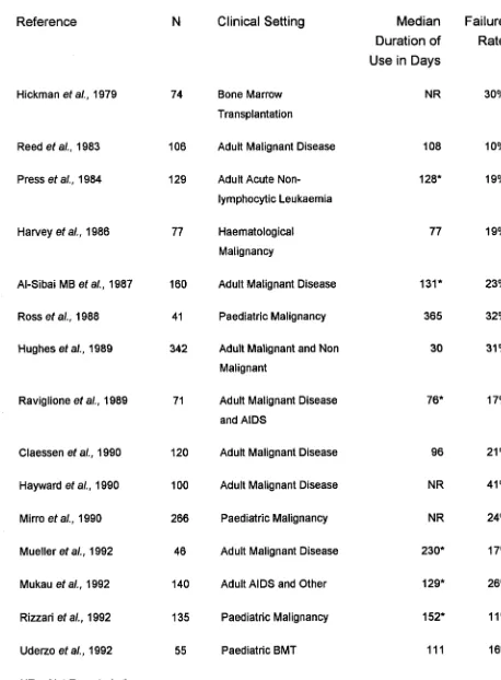
Selected studies describing the use and related adverse events of Hickman catheters are listed in table 1. Most of these observational studies have included, within the study cohort, ambulatory subjects for whom some of the catheter care has been performed at home. The median duration of catheter use in these studies ranges from 30 days up to 365 days. The large majority of catheters have been used without major problems and then removed electively when no longer required, although in one study 41% of catheters were removed because of complications (Haywood et ai, 1990).
Hickman catheters have been well accepted by patients who appreciate the ease of vascular access and the avoidance of repeated venipuncture and needle phobia (Claessen et al., 1990). Fully implantable ports may be preferred particularly by children requiring repeated intravenous access (Ross et ai, 1988).
While the use of Hickman catheters has been an innovation in cancer
medicine, their widespread introduction has been accompanied by significant adverse events and attendant morbidity. Complications reported in relation to Hickman catheter use include, in descending order of frequency: infection, central venous thrombosis, catheter blockage, catheter migration or
Table 1
Hickman Catheter Failure Rate and Duration Of Use
Reference N Clinical Setting Median
Duration of Use in Days
Failure Rate
Hickman e t al., 1979 74 Bone Marrow
Transplantation
NR 30%
Reed e t al., 1983 106 Adult Malignant Disease 108 10%
Press e t al., 1984 129 Adult Acute
Non-lymphocytic Leukaemia
128* 19%
Harvey e t al., 1986 77 Haematological
Malignancy
77 19%
Al-Sibai MB e t a i , 1987 160 Adult Malignant Disease 131* 23%
Ross e t al., 1988 41 Paediatric Malignancy 365 32%
Hughes e t al., 1989 342 Adult Malignant and Non
Malignant
30 31%
Raviglione e t al., 1989 71 Adult Malignant Disease
and AIDS
76* 17%
Claessen et al., 1990 120 Adult Malignant Disease 96 21%
Hayward e t al., 1990 100 Adult Malignant Disease NR 41%
Mirro e t al., 1990 266 Paediatric Malignancy NR 24%
Mueller e t al., 1992 46 Adult Malignant Disease 230* 17%
Mukau e t al., 1992 140 Adult AIDS and Other 129* 26%
Rizzari e t al., 1992 135 Paediatric Malignancy 152* 11%
Uderzo e t al., 1992 55 Paediatric BMT 111 16%
NR = Not Reported; * = mean
[image:20.560.66.524.116.738.2]Hickman Catheter Related Infection
Infection remains the most important problem associated with the long term use of Hickman catheters. The presence of a semi-permanent central
venous catheter has been shown by von Hoff et al., (1990) to be a major risk factor for bacteraemia in children receiving chemotherapy. In their
retrospective cohort study of children receiving chemotherapy in a single institution, those children with central venous catheters (Hickman or Broviac catheters) experienced a risk of infection 6.4-times that of children without catheters after controlling for other risk factors in a logistic regression model. These investigators found that children with central venous catheters spent an average of 15.4 more days per year in hospital for the treatment of catheter related complications, mostly infections.
In a retrospective, matched case control study conducted in a large adult hospital, the presence of a Hickman catheter was a major risk factor for the development of nosocomial candidaemia, with an estimated odds ratio of 7.23 (Wey et al, 1989). Other identified risk factors for candidaemia in this study were: treatment with multiple antibiotics, prior haemodialysis, and the isolation of Candida species from other sites. Karabinis et al., (1988) have reported a retrospective case-control study in adult patients with cancer examining the determinants of candidaemia. Central venous catheters were a significant risk factor for candidaemia with a matched relative risk of 6 in this study when estimated using a logistic regression model.
Pathogenesis of Catheter Infection
Direct access to the circulation provides an easy portal of entry for
organisms, bypassing the physical barriers to infection. Frequent use of the catheter for intravenous infusions, irrigations to prevent clotting in the
catheter and blood sampling might increase access of pathogenic organisms. Body movements can facilitate the migration of organisms along the external surface of the tunnelled portion of the catheter, eventually reaching the circulation.
Bacterial Biofilms:
Intravascular catheters are known to develop bacterial biofilms of slime or glycocalyx, in which colonising bacteria, usually Staphylococcus epidermidis, may be observed using electron microscopy (Newman et a!., 1993). Such biofilms are almost universally present, even on catheters in situ for only 1 day (Passerini et al., 1992). In long term catheters, the colonisation is on the luminal surface (Raad et al., 1993B). The significance of biofilms and
bacterial colonisation of the Hickman catheters remains unclear. Adherent fibrin at the catheter tip may also be important. Several reports suggest treatment with low dose thrombolytic therapy and antibiotics may be more effective than the same antibiotics given alone for some patients with catheter related infection (Ascher et al., 1993; Jones et al., 1993). Further research on the significance of biofilms and fibrin as factors in catheter related infection is awaited.
Source of Organisms Causing Catheter Related Infection:
Infection of central venous catheters may occur with the migration of adherent organisms along the external catheter surface after infecting or colonising the skin catheter entry site. The subcutaneous tunnel and cuff of the Hickman catheter were designed to minimise this mode of infection. Alternatively, organisms may invade by migrating down the luminal surface, after first contaminating the catheter hub (Collignon & Munro, 1989). Careful protocols to prevent catheter hub contamination would be expected to reduce this risk (reviewed by Putterman, 1990). Catheter related infection may develop rarely from the infusion of contaminated fluids, sometimes as outbreaks due to a single contaminated source (Pegues et al., 1993). Patients in whom Hickman catheters are inserted are often debilitated, immune suppressed, and frequently experience prolonged neutropenia after chemotherapy, enhancing the risk of infection. In these patients, particularly
if mucosal barriers are injured by chemotherapy, there may be translocation to, and colonisation of, the catheter by gut bacteria, occurring presumably during periods of asymptomatic bacteraemia (Tancrede & Andremont 1985). A similar finding has been reported in children with short bowel syndrome and central venous catheters. In one study, 19 of 28 cases of catheter sepsis the offending organism was present within the faecal flora, supporting
translocation as an important mechanism of catheter infection in this patient group (Kurkchubasche et at., 1992).
Pathogenic organisms may be introduced at the time of catheter insertion if a break down in aseptic technique occurs leading to early nosocomial infection.
Diagnosis and Definition of Catheter Related Infection
Catheter related infection can be divided into three categories based on the predominant site of infection. Exit site infection refers to catheter infections localised to the exit site. Subcutaneous tunnel infections involve that part of the catheter which is placed subcutaneously, before entering the circulation. Catheter associated bacteraemia orfungaemia implies infection involving the intravascular portion of the catheter or, alternatively, the intraluminal surface. The reported incidence of catheter related infection has varied from 2.7% to 60% (Groeger et a/., 1993). Variation between studies of the definitions of infection used make direct comparisons of infection rates impossible.
Press et al. (1984) have provided a set of definitions for Hickman catheter infections which have provided a basis for subsequent studies. Their definitions are given here.
Hickman catheter exit site infection: development of erythema, tenderness, induration, and/or purulence within 2 cm of the skin exit of the catheter.
Hickman catheter tunnel infection: development of erythema, tenderness and induration along the subcutaneous tract ("tunnel") of the Hickman catheter at a distance >2 cm from the skin exit site with or without signs of inflammation or purulence at the exit site. Insertion site infections (that is the site used to insert the catheter through the vein wall) involve the tunnel and are considered in this category.
Hickman catheter septic thrombophlebitis: development of septic venous occlusion in proximity to the Hickman catheter associated with bacteraemia and fever.
The definition for catheter related sepsis has been modified by Mirro et al to include information from the catheter tip culture and from blood cultures performed on samples drawn simultaneously from the catheter and from a venipuncture (Mirro et al., 1989). Catheter tip culture is performed usually by the semi-quantitative method of Maki (Maki et al., 1977). In this method, an intravascular segment of the removed catheter is rolled on a sheep blood agar plate. The culture is considered to be positive if 15 or more colonies are present. Several investigators have suggested a lower cut off number of colonies to be significant in indicating catheter colonisation and/or infection of Hickman catheters (reviewed by Collignon & Munro, 1989).
Cultures of the catheter tip may provide retrospective confirmation of a clinical diagnosis of catheter related sepsis. Semi-quantitative cultures of intravascular catheters have been included in the Centers for Disease Control definitions for nosocomial venous infection (Garner et al., 1988). Similar information may be obtained by direct staining and microscopic examination of a catheter segment (Cooper & Hopkins, 1985). The clinical utility of these retrospective methods, as assessed by the degree in which test results effect management decisions, is low (Widmer et al., 1992).
Simultaneous peripheral and catheter blood cultures have been useful in the diagnosis of catheter related sepsis with high specificity (89%) but low
sensitivity (Andremont et al., 1988). Catheter drawn specimens for culture are also very suitable for the diagnosis of non catheter related infections (Wormser et al., 1990). The definitions of Mirro et al for septicaemia and catheter related septicaemia extend those of Press et al. (1984) and are summarised here.
Septicaemia: growth of the same organism from two separate blood cultures or isolation of an organism from a single blood culture accompanied by symptoms and signs of infection.
the same organism isolated from the blood and the catheter tip when the catheter was removed.
Quantitative blood culture techniques have been developed which allow the number of organisms per millilitre to be directly estimated (Yagupsky & Nolte, 1990). Such diagnostic methods, although labour intensive, have some putative advantages over the conventional, broth media based method. For example, in Staphylococcus aureus bacteraemia from any cause, high
bacterial colony counts are associated with a poor prognosis (Whimbey et al., 1987; Dugdale & Ramsey, 1990). In the diagnosis of catheter related sepsis, the comparison of quantitative blood cultures obtained from peripheral veins and Hickman catheters has been studied (Fan et al., 1989). A central
catheter to peripheral bacterial colony count ratio of greater than 7 correlated with a subsequent positive catheter tip culture. The sensitivity and specificity of quantitative paired central-peripheral blood cultures in the diagnosis of catheter related septicaemia remain to be defined (Yagupsky & Nolte, 1990). Recently it has been suggested that a central catheter to peripheral bacterial colony count ratio of greater than 4 should be used (Capdevila et al., 1992).
Benezra et al (1988) have incorporated quantitative paired blood cultures into a definition of catheter related septicaemia. In their study of 488 Hickman catheters, exit site infection and catheter tunnel infection were defined as previously described by Press et al (1984).
Catheter related septicaemia was defined as present when:
(1) positive blood cultures collected through the central venous (Hickman) catheter showed a 10-fold or greater colony count compared with peripheral quantitative blood cultures or
(2) if peripheral cultures were not collected , a colony count of 100 colony forming units per ml or more in blood from the central catheter. In addition, if any other clinically or microbiolically apparent source of
bacteraemia was present then the diagnosis of catheter related septicaemia was not made.
infection alone. This test has yet to be applied widely in studies of Hickman catheter related infection.
Microbiology of Hickman Catheter Related Infection
A diverse range of organisms have been associated with Hickman catheter related infection. Bacteria found in skin flora, particularly coagulase negative staphylococci and aerobic diptheriods, have become increasingly important as pathogens in patients with Hickman catheters in situ. These organisms, along with Staphylococcus aureus, have replaced aerobic gram negative rods as the major pathogens in catheter related infection. Table 2 lists organisms responsible for catheter infection from 13 combined studies reviewed by Clarke and Raffin (1990).
Staphylococcus epidermidis has been shown to migrate rapidly along the external surface of a tunnelled central venous catheter in an experimental model (Cooper et at., 1988). It is the most common organism causing
catheter related infection in many studies (Clarke & Raffin 1990 and Table 2). Staphylococcus epidermidis has been associated with catheter related, right sided, endocarditis in severely immune suppressed bone marrow transplant recipients (Martino et at, 1990). In older studies, isolates of Staphylococcus epidermidis may have been erroneously discounted as contaminants.
Infection with Staphylococcus aureus has been accompanied by especially grave sequelae, including septic thrombosis, acute endocarditis and
metastatic infection (George & Cornel, 1992; Raad et al., 1992). In one study of Hickman catheter infection due to this organism, 3 of 36 patients died due to progressive sepsis despite intensive treatment (Dugdale & Ramsey, 1990). Moreover, only 18% of these infections were cured without removal of the catheter. Infection involving the catheter tunnel, in particular, required catheter removal.
Pseudomonas species are also an important cause of catheter related infection in patients who are neutropenic or who suffer from AIDS. The majority of infections can be controlled with antibiotic therapy alone, avoiding catheter removal. Catheter tunnel infection with Pseudomonas aeruginosa has usually required catheter removal (Benezra et at, 1988). In a
retrospective study of 584 patients with AIDS presenting over a 5 year period, 19 bacteraemic infections with Pseudomonas species were observed, of which 11 were Hickman catheter related (Nelson et at, 1991). In this study,
Pseudomonas aeruginosa was second only to Staphylococcus aureus as a cause of catheter related infection. Even in children with underlying HIV infection, the majority of catheter related Pseudomonas species infections can be controlled without catheter removal (Roilides et a!., 1992).
Catheter associated fungaemia is a particularly dangerous adverse event. In a retrospective study by Lecciones et al. (1992) of 155 episodes of catheter related fungaemia and cancer, the mortality rate among infected patients was 52%. Almost all infections were due to Candida species, predominantly
Candida albicans. The high mortality rate was observed despite aggressive therapy with amphotericin B and catheter removal in most patients.
Table II
Spectrum of Organisms Responsible for Central Venous Catheter Infections (from a review of 13 studies by Clarke & Raffin, 1990)
Organism Responsible %
Coagulase-negative staphylococci 31
S ta p h y lo c o c c u s a u re u s 14
Group D streptococcus 3
Other streptococci 6
C o ryn e b a cte riu m species 11
B a cillu s species 3
P s e u d o m o n a s species 7
A c in e to b a c te r 3
E n te ro b a c te r species 4
K le b sie lla species 4
E sch e rich ia co li 6
Other Gram-negative 3
C an dida species 7
Mixed infections 8
Other 2
Total 112
Putative Risk Factors for Infection
A number of potential risk factors for infection complicating Hickman catheter use have been identified in previous studies (including randomised controlled trials). The most frequently observed risk factors are summarised here.
Nature of Underlying Illness:
Many people requiring Hickman or similar semi-permanent intravenous catheters suffer from chronic illnesses which are associated with impaired immunity. Immunocompromised patients which have been observed to have a particularly high risk of catheter related infection include patients suffering from AIDS (Raviglione et al., 1989; Mukau eta!., 1992), prolonged severe neutropenia (Harvey et al., 1986), acute leukaemia and bone marrow transplantation (Groeger et al., 1993; Ranson et al., 1990).
Catheter Exit Site Dressing:
The type of dressing used over the catheter exit site may influence the risk of infection. Transparent semipermeable dressings have been widely used. They form an occlusive dressing and allow the exit site to be observed. A meta-analysis of 15 randomised trials comparing transparent dressings to conventional gauze dressings for the exit site of non-tunnelled central venous catheters has been reported (Hoffman et al., 1992). Transparent dressings were associated with an increased relative risk of infection (RR = 1.69), and of bacteraemia (RR = 1.63) compared to gauze dressings. Similar studies have not been performed with Hickman catheters.
Duration of Placement:
As would be expected, a positive relationship has been observed between the overall risk of Hickman catheter infection and the duration of placement (Fuchs et al., 1984).
Catheter Use:
The use of Hickman catheters for total parenteral nutrition has been observed to elevated the risk of infection when compared to other uses (Mulloy et al., 1991)
Single Versus Double Lumen Hickman Catheters:
lumen catheters compared to single lumen catheters in retrospective non- randomised studies (Early etal., 1990; Henriques etal., 1993). In a
randomised controlled trial of non-tunnelled central venous catheters used for total parenteral nutrition, triple lumen catheters were more frequently
infected than single lumen catheters (Clark-Christoff et al., 1992). Similar randomised trials with Hickman catheters have not been performed.
Criteria for Hickman Catheter Removal for Infection
Many Hickman catheter infections can be controlled with antibiotic therapy without removal of the catheter and guidelines for catheter salvage have been suggested (reviewed by Wickham et al., 1992). In brief, most authors suggest removal if there is catheter related bacteraemia and no response after 48 hours treatment, or if there is evidence of hypotension due to sepsis, or evidence of septic emboli or endocarditis. Catheter removal is required for bacteraemia due to Bacillus species, Corynebacteriurn species and Candida species. Tunnel infections due to Staphylococcus aureus or Pseudomonas species also require the Hickman catheter to be removed. Hickman catheter related septicaemia can often be managed successfully, even in the
presence of neutropenia, using parenteral antibiotics without catheter
removal (Press et al., 1984; Newman et al., 1989; Benezra et al., 1988). The outcome may be improved, however, by early catheter removal when
fungaemia or Staphylococcus aureus bacteraemia is present (Dato & Dajani, 1990; Lecciones et al., 1992; Dugdale & Ramsey, 1990). In a prospective study by Benezra et al. (1988) 33 of 54 patients with catheter related sepsis were cured with antibiotic therapy alone, while in the remaining 21 instances the catheter was removed. Other studies in patients with malignant disease have shown catheter retention rates after antibiotic therapy of between 60% and 90% (Mirro et al., 1989; Press et al., 1984). Similar success rates with Hickman catheter retention have been reported in patients with HIV infection and catheter related septicaemia (Raviglione et al., 1989).
Venous Thrombosis and Hickman Catheter Occlusion
Thrombosis of any of the central veins due to Hickman catheter placement can cause substantial morbidity and may be, rarely, life threatening.
Thrombosis of the superior vena cava is accompanied by clinical signs of bilateral engorgement of neck veins, facial swelling, and the appearance of collateral vessels over the chest wall. The syndrome may lead to cerebral oedema, coma and death. Thrombosis of the brachiocephalic vein is
accompanied by ipsilateral swelling of the neck and arm. Thrombosis of the subclavian or axillary veins produces venous engorgement in the affected limb.
Venous thrombosis is most often detected when upper limb venograms and or catheter contrast studies are performed for symptoms of thrombosis or for catheter blockage. In studies where venograms have been routinely
performed in asymptomatic patients, very high rates of unsuspected
thrombosis (often incomplete) have been found. In such a study by Haire et al. in adults treated with autologous bone marrow transplantation, 22 of 35, or 64%, of venograms showed evidence of thrombosis, with complete blockage of the subclavian vein in 10 (Haire et al., 1991). Most patients were
asymptomatic even when the subclavian vein was completely occluded. The diagnosis of thrombosis may also be achieved using two-dimensional
echocardiography supplemented with colour flow Doppler sonography (Hammerli & Meyer, 1993).
Hickman catheter occlusion may be complete with total obstruction of the catheter. More commonly however, there is simply increased resistance to flow. It is sometimes possible to infuse fluids through the catheter but not to withdraw blood, a so called "one-way catheter". Such catheter malfunction can afflict up to 25% of Hickman catheters (Lokich et al., 1985).
Bern et al. (1990) have conducted a randomised controlled trial of mini-dose warfarin to prevent venous thrombosis in adult patients with solid tumours and Hickman catheters. Again, routine upper limb venography was
performed. The observation period was 90 days. In the warfarin treated group 4 of 42 (10%) catheters were complicated by venous thrombosis versus 15 of 40 (38%) catheters in the control group.
Major venous thrombosis, detected clinically or at post-mortem examination was found to complicate Hickman catheter placement in 17% of patients with solid tumour malignant disease (Anderson et al., 1989). Thrombosis may be accompanied by bacteraemia, which is often resistant to therapy until the thrombotic lesion resolves (Rupar et al., 1990).
Blood clots can be frequently aspirated from asymptomatic, normally
Hickman catheter related central vein thrombosis has been reported to cause clinical pulmonary embolus (Leiby et al., 1989: Hughes et al., 1989).
Pulmonary embolus in this setting may be fatal (Anderson et al., 1989).
Thus, venous thrombosis is a very common accompaniment of Hickman catheter placement. It is often asymptomatic. The significance, clinically, of asymptomatic thrombosis is unclear. Catheter occlusion can occur due to deposition of fibrin and blood clot within the catheter lumen itself. Frequently, a fibrin sheath will develop over the catheter tip without any associated
venous thrombosis. Data from a single trial suggest catheter related thrombosis can be largely prevented by mini-dose warfarin prophylaxis.
Pathogenesis of Hickman Catheter Related Thrombosis
Hickman catheters are constructed from a soft silicone elastomer. Catheters constructed from this material have been shown to have a lower risk of inducing thrombosis than polyethylene catheters (Pottecher et al., 1984). A small randomised trial comparing Hickman or Broviac catheters to polyvinyl chloride catheters found a thrombosis rate of 5% in both patient groups (Wagman et al., 1984). Thus, although an improvement upon older catheter designs, Hickman catheters remain thrombogenic. Platelet aggregation and initiation of the coagulation cascade may triggered by the catheter itself, or by associated endothelial damage. Infusion of some fluids including
hypertonic parenteral nutrition solutions may initiate thrombosis (Brennan, 1985).
Fibrin Sheath:
The formation of a fibrin sheath enveloping the distal intravascular segment is a frequent cause of catheter blockage. In one radiological study of
malfunctioning Hickman catheters, an occluding fibrin sheath was present in 57% of cases (Cassidy et al., 1987). Rarely, the enveloping fibrin sheath can be so extensive as to cause retrograde flow of infusate within the fibrin
sheath to the catheter entry site with extravasation (Gemlo et al., 1988). The development of fibrin sheath can be detected early by measuring the
resistance to flow within the catheter directly (Stokes et al., 1989). Catheter management protocols have generally included frequent bolus flushes of low dose heparin in order to prevent the development of intraluminal clots and fibrin sheaths (Fry, 1992). Despite these protocols, intraluminal blood clots are common, although the consequences of these in terms of rates of catheter blockage remain uncertain (Anderson et al., 1987)
The relationship of malignancy to an increased risk of thromboembolic
diseases was described initially by Trousseau in 1865 and has subsequently been confirmed in many studies (Bunn & Minna, 1985). Carcinomas,
particularly mucin producing adenocarcinomas, have the highest risk. Chemotherapy may contribute to a transient hypercoagulable state in patients with solid tumours, contributing to thrombosis risk (Levine et al., 1988).
Putative Risk Factors for Thrombosis and Catheter Malfunction
The determinants of central venous thrombosis related to Hickman catheters have been less carefully studied than the determinants of infectious
complications. Risk factors that have been observed are discussed here. Nature of Underlying Illness:
In a study of Hickman catheters in patients with solid tumours, a diagnosis of adenocarcinoma of the lung was associated with the highest risk of catheter related thrombosis (Anderson et al., 1989). Thrombosis rates, reported from cohort studies where the majority of patients suffered from haematological disorders, are lower than those reported where the majority suffered from solid tumours. Such indirect comparisons suggest a higher risk of major thrombosis in patients with solid tumours (reviewed by Leiby et al., 1989).
Further studies are required to test this hypothesis. Distortion of the superior vena cava due previous thrombotic episodes or due to large mediastinal tumours can elevate the risk of thrombosis.
Catheter Use:
Continuous infusions of chemotherapeutic drugs such as 5-flourouracil may lead to thrombosis more frequently than bolus injections through the catheter (Lokich et al., 1985). Use of the catheter for total parenteral nutrition may be accompanied by an elevated risk of thrombosis although direct evidence is lacking (Mughal 1989).
Position of the Catheter Tip and Site of Insertion:
In a large retrospective study of implanted ports in patients with cancer, Puel et al found a strong relationship between catheter tip malposition and
study of both Hickman catheters and ports, catheter tip position high within the superior vena cava or within the brachiocephalic veins led to thrombosis (Stanislav et al., 1987).
Migration of the catheter tip to an unsatisfactory intravascular or
extravascular position is discussed in the next section. Incomplete occlusion, with failure of blood to be aspirated may occasionally be due to abutment of the catheter tip against the wall of the central vein, usually the superior vena cava (Cassidy et a!., 1987). Improvement in catheter function can sometimes be obtained in these circumstances by changing the posture of the patient during catheter use.
Catheter Compression:
With percutaneously inserted Hickman catheters, compression of the catheter can occur as it passes between the clavicle and first rib prior to entering the subclavian vein (Aitken & Minton, 1984). Such compression can lead to postural catheter occlusion and, more importantly, catheter fracture, as discussed below.
Catheter Salvage After Central Vein Thrombosis
The traditional management of central venous thrombosis has included systemic anticoagulation and catheter removal (Lokich et al., 1985). Several investigators have demonstrated favourable outcomes with anticoagulation only, while maintaining the catheter in situ (Anderson et al., 1989). The local infusion of low dose urokinase has also been suggested to assist catheter salvage and to reduce morbidity from central vein thrombosis (Fraschini et al., 1987)
Hickman catheter occlusion, not associated with major venous thrombosis or catheter malposition, has been successfully relieved with low dose urokinase bolus injections (Anderson et al., 1987). This manoeuvre may fail in up to 68% of cases (Monturo et al., 1990). In these resistant cases, low dose infusions of urokinase, streptokinase or tissue plasminogen activator have been successful (Haire et al., 1990: Wickham et al., 1992). Although re occlusion may occur, many Hickman catheters can be safely salvaged with these methods (Haire & Lieberman, 1992).
Catheter Migration And Dislodgement
Despite accurate placement of a Hickman catheter, it is possible for the catheter tip to migrate to an unsatisfactory position, such as the internal jugular vein (Rasuli et al., 1992). Migration may be triggered by paroxysmal coughing, and has been reported as a complication of cystic fibrosis (Jacobs & Zaroukian, 1991). Such catheters often function poorly and are more likely to cause venous thrombosis (Cassidy et al., 1987). More serious sequelae have been reported following erosion of a vein wall by the catheter tip with migration to an extravascular site. Subsequent infusions through the catheter can lead to catastrophic extravasation of infusate (Krasna & Krause, 1991; Cathcart-Rake & Mowery, 1991). Lateral orientation of the catheter tip, abutting the vein wall, may be a precursor to extravascular migration (Manheimer et al., 1992).
Catheter dislodgement due to accidental traction on the external portion of the catheter has caused significant catheter loss, both in adult and paediatric populations (Carde et al., 1989). The subcutaneous Dacron cuff is not firmly adherent to the subcutaneous tissues for several weeks and early
dislodgement can therefore occur. Even minor degrees of dislodgement might be expected to cause migration of the Hickman catheter tip to a higher and possibly less satisfactory position within the superior vena cava or subclavian veins (Cassidy et al., 1987).
Management of Catheter Migration
Migratory catheter tips can sometimes be repositioned with guide wire manipulation (Rasuli et al., 1992). Minor dislodgement may leave the catheter in a usable position, however most dislodgements or catheter tip migrations lead to catheter loss.
Mechanical Failure or Fracture
As Hickman catheters may be in use for many months, they are subject to wear and tear, and particularly to accidental puncture. Repair of damaged
Hickman catheters with silicone adhesive and replacement hub kits is
possible. Rarely Hickman catheters have fractured at the site of compression between the first rib and the clavicle, leading to embolisation of the
radiographs as the "pinch-off sign". A scoring system has been suggested (Hinke et at., 1990). Removal after a maximum of 6 months use has been recommended for "pinched-off" Hickman catheters (Lafreniere 1991). Acute Complications of Hickman Catheter Insertion
The insertion procedure may be accompanied by bleeding often in association with accidental arterial puncture or traumatic pneumothorax. Although accidental arterial puncture has been reported as frequently as 6% of placements (Wisborg et al., 1990), significant bleeding is rare, even in populations with a high prevalence of coagulation disorders (Harvey et al., 1986). Pneumothorax is also uncommon, complicating 1-5% of
percutaneously inserted Hickman catheters (Hughes et al., 1989: Wisborg et al., 1990). Pneumothorax of sufficient severity to require intercostal tube drainage remains infrequent. Higher rates of detection of unsuspected iatrogenic pneumothorax can be obtained if delayed chest radiographs are taken (Spiliotis et al., 1992).
Venous air embolism has been reported during use or removal of central venous catheters as has a single instance of intra cerebral air embolism during insertion of a Hickman catheter (Mennim et al., 1992; Dukes et al., 1991).
6.
METHODS
Design
A retrospective records based cohort study of consecutive Hickman catheters placed in a population of patients with malignant or haematological disease attending the medical oncology and clinical haematology units within a large hospital was conducted. The unit of study was each individual Hickman catheter successfully inserted during the study period.
Ethical Considerations
The written permission of the ACT Health Ethics Committee was obtained to conduct the study. Clinical records and information used during the study was stored securely within a medical records storage room within Woden Valley Hospital. A computer data file, with patient identifying information removed, was used within the Australian National University (in a secure facility) to perform part of the data analysis.
Case Ascertainment
All Hickman catheters placed during a 36 month study period, from January 1989 to December 1991 inclusive, were identified by reviewing the clinical departments' records. Cross-checking of these records was performed by reviewing log books held in the angiography suites of the Radiology
Department. Following the 36 month study period, study catheters remaining in situ were followed for a further 8 months. Data collection was performed in September 1992 and events included if they occurred prior to August 31, 1992.
Catheter Eligibility
Technique of Catheter Insertion
All catheters were inserted percutaneously using local anaesthesia and fluoroscopic control in the Radiology Department (see Section 4 for
discussion). Placement procedures were performed by one of two consultant radiologists or by senior radiology registrars trained in the technique. Single lumen Hickman catheters were used unless a double lumen catheter was requested by the clinical team. Such requests were based on the expectation of a requirement for more than one intravenous infusion concurrently. The site of insertion (left or right) was chosen by the radiologist performing the placement. The inserting radiologists generally preferred the right sided approach although left sided catheters were freely inserted if preferred by the patient. The right sided approach was considered relatively contraindicated if previous surgery had occurred within the right shoulder region or if a right subclavian vein catheter had been used in the past. In these circumstances the left sided approach was used.
The insertion method has been described (Cockburn eta!., 1992; Page et a/., 1990; Robertson et a!., 1989). The procedure was performed with the patient supine. Under aseptic conditions, a stab incision was made at the entry site below the junction of the lateral and middle thirds of the clavicle. The
subclavian vein was punctured with a Chub needle and J-wire inserted, over which a peel-away sheath was introduced. The subcutaneous track was created with a tunnelling instrument (Heyd & Rosser, 1991). The catheter length was determined by laying it along the line on of the j-wire using
fluoroscopy, with the aim of leaving the catheter tip at the right atrial-superior vena cava junction. The catheter was then filled with saline containing
heparin and inserted through the peel away sheath and positioned with fluoroscopic control. Catheter function was assessed immediately after the
procedure. The catheter exit site was covered with a semi-permeable transparent dressing (OpsiteR). An erect posterioanterior chest radiograph was taken after insertion to check the final position of the catheter tip and to exclude the immediate complications of pneumothorax and haemothorax. Catheter Use and Care
There were no specific restrictions on clinical use of the catheters during the study period. Hickman catheters were used to infuse intravenous drugs and fluids including cytotoxic chemotherapy, blood products, and parenteral nutrition as required. Blood samples for testing were routinely drawn from