www.ecmjournal.org GA Feichtinger et al. Non-viral osteoinductive gene therapy European Cells and Materials Vol. 27 2014 (pages 166-184) ISSN 1473-2262
Abstract
Tissue regenerative gene therapy requires expression strategies that deliver therapeutic effective amounts of transgenes. As physiological expression patterns are more complex than high-level expression of a singular therapeutic gene, we aimed at constitutive or inducible co-expression of 2 transgenes simultaneously.
Co-expression of human bone morphogenetic protein 2 and 7 (BMP2/7) from constitutively expressing and doxycycline inducible plasmids was evaluated in vitro in C2C12 cells with osteocalcin reporter gene assays and standard assays for osteogenic differentiation. The constitutive systems were additionally tested in an in vivo pilot for ectopic bone formation after repeated naked DNA injection to murine muscle tissue.
Inductor controlled differentiation was demonstrated in vitro for inducible co-expression. Both co-expression systems, inducible and constitutive, achieved significantly better osteogenic differentiation than single factor expression. The potency of the constitutive co-expression systems was dependent on relative expression cassette topology. In vivo, ectopic bone formation was demonstrated in 6/13 animals (46 % bone formation efficacy) at days 14 and 28 in hind limb muscles as proven by in vivo µCT and histological evaluation.
In vitro findings demonstrated that the devised single vector BMP2/7 co-expression strategy mediates superior osteoinduction, can be applied in an inductor controlled fashion and that its efficiency is dependent on expression cassette topology. In vivo results indicate that co-expression of BMP2/7 applied by non-viral naked DNA gene transfer effectively mediates bone formation without the application of biomaterials, cells or recombinant growth factors, offering a promising alternative to current treatment strategies with potential for clinical translation in the future.
Keywords: Bone morphogenetic protein; co-expression; non-viral gene therapy; inducible, BMP2/7; heterodimer; intramuscular.
*Address for correspondence: Georg A. Feichtinger,
Ludwig Boltzmann Institute for Experimental and Clinical Traumatology,
Donaueschingenstrasse 13, A-1200 Vienna, Austria
Telephone Number: +43 1 33110 461 FAX Number: +43 1 33110 460 Email: georg.feichtinger@trauma.lbg.ac.at
Introduction
Transient somatic gene transfer, enabling temporally and spatially restricted, safe expression of transgenes in vivo, could revolutionise the clinical treatment of organ and tissue specific diseases. Current gene transfer methods are divided into viral (high efficiency, high immunogenicity) and non-viral (low efficiency, low immunogenicity) gene transfer for transient expression of therapeutic transgenes (Gelehrter et al., 1998; Bleiziffer et al., 2007). Transient gene therapeutics can augment local levels of cytokines and morphogens to compensate for pathologically down-regulated gene expression or locally augment growth factors to therapeutic levels. In contrast to the therapy of monogenetic diseases with stable integrating viral vectors, that have to provide life-long stable expression of therapeutic genes, non-viral non-integrating vectors – such as plasmid DNA – are active as non-replicating episomal entities in vivo. The information is lost within a specific time frame, with a very rare frequency of random chromosomal integration (Martin et al., 1999; Ledwith et al., 2000; Coelho-Castelo et al., 2006) and is therefore not associated with vector-associated genotoxicity.
For tissue regenerative gene therapy approaches, the inherent drawback of loss of transgene expression in transient gene therapy is actually a desired safety feature. Temporally regulated, or restricted expression, of the therapeutic is mandatory to prevent systemic effects, aberrant cellular growth and to allow complex tissue growth (Franceschi, 2005; Bleiziffer et al., 2007). Plasmid DNA is cleared relatively quickly from the system and does not show substantial systemic spread or ectopic expression at off-target sites (Hengge et al., 2001; Hohlweg and Doerfler, 2001; Gehl, 2003; Coelho-Castelo et al., 2006). Low production costs of the non-viral plasmid therapeutic are another potential advantage (Bleiziffer et al., 2007). Plasmid DNA is produced with sufficient purity and in sufficient amounts for clinical application at relatively low cost, compared to recombinant growth factors (Bonadio et al., 1999; Johnson and Urist, 2000; Tepper and Mehrara, 2002; Einhorn, 2003) as advanced therapeutics. Gene therapeutics lead to an inherent sustained release of expressed therapeutic genes by host cells in situ, which is more effective due to higher bioactivity of the host-produced growth factor (Bonadio et al., 1999; Bleiziffer et al., 2007). Plasmid DNA has been successfully applied in direct in vivo gene transfer for wound healing and angiogenesis (Michlits et al., 2007; Mittermayr et al., 2008), cardiac regeneration (Sundararaman et al., 2011) and musculoskeletal regeneration (Grossin et al., 2003; Kawai et al., 2006; Osawa et al., 2009; Osawa et al., 2010).
CONSTITUTIVE AND INDUCIBLE CO-EXPRESSION SYSTEMS FOR NON-VIRAL
OSTEOINDUCTIVE GENE THERAPY
G.A. Feichtinger1, A.T. Hofmann1, K. Wassermann1, A. Zimmermann1, M. van Griensven1,2 and H. Redl1
1 Ludwig Boltzmann Institute for Experimental and Clinical Traumatology, AUVA Research Centre, Austrian Cluster
for Tissue Regeneration, European Institute of Excellence on Tissue Engineering and Regenerative Medicine Research (Expertissues EEIG) Vienna-Branch, Vienna, Austria
Taking these advantages into account, non-viral vectors, such as plasmid DNA, are considered the most likely candidates for clinical translation of tissue regenerative gene therapies.
The major drawback of non-viral vectors, however, is their low transfection efficacy if used without adjuvant measures (naked) (Herweijer and Wolff, 2003; Schertzer et al., 2006). A wide range of agents, materials and physical methods to enhance the in vivo transfer of plasmid DNA have been developed with varying success (Bleiziffer et al., 2007). An alternative strategy to increase the therapeutic effectiveness is plasmid modification (Tolmachov, 2009; Tolmachov, 2011) and the selection of an optimal therapeutic gene to compensate for the low transfection efficacy of plasmid vectors in vivo. Furthermore, gene combinations are beneficial in experimental models (Zhu et al., 2004; Kawai et al., 2006; Steinert et al., 2009; Wan et al., 2012). Several multi-gene approaches have already been applied for tissue regenerative therapies. In bone regeneration it has been shown that combinatorial gene therapy using multiple bone morphogenetic protein (BMP) genes – especially the combination of BMP2 and BMP7 – leads to higher osteogenic bioactivity compared to single factor expression (Zhu et al., 2004; Kawai et al., 2006). This BMP2/7 co-expression strategy has been shown to lead to the expression of a heterodimeric growth factor with higher bioactivity (Israel et al., 1996) compared to the respective single gene derived homodimeric variants (Kawai et al., 2009).
Gene combinations can be co-delivered as a mixture of different single expression cassette plasmids, as co-expression plasmids with multiple independent transcriptional entities (Kawai et al., 2009) and as plasmids encoding poly-cistronic mRNA with intra-ribosomal entry sites (IRES) (Gurtu et al., 1996; Guo-ping et al., 2010). Multi-cistronic plasmids allow the co-expression of multiple factors from the same plasmid molecule via bidirectional, or separated unidirectional, gene expression cassettes and enable the simultaneous expression of multiple therapeutic genes at the target site.
The achievable functional expression levels of multi-cassette co-expression systems could be strongly related to expression cassette topology, due to potential transcriptional interference phenomena (Callen et al., 2004; Shearwin et al., 2005) between the promoters. Furthermore, multiple expression cassettes using the same heterologous promoters could be prone to competitive effects, reducing total transgene expression (Ngo et al., 1993). Nevertheless, at least within the scope of the co-delivery of 2 therapeutic genes, single molecule transcriptional entities can still be advantageous in vivo. If both transgenes are encoded on the same molecule, then the correlated expression of both factors obtained from co-expression plasmid transfer should theoretically be higher compared to co-transfection of 2 individual plasmids. This correlated co-expression from one single molecule can be advantageous for the production of heterodimeric growth factor variants (Kawai et al., 2009) or for balanced co-expression of 2 individual synergetic factors (Banfi et al., 2012).
Besides constitutive expression strategies, there are plasmid systems available with response elements that enable regulated, inductor controlled levels of therapeutic gene expression. Such, particular regulated expression kinetics could provide enhanced safety (on/off inductor dependence) and allow mimicking of physiological gene expression patterns for increased therapeutic efficacy. Systems such as the dual-component (activator and response plasmid) tetracycline (Tet) inducible or repressed (TetON, TetOFF) regulated gene expression systems (Gossen and Bujard, 1992; Gossen et al., 1995; Goverdhana et al., 2005) can be applied for this purpose, even for co-expression of 2 therapeutic genes (Baron, 1995).
For therapeutic in vivo over-expression studies, the tetracycline-inducible system (TetON) appears to be more suitable than the tetracycline repressible TetOFF system, since the TetON system is induced by application rather than withdrawal of a small molecule and in a default off-state in its absence.
These inducible systems could help to determine the minimally required gene dose and to fine-tune therapeutic expression levels in experimental models. However, their translatability to the clinic remains questionable given the complexity of such systems and the need to additionally express a heterologous transactivator transgene with potential immunogenicity (Le Guiner et al., 2007). Simple constitutive co-expression systems, ideally backbone-devoid minicircle (Chen et al., 2003) variants, on the other hand are more likely to be clinically translated.
With regards to the above mentioned opportunities to enhance the therapeutic efficacy of non-viral plasmid based gene delivery through modification of the vector, it was the aim of this study to design and evaluate constitutive and inductor-controlled inducible BMP2/7 co-expression plasmids for osteoinductive non-viral in vivo gene therapy. The TetON-inducible bidirectional co-expression system was modified to contain all elements (response and activator sequences) on one single plasmid. Subsequently it was evaluated for its inductor dose dependent expression kinetics. Its osteoinductive capacity was compared to single factor expression, constitutive co-expression or application of recombinant growth factors in vitro. The constitutive systems were designed based on the plasmid backbone pVAX1, which is compliant with FDA safety guidelines for plasmid DNA vaccines (FDA Docket No. 96N-0400), and the employed therapeutic BMP 2 and 7 cDNAs were of human origin, in order to generate constitutive systems with potential for clinical translation. These “preclinical” constitutive systems were evaluated in vitro for their osteoinductive capacity and tested in vivo in an ectopic bone formation assay using repeated non-viral intramuscular in vivo gene transfer.
Materials and Methods
Primers
www.ecmjournal.org GA Feichtinger et al. Non-viral osteoinductive gene therapy
Growth factors
Recombinant CHO-derived human BMP-2 was purchased from Medtronic Biopharma (Neuchatel, Switzerland, PZN#20238). Recombinant CHO-derived human BMP7 and E. coli-derived BMP-2/7 heterodimer were purchased from R&D Systems (Minneapolis, MN, USA, #354-BP, #3229-BM). The lyophilised growth factors were dissolved according to manufacturers’ instructions and then diluted to working stocks 100 ng/µL and stored at -80 °C prior to use.
Plasmids
The constitutive expression plasmid pVAX1 was purchased from Life Technologies (Paisley, UK, #V260-20). The two-component tetracycline (Tet) inducible co-expression system, consisting of the pTREtight-BI response and the pTetON-Advanced activator plasmid, was purchased from Takara Bio Europe/Clontech (Saint-Germain-en-Laye, France, #631068, #631069). The click beetle luciferase (CBR) plasmid pCBR-Control was purchased from Promega (Madison, WI, USA, #E1421). The CMV-enhanced osteocalcin reporter plasmid pCMVE/mOCP-EYFPHis was described previously (Feichtinger et al., 2010). All plasmids were maintained in E.coli TOP10 (Life Technologies #C404003) unless indicated otherwise and prepared as endotoxin-free maxipreps using the EndoFree Plasmid Maxi or Giga kits from Qiagen (Hilden, Germany, #12362) for in vitro and in vivo use.
Cloning of human BMP2 and BMP7 cDNAs
Full-length human BMP2 and BMP7 cDNA were cloned from human total lung RNA or kidney RNA (Takara Bio Europe/Clontech, #636524, #636529) using AMV based reverse transcription of poly-a RNA to cDNA (Promega, #M5108) according to manufacturers’ instructions and primers in Table1. Both cDNAs were subcloned and
verified into pVAX1 by means of sequencing (Microsynth, data not shown).
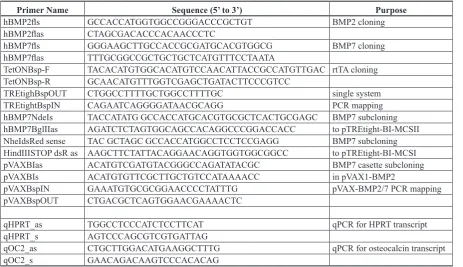
Preparation of constitutive co-expression plasmids The co-expression plasmids pVAX1-BMP2/7+ (tandem organisation of expression cassettes, Fig. 1C) and pVAX1-BMP2/7– (divergent organisation of expression cassettes, Fig. 1D) were constructed by transferring the complete expression cassette for BMP7 expression from pVAX1-BMP7 to pVAX1-BMP2 (Fig. 1A, restriction sites for insertion marked with red lines) to the BspH1 backbone site in pVAX1-BMP2 using standard PCR (primers see Table 1) and cloning procedures.
Preparation of inducible single-vector co-expression plasmids
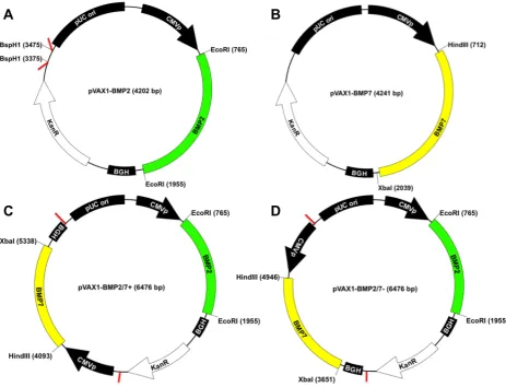
An inducible expression kinetics reporter system was designed by PCR-amplification and cloning of dsRed and EYFP in pTREtight-BI using standard cloning procedures. This response plasmid pTRE-EYFP/dsRed (Fig. 2A) was then modified to a single-vector inducible system by cloning the entire reverse tet-transactivator (rtTA) expression cassette, obtained through high-fidelity Phusion® polymerase (New England Biolabs, Frankfurt,
Germany, #M0530S) PCR amplification (primers see Table 1), from the pTetON Advanced activator plasmid (Fig. 2B) into the BspLU11I site of pTRE-EYFP/dsRed (Fig. 2A, insertion site marked red) to form pTetON-EYFP/ dsRed (Fig. 2C). Both plasmids were used for expression kinetics studies and for a comparison of single vector TetON systems with standard double vector systems. The therapeutic inducible co-expression plasmid pTetON-BMP7/2 (Fig. 2D) was created by transferring the cDNAs of human BMP2 and human BMP7 into pTREtight-BI using standard cloning procedures in the E. coli cloning
Primer Name Sequence (5’ to 3’) Purpose
hBMP2fls GCCACCATGGTGGCCGGGACCCGCTGT BMP2 cloning
hBMP2flas CTAGCGACACCCACAACCCTC
hBMP7fls GGGAAGCTTGCCACCGCGATGCACGTGGCG BMP7 cloning
hBMP7flas TTTGCGGCCGCTGCTGCTCATGTTTCCTAATA
TetONBsp-F TACACATGTGGCACATGTCCAACATTACCGCCATGTTGAC rtTA cloning TetONBsp-R GCAACATGTTTGGTCGAGCTGATACTTCCCGTCC
TREtighBspOUT CTGGCCTTTTGCTGGCCTTTTGC single system
TREtightBspIN CAGAATCAGGGGATAACGCAGG PCR mapping
hBMP7NdeIs TACCATATG GCCACCATGCACGTGCGCTCACTGCGAGC BMP7 subcloning hBMP7BglIIas AGATCTCTAGTGGCAGCCACAGGCCCGGACCACC to pTREtight-BI-MCSII NheIdsRed sense TAC GCTAGC GCCACCATGGCCTCCTCCGAGG BMP7 subcloning HindIIISTOP dsR as AAGCTTCTATTACAGGAACAGGTGGTGGCGGCC to pTREtight-BI-MCSI
pVAXBIas ACATGTCGATGTACGGGCCAGATATACGC BMP7 casette subcloning
pVAXBIs ACATGTGTTCGCTTGCTGTCCATAAAACC in pVAX1-BMP2
pVAXBspIN GAAATGTGCGCGGAACCCCTATTTG pVAX-BMP2/7 PCR mapping
pVAXBspOUT CTGACGCTCAGTGGAACGAAAACTC
qHPRT_as TGGCCTCCCATCTCCTTCAT qPCR for HPRT transcript
qHPRT_s AGTCCCAGCGTCGTGATTAG
qOC2_as CTGCTTGGACATGAAGGCTTTG qPCR for osteocalcin transcript
[image:3.595.70.534.68.335.2]qOC2_s GAACAGACAAGTCCCACACAG
strain SURE2 (Stratagene/Agilent Technologies, Santa Clara, CA, USA, #200152). The system was then modified to a single-vector inducible system as described for the kinetics reporter system.
Cell culture and transfection
The mouse cell line C2C12, capable of osteogenic differentiation (Katagiri T, 1997; Katagiri T, 1994) was purchased from the German Collection of Microorganisms and Cell Cultures (DSMZ, Braunschweig, Germany, #ACC565) and cultured in Dulbecco’s modified Eagle’s medium (DMEM; Sigma-Aldrich, Vienna, Austria, #D6546) containing 4.5 g/L D-glucose, supplemented with 2 mM L-glutamine (Sigma-Aldrich, #G6392) and 5 % foetal bovine serum (FBS; Lonza, Basel, Switzerland, #14-502E).
The cells were kept sub-confluent during expansion and seeded at 0.5 x 10⁵ cells/mL/well in 24-well plates
to enable transfection at approx. 80 % confluence unless otherwise stated. Differentiation experiments were carried out in serum-reduced DMEM+ 1 % FCS for 6 d.
Constitutive systems
Initial transfection studies were carried out in C2C12 cells at a cell density of 0.5 x 10⁵ cells/mL for 6 d using
Lipofectamine 2000 (LifeTechnologies, #11668019), according to the manufacturer’s instructions. 2 µg total pDNA : 2 µL Lipofectamine 2000 per well in a 24-well plate was used in transfections with vectors expressing only one factor. 1 µg: 1 µg (2 µg total pDNA) of these plasmids were used in co-transfection incubated with 2 µL Lipofectamine 2000. Co-expression plasmids were transfected at an amount of 2 µg: 2 µL of Lipofectamine 2000.
The constitutive systems were tested with the osteocalcin reporter plasmid pCMVE/mOCP-EYFPHis, in a super-transfection experiment. C2C12 cells were transfected with the reporter plasmid 24 h prior to seeding in a T175 flask at 80 % confluence. 24 h post transfection with the reporter, the cells were seeded into 24-well plates at a density of 0.5 x 10⁵ cells/ml. 24 h post
[image:4.595.66.530.56.410.2]www.ecmjournal.org GA Feichtinger et al. Non-viral osteoinductive gene therapy
following transfection, the medium was changed to DMEM with a reduced serum amount of 1 % FCS. Fluorescence microscopy for osteocalcin-linked EYFP expression was carried out 6 d post expression vector transfection.
Inducible systems
The expression levels of the inducible reporter systems pTRE-EYFP/dsRed, pTetON-Advanced (double vector system) and pTetON-EYFP/dsRed were compared by fluorescence microscopy after transfection and induction in C2C12 cells. The double vector system was co-transfected as 1 µg pTRE-EYFP/dsRed response plasmid combined with 1 µg of pTetON-Advanced activator plasmid using 2 µL of Lipofectamine 2000 per well in a 24-well plate. In the other group, 2 µg of the single vector system pTetON-EYFP/dsRed was transfected using 2 µL Lipofectamine 2000 per well in a 24-well plate.
Both systems were induced with a single dose of doxycycline (0 ng, 250 ng and 1000 ng/mL/well) (Takara Bio Europe/Clontech, #631311) at the start of the experiment (following transfection) and expression of the fluorescent reporter genes was compared 48 h post induction. The expression kinetics of the single vector system were investigated after transfection into C2C12 cells and induction with a single dose of 0 ng, 250 ng and 1000 ng/mL/well of doxycycline for up to 14 d with fluorescence microscopy.
[image:5.595.67.535.56.410.2]The inducible system was tested with the osteocalcin reporter plasmid pCMVE/mOCP-EYFPHis in a co-culture experiment. 0.25 x 10⁵ pCMVE/mOCP-EYFPHis
transfected “reporter cells” were mixed with 0.25 x 10⁵
pTetON-BMP2/7 “osteogenic cells” per well and cultured for 6 d in DMEM+ 1 % FCS. Co-Expression of BMP2 and BMP7 was induced with a single dose of 0 ng, 250 ng and 1000 ng/mL/well of doxycycline. Osteocalcin-coupled reporter gene expression by pCMVE/mOCP-EYFPHis was observed by fluorescence microscopy on day 6.
Alkaline phosphatase (ALP) assay
Enzymatic ALP assays were carried out as previously described (Feichtinger et al., 2010). Enzyme activity was calculated as fold induction relative to negative control samples, 6 d after induction.
Osteocalcin gene expression analysis
Osteocalcin gene expression was determined as fold induction (comparative CT method, (Schmittgen and Livak, 2008)) relative to negative control samples by quantitative real-time PCR using SYBR green as previously described (Feichtinger et al., 2010) using the primers qOC2_s, qOC2_as and qHPRT_s and qHPRTas (Table 1).
BMP-ELISAs
BMP2 and BMP7 concentrations were assessed in samples from C2C12 cells transfected with the constitutive systems BMP2, BMP7 and pVAX1-BMP2/7- 24 h after transfection using standard Anti-BMP2 (BMP-2 Quantikine ELISA Kit, #DBP200, R&D Systems/Biomedica, Vienna, Austria) and 7 (Human BMP-7 Quantikine ELISA Kit, #DBP700, R&D Systems/ Biomedica, Vienna, Austria) ELISA kits and protocols according to the manufacturer’s instructions.
In vivo non-viral naked DNA gene transfer
The animal protocol review board of the City Government of Vienna, Austria approved all experimental procedures in accordance with the Guide for the Care and Use of Laboratory Animals as defined by the National Institute of Health. Female nude mice (Hsd:Athymic Nude-Foxn1nu,
Harlan Laboratories, Bresso, Italy, #069(nu)/070(nu/+)) of approx. 10-12 weeks age weighing approx. 30 g were used in this study (n = 18).
Plasmid Injections
20 µg of either pVAX1-BMP2/7 or pTetON-BMP2/7 both combined with 20 µg of pCBR plasmid as internal luciferase transfection control were injected as a mixture (50 µL total) into gastrocnemius hind-limb muscles under short inhalation anaesthesia (2 vol.% isoflurane (Forane, Abbott GmbH, Vienna, Austria) and 3 L/min air) at each day of the treatment protocol. The treatment was repeated to a total of 5 injections, resulting in a final total dose of 200 µg plasmid DNA per hindlimb muscle (100 µg therapeutic plasmid: approx. 3.3 mg/kg therapeutic DNA per animal). Tet-inducible BMP2/7 co-expression by pTetON-BMP2/7 was induced in vivo using 2 mg doxycycline (Takara Bio Europe/Clontech, Saint-Germain-en-Laye, France, #631311) in 300 mL ringer solution
(Mayerhofer Pharmazeutika GmbH, Linz, Austria, license #16.609) intraperitoneally per day for a total of 7 d.
Micro Computer Tomography (µCT) analysis
In vivo µCT images of hindlimbs were obtained using a vivaCT 75 (Scanco Medical, Brütisellen, Switzerland) 14 and 28 d post last gene transfer under short inhalation anaesthesia. Bone volume (mm3) and bone mineral
density (mg hydroxyapatite per cm3) for 14 and 28 d were
calculated using Scanco software and a standard density calibration phantom.
Bioluminescence imaging
Bioluminescence imaging of CBR luciferase activity after in vivo gene transfer was carried out under short inhalation anaesthesia 24 h post last plasmid DNA injection using a Xenogen IVIS100 Imaging system (Caliper Life Sciences, Mainz, Germany). Mice received 0.5 mg D-luciferin potassium salt (Caliper Life Sciences, # 122796) in 300 µL ringer solution (Mayerhofer Pharmazeutika, Linz, Austria, license #16.609) intraperitoneally before imaging. 20 min post D-luciferin administration, the mice were imaged for 2 min. Imaging was carried out at 24 h, 48 h, 72 h, 9 d, 14 d and 28 d post last gene transfer. Luminescence intensity was depicted in overlay images in false colour.
Histological examination
Gastrocnemius muscles were excised and fixed with 4 % paraformaldehyde in PBS for 24 h, dehydrated in 50 % ethanol and stored in 70 % ethanol at 4 °C. Samples were embedded in paraffin without decalcification, and several sections of the same sample were stained with haematoxylin and eosin (H&E) and von Kossa staining for mineralisation according to standard histology protocols.
Statistical analysis
Results are represented as average ± standard deviation (AVG ±SD) unless otherwise stated. Statistical testing was carried out using the non-parametric Kruskal-Wallis test in conjunction with Dunn’s multiple comparison test as post-test for ALP data.
Real-time PCR data passed normality tests and was tested with parametric ANOVA analysis followed by Tukey’s multiple comparison test. Bone mineral density values for 14 d and 28 d passed normality tests and were evaluated with a two-tailed t-test for significance. p < 0.05 was considered statistically significant (* p < 0.05, *** p < 0.001).
Results
Constitutive co-expression systems in vitro
Osteogenic differentiation using single factor expression vs. co-expression
www.ecmjournal.org GA Feichtinger et al. Non-viral osteoinductive gene therapy
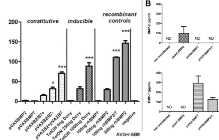
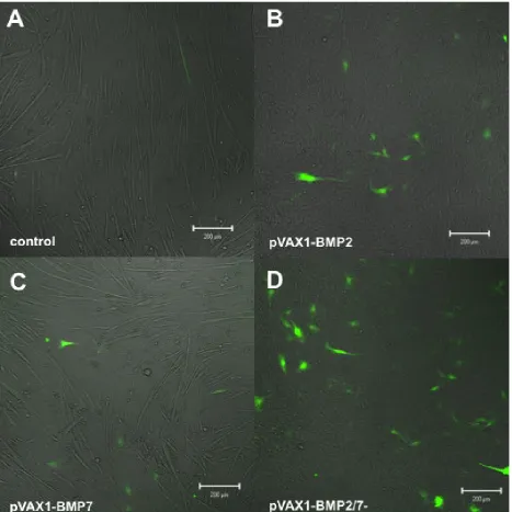
had the strongest osteoinductive capacity of the tested constitutive systems. The co-expression plasmid pVAX1-BMP2/7- (divergent organisation of expression cassettes, Fig. 1D) showed moderate ALP expression (comparable to 100 ng/mL recombinant BMP2), significantly elevated if compared to the negative control. pVAX1-BMP2/7+ (tandem organisation of expression cassettes, Fig. 1C) displayed weaker ALP-induction than pVAX1-BMP2/7- and failed to induce ALP levels significantly higher than the negative control. Single factor expression via pVAX1-BMP2 or pVAX1-BMP7 did not induce significant up-regulation of ALP compared to the negative control, although low-level induction was observable in pVAX1-BMP2 transfected cells. The strongest induction of ALP was achieved in the recombinant growth factor controls in which 300 ng/mL recombinant BMP2 homodimer and 100 ng recombinant BMP2/7 heterodimer were applied.
No induction of ALP was achieved in samples treated with 100 ng/mL recombinant human BMP7.
Osteocalcin reporter transfected C2C12 cells showed highest expression of the fluorescent reporter in cultures that were super transfected with the constitutive BMP2/7 co-expression plasmid pVAX1-BMP2/7- (Fig. 4D). The cultures furthermore displayed altered morphology compared to the negative control. Single factor delivery of pVAX1-BMP2 or pVAX1-BMP7 induced detectable reporter gene expression at lower EYFP positive cell numbers (Figs. 4B,C). The frequency of reporter positive cells was slightly higher in BMP2 transfected cells compared to BMP7 transfected cells. pVAX1-BMP2 transfected cells (Fig. 4B) displayed an altered morphology compared to the negative control.
[image:7.595.87.528.59.341.2]BMP-ELISAs
Anti-BMP2 and BMP7 ELISAs (Fig. 3B) showed production of BMP2 and BMP7 according to the chosen single gene expression system. Interestingly, when the constitutive BMP2/7 co-expression system pVAX1-BMP2/7- was evaluated by ELISA, it was possible to detect the formation of BMP7 (reduced in comparison to single gene expression) but no formation of BMP2 could be detected in contrast to single gene expression.
Inducible co-expression systems in vitro
Single vs. double vector TetON co-expression systems Both Tet-inducible co-expression systems, the
two-component (response + activator plasmid, Figs. 2A,B) and the one component (response/activator plasmid, Fig. 2C) system, displayed comparable, dose dependent activation of the EYFP and dsRed co-expression reporter genes 48 h post treatment with a single dose of doxycycline (Fig. 5 B&E, C&F). Basal expression was absent in inactive controls not treated with doxycycline (Fig.5A&D). The majority of reporter expressing cells emitted both, EYFP and dsRed fluorescence.
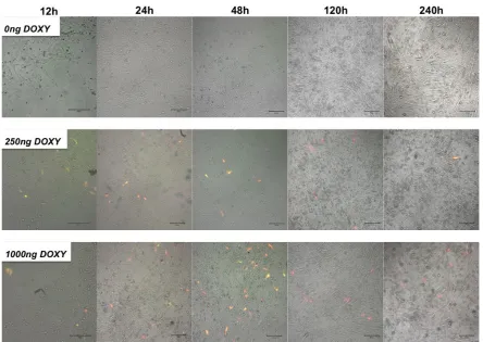
TetON co-expression kinetics
[image:8.595.66.534.55.522.2]Fluorescence imaging of the inducible single vector co-expression plasmid pTetON-EYFP/dsRed for 10 d Fig. 4. Fluorescence microscopy based pCMVE/mOCP-EYFPHis osteocalcin reporter gene assay with
www.ecmjournal.org GA Feichtinger et al. Non-viral osteoinductive gene therapy
Fig. 5. Inducible EYFP/dsRed co-expression systems. Fluorescence microscopy of EYFP/dsRed expression of the dual component TetON system pTRE-EYFP/dsRed+pTetON-Advanced (A-C) and the single component response/activator EYFP/dsRed coexpression system pTetON-EYFP/dsRed (D-F). Scale bars represent 200μm.
[image:9.595.75.520.428.743.2]revealed doxycycline dose dependent expression kinetics (Fig. 6). Expression was observable as early as 12 h post administration of 250 ng/mL or 1000 ng/mL of doxycycline. A peak of fluorescence positive cells was observed at 48 h post administration of a single dose of 1000 ng/mL doxycycline. After 120 h post administration lower numbers of cells were positive and exhibited
exclusively dsRed fluorescence. This fluorescence decreased further on day 10.
No basal expression of EYFP or dsRed fluorescence was detected in control samples not induced with doxycycline.
[image:10.595.63.534.53.504.2]www.ecmjournal.org GA Feichtinger et al. Non-viral osteoinductive gene therapy
Inducible osteogenic differentiation
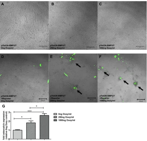
Induction of osteogenic differentiation (ALP assays, Fig. 3) by BMP2/7 co-expression showed a doxycycline dose dependency. Induction with 250 ng/mL doxycycline stimulated expression of ALP at levels comparable to cells treated with 100 ng/mL recombinant BMP2. 1000 ng/ mL of doxycycline triggered ALP expression to similar levels as treatment with 100 ng/mL recombinant BMP2/7 heterodimer or 300 ng/mL recombinant BMP2 homodimer and was significantly higher than the negative control.
No induction of ALP expression was detectable in samples not treated with doxycycline. These findings were paralleled by morphologic observations (Fig. 7A-C) that revealed changes only in samples treated with 250 ng/mL or 1000 ng/mL of doxycycline.
[image:12.595.70.524.59.571.2]TetON-BMP2/7 cells co-cultured with osteocalcin reporter transfected cells (Fig. 7D-F) induced osteocalcin-specific fluorescence reporter gene expression upon treatment with doxycycline. Fluorescence accumulated in cellular clusters of differentiation.
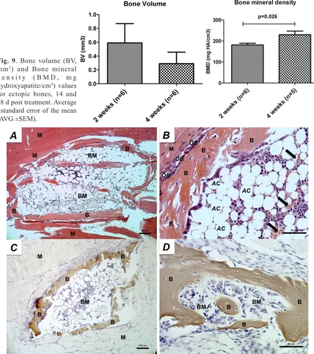
Fig. 9. Bone volume (BV, mm3) and Bone mineral
d e n s i t y ( B M D , m g hydroxyapatite/cm3) values
for ectopic bones, 14 and 28 d post treatment. Average ±standard error of the mean (AVG ±SEM).
www.ecmjournal.org GA Feichtinger et al. Non-viral osteoinductive gene therapy
Endogenous expression of osteocalcin, quantified by qPCR, increased significantly in TetON-BMP2/7 transfected cells upon treatment with increasing doses doxycycline (Fig. 7G). A statistically significant, dose dependent difference in osteocalcin expression was observed between 250 ng and 1000 ng/mL doxycycline. Both induced groups exhibited significantly higher osteocalcin expression compared to the control without doxycycline.
In vivo gene transfer
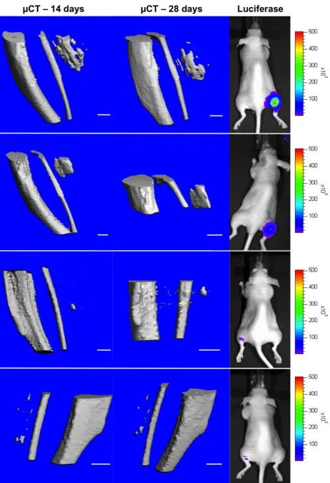
Bioluminescence monitoring of gene transfer efficacy
Detection of luciferase expression (Fig. 8) 24 h post last plasmid injection confirmed gene transfer and expression for 8 out of 13 animals (gene transfer efficacy of 61 %) treated. Luciferase expression levels were highly variable among animals, no specific peak was detected within the observed period of 28 d and some animals showed luciferase activity for more than 21 d (data not shown).
In vivo µCT Imaging
µCT images of hindlimbs at day 14 and day 28 (Fig. 8) confirmed ectopic bone formation in gastrocnemius muscles after treatment with pVAX1-BMP2/7- for both time points in 6 out of 13 animals (46 % bone formation efficacy). Furthermore, resorption and remodelling of ectopic bones was observable in 28 d images in comparison with the 14 d images. The ectopic bones displayed varying morphology, ranging from singular structures of notable size in some animals to several smaller centres of ossification in others. Total bone volumes were variable for both time points (14 d: 0.59 ±0.29 mm3; 28 d:
0.29 ±0.17 mm3). Bone mineral density significantly
increased from 181.5 ±7.9 mg/cm3 hydroxyapatite (14 d)
to 229.8 ±17.6 mg/cm3 (28 d) during the observed time
period (Fig. 9).
µCT and bioluminescence data were furthermore analysed for correlation, given that initial results indicated a potential correlation and bone formation was expected to correlate with gene transfer efficacy and gene expression levels. However, this correlation was not significant. No bone formation was observed after treatment with pTetON-BMP2/7 and doxycycline in any of the 6 animals in the inducible system group.
Histological examination
The formed ectopic bone structures displayed a compact layer of bone, including osteocytes and bone synthesising lining osteoblasts, haematopoiesis and a bone marrow like lumen with progenitor cells and adipose cells observed in HE stained samples (Fig. 10A, B). Von Kossa staining for mineralisation confirmed ectopic structures as mineralised bone tissue (Fig. 10C, D).
Discussion
The study presented herein was aimed at designing and evaluating co-expression systems for the constitutive and inducible co-delivery of 2 different BMP genes for
induction of osteogenic differentiation by means of non-viral gene therapy. Co-expression of BMP2 and BMP7 has been demonstrated to be more effective than single factor expression in vitro. This particular co-expression strategy displayed potent osteoinductive capacity, with a potency comparable to the application of recombinant growth factors in vitro. The potency of the differentiation induced by expression from constitutive single molecule co-expression plasmids was dependent on expression cassette topology and associated potential transcriptional interference.
Furthermore, it was possible to demonstrate the feasibility of applying this strategy in an inducible fashion, triggering the expression of 2 transgenes simultaneously in a doxycycline dependent manner. This system enabled tight control of BMP expression and induction of differentiation in vitro, being silent in the non-induced state. In vivo ectopic evaluation of the constitutive co-expression system with divergent orientation of co-expression cassettes clearly demonstrated an osteoinductive potential. Repeated injection into mouse hind-limb muscles was leading to formation of ectopic bone in 46 % of treated animals after 5 injections of only 20 µg therapeutic DNA per day, displaying similar efficacy at lower daily and total doses when compared to approaches using BMP2 gene delivery only (Osawa et al., 2010). The inducible system in contrast, did not show in vivo ectopic bone formation after 5 injections of 20 µg therapeutic DNA per day and induction of gene expression for 7 d using 2 mg doxycycline i.p. per day, indicating a limiting threshold of BMP2/7 co-expression levels required for successful induction of bone formation.
In vitro evaluation
Constitutive co-expression systems
The influence of transcriptional interference phenomena (Shearwin et al., 2005) needs to be taken into account when working with multiple expression cassettes. CMV-enhancer elements were shown to compete for transcription initiation complex formation in plasmids, through interference of their respective enhancer elements (Andersen et al., 2011), which might partially account for the lower expression efficacy and osteoinductive potential of both constitutive co-expression systems in vitro. However, we observed better osteoinduction with the divergent system compared to tandem organisation, indicating the presence of stronger downstream interference phenomena in the tandem configuration. Given the limited predictability of transcriptional interference and the potential compound effect of multiple forms of transcriptional interference on one specific construct, such effects have to be elucidated empirically (Curtin et al., 2008) with different cassette arrangements as demonstrated in the current study. Another potential issue regarding multi-cassette expression plasmids is the potential for intramolecular recombination (Rubnitz and Subramani, 1985) after transfection of plasmids containing direct (tandem) repeated sequences. Such recombination events, although expected to be relatively rare (Chakrabarti et al., 1985; Chakrabarti and Seidman, 1986; Timme et al., 1989) are non-conservative (Chakrabarti and Seidman, 1986) and could lead to the elimination of sequences from the plasmids. In this study this could be the especially the case for the tandem constitutive co-expression system pVAX1-BMP2/7+, given that such intramolecular recombination events in episomal elements have been proven to be more likely in direct repeat constructs than in inverted repeat constructs (Rubnitz and Subramani, 1985) such as pVAX-BMP2/7-. Given the low frequency of recombination, however, the authors assume that transcriptional interference might be the prevalent reason for reduced efficacy of the multi-cassette constitutive co-expression systems, eventually aggravated by potential non-conservative recombination events in the direct (tandem organisation of expression cassettes) repeat plasmid.
Given that BMP-heterodimer formation relies on BMP dimerisation within the endoplasmic reticulum of the cell (Degnin et al., 2004), co-transfection of a single molecule is still preferable, when aiming at heterodimer production for maximum bioactivity. Co-transfection efficacy and balanced expression of 2 transgenes in vitro has been demonstrated to be highly variable (Schwake et al., 2010), with only a low number of cells positive for both transgenes when using individual plasmids for co-transfection, thus indicating a potential advantage of delivering 2 transgenes via single vector strategies (Kerrigan et al., 2011).
BMP-ELISAs
BMP-ELISAs for BMP2 and BMP7 production showed consistent formation of the respective BMP homodimers in single gene expression system transfected C2C12 cells, but not in cells that were transfected with the constitutive co-expression system pVAX1-BMP2/7- for BMP2/7 heterodimer formation. It was not possible to detect BMP2 in these samples, which could be attributed to issues in detecting homodimeric BMPs in samples that
produce heterodimeric BMP variants. It is not clear how selective the respective kits used in this study are in terms of detecting only homodimeric variants of BMPs and if the epitopes used for detection are conformationally linked to homodimeric BMPs. As the used kits are not supposed to be extensively cross-reactive with the corresponding BMP detected by the other kit used it can be assumed that, at least for the BMP2 ELISA kit, there are issues in detecting heterodimeric BMPs given that formed molecules represent only one half of the target epitope of the ELISA kit. Indeed, work by Kaito et al. (Kaito et al., 2013) has recently demonstrated that overall detectable BMP homodimer levels in BMP2/7 co-transfected cultures were markedly reduced (approx. 4x) compared to single factor expression, which is basically replicating our ELISA results. This might be due to issues in detecting heterodimeric BMPs with commercial ELISA kits for homodimeric BMP detection. The observed reduction for homodimeric BMP7 in BMP2/7 co-transfected cultures (see Fig. 3, B lower graph) was approximately 3x. If this also was the case for BMP2 levels (Fig. 3, B upper graph), BMP2 levels in the BMP2/7 co-transfected cultures might as well have dropped below the limit of detection and therefore led to a negative result for BMP2 quantification in the BMP2/7 co-transfected wells. The development of sensitive ELISA kits for heterodimeric BMPs could help determining if exclusively heterodimeric or if a mixture of homodimeric and heterodimeric BMPs are formed upon co-transfection with individual or co-expressing BMP2/7 plasmid systems.
Inducible co-expression systems
Regulation of therapeutic gene expression levels and kinetics allows controlling of optimal dosing and can help to mimic physiological gene expression patterns. Furthermore, bi-directional inducible systems (Baron et al., 1995) can deliver 2 transgenes simultaneously under the control of an inducible bidirectional promoter. These systems, however, require the co-transfection of an activator plasmid, expressing the doxycycline responsive transactivator protein required for expression from the response plasmid. This situation represents another setup, where co-delivery of all transcriptional units on the same molecule might offer an advantage over simple co-transfection of 2 individual plasmids. Therefore, we investigated whether modification of the original 2-component system to a single plasmid system would substantially alter performance. The comparison of fluorescence microscopy images of doxycycline controlled co-expression of EYFP and dsRed from the original 2-component system and the novel single molecule system, showed no substantial differences in expression levels as well as in background expression without induction. This suggests that the novel single molecule TetON co-expression system performs in a similar way as the original system.
www.ecmjournal.org GA Feichtinger et al. Non-viral osteoinductive gene therapy
with expression of functional fluorescent proteins as early as 12 h post induction, a substantial drop of positive cells already after 120 h and no basal expression without addition of doxycycline. These findings are in line with the kinetic data obtained in similar studies (Puttini et al., 2001), where maximal expression was obtained with 1000 ng/mL doxycycline at 48 h and returned to the off-state within 36 h. Individual turnover rates of the employed reporter genes need to be taken into account for kinetic data and indeed it has been shown that Tet-regulated gene expression studied with enhanced high turnover reporter constructs (Voon et al., 2005) responds even faster (within 4 h) than detectable with common reporters. We therefore conclude, according to the mentioned studies and our findings that transcriptional activation of Tet-regulated co-expression can be very fast within few hours and that gene regulation turns effective with a specific delay accountable to mRNA half-life, protein maturation time and turnover rate (Voon et al., 2005). In our experiments using EYFP (maturation time: 8-12 h; half-life: ~24 h, (Clontech protocol PT2040-1, Clontech application note 2003)) and dsRed (maturation time: 8-12 h; half-life: ~4.5 d, (Verkhusha et al., 2003; Mirabella et al., 2004)), kinetics were in the range of 12-48 h for EYFP and 12-240 h for dsRed. For therapeutic regulated co-expression such time windows will have to be determined for each individual therapeutic gene of interest as expression time windows will depend on individual protein half-life & stability as well as individual mRNA half-lives of different cloned cDNAs. Therefore, the obtained kinetics for EYFP and dsRed only represent a rough estimate for the effective therapeutic time window of gene-expression.
Co-delivery of BMP2 and BMP7 with this expression strategy enabled tightly inductor controlled expression, which lead to subsequent differentiation. The extent of osteogenic differentiation, as determined by microscopy, ALP assays, osteocalcin reporter gene assay and quantitative real-time PCR clearly indicated a direct link between doxycycline dose and differentiation outcome. 1000 ng/mL induced cultures showed the most potent osteoinduction of all tested systems in vitro and no induction of osteogenic differentiation was observed without induction with doxycycline. Tightly regulated gene co-expression was observed after transfection of 1 singular plasmid unit in vitro, narrowing the effective time window of BMP2/7 co-expression down to approximately 4 d for C2C12 cells. This system will provide a useful tool in future in vivo studies to demonstrate control over in vivo bone formation via doxycycline triggered gene expression and to determine minimally required therapeutic BMP-expression time windows for bone induction.
In vivo gene transfer
Intramuscular gene transfer by naked plasmid injection is a method already used for a long time in vivo (Herweijer and Wolff, 2003) even clinically (Rauh et al., 2001) and muscle tissue is an excellent ectopic environment for preliminary bone formation assays (Scott et al., 2012), as well as for the generation of transplantable bone in clinical applications (Warnke et al., 2004). It has previously been shown that repeated non-viral BMP2 gene transfer can
induce bone formation at ectopic sites in vivo (Osawa et al., 2010), with fair efficacy depending on the amount of repetitive treatments. BMP2/7 co-delivery has already been shown to outperform BMP2 delivery after electroporative gene transfer in vivo and is perceived as an ideal gene combination for osteoinductive gene therapies ( Zhu et al., 2004; Zhao et al., 2005; Kawai et al., 2006).
In line with these studies, we tested how the constitutive pVAX1 BMP2/7 co-expression plasmid would perform in an in vivo test for ectopic bone formation using passive DNA delivery. Ideally, by using this BMP2/7 co-expression strategy, we aimed at generating results comparable to the work of Osawa et al. at a reduced number of repetitive treatments (5 vs. 8) and lower total (100 µg vs. 500 µg) and daily DNA dose (20 µg vs. 62.5 µg). Furthermore, to enable non-invasive gene expression monitoring, we incorporated an internal luciferase control plasmid. The bone formation efficacy of 46 % observed in this study is within the range of efficacy reported by Osawa et al. for repeated BMP2 gene delivery, where 4 injections of 125 µg BMP2 DNA led to 57 % and 8 injections of 62.5 µg BMP2 DNA led to 62 % bone formation. Therefore, we conclude that BMP2/7 co-expression in vivo induces bone formation at lower DNA doses to a similar extent as BMP2 delivery. Furthermore, we observed higher bone formation efficacy using BMP2/7 co-expression from a CMV-driven plasmid, whereas Osawa et al. (as Kawai et al.) employed plasmids using the CMV-enhancer chicken b-actin (CAG)-promoter (Kawai et al., 2006; Kawai et al., 2009; Osawa et al., 2010). The CAG-promoter (Miyazaki et al., 1989; Niwa et al., 1991) is a very powerful promoter employed for ubiquitous sustained gene expression and does not exhibit rapid inactivation as observed for the CMV-promoter (Yew, 2005). Furthermore, its activity is especially high in muscle cells (a potential target in our study) (Robert et al., 2012). Therefore, we hypothesise that gene expression levels in those studies were matching levels obtained in our study, but due to the CMV-promoter in our constructs we potentially did not obtain extended therapeutic gene expression windows as in the mentioned papers employing the CAG-promoter. This further highlights the higher bioactivity of the chosen gene combination, as our less-powerful expression system was still able to produce higher bone formation efficacies and modification of our system to replace the CMV promoter by the CAG-promoter – similar to the BMP2/7 co-expression system used by Kawai et al. (Kawai et al., 2009) – could substantially improve its performance. The primary aim of this study, however, was to develop a system for potential clinical translation and therefore we selected the pVAX system, although plasmids containing potentially superior promoters could have been used.
gene dose is limited due to low gene transfer efficacy and therefore not sufficient for crossing the threshold for ectopic bone formation within the limited time period of 7 d. Indeed, it has been possible to show that by improving gene delivery efficacy, by using sonoporation (Feichtinger et al., 2013) for the delivery of the inducible system, it is possible to achieve ectopic bone formation and therefore linking the observed results to limited gene transfer efficacy in this work rather than to limited induction of the TetON system.
Direct comparison of BMP2 with BMP2/7 co-expression in vivo by Kawai et al. using intramuscular electroporation already clearly demonstrated the higher osteoinductive potential of BMP2/7 co-delivery and thus supports this interpretation (Kawai et al., 2006) although using a more invasive approach to gene delivery than applied in the present study.
Similar results for gene transfer efficacy estimation based on luciferase expression (61 % positive, 8/13 animals) and bone formation (46 %, 6/13 animals) demonstrated fair gene transfer efficacies and the feasibility of employing an internal luciferase control in gene transfer studies. However, if compared to previous unpublished single-injection data of only luciferase plasmids from our group, the luciferase levels reported here are lower than expected after gene transfer in vivo – if co-delivered with the therapeutic plasmid. This could indicate that competitive effects (Ngo et al., 1993) between two independent expression systems (CBR luciferase system & BMP2/7 co-expression system) can occur in vivo, thus potentially hampering luciferase expression monitoring or therapeutic efficacy and this is currently under investigation both in vitro and in vivo. The generally high variability of luciferase expression and bone volumes among animals within this study, however, should be mainly attributed to the plethora of parameters that affect passive naked DNA intramuscular gene delivery and its variability (Wolff, 1991; Wolff and Budker, 2005), which are difficult to control, especially in a treatment protocol that relies on multiple injections per site. Injection volumes and injection speed have been shown to influence transgene expression in vivo (Andre et al., 2006), potentially through additional hydrodynamic poration. This can substantially increase DNA uptake, in addition to the postulated default receptor-mediated endocytosis of DNA in muscle (Satkauskas et al., 2001) mainly responsible for intramuscular naked DNA transfer.
Another caveat of passive BMP gene transfer is that the employed DNA doses are still relatively high (~3 mg/ kg for BMP2/7 up to ~15 mg/kg for BMP2 delivery) and that a single injection of BMP-plasmids is not sufficient in order to obtain efficient expression for ectopic bone formation (Osawa et al. and own unpublished data). Therefore, given that passive intramuscular gene transfer is difficult to control and still requires multiple treatments, it might be of higher practical use to introduce an additional, preferably non-invasive physical gene transfer method, which has been successfully investigated in another study (Feichtinger et al., 2013). Such methods can increase and unify global transfection efficacy, while reducing the total DNA amount applied. Sonoporation (Zhang et al., 2006;
Osawa et al., 2009) and electroporation (McMahon and Wells, 2004; Kusumanto et al., 2007) are such minimally invasive methods employing naked DNA without chemical agents that can provide increased transfection efficacy in vivo and indeed electroporation has already been used for the delivery of BMP2/7 co-expression plasmids in vivo (Kawai et al., 2006; Kawai et al., 2009).
The developed, inducible and constitutive BMP2/7 co-expression plasmids are useful tools for investigating the potential of non-viral gene therapeutics and the results obtained with these systems provide further evidence for the feasibility of developing co-expression based gene therapeutics, paving the way for further preclinical research on the performance of BMP2/7 co-expression in orthotopic models for functional bone regeneration. Gene transfer efficacy, consistency and expression vector design are variables critically influencing success of such strategies and still require further improvement in future studies in order to enable translation of this promising approach for clinical application.
Acknowledgements
This work was partially funded by the FP7 project NMP LA 2008 214402 – ANGIOSCAFF and the EUROSTARS project E!5650 UGen. The authors would like to thank Dr. Alexandra Meinl for her support with histology, Mr. Martin Mayer for assistance with µCT evaluation and Prof. Dr. Anthony McHale for his excellent assistance with the preparation of the manuscript.
References
Andersen CR, Nielsen LS, Baer A, Tolstrup AB, Weilguny D (2011) Efficient expression from one CMV enhancer controlling two core promoters. Mol Biotechnol 48: 128-137.
Andre FM, Cournil-Henrionnet C, Vernerey D, Opolon P, Mir LM (2006) Variability of naked DNA expression after direct local injection: the influence of the injection speed. Gene Ther 13: 1619-1627.
Banfi A, von Degenfeld G, Gianni-Barrera R, Reginato S, Merchant MJ, McDonald DM, Blau HM (2012) Therapeutic angiogenesis due to balanced single-vector delivery of VEGF and PDGF-BB. FASEB J 26: 2486-2497. Baron U Freundlieb S, Gossen M, Bujard H (1995) Co-regulation of two gene activities by tetracycline via a bidirectional promoter. Nucleic Acids Res 23: 3605-3606. Bleiziffer O, Eriksson E, Yao F, Horch F, Kneser U (2007) Gene transfer strategies in tissue engineering. J Cell Mol Med 11: 206-223.
Bonadio J, Smiley E, Patil P, Goldstein S (1999) Localized, direct plasmid gene delivery in vivo: prolonged therapy results in reproducible tissue regeneration. Nat Med 5: 753-759.
www.ecmjournal.org GA Feichtinger et al. Non-viral osteoinductive gene therapy
Tet-inducible gene expression in a subcutaneous xenograft model. J Biomol Tech 18: 120-123.
Chakrabarti S, Joffe S, Seidman MM (1985) Recombination and deletion of sequences in shuttle vector plasmids in mammalian cells. Mol Cell Biol 5: 2265-2271. Chakrabarti S, Seidman MM (1986) Intramolecular recombination between transfected repeated sequences in mammalian cells is nonconservative. Mol Cell Biol 6: 2520-2526.
Chen ZY, He CY, Ehrhardt A, Kay MA (2003) Minicircle DNA vectors devoid of bacterial DNA result in persistent and high-level transgene expression in vivo. Mol Ther 8: 495-500.
Coelho-Castelo AA, Trombone AP, Rosada RS, Santos RR, Jr., Bonato VL, Sartori A, Silva CL (2006) Tissue distribution of a plasmid DNA encoding Hsp65 gene is dependent on the dose administered through intramuscular delivery. Genetic Vaccines Ther 4: 1.
Curtin JA, Dane AP,, Swanson A, Alexander IE, Ginn SL (2008) Bidirectional promoter interference between two widely used internal heterologous promoters in a late-generation lentiviral construct. Gene Ther 15: 384-390. Degnin C, Jean F, Thomas G, Christian JL (2004) Cleavages within the prodomain direct intracellular trafficking and degradation of mature bone morphogenetic protein-4. Mol Biol Cell 15: 5012-5020.
Einhorn T (2003) Clinical applications of recombinant human BMPs: Early experience and future development. J Bone Joint Surg 85-A: 82-88.
Feichtinger GA, Hofmann AT, Slezak P, Schuetzenberger S, Kaipel M, Schwartz E, Neef AB, Nomikou N, Nau T, van Griensven M, McHale AP, Redl H (2014) Sonoporation increases therapeutic efficacy of inducible and constitutive BMP2/7 in vivo gene delivery. Hum Gene Ther Meth 25: 57-71.
Feichtinger GA, Morton TJ, Zimmermann A, Dopler D, Banerjee A, Redl H, van Griensven M (2010) Enhanced reporter gene assay for the detection of osteogenic differentiation. Tissue Eng Part C Methods 17: 401-410. Franceschi RT (2005) Biological approaches to bone regeneration by gene therapy. J Dent Res 84: 1093-1103. Gehl J (2003) Electroporation: theory and methods, perspectives for drug delivery, gene therapy and research. Acta Physiol Scand 177: 437-447.
Gelehrter T, Collins F, Ginsburg D (1998) Gene therapy. In: Principles of Medical Genetics (Kelly P, ed), Williams & Wilkins, Baltimore, MD, pp 311-328.
Gossen M, Bujard H (1992) Tight control of gene expression in mammalian cells by tetracycline-responsive promoters. Proc Natl Acad Sci USA 89: 5547-5551. Gossen M, Freundlieb S, Bender G, Muller G, Hillen W, Bujard H (1995) Transcriptional activation by tetracyclines in mammalian cells. Science 268: 1766-1769.
Goverdhana S, Puntel M, Xiong W, Zirger JM, Barcia C, Curtin JF, Soffer EB, Mondkar S, King GD, Hu J, Sciascia SA, Candolfi M, Greengold DS, Lowenstein PR, Castro MG (2005) Regulatable gene expression systems for gene therapy applications: progress and future challenges. Mol Ther 12: 189-211.
Grossin L, Cournil-Henrionnet C, Mir LM, Liagre B, Dumas D, Etienne S, Guingamp C, Netter P, Gillet P (2003)
Direct gene transfer into rat articular cartilage by in vivo electroporation. FASEB J 17: 829-835.
Guo-ping W, Xiao-chuan H, Zhi-hui Y, Li G (2010) Influence on the osteogenic activity of the human bone marrow mesenchymal stem cells transfected by liposome-mediated recombinant plasmid pIRES-hBMP2-hVEGF165 in vitro. Ann Plastic Surg 65: 80-84.
Gurtu V, Yan G, Zhang G (1996) IRES bicistronic expression vectors for efficient creation of stable mammalian cell lines. Biochem Biophys Res Commun 229: 295-298.
Hengge UR, Dexling B, Mirmohammadsadegh A (2001) Safety and pharmacokinetics of naked plasmid DNA in the skin: studies on dissemination and ectopic expression. J Invest Dermatol116: 979-982.
Herweijer H, Wolff JA (2003) Progress and prospects: naked DNA gene transfer and therapy. Gene Ther 10: 453-458.
Hohlweg U, Doerfler W (2001) On the fate of plant or other foreign genes upon the uptake in food or after intramuscular injection in mice. Mol Genet Genomics 265: 225-233.
Israel DI, Nove J, Kerns KM, Kaufman RJ, Rosen V, Cox KA, Wozney JM (1996) Heterodimeric bone morphogenetic proteins show enhanced activity in vitro and in vivo. Growth Factors 13: 291-300.
Johnson E, Urist M (2000) Human bone morphogenetic protein allografting for reconstruction of femoral nonunion. Clin Orthopaed 371: 61-74.
Kaito T, Johnson J, Ellerman J, Tian H, Aydogan M, Chatsrinopkun M, Ngo S, Choi C, Wang JC (2013) Synergistic effect of bone morphogenetic proteins 2 and 7 by ex vivo gene therapy in a rat spinal fusion model. J Bone Joint Surg Am 95: 1612-1619.
Katagiri T, Akiyama S, Namiki M, Komaki M, Yamaguchi A, Rosen V, Wozney JM, Fujisawa-Sehara A, Suda T (1997) Bone morphogenetic protein-2 inhibits terminal differentiation of myogenic cells by suppressing the transcriptional activity of MyoD and myogenin. Exp Cell Res 230: 342-351.
Katagiri T Yamaguchi A, Komaki M, Abe E, Takahashi N, Ikeda T, Rosen V, Wozney JM, Fujisawa-Sehara A, Suda T (1994) Bone morphogenetic protein-2 converts the differentiation pathway of C2C12 myoblasts into the osteoblast lineage. J Cell Biol 127: 1755-1766.
Kawai M, Bessho K, Maruyama H, Miyazaki J, Yamamoto T (2006) Simultaneous gene transfer of bone morphogenetic protein (BMP) -2 and BMP-7 by in vivo electroporation induces rapid bone formation and BMP-4 expression. BMC Musculoskelet Disord 7: 62.
Kawai M, Maruyama H, Bessho K, Yamamoto H, Miyazaki J, Yamamoto T (2009) Simple strategy for bone regeneration with a BMP-2/7 gene expression cassette vector. Biochem Biophys Res Commun 390: 1012-1017. Kerrigan JJ, Xie Q, Ames RS, Lu Q (2011) Production of protein complexes via co-expression. Protein Expr Purif 75: 1-14.
Le Guiner C, Stieger K, Snyder RO, Rolling F, Moullier P (2007) Immune responses to gene product of inducible promoters. Curr Gene Ther 7: 334-346.
Ledwith BJ, Manam S, Troilo PJ, Barnum AB, Pauley CJ, Griffiths TG 2nd, Harper LB, Schock HB, Zhang H, Faris JE, Way PA, Beare CM, Bagdon WJ, Nichols WW (2000) Plasmid DNA vaccines: assay for integration into host genomic DNA. Dev Biol 104: 33-43.
Martin T, Parker SE, Hedstrom R, Le T, Hoffman SL, Norman J, Hobart P, Lew D (1999) Plasmid DNA malaria vaccine: the potential for genomic integration after intramuscular injection. Human Gene Ther 10: 759-768. McMahon JM, Wells DJ (2004) Electroporation for gene transfer to skeletal muscles: current status. BioDrugs 18: 155-165.
Michlits W, Mittermayr R, Schafer R, Redl H, Aharinejad S (2007) Fibrin-embedded administration of VEGF plasmid enhances skin flap survival. Wound Repair Regen 15: 360-367.
Mirabella R, Franken C, van der Krogt GN, Bisseling T, Geurts R (2004) Use of the fluorescent timer DsRED-E5 as reporter to monitor dynamics of gene activity in plants. Plant Physiol 135: 1879-1887.
Mittermayr R, Morton T, Hofmann M, Helgerson S, van Griensven M, Redl H (2008) Sustained (rh) VEGF(165) release from a sprayed fibrin biomatrix induces angiogenesis, up-regulation of endogenous VEGF-R2, and reduces ischemic flap necrosis. Wound Repair Regen 16: 542-550.
Miyazaki J, Takaki S, Araki K, Tashiro F, Tominaga A, Takatsu K, Yamamura K (1989) Expression vector system based on the chicken beta-actin promoter directs efficient production of interleukin-5. Gene 79: 269-277.
Ngo V, Laverriere JN, Gourdji D (1993) Binding capacity and cis-acting efficiency of DNA regulatory sequences can be distinguished in an in vivo competition assay. Nucleic Acids Res 21: 5795-5796.
Niwa H, Yamamura K, Miyazaki J (1991) Efficient selection for high-expression transfectants with a novel eukaryotic vector. Gene 108: 193-199.
Osawa K, Okubo Y, Nakao K, Koyama N, Bessho K (2009) Osteoinduction by microbubble-enhanced transcutaneous sonoporation of human bone morphogenetic protein-2. J Gene Med 11: 633-641.
Osawa K, Okubo Y, Nakao K, Koyama N, Bessho K (2010) Osteoinduction by repeat plasmid injection of human bone morphogenetic protein-2. J Gene Med 12: 937-944.
Perez N, Plence P, Millet V, Greuet D, Minot C, Noel D, Danos O, Jorgensen C, Apparailly F (2002) Tetracycline transcriptional silencer tightly controls transgene expression after in vivo intramuscular electrotransfer: application to interleukin 10 therapy in experimental arthritis. Hum Gene Ther 13: 2161-2172.
Puttini S, Beggah AT, Ouvrard-Pascaud A, Legris C, Blot-Chabaud M, Farman N, Jaisser F (2001) Tetracycline-inducible gene expression in cultured rat renal CD cells and in intact CD from transgenic mice. Am J Physiol 281: F1164-1172.
Rauh G, Pieczek A, Irwin W, Schainfeld R, Isner JM (2001) In vivo analysis of intramuscular gene transfer in
human subjects studied by on-line ultrasound imaging. Hum Gene Ther 12: 1543-1549.
Robert MA, Lin Y, Bendjelloul M, Zeng Y, Dessolin S, Broussau S, Larochelle N, Nalbantoglu J, Massie B, Gilbert R (2012) Strength and muscle specificity of a compact promoter derived from the slow troponin I gene in the context of episomal (gutless adenovirus) and integrating (lentiviral) vectors. J Gene Med 14: 746-760.
Rubnitz J, Subramani S (1985) Rapid assay for extrachromosomal homologous recombination in monkey cells. Mol Cell Biol 5: 529-537.
Satkauskas S, Bureau MF, Mahfoudi A, Mir LM (2001) Slow accumulation of plasmid in muscle cells: supporting evidence for a mechanism of DNA uptake by receptor-mediated endocytosis. Mol Ther 4: 317-323.
Schertzer JD, Plant DR, Lynch GS (2006) Optimizing plasmid-based gene transfer for investigating skeletal muscle structure and function. Mol Ther 13: 795-803. Schmittgen TD, Livak KJ (2008) Analyzing real-time PCR data by the comparative C(T) method. Nature Protoc 3: 1101-1108.
Schwake G, Youssef S, Kuhr JT, Gude S, David MP, Mendoza E, Frey E, Radler JO (2010) Predictive modeling of non-viral gene transfer. Biotechnol Bioeng 105: 805-813.
Scott MA, Levi B, Askarinam A, Nguyen A, Rackohn T, Ting K, Soo C, James AW (2012) Brief review of models of ectopic bone formation. Stem Cells Dev 21: 655-667. Shearwin KE, Callen BP, Egan JB (2005) Transcriptional interference--a crash course. Trends Genet 21: 339-345. Steinert AF, Palmer GD, Pilapil C, Noth U, Evans CH, Ghivizzani SC (2009) Enhanced in vitro chondrogenesis of primary mesenchymal stem cells by combined gene transfer. Tissue Eng Part A 15: 1127-1139.
Sundararaman S, Miller TJ, Pastore JM, Kiedrowski M, Aras R, Penn MS (2011) Plasmid-based transient human stromal cell-derived factor-1 gene transfer improves cardiac function in chronic heart failure. Gene Ther 18: 867-873.
Tepper OM, Mehrara BJ (2002) Gene therapy in plastic surgery. Plast Reconstruct Surg 109: 716-734.
Timme TL, Wood CM, Moses RE (1989) Intermolecular plasmid recombination in fibroblasts from humans with DNA damage-processing defects. Plasmid 22: 1-9. Tolmachov O (2009) Designing plasmid vectors. Meth Mol Biol 542: 117-129.
Tolmachov OE (2011 ) Building mosaics of therapeutic plasmid gene vectors. Curr Gene Ther 11: 466-478. Verkhusha VV, Kuznetsova IM, Stepanenko OV, Zaraisky AG, Shavlovsky MM, Turoverov KK, Uversky VN (2003) High stability of Discosoma DsRed as compared to Aequorea EGFP. Biochemistry 42: 7879-7884.
Voon DC, Subrata LS, Baltic S, Leu MP, Whiteway JM, Wong A, Knight SA, Christiansen FT, Daly JM (2005) Use of mRNA- and protein-destabilizing elements to develop a highly responsive reporter system. Nucleic Acids Res 33: e27.
www.ecmjournal.org GA Feichtinger et al. Non-viral osteoinductive gene therapy
new hope for the treatment of osteoarthritis (review). Mol Med Rep 6: 16-18.
Warnke PH, Springer IN, Wiltfang J, Acil Y, Eufinger H, Wehmoller M, Russo PA, Bolte H, Sherry E, Behrens E, Terheyden H (2004) Growth and transplantation of a custom vascularised bone graft in a man. Lancet 364: 766-770.
Wolff JA, Budker V (2005) The mechanism of naked DNA uptake and expression. Adv Genet 54: 3-20. Yew NS (2005) Controlling the kinetics of transgene expression by plasmid design. Adv Drug Deliv Rev 57: 769-780.
Zhang X, Yang M, Lin L, Chen P, Ma KT, Zhou CY, Ao YF. (2006) Runx2 overexpression enhances osteoblastic differentiation and mineralization in adipose--derived stem cells in vitro and in vivo. Calcif Tissue Int 79: 169-178. Zhao M, Zhao Z, Koh JT, Jin T, Franceschi RT (2005) Combinatorial gene therapy for bone regeneration: cooperative interactions between adenovirus vectors expressing bone morphogenetic proteins 2, 4, and 7. J Cell Biochem 95: 1-16.
Zheng Y, Wu G, Zhao J, Wang L, Sun P, Gu Z (2010) rhBMP2/7 heterodimer: an osteoblastogenesis inducer of not higher potency but lower effective concentration compared with rhBMP2 and rhBMP7 homodimers. Tissue Eng Part A 16: 879-887.
Zhu W, Rawlins BA, Boachie-Adjeu O, Myers ER, Arimizu J, Choi E, Liebermann JR, Crystal RG, Hidaka C (2004) Combined bone morphogenetic protein-2 and -7 gene transfer enhances osteoblastic differentiation and spine fusion in a rodent model. J Bone Miner Res 19: 2021-2032.
Discussion with Reviewers
R. Porter: While the negative inducible in vivo data are now mentioned in the text, why are they not included as part of Fig. 8?
Authors: As there was no ectopic bone formation observable in these samples the authors omitted the inclusion of µCT images, which do not contain ectopic bone in order to save space for the more relevant images of the constitutive system that do actually contain ectopic bone structures of interest to the reader. If needed, the images of the inducible system negative result µCT scans are available and can be provided by the authors.
M. Anton: Your results suggest that passive gene transfer will not be practical for generating therapeutic levels of BMP expression. To improve gene transfer efficiency, you point to promising physical transfer methods, such as electroporation or sonoporation.
For clinical cases where the transfection target is relatively deep, such as injury to a long bone, can these physical transfer methods be employed without open surgery to circumvent the overlying skeletal muscle? What
are the principal challenges to their application in bone repair and regeneration?
Authors: In principle (using X-ray or Ultrasound Imaging guided injection at deep within tissue target sites), it would be possible to treat such defects with electroporation or sonoporation. Sonoporation, however, would be more suitable for this approach as it represents a minimally invasive stimulus that can be applied from outside the body without the need for insertion of needle electrodes as for example in electroporation. Although such devices are still in development it would be theoretically possible to limit the ultrasound stimulus to a defined point in space deep within the tissue at the target site by employing phased-array focussed or convergent beam devices that target high intensity ultrasound at specific sites through constructive interference. The main challenges of using either physical stimulus (electroporation or sonoporation) will be to limit potential tissue damage of surrounding tissues exposed to strong electrical fields or difficult to control reflection and resonance of ultrasound within the body.
M. Anton: Certain indications from rodent bone healing models will not readily translate to the treatment of patients. It is very possible that the effective timeframe for BMP stimulation is one such indication. Consequently, larger animal models (e.g., sheep) may have to be used to adjust initial suggestions from rodent models. Can your inducible expression system be applied to large animal models, and what might be the associated challenges? Authors: The inducible expression system could be applied to large animal models given that feasible gene delivery methods for large animals are available as with any expression system. The specific challenge associated with the use of a doxycycline inducible system in large animals are the higher total amounts of doxycycline that have to be administered and is potentially different pharmacokinetics in a large animal.
M. Anton: What reporting alternatives to luciferase might be suitable for evaluating gene expression noninvasively in these models?
Authors: Luciferases are still the most suitable reporter genes for noninvasively monitoring of gene expression in vivo. However, anticipating significant advances in this area of research, it might be possible in the future to establish transgenes suitable for magnetic resonance imaging (see Kang and Chung, 2008, additional reference), which would be the non-invasive method of choice for in vivo imaging of gene expression in large animals.
Additional Reference