R E S E A R C H A R T I C L E
Open Access
Evidence for a multipotent mammary progenitor
with pregnancy-specific activity
Alice S Kaanta
1,2, Carl Virtanen
3, Laura M Selfors
2, Joan S Brugge
2*and Benjamin G Neel
1,3,4*Abstract
Introduction:The mouse mammary gland provides a powerful model system for studying processes involved in epithelial tissue development. Although markers that enrich for mammary stem cells and progenitors have been identified, our understanding of the mammary developmental hierarchy remains incomplete.
Methods:We used the MMTV promoter linked to the reverse tetracycline transactivator to induce H2BGFP expression in the mouse mammary gland. Mammary epithelial cells (MECs) from virgin mice were sorted by flow cytometry for expression of the mammary stem cell/progenitor markers CD24 and CD29, and H2BGFP. Sorted populations were analyzed forin vivorepopulation ability, expression of mammary lineage markers, and differential gene expression. Results:The reconstituting activity of CD24+/CD29+cells in cleared fat pad transplantation assays was not distinguished in GFP+compared to GFP-subpopulations. However, within the CD24+/CD29loluminal
progenitor-enriched population, H2BGFP+, but not H2BGFP-, MECs formed mammary structures in transplantation assays; moreover, this activity was dramatically enhanced in pregnant recipients. These outgrowths contained luminal and myoepithelial mammary lineages and produced milk, but lacked the capacity for serial transplantation.
Transcriptional microarray analysis revealed that H2BGFP+/CD24+/CD29loMECs are distinct from H2BGFP-/CD24+/CD29lo MECs and enriched for gene expression signatures with both the stem cell (CD24+/CD29+) and luminal progenitor (CD24+/CD29lo/CD61+) compartments.
Conclusions:We have identified a population of MECs containing pregnancy-activated multipotent progenitors that are present in the virgin mammary gland and contribute to the expansion of the mammary gland during pregnancy.
Keywords:Mammary progenitors, Mammary stem cells, Pregnancy, MMTVrtTA, H2BGFP
Introduction
During the lifetime of a female mammal, the mammary gland undergoes three distinct modes of growth [1]. In puberty, ductal elongation and branching establish the overall mammary architecture. After the female has reached sexual maturity, the mammary gland undergoes cyclic ex-pansion and regression during the estrus/menstrual cycle. Pregnancy features dramatic proliferation and alveolar dif-ferentiation, which are critical for lactation. These processes are governed by different signals and hormones, and could involve the activity of different cell types [2,3].
The ability of the mammary gland to undergo repeated rounds of proliferation and remodeling in response to pregnancy was long cited as evidence for the existence of mammary stem cells. The Visvader and Eaves labora-tories reported that subpopulations of mammary epithelial cells (MECs) within the CD24+/CD29+or CD24+/CD49f+ compartments are capable of forming a fully functional mammary gland that can be serially transplanted [4,5]. These groundbreaking studies were followed by the discovery of additional markers for the purification and characterization of several MEC subtypes [6-9].
Whereas a mammary epithelial stem cell can be defined by its ability to establish a fully functional mammary tree in vivo, the identification and characterization of mammary progenitors (which are defined by their ability to replicate and differentiate, but not self-renew) require different types of assays. Lineage-restricted progenitors that differentiate * Correspondence:[email protected];[email protected]
2Department of Cell Biology, Harvard Medical School, 240 Longwood
Avenue, Boston, MA 02115, USA
1Department of Medicine, Cancer Biology Program, Division of
Hematology-Oncology, Beth Israel Deaconess Medical Center, Harvard Medical School, 330 Brookline Avenue, Boston, MA 02215, USA Full list of author information is available at the end of the article
into myoepithelial or luminal MECs have been identified, principally using in vitro assays [4-7]. The CD24+/CD29lo population can be subdivided into populations of luminal progenitors and differentiated luminal MECs, depending on the expression or absence of CD61 (β3 integrin), respect-ively [6]. Luminal epithelial cells with different in vitro proliferative potential can be distinguished based on CD49b (α2 integrin) expression; CD24+/CD49b+, but not CD24+/ CD49blo, MECs form colonies on NIH 3T3 feeder cells [7]. CD14 and c-Kit expression have been used to identify prospective alveolar progenitors [8,9]. Although it was reported initially that CD24+/CD29loand CD24+/CD49flo MECs are unable to form outgrowthsin vivo,more recent studies have demonstrated that these MECs can form small, branched mammary structures when co-injected with Matrigel into mammary fat pads [10,11]. Other groups transplanted mixed populations and inferred that the mammary gland contains progenitors that can give rise to mammary structures of different sizes and morphological characteristics [12,13].
Another approach to defining the in vivo activity of mammary stem cells and progenitors is to track MEC populations by lineage tracing. Van Keymeulen et al. recently conducted extensive studies of transgenic mice that inducibly express fluorescent proteins driven by pro-moters for known mammary lineage markers, including CK14, CK8 and CK18 [14]. This study provided evidence for the presence in the adult mammary gland of long-lived, unipotent luminal and myoepithelial progenitors that could contribute significantly to mammary gland expansion during puberty and pregnancy.
Adult stem cells also have been identified using pulse-chase assays that are based on the ability of stem cells to re-tain molecular labels significantly longer than bulk cells in tissues. Such‘label retention’is attributed either to slower cycling of stem cells or to asymmetric distribution of paren-tal and daughter chromatids to stem cells and their pro-genitor progeny (the immortal strand hypothesis). Until recently, most label retention studies had been performed using DNA-based labels such as bromodeoxyuridine (BrdU) or tritiated thymidine. Previous reports that used such methods suggested the existence of label-retaining cells in the mammary gland that contribute to pregnancy, undergo mitosis and express steroid hormone receptors [15-17].
The development of transgenic mice expressing histone 2b fused to eGFP (H2BGFP) has permitted the isolation of live label-retaining cells, including epidermal and hematopoietic stem cells, by fluorescence-activated cell sorting (FACS) [18-20]. Using an analogous approach in an attempt to identify and purify mammary stem cells, we crossed H2BGFP transgenic mice with a strain expressing the reverse tetracycline transactivator under the control of the mouse mammary tumor virus promoter (MMTVrtTA). We chose this MMTVrtTA line because it reportedly
expresses tet element-controlled transgenes in all MECs [21]. We found no evidence that label retention enriches for mammary stem cell activity, although the length of the
‘chase’in our experiment might have been insufficient to isolate a mammary population of interest.
Nevertheless, in control experiments analyzing pubertal MMTVrtTA/H2BGFP mice, we made the unexpected discovery that H2BGFP was expressed primarily within the CD24+/CD29+ and CD24+/CD29lo compartments, which contain mammary stem cells and progenitors, respectively. This unusual expression pattern led us to test whether the H2BGFP+ and H2BGFP- MEC populations had distinct properties. Using mammary fat pad transplantation assays, we found that H2BGFP+/CD24+/CD29locells contain a population of multipotent progenitors that give rise to mammary structures capable of differentiation and lacta-tion, but which cannot be serially transplanted. The ability of these cells to produce such structures increases 5- to 10-fold if recipient mice are made pregnant. Therefore, our data suggest that the mammary glands of virgin pu-bertal mice contain a population of pregnancy-activated multipotent mammary progenitor cells.
Methods Mice
MMTVrtTA mice [21] on FVB background were obtained from L. Chodosh (Abramson Cancer Institute). H2BGFP mice [18] in a CD-1 background were provided by E. Fuchs (Rockefeller University) and backcrossed onto FVB for a minimum of five generations prior to crossing with MMTVrtTA mice. To induce transgene expression, breeders and experimental mice were maintained on 2 mg/ml doxycycline (Research Products International Corp., Mount Prospect, IL, USA; 50213285). Mouse colonies were maintained according to federal, state, and institutional regulations and ethical approval for this study was obtained by the institutional review boards of Beth Israel Deaconess Medical Center and Harvard Medical School, respectively.
Genotyping
Tail biopsies were obtained from transgenic mice, and dissolved in 0.5 ml tail lysis buffer (10 mM Tris + 100 mM NaCl + 10 mM EDTA + 0.5% SDS + 1 mg/ml proteinase K) at 60°C for >4 hours. After proteinase K treatment, 250μl of 6 M NaCl were added to each sample, which was then inverted two to three times and incubated on ice for 10 minutes. Samples were centrifuged at 10,000 rpm in a microfuge for 10 minutes, and supernatants were added to 650 μl of isopropanol, inverted two to three times, and then centrifuged at top speed in a microfuge for 10 minutes. Pellets were air-dried and dissolved in 100μl H2O. For H2BGFP genotyping, primers were: GFP-F:
5′-GGCGAGCTGCACGCTGCCGTCCTC-3′, expected band size 500 base pair (bp). Fortetgenotyping, primers were: tTA-F: 5′-CGCTGTGGGGCATTTTACTTTAG-3′,tTA -R: 5′-CATgTCCAgATCgAAATCgTC-3′, expected band size 450 bp. Control genotyping PCR (T-cell receptor delta) primers were: TCRD-F: 5′-CAAATGTTGCTTGTCTGGTG-3′, TCRD-R: 5′-gTCAgTCgAgTgCACAgTTT-3′, expected band size 200 bp. All genotyping PCRs were carried out as follows: 95°C 5 min, 95°C 45 sec, 58°C 45 sec, 72°C 60 sec, repeat from second 95°C step 25X, 72°C 5 min, 4°C final. PCR products were resolved on 2% agarose gels.
Mammary gland harvesting and preparation
Mice were sacrificed and mammary glands were harvested. Lymph nodes were removed from inguinal gland num-ber 4, mammary tissue was manually minced, placed in culture medium (DMEM/F12 + 500 ng/ml hydrocorti-sone + 10 ng/ml EGF + 10μg/ml insulin + 20 ng/ml chol-era toxin + 1% Pen/Strep) [4] with 600 U/ml collagenase and 200 U/ml hyaluronidase, and shaken at 37°C for 1 to 1.5 hrs. Cells from digested tissue were recovered by centri-fugation and resuspended in a mixture of 0.25% Trypsin-EDTA (Gibco number 15050-065; Invitrogen, Carlsbad, CA, USA), 5 mg/ml dispase (Roche 04942078001; Roche, Basel, Switzerland) and 0.1 mg/ml DNAse (Sigma number D5319; Sigma-Aldrich, St Louis, MO, USA) for 5 to 7 minutes. Reactions were quenched with DMEM/F12 + 10% FBS, and cells were recovered by centrifugation, resuspended in Tris-buffered 0.64% NH3Cl for 3 minutes on ice, and then passed sequentially through 100 um and 40 um filters.
Flow cytometry
Single cell suspensions were blocked in FACS buffer (PBS + 2 mM EDTA + 0.1% BSA) containing 1:25 normal rat serum (eBioscience, Inc., San Diego, CA, USA; 24-5555) and 1:25 anti-mouse FC receptor block (eBioscience 16-0161) for 10 minutes, and stained in FACS buffer containing directly con-jugated antibodies for 20 minutes. Cells were resuspended in FACS buffer (containing 1μg/ml DAPI for viability analysis) and analyzed on a BD Aria II or FACS Canto II using BD Aria software (BD Biosciences, San Jose, CA, USA). Anti-bodies included: CD24•PE (eBioscience 12-0242), CD29•APC (Biolegend, San Diego, CA, USA; 102215), CD49f•Biotin (eBioscience 13-0495), CD31•PeCy7 (eBioscience; 25-0311), CD45•PeCy7 (eBioscience 25-0451), Ter119•PeCy7 (eBioscience 25-5921) and Streptavidin APC-eFluor 780 (eBioscience 47-4317-82). Antibodies were used at the dilutions recommended by their manufacturer. Single cells were ana-lyzed after gating out dead (DAPI+), hematopoietic (CD45+, Ter119+), and endothelial (CD31+) cells.
Antibodies tested in Figure S1 in Additional file 1: AlCam•PE (eBioscience 12-1661-81), amphiregulin (R&D Systems, Minneapolis, MN, USA; BAF989), CD14 (eBioscience 13-0141-80), CD24 (eBioscience 13-0242-80),
CD44 (eBioscience 0441-81), CD49b (eBioscience; 13-5971-80), CD49f (eBioscience 13-0495), CD55 (eBioscience 12-0559-71), CD61 (eBioscience 13-0611-80), CD86 (eBioscience 13-0862-80), CD138/Syndecan•PE (R&D FAB2966P), CD164 (R&D BAF3118), c-Kit (eBioscience 13-1171-81), CXCL16 (R&D BAF503), E-cadherin (eBioscience 13-3249-80), EpCAM (eBioscience 13-5791-80), FGFR2•PE (R&D FAB684A), IL1R1•PE (R&D FAB7712P), IL33 (R&D BAF3626), Jag1•PE (eBioscience 12-3391-80), Muc1 (AbCam, Cambridge, UK; ab13970), Osteoactivin (R&D BAF2330), PRLR (R&D BAF1445), and Sca-1 (eBioscience). Secondary antibodies included: Streptavidin APC-eFluor 780 (eBioscience 47-4317-82), anti-rabbit Alexa750 (Invitrogen A-21039).
Mammary fat pad transplants
Sorted cells were counted and resuspended in growth factor-reduced Matrigel (BD Biosciences, Franklin Lakes, NJ, USA; 354230). Three-week-old female mice between 10 to 12 g were obtained from Charles River Laboratories, Wilmington, MA, USA. Mice were anesthetized with 4 mg/ 10 g body weight Avertin (Sigma-Aldrich; T48402), and ab-domens were shaved and cleaned with 70% ethanol and Betadine. An inverted Y-shaped incision was made along the thoracic-inguinal region, and the mammary glands were exposed. The nipple and mammary artery connecting the number 4 and number 5 glands were cauterized, and the epithelium was removed from the inguinal (number 4) mammary glands. The removed epithelium was fixed and stained with Carmine Alum solution (2 g/l Carmine + 5 g/l aluminum potassium sulfate) to verify complete clear-ing. Cells were injected into cleared fat pad pads in 10μl vol-umes, using a Hamilton syringe (Hamilton, Reno, NV, USA). Incisions were closed with wound clips. For pain control, two doses of 0.5 mg/kg body weight Buprenorphine (Webster Veterinary, Devens, MA, USA; 07-867-1196) were adminis-tered in the 24 hrs after surgery. Mammary fat pads were harvested six weeks after injection, unless pregnancy induc-tion required an earlier harvest date. Harvested tissues were either whole-mounted and scored for mammary outgrowths or dissociated for reanalysis and/or serial transplants [22].
Pregnancy induction
Adult males were housed with post-transplant females three to four weeks after surgery. Plugs were checked daily to verify timing of pregnancies. Females were sacrificed and mammary glands were harvested between days 14 to 20 of the pregnancy.
Whole mounts
25% EtOH, followed by distilled water, and then stained overnight in Carmine Alum solution. Mammary glands were dehydrated sequentially in 70% and 100% EtOH, subjected to two washes with xylenes, and then mounted in Permount (Thermo Fisher Scientific, Waltham, MA, USA; SP15-100). Whole mounts were scored and imaged under a dissecting microscope [22]. All mammary outgrowths observed by Carmine Alum stain were confirmed by tissue sectioning and immunostaining.
Statistical analysis
Mammary repopulating unit MRU frequencies were calcu-lated using the ELDA (extreme limiting dilution analysis) method (http://bioinf.wehi.edu.au/software/elda/) [23].
RNA analysis
RNA was harvested from sorted cells using the Qiagen RNeasy Micro Kit (Qiagen, San Diego, CA, USA; 74004), amplified using the NuGen RNA amplification system, and analyzed for gene expression on Illumina Whole Genome Mouse chips (Illumina, Inc., San Diego, CA, USA; WG6 v2r2). Processed arrays were checked for overall quality using R (v2.10.0) with the Bioconductor [24] and LUMI packages [25]. Data were imported into GeneSpring (v12.0, Agilent Technologies, Santa Clara, CA, USA) and normal-ized with a quantile-based algorithm. Because experimental samples were collected on two separate dates, a further batch correction was applied using a parametric empirical Bayes framework [26]. Pfiltering was performed to re-move probes with no expression in any of the groups under consideration (at least 80% of samples having an ex-pression value greater than the 20th percentile). Hierarch-ical cluster diagrams were built using a Pearson centered distance measure under average linkage rules. Differentially expressed genes in GFP+ and GFP- CD24+/CD29lo cells were identified on a re-normalized and filtered subset of the whole data set using unpaired Student’s t test with Benjamini and Hochberg false discovery rate (FDR) [27] multiple testing correction (q <0.05). Microarray data have been made publicly available at the Gene Expression Omnibus (www.ncbi.nlm.nih.gov/geo) under the series identifier GSE42055.
For analysis of mammary subpopulations defined in Vaillantet al. [28], pre-processed data were downloaded from the Gene Expression Omnibus (GSE19446, http:// www.ncbi.nlm.nih.gov/geo/) and analyzed using R (v.2.13.1). Differentially expressed genes in the CD24+/CD29+, CD24+/CD29lo/CD61+and CD24+/CD29lo/CD61+ subpop-ulations were identified using one-way analysis of variance, post hoc Tukey honestly significant differences tests and a P value threshold of 0.05. These results were merged with the differentially expressed genes identified by the GFP+ and GFP-CD24+/CD29loarrays, using the manufacturers’ unique probe identifiers. A representative single probe per
gene was selected by highest variance across the GFP+ and GFP-CD24+/CD29losamples. Fold-enrichment was defined as the observed frequency divided by the expected frequency. Enrichment P values were calculated with the hypergeometric distribution implemented in the phyper function in R. The background population set was the 16,245 unique genes that were identified after pre-filtering the GFP+and GFP-CD24+/CD29lodata.
qPCR
RNA was harvested from sorted cells using the Qiagen RNeasy Micro Kit, amplified using the NuGen RNA ampli-fication system, and labeled using SYBR Green Master Mix (Applied Biosystems, Foster City, CA, USA; 4309155). qPCR reactions were performed by using an Applied Biosystems 7900HT. Primers were:Esr1-F 5′-TGGGCGA CATTCTTCTCAA-3′, Esr1-R 5′-TGGACCAGAGGTAC ATCCATT-3′, Prlr-F 5′-GGTTATAGCATGATGACCT GCAT-3′, Prlr-R 5′- CAGTTCTTCAGACTTGCCC TTC-3′, Areg-F 5′-GCGAATGCAGATACATCGAG-3′, Areg-R 5′- CCACACCGTTCACCAAAGTA-3′, Fgfr2-F 5′-GAGCGCTGCCATTCAAGT-3′, Fgfr2-R 5′- TTGC TGTTGTTACTGCTGTTCC-3′, Muc1-F 5′-CTGTTCA CCACCACCATGAC-3′, Muc1-R 5′-CTTGGAAGGGC AAGAAAACC-3′, Wnt5a-F 5′- ACGCTTCGCTTGA ATTCCT-3′, Wnt5a-R 5′- CCCGGGCTTAATATTC CAA-3′,Il1R1-F 5′-CGAACCGTGAACAACACAAA-3′,
Il1R1-R 5′- AATCTCCAGCGACAGCAGAG-3′, Cav1-F
5′-ACGACGACGTGGTCAAGATT-3′, Cav1-R 5′-CAC AGTGAAGGTGGTGAAGC-3′, Robo1-F 5′-CCACCCA CCAGACAGGAG-3′, Robo1-R 5′-GTATGAGGTGGG GAAATTGG-3′.
Immunostaining
For tissue sections, mammary glands were fixed in methyl Carnoy, or, if previously stained with Carmine Alum, first un-mounted in xylenes. Tissues were paraffin-embedded and sec-tioned. For antigen retrieval, slides were deparaffinized in xylenes, and rehydrated in decreasing concentrations of EtOH, followed by boiling for 20 minutes in 10 mM sodium citrate. For frozen sections, freshly dissected tissues were mounted in OCT compound (Sakura Finetek USA, Inc., Torrance, CA, USA; 4583), flash-frozen in liquid nitrogen and sectioned.
For cytospins, slides and coverslips were fixed in 4% PFA and rinsed with PBS for staining. Three-dimensional Matrigel cultures were fixed and stained as described by Debnath et al. [29]. Primary antibody staining was performed at 4°C overnight; secondary antibody staining was performed at room temperature for 1 hr.
β1 integrin (BD Biosciences 550531), estrogen receptor (Santa Cruz Biotechnology, Santa Cruz, CA, USA; SC-542), progesterone receptor (DAKO, Glostrup, Denmark; A0098), glucocorticoid receptor (Santa Cruz SC-1004), milk (Nordic Immunological Laboratories, Tilburg, The Netherlands; RAM/MSP), amphiregulin (Santa Cruz sc-25436). AlexaFluor™secondary antibodies were used for visualization and imaging.
Results
MMTVrtTA induces H2BGFP expression in the mammary stem cell and progenitor compartments
We expressed H2BGFP in the mouse mammary gland by crossing mice that carry a tetracycline-inducible H2BGFP fusion gene [30] to mice expressing the reverse tetracycline transactivator under the control of the mouse mammary tumor virus promoter (MMTVrtTA) [21] (Figure S2 in Additional file 2). To induce H2BGFP expression, breeders and pups were treated with doxycycline throughout breeding, pregnancy, lactation, and after weaning until sacrifice. Mammary glands were harvested from double transgenic (MMTVrtTA/H2BGFP) virgin females at four weeks post-partum and analyzed by flow cytometry and immunostaining.
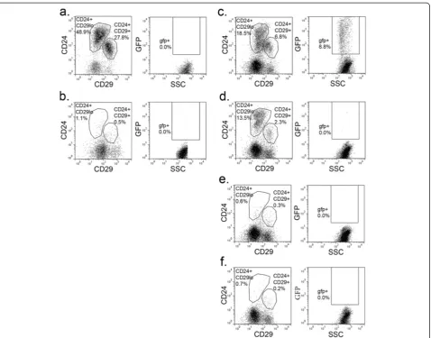
For flow cytometric analysis, mammary glands were dis-sociated to single cell suspensions and stained for CD24, CD29, and CD49f (Figure 1). These markers divided MECs into the subpopulations (Figure 1b, c) reported previously [4,5]. As expected, H2BGFP was not detected in mammary cells from single transgenic or uninduced double transgenic mice (data not shown). In contrast to a previous report that this MMTVrtTA transgene drives ubiquitous expression in MECs [21], H2BGFP expression was detected in only a fraction (18 ± 3.1%) of MECs from double transgenic mice (Figure 1a; also see Discussion).
The vast majority of H2BGFP+MECs were found in either the mammary stem cell (CD24+/CD29+, CD24+/CD49f+) or progenitor (CD24+/CD29lo, CD24+/CD49flo) compartments. Of the H2BGFP+cells, 47 ± 11% were CD24+/CD29+, while 39 ± 11% were CD24+/CD29lo (n = 10; representative flow cytometric profile is shown in Figure 1d). Conversely, 55 ± 6.7% of CD24+/CD29+ MECs and 15 ± 4.4% of CD24+/CD29loMECs were H2BGFP+, respectively (n = 10; representative flow cytometric profiles are shown in Figures 1e, f). Similarly, 36 ± 7.1% of the H2BGFP+ cells were CD24+/CD49f+ and 50 ± 1.7% were CD24+/CD49flo (n = 4; representative flow cytometry profile is shown in Figure 1g), while 46 ± 3.6% of CD24+/CD49f+ MECs and 21 ± 2.7% of CD24+/CD49floMECs were H2BGFP+(n = 4; representative flow cytometric profiles are shown in Figures 1h, i). Although each subpopulation exhibited a range of H2BGFP levels, the maximum fluorescence intensity was higher in the CD24+/CD29loand CD24+/ CD49flo(progenitor-containing) populations than in the
CD24+/CD29+ and CD24+/CD49f+ (stem cell-containing) compartments (Figure 1e, f, h, i).
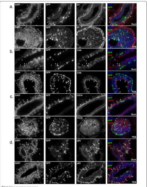
To further characterize H2BGFP+MECs, we also analyzed the pattern of H2GFP expressionin situ. Mammary glands from four-week-old, doxycycline-induced, MMTVrtTA/ H2BGFP females were sectioned and immunostained forβ1 integrin (CD29), CK8, CK14, and p63. Consistent with the flow cytometry data, H2BGFP was expressed mosaically and at a range of levels, and the majority of H2BGFP+cells co-expressed β1 integrin (Figure 2a). H2BGFP+ MECs were detected in the basal layer of the ductal tree and in the cap cells of the terminal end buds (TEB), as well as in the luminal layer. Moreover, the luminal (CK8+; Figure 2b), myoepithelial (CK14+; Figure 2c), and basal/cap cell (p63+; Figure 2d) populations each contained H2BGFP + MECs (arrowheads). These data are consistent with our finding that H2BGFP+cells fall predominantly within the CD24+/CD29+and CD24+/CD29locompartments.
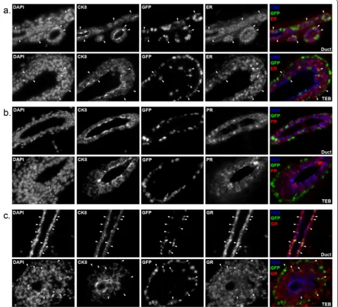
We also examined how H2GFP expression correlated with the expression of steroid hormone receptors that are associated with differentiated MEC populations and play important roles in mammary biology [4,5,31]. Mammary glands from four-week-old double transgenic mice were sectioned and immunostained for CK8 to mark the luminal compartment, along with estrogen receptor (ER) (Figure 3a), progesterone receptor (PR) (Figure 3b) or glucocorticoid receptor (GR) (Figure 3c), which activates the MMTV promoter and is important for mammary proliferation during pregnancy [32]. In these pubertal mice, most cells in the ducts and TEBs were ER+and GR+, and H2BGFP was co-expressed with ER and GR in some of these cells (Figure 3a, c, arrowheads). By contrast, H2BGFP+cells consistently failed to express PR (Figure 3b).
MMTVrtTA/H2BGFP expression identifies MECs with differential repopulation activity
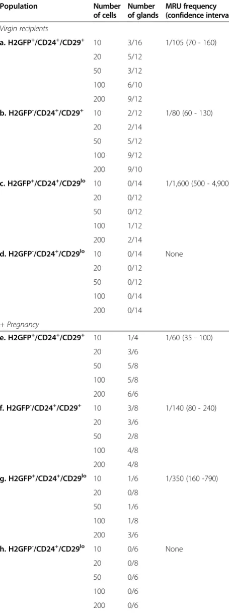
Fat pads that had received transplants were harvested six weeks post-transplantation, stained with Carmine Alum to visualize outgrowths and scored for size (Table 1, Figure S3 in Additional file 3, Table S1 in Additional file 4). Because some outgrowths were quite small, recipient fat pads also were sectioned and immunostained for mammary epithelial markers to verify their mammary origin and to visualize the structure of all outgrowths. The MRU frequency of each population was calculated, using the ELDA method [23,33,34]; similar results were obtained by assuming single-hit Poisson statistics (data not shown). In order to increase the size and visi-bility of the mammary glands, pregnancy was induced in some recipients, three to four weeks after trans-plantation. To determine whether pregnancy affected repopulation, data from virgin and pregnant recipients were analyzed separately.
We found that the repopulation abilities of H2BGFP+/ CD24+/CD29+ and H2BGFP-/CD24+/CD29+ popula-tions were similar in virgin and pregnant recipients (Table 1a, b, e, f ) and comparable to established repopula-tion frequencies for (unfracrepopula-tionated) CD24+/CD29+ cells [4]. Based on these data, we conclude that MMTVrtTA/ H2BGFP expression in the CD24+/CD29+ compartment does not label MECs with different repopulating activity.
To determine whether MMTVrtTA/H2BGFP expression could identify previously unidentified mammary progeni-tors within vivoactivity, we also tested the H2BGFP+and H2BGFP-CD24+/CD29lopopulations in transplantation experiments. Although CD24+/CD29lo MECS were ini-tially believed to be incapable ofin vivomammary gland reconstitution [4], other studies have since discovered that this population can develop mammary outgrowths upon transplantation, under certain conditions [10,11]
a.
b.
c.
d.
e.
f.
[image:6.595.59.538.88.455.2]g.
h.
i.
(See figure on previous page.)
Figure 2Expression of lineage markers in mammary glands of MMTVrtTA/H2BGFP mice.Mammary glands were harvested from four-week-old double transgenic females treated with doxycycline, sectioned and immunostained for:(a)β1 integrin(b)CK8(c)CK14 or(d)p63. Each pair of micrographs depicts ducts (upper) and terminal end buds (lower), at 60× magnification. Note that, as expected, cytoplasmic (CK8, CK14) and cell surface (β1 integrin) lineage markers do not co-localize with H2BGFP in cells that co-express both proteins; only with p63, which is a nuclear protein, does co-expression result in co-localization with H2BGFP. In all panels, representative co-expressing cells are indicated by arrowheads. H2BGFP, histone 2B-eGFP; MMTV, mouse mammary tumor virus promoter; rtTA, reverse tetracycline transactivator.
(see Discussion). We discovered that, in contrast to CD24+/ CD29+cells, the ability of H2BGFP+/CD24+/CD29loMECs to give rise to multilineage outgrowths was nearly 5-fold higher in pregnant (1/350), compared with virgin (1/1,600), mice (Table 1c, g). Outgrowths from H2BGFP+/CD24+/ CD29loMECs were much smaller than glands derived from CD24+/CD29+cells (Figure S3 in Additional file 3, Table S1 in Additional file 4), but contained all mammary lineages and were able to produce milk (Figure 4).
H2BGFP+/CD24+/CD29loMECs give rise to mammary outgrowths preferentially in pregnant mice
To directly address whether H2BGFP+/CD24+/CD29lo MECs expand selectively during pregnancy, we performed an additional set of transplantation experiments (n = 3), focusing specifically on the H2BGFP+/CD24+/CD29lo population. Half of the mice injected with H2BGFP+/ CD24+/CD29loMECs were made pregnant post-transplant to allow direct comparison of H2BGFP+/CD24+/CD29lo cell repopulating activity in virgin vs. pregnant mice (Table 1, bottom). In agreement with the first set of ex-periments, the MRU frequency of H2BGFP+/CD24+/ CD29lo MECs was 1/280 in pregnant mice, while in virgin recipients, H2BGFP+/CD24+/CD29lo MECs produced only a single outgrowth (Table 1i, j). We conclude that MMTVrtTA-driven expression of H2BGFP in the CD24+/ CD29lo compartment labels a population containing multipotent stem cells or progenitors that expand pref-erentially during pregnancy.
H2GFP+/CD24+/CD29locells do not self-renew
[image:9.595.57.285.112.721.2] [image:9.595.304.539.113.267.2]Stem cells, unlike progenitors, have the ability to self-renew. In the hematopoietic system, there is a hierarchy of stem cells with long-term, intermediate-term and short-term
Table 1 Limiting dilution transplants of MMTVrtTA/ H2BGFP populations Population Number of cells Number of glands MRU frequency (confidence interval) Virgin recipients
a. H2GFP+/CD24+/CD29+ 10 3/16 1/105 (70 - 160)
20 5/12
50 3/12
100 6/10
200 9/12
b. H2GFP-/CD24+/CD29+ 10 2/12 1/80 (60 - 130)
20 2/14
50 5/12
100 9/12
200 9/10
c. H2GFP+/CD24+/CD29lo 10 0/14 1/1,600 (500 - 4,900)
20 0/12
50 0/12
100 1/12
200 2/14
d. H2GFP-/CD24+/CD29lo 10 0/14 None
20 0/12
50 0/12
100 0/14
200 0/14
+ Pregnancy
e. H2GFP+/CD24+/CD29+ 10 1/4 1/60 (35 - 100)
20 3/6
50 5/8
100 5/8
200 6/6
f. H2GFP-/CD24+/CD29+ 10 3/8 1/140 (80 - 240)
20 3/6
50 2/8
100 4/8
200 4/8
g. H2GFP+/CD24+/CD29lo 10 1/6 1/350 (160 -790)
20 0/8
50 1/6
100 1/8
200 3/6
h. H2GFP-/CD24+/CD29lo 10 0/6 None
20 0/8
50 0/6
100 0/6
200 0/6
Table 1 Limiting dilution transplants of MMTVrtTA/ H2BGFP populations(Continued)
Virgin Recipients vs. Pregnancy - 2ndExperimental Set
i. H2GFP+/CD24+/CD29lo 10 0/14 1/3,800 (500 - 27,000)
Virgin 20 0/12
50 1/12
100 0/12
200 0/8
j. H2GFP+/CD24+/CD29lo 10 2/12 1/280 (170 - 480)
+Pregnancy 20 2/14
50 4/14
100 4/14
200 4/14
Mammary repopulating ability of H2BGFP+
/CD24+
/CD29+
(a, e), H2BGFP
-/CD24+
/ CD29+
(b, f), H2BGFP+
/CD24+
/CD29lo
(c, g, i) and H2BGFP
-/CD24+
/CD29lo
reconstitution ability, respectively [35]. To test whether MMTVrtTA-driven expression of H2GFP might mark MECs with differential self-renewal ability, we compared the serial transplantation ability of the three cell popula-tions (H2BGFP+/CD24+/CD29+, H2BGFP-/CD24+/CD29+, and H2BGFP+/CD24+/CD29lo) capable of forming mam-mary outgrowths. Each MEC population was isolated by FACS from four-week-old MMTVrtTA/H2BGFP females and injected in 500-cell aliquots into 10 cleared fat pads of syngeneic mice. Recipients were treated with doxycycline to promote H2BGFP expression. After six weeks, transplanted fat pads were harvested and pooled according to transplanted cell type, analyzed by flow cytometry, and re-transplanted. MMTVrtTA/H2BGFP populations were serially transplanted twice in two experiments, and tertiary transplants were performed in one experiment.
The H2BGFP+/CD24+/CD29+ and H2BGFP-/CD24+/ CD29+populations each gave rise to serially transplantable mammary glands (Figure 5). Surprisingly, only H2BGFP+/ CD24+/CD29+ cells gave rise to mammary glands that
expressed H2BGFP. To eliminate the possibility that the apparent H2BGFP-/CD24+/CD29+ outgrowths were the product of endogenous MECs from incompletely cleared mammary fat pads, genomic DNA was harvested from mammary glands after tertiary transplants and tested by PCR for the MMTVrtTA and H2BGFP transgenes. Both transgenes were present in H2BGFP+/CD24+/CD29+- and H2BGFP-/CD24+/CD29+-derived glands, demonstrating their transgenic origin (data not shown). These results suggest that H2BGFP expression was repressed perman-ently in the H2BGFP-/CD24+/CD29+ population. We conclude that both CD24+/CD29+populations are capable of long-term stem cell activity, and H2BGFP expression within this compartment does not, at least at this level of analysis, distinguish subpopulations of CD24+/CD29+cells with differential biological activity.
[image:10.595.57.539.89.399.2]Gene expression analysis is consistent with biologically distinct roles for H2BGFP+/CD24+/CD29loand H2BGFP-/ CD24+/CD29lopopulations
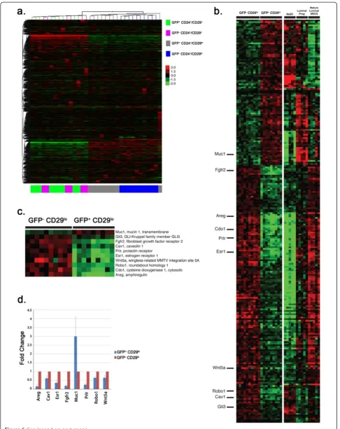
The above data provide evidence that H2BGFP+CD24+/ CD29lo MECs contain a subpopulation of pregnancy-activated multipotent progenitors. To characterize these cells further, RNA was isolated from the four MEC subpopulations of four-week-old mice, and transcrip-tional profiling was performed using the Illumina Mouse Microarray platform. Analysis of the resulting data by unsupervised hierarchical clustering segregated the samples into subpopulations defined by CD24, CD29 and H2BGFP expression (Figure 6a). Because we observed no functional differences between the H2GFP+ and H2GFP- CD24+/CD29+ populations, their failure to form a separate cluster by unsupervised analysis was not surprising, and suggests that H2BGFP expression in the CD24+/CD29+ population is a stochastic phenomenon.
By contrast, H2BGFP+/CD24+/CD29lo and H2BGFP-/ CD24+/CD29loMECs segregated into distinct clusters, consistent with their different behavior in transplant-ation experiments.
[image:11.595.61.540.87.462.2]well-documented mediators of mammary gland develop-ment as theEstrogen receptor 1(Esr1),Fibroblast growth factor receptor 2 (Fgfr2), Prolactin receptor (Prlr), and Amphiregulin(Areg) genes. To validate these observations, mRNA from H2BGFP+/CD24+/CD29lo and H2BGFP-/ CD24+/CD29loMECs was analyzed by qPCR (Figure 6d). These assays confirmed the microarray data for eight of the ten mammary developmental genes: Areg, Caveolin 1 (Cav1), Esr1, Fgfr2, Mucin 1 (Muc1), Prlr, Roundabout
homolog 1 (Robo1) and Wingless-type MMTV integration
site family, member 5A(Wnt5a). In addition, MMTVrtTA/ H2BGFP MECs from each of the four populations were cytospun onto slides and immunostained for proteins whose transcripts were expressed differentially in the H2BGFP+/CD24+/CD29loand H2BGFP-/CD24+/CD29lo populations and for mammary lineage markers (Figure S4 in Additional file 7). Again, higher levels ofAregandEsr1 were found in H2BGFP-/CD24+/CD29loMECs than in H2BGFP+/CD24+/CD29loMECs.
We next compared the gene expression profiles of the H2GFP+/CD24+/CD29lo cells and three mammary cell subpopulations defined by Shackletonet al.[4] and Vaillant et al.[28], and characterized by Limet al.[36]: mammary stem-cell enriched (MaSC) (CD24+/CD29e/CD61+), luminal progenitor (CD24+/CD29lo/CD61+), and mature luminal (CD24+/CD29+/CD61-) cells (Figure 6b). Differentially expressed genes in H2BGFP+/CD24+/CD29loand H2BGFP-/ CD24+/CD29loMECs were enriched for genes that dis-tinguish the CD24+/CD29+, CD24+/CD29lo/CD61+and CD24+/CD29e/CD61+ subpopulations (P = 1.07e-06). Transcripts expressed at higher levels in H2BGFP-/ CD24+/CD29lo cells (compared with H2GFP-/CD24e/ CD29lo cells) were 2.8-fold enriched for genes that were expressed highly in CD24+/CD29lo/CD61+ MECs, at lower levels in CD24+/CD29lo/CD61-mature cells and at low-est levels in the CD24+/CD29+ stem cell compartment (P= 9.91e-08). The transcripts expressed at higher levels in H2BGFP+/CD24+/CD29lo MECs also were 2.8-fold enriched for genes expressed highly in the stem cell compartment, at lower levels in the CD24e/CD29lo/ CD61+ population, and at lowest levels in the mature luminal cells (P= 8.75e-06).
Although these data indicate that H2BGFP+/CD24+/ CD29lo cells are more similar to CD24+/CD29lo/CD61+ MECs than to CD24+/CD29lo/CD61-MECs, most notably in their lower levels of transcripts for mammary develop-mental genes such as Areg, Esr1,Fgfr2and Prl, H2BGFP expression in the CD24+/CD29lopopulation did not correl-ate with CD61 expression (Figure S1 in Additional file 1). Therefore, H2BGFP+/CD24+/CD29lo cells share tran-scriptional characteristics with mammary stem cells and luminal-restricted progenitors, a phenotype that might be predicted for a multipotent progenitor, but one clearly distinct from the CD24+/CD29+ and CD24+/ CD29lo/CD61+populations.
Discussion
We have identified and characterized a MEC population with properties of a pregnancy-activated multipotent progenitor. By studying mammary development in MMTVrtTA/H2BGFP transgenic mice, we discovered a subset of CD24+/CD29locells possessing the unique abil-ity to develop small, multilineage mammary outgrowths, ranging in size from a small alveolar cluster to a half-grown mammary gland, that can produce milk, yet have no self-renewal activity. The ability of these cells to form glandular outgrowths increases 5- to 10-fold when recipient mice are made pregnant. Transcriptional profiling pro-vides additional evidence that the H2BGFP+and H2GFP -CD24+/CD29lo populations are biologically distinct, with H2GFP+ cells expressing lower levels of key transcripts involved in mammary differentiation and higher levels of transcripts found in the CD24+/CD29+, mammary stem cell-enriched, compartment.
These H2BGFP+/CD24+/CD29loMECs are identifiable in mice in early puberty, which suggests that they arise during or before pubertal development and remain dor-mant until pregnancy. The ability of these progenitors to give rise to mammary outgrowths three to four weeks after they are transplanted also is consistent with an ability to remain dormant until activated by pregnancy stimuli.
Our observation that MMTVrtTA is active in mammary stem cells and pregnancy-activated progenitors conflicts with a previous report that the same strain of MMTVrtTA (See figure on previous page.)
Figure 6Gene expression differences segregate MMTVrtTA/H2BGFP MECs into distinct subpopulations. (a)Gene expression data from the four MMTVrtTA/H2BGFP populations were analyzed using an unsupervised two-way hierarchical clustering of 35,927 filtered probes on the Illumina Mouse Whole Genome (WG-6 version 2 release 2) microarray. Samples in the CD29+CD24-populations can be seen to segregate into two distinct subpopulations based on the H2BGFP marker, whereas H2BGFP does not affect the clustering of CD29+CD24+samples.(b-d)
Differentially expressed genes between the H2BGFP+/CD24+/CD29loand H2BGFP-/CD24+/CD29lopopulations were compared with those found in a publicly available data set [36] consisting of isolated mammary stem cell-enriched (MaSC), luminal progenitor (lum prog) and mature luminal mammary epithelial cell (lum mature MEC) populations. The resulting 247 genes in common were plotted as a heat map(b). Ten genes(c)were chosen for further validation and follow-up analyses based on their association with mammary gland development and differentiation.(d)
is expressed homogeneously in all MECs [21]. In that study, the MMTVrtTA transgene-induced expression of tetracycline-driven LacZ, and an enzymatic assay was used to detect transgene expression. The enzymatic LacZ assay is probably more sensitive than flow cytometric detection of H2BGFP. Moreover, if theβ-galactosidase reaction was not in the linear range in the earlier experiments, it might not have differentiated between levels of protein expression, resulting in the erroneous conclusion that MMTVrtTA transgene activity was uniform in the mammary gland. In any event, the interpretation of studies that assumed that this MMTVrtTA strain directs transgene expression ‘ uni-formly’ in the mammary gland should be reconsidered in light of our data that expression is largely restricted to stem cells and progenitors.
The molecular basis for selective MMTV-driven label-ing of stem cells and pregnancy-activated progenitors re-mains to be elucidated. In separate experiments, wherein the H2BGFP strain was crossed to other rtTA transgenic mice, H2BGFP expression was found in significantly fewer MECs and did not correlate with specific mammary line-ages (data not shown). Therefore, the pattern of H2BGFP expression observed in our study appears to be determined by the MMTVrtTA transgene.
The MMTV promoter contains PR and GR binding sites, and mammary development during pregnancy is regulated by progesterone and glucocorticoids, suggesting a possible mechanism behind MMTV activity in the mouse mammary gland; however, we found no correlation between H2BGFP expression and PR or GR expression. Al-ternatively, the insertion site of this particular MMTVrtTA transgene could underlie the expression pattern observed in this study.
Some investigators have hypothesized that mammary stem cells, rather than progenitors, are activated during pregnancy for alveoli production [37,38]. Others, however, suggest that the establishment of the mammary architec-ture of pregnancy utilizes a process distinct from the prolif-eration of puberty and estrus, involving the activity of different cell types [39,40]. Wagneret al.suggested the exist-ence of pregnancy-activated progenitors by using WAP-Cre transgenic mice in combination with a Rosa26-lox-lacZ allele to tag MECs in pregnant glands [40]. After an initial pregnancy to activate the WAP-Cre transgene, lineage tracing was used to identify a population of pregnancy-initiated MECs (PI-MECs) that could give rise to alveoli in successive pregnancies. These cells also could form fully functional mammary glands and were found subsequently to be CD24+/CD49f+[41,42], leading these authors to conclude that PI-MECs represent a mixed population of alveoli-forming progenitors and stem cells. Our H2BGFP+/CD24+/CD29-cells, by contrast, do not give rise to serially transplantable mammary outgrowths, demonstrating that this population lacks
mammary stem cells. Because we did not trace the dy-namics of H2BGFP+/CD24+/CD29lo MECs, we cannot determine the fate of these progenitor-derived structures following parturition.
While this work was in progress, Vaillantet al. reported that CD24+/CD29-cells can form limited mammary struc-turesin vivoonly when co-injected with Matrigel, while the repopulating ability of CD24+/CD29+cells is the same with or without Matrigel co-injection [10]. Jeselsohn et al.also found that CD24+/CD49flo MECs developed mammary outgrowths only in the presence of Matrigel [11], whereas Spike et al. reported that Matrigel could increase the mammary repopulation activity content of embryonic mammary cells [43]. These results suggest that the mam-mary gland contains progenitors that require additional sig-nals or growth factors to survive or proliferate, and could explain why the ability of CD24+/CD29locells to form out-growths was missed in earlier studies [4]. Although these studies demonstrated that CD24+/CD29lo MECs can form mammary outgrowths following transplant into virgin females, they did not assess the effects of pregnancy on the rate (or detection) of repopulation.
Our data show that this Matrigel-dependent outgrowth-generating ability is restricted to a subpopulation within the CD24+/CD29locompartment. Furthermore, the prolif-eration and development of outgrowths from these cells is increased by pregnancy. It is possible that Matrigel is required for the engraftment of H2BGFP+/CD24+/CD29lo cells in vivo, but based on our data, we conclude that pregnancy is still the critical trigger for development of mammary structures able to pass the threshold of de-tection in most outgrowths.
The existence of mammary progenitors with hormone-or environment-dependent activity is supphormone-orted by a recent and extensive lineage-tracing study, which concluded that mammary stem cells derive from an embryonic CK14+ lineage, whereas CK8 cells only give rise to luminal progen-itors [14]. Cells from the CK8+lineage also can contribute to alveoli in successive pregnancies, but are unable to form mammary structures in vivowithout co-injection of other MECs. In another study, Sleemanet al.showed that CD24+ MECs co-transplanted with Matrigel can give rise to mam-mary outgrowths with a wide range of sizes, suggesting that mammary stem cells/progenitors with different
in vivo growth potentials exist within the CD24+
The functional differences in MEC subpopulations identi-fiable by MMTVrtTA/H2BGFP expression were verified by gene expression analysis. By unsupervised hierarchical clustering, samples from the H2BGFP+ and H2GFP -CD24+/CD29lo populations segregated independently, consistent with their functional differences in transplant-ation experiments. Significantly, genes with critical roles in mammary development, includingAreg,Esr1andPrlr, were expressed at lower levels in H2BGFP+/CD24+/CD29e cells, which suggests that these cells are less differentiated than H2BGFP-/CD24+/CD29lo MECs. H2BGFP+/CD24+/ CD29lo cells also shared partial gene signatures with the CD24+/CD29+ (stem cell) and CD24+/CD29lo/CD61+ (luminal progenitor) populations. In particular, genes expressed at lower levels in the H2BGFP+/CD24+/CD29lo cells were expressed at low levels in CD24+/CD29+ (stem cell-enriched) MECs, whereas genes expressed at higher levels in H2BGFP+/CD24+/CD29lo cells were expressed at higher levels in CD24+/CD29lo/CD61+ MECs. Because H2BGFP expression in the CD24+/CD29lo population did not correlate with CD61 expression (Figure S1 in Additional file 1), these results suggest that H2BGFP+/CD24+/CD29locells have an intermediate phenotype between mammary stem cells and luminal pro-genitors, as expected for a multipotent progenitor.
We attempted to identify a marker that could separ-ate the H2BGFP+ and H2BGFP- populations in the CD24+/CD29locompartment by FACS, without use of our transgenic model. To this end, we tested antibodies against antigens found by the microarray (CD14, AREG, AlCAM, CD55, CXCL16, CD164, CD138, PRLR) or that have been identified as potential mammary lineage markers (CD49b, CD24, Sca-1, c-kit). However, no marker proved suitable for isolation of the H2BGFP+/CD24+/CD29lo population (Figure S1 in Additional file 1). The only notable result was that all H2BGFP+/CD24+/CD29lo MECs, as well as a significant fraction of H2BGFP-/ CD24+/CD29loMECs, were found to be CD14+. Other studies have found that CD14+ MECs demonstrate an alveolar phenotypein vitro, but thein vivorole of these cells is currently unknown [8,9].
While this manuscript was in review, Dos Santos and colleagues used an H2BGFP label retention approach to isolate an enriched mammary stem cell population employing the K5 promoter to induce H2BGFP expression rather than the MMTV promoter. The CD24+/CD29+ subpopulation that was isolated using label retention was found to have a mammary repopulation frequency of 1/33, as compared to the 1/70 described by Shackleton et al.[4]. Transcriptional profiling of these cells led to the identification of additional cell surface markers CD1d, CD22, and CD59a that could further purify mammary stem cells [20]. In our studies, we were unable to find CD24+/ CD29+ MECs with enhanced mammary repopulation
abilities, through either MMTVrtTA/H2BGFP expres-sion or MMTVrtTA/H2BGFP label retention, though it is possible our‘chase’was insufficient in the label re-tention studies.
It has been hypothesized that the mammary stem cells responsible for establishing the mammary architecture during puberty are directly responsible for all subse-quent mammary proliferation [37,38]. However, our data suggest that the developing mammary gland contains multipotent pregnancy-activated progenitors that give rise to milk-producing mammary structures in vivo. We conclude that the mammary stem cell hierarchy contains progenitors not only for different MEC lineages, but also for the specific functions and structures involved in lactation. These progenitors are of particular interest in light of the evidence that early pregnancy can pro-tect from breast cancer, presumably from a reduction in the proliferative potential of stem cells and/or the likelihood that progenitors that are potential targets for ma-lignant transformation. MECs that proliferate actively in response to pregnancy might be prone to oncogenesis, and therefore merit further investigation on both scien-tific and medical grounds.
Conclusions
We have provided evidence for a multipotent mammary progenitor present in the glands of pre-pubertal virgin females, which, when transplanted into the mammary fat pad, gives rise to milk-producing mammary structures containing multiple cell lineages at a dramatically higher frequency during pregnancy. This progenitor population is distinguished from previously identified mammary lineages byin vivofunction and gene expression analysis, and might represent a target cell in mammary tumorigenesis. Further study is required to better elucidate this progenitor’s role in normal mammary development and oncogenesis.
Additional files
Additional file 1: Figure S1.Candidate markers for separating H2BGFP+/CD24+/CD29loand H2BGFP-/CD24+/CD29lopopulations. Mammary epithelial cells (MECs) from 4-week-old MMTVrtTA/H2BGFP females were isolated, stained with a panel of antibodies against surface proteins, and analyzed by flow cytometry for their ability to distinguish the H2BGFP+/CD24+/CD29lo(green) and H2BGFP-/CD24+/CD29lo(red) populations. Markers included previously identified stem cell/progenitor markers and differentially expressed genes identified from microarray analyses (see Figure 6). H2BGFP, histone 2B-eGFP; MMTV, mouse mammary tumor virus promoter; rtTA, reverse tetracycline transactivator.
Additional file 2: Figure S2.Schematic of MMTVrtTA/H2BGFP mice. The MMTVrtTA strain (tet-on) was crossed with tet-responsive H2BGFP transgenic mice, resulting in tetracycline/doxycycline-activated, MMTV-driven expression of H2BGFP. H2BGFP, histone 2B-eGFP; MMTV, mouse mammary tumor virus promoter; rtTA, reverse tetracycline transactivator.
size based on the percentage of the mammary fat pad filled with epithelium, as indicated.
Additional file 4: Table S1.Mammary outgrowth sizes in limiting dilution transplants. Size of mammary outgrowths from H2BGFP+/CD24+/CD29+ (a,e), H2BGFP-/CD24+/CD29+(b,f), H2BGFP+/CD24+/CD29lo(c,g,i)and H2BGFP-/CD24+/CD29lo(d,h,j)populations in virgin mice(a-d,i)and pregnant mice(e-h,j). H2BGFP, histone 2B-eGFP.
Additional file 5:Hierarchical clustering of probes found to be significantly differentially expressed between the CD29 + CD24- GFP + versus CD29 + CD24- GFP- populations.
Additional file 6:Differentially expressed genes between the H2BGFP+/CD24+/CD29loand H2BGFP-/CD24+/CD29lopopulations compared with those found in a publicly available data set consisting of isolated mammary stem cell-enriched (MaSC), luminal progenitor (lum prog) and mature luminal mammary epithelial cell (lum mature MEC) populations.H2BGFP, histone 2B-eGFP.
Additional file 7: Figure S4.Cytospins of MMTVrtTA/H2BGFP populations. MMTVrtTA/H2BGFP mammary epithelial cell (MEC) populations were isolated by fluorescence-activated cell sorting (FACS), cytospun onto slides, fixed and immunostained for mammary lineage markers and microarray hits. The percentage of cells from each subpopulation that stain positive for each marker is indicated. H2BGFP, histone 2B-eGFP; MMTV, mouse mammary tumor virus promoter; rtTA, reverse tetracycline transactivator.
Abbreviations
bp:Base pair; BSA: Bovine serum albumin; CI: Confidence interval; DAPI: 4',6-diamidino-2-phenylindole; DMEM: Dulbecco’s modified Eagle’s medium; EGF: Epidermal growth factor; ER: Estrogen receptor; FACS: Fluorescence-activated cell sorting; FBS: Fetal bovine serum; FDR: False discovery rate; GFP: Green flourescent protein; GR: Glucocorticoid receptor; H2BGFP: Histone 2B enhanced GFP; MaSC: Mammary stem cell; MEC: Mammary epithelial cell; MMTV: Mouse mammary tumor virus promoter; MRU: Mammary
repopulating unit; PBS: Phosphate-buffered saline; PR: Progesterone receptor; rtTA: Reverse tetracycline transactivator; TEB: Terminal end bud.
Competing interests
The authors declare that they have no competing interests.
Authors’contributions
ASK conducted the experiments, analyzed data, and drafted the manuscript. LMS and CV performed the bioinformatic/statistical analysis of the microarray data. BGN conceived the study, and BGN and JSB participated in its design, coordination, and data analysis and helped to draft and edit the manuscript. All authors read and approved the final manuscript.
Acknowledgements
We are grateful to M. Bentires-Alj for instruction in the cleared mammary fat pad assay, J. Daley and S. Lazo for their FACS expertise, L. Chodosh for the MMTVrtTA mouse strain and E. Fuchs for H2GFP mice. This work was supported by a Terry Fox Program Project Grant and NIH R37 CA49152 to BGN as well as a grant from the Breast Cancer Research Foundation to JSB. BGN is a Canada Research Chair, Tier 1, and work in his laboratory is partially supported by the Ontario Ministry of Health and Long-Term Care and the Princess Margaret Foundation.
Author details
1
Department of Medicine, Cancer Biology Program, Division of Hematology-Oncology, Beth Israel Deaconess Medical Center, Harvard Medical School, 330 Brookline Avenue, Boston, MA 02215, USA.2Department of Cell Biology, Harvard Medical School, 240 Longwood Avenue, Boston, MA 02115, USA. 3
Campbell Family Cancer Research Institute, Ontario Cancer Institute, Princess Margaret Cancer Center, University Health Network, 610 University Avenue, Toronto ON M5G 2M9, Canada.4Department of Medical Biophysics, University of Toronto, Ontario Cancer Institute, Princess Margaret Hospital, 610 University Avenue, Toronto, Ontario M5G 2M9, Canada.
Received: 16 November 2012 Accepted: 22 July 2013 Published: 15 August 2013
References
1. Cole H:The mammary gland of the mouse, during oestrus cycle, pregnancy and lactation.Proc R Soc London: Biol Sci1933,114:136–161. 2. Tiede B, Kang Y:From milk to malignancy: the role of mammary stem cells
in development, pregnancy and breast cancer.Cell Res2011,21:245–257. 3. Hennighausen L, Robinson GW:Signaling pathways in mammary gland
development.Dev Cell2001,1:467–475.
4. Shackleton M, Vaillant F, Simpson KJ, Stingl J, Smyth GK, Asselin-Labat ML, Wu L, Lindeman GJ, Visvader JE:Generation of a functional mammary gland from a single stem cell.Nature2006,439:84–88.
5. Stingl J, Eirew P, Ricketson I, Shackleton M, Vaillant F, Choi D, Li HI, Eaves CJ: Purification and unique properties of mammary epithelial stem cells. Nature2006,439:993–997.
6. Asselin-Labat ML, Sutherland KD, Barker H, Thomas R, Shackleton M, Forrest NC, Hartley L, Robb L, Grosveld FG, van der Wees J, Lindeman GJ, Visvader JE:Gata-3 is an essential regulator of mammary-gland morphogenesis and luminal-cell differentiation.Nat Cell Biol2007,9:201–209.
7. Li W, Ferguson BJ, Khaled WT, Tevendale M, Stingl J, Poli V, Rich T, Salomoni P, Watson CJ:PML depletion disrupts normal mammary gland
development and skews the composition of the mammary luminal cell progenitor pool.Proc Natl Acad Sci U S A2009,106:4725–4730. 8. Asselin-Labat ML, Sutherland KD, Vaillant F, Gyorki DE, Wu D, Holroyd S,
Breslin K, Ward T, Shi W, Bath ML, Deb S, Fox SB, Smyth GK, Lindeman GJ, Visvader JE:Gata-3 negatively regulates the tumor-initiating capacity of mammary luminal progenitor cells and targets the putative tumor suppressor caspase-14.Mol Cell Biol2011,31:4609–4622.
9. Stingl J:Detection and analysis of mammary gland stem cells.J Pathol 2009,217:229–241.
10. Vaillant F, Lindeman GJ, Visvader JE:Jekyll or Hyde: does Matrigel provide a more or less physiological environment in mammary repopulating assays?Breast Cancer Res2011,13:108.
11. Jeselsohn R, Brown NE, Arendt L, Klebba I, Hu MG, Kuperwasser C, Hinds PW:Cyclin D1 kinase activity is required for the self-renewal of mammary stem and progenitor cells that are targets of MMTV-ErbB2 tumorigenesis.Cancer Cell2010,17:65–76.
12. Smith GH:Experimental mammary epithelial morphogenesis in an in vivo model: evidence for distinct cellular progenitors of the ductal and lobular phenotype.Breast Cancer Res Treat1996,39:21–31.
13. Sleeman KE, Kendrick H, Ashworth A, Isacke CM, Smalley MJ:CD24 staining of mouse mammary gland cells defines luminal epithelial, myoepithelial/ basal and non-epithelial cells.Breast Cancer Res2006,8:R7.
14. Van Keymeulen A, Rocha AS, Ousset M, Beck B, Bouvencourt G, Rock J, Sharma N, Dekoninck S, Blanpain C:Distinct stem cells contribute to mammary gland development and maintenance.Nature2011,479:189–193. 15. Smith GH:Label-retaining epithelial cells in mouse mammary gland
divide asymmetrically and retain their template DNA strands. Development2005,132:681–687.
16. Booth BW, Smith GH:Estrogen receptor-alpha and progesterone receptor are expressed in label-retaining mammary epithelial cells that divide asymmetrically and retain their template DNA strands. Breast Cancer Res2006,8:R49.
17. Booth BW, Boulanger CA, Smith GH:Selective segregation of DNA strands persists in long-label-retaining mammary cells during pregnancy. Breast Cancer Res2008,10:R90.
18. Tumbar T, Guasch G, Greco V, Blanpain C, Lowry WE, Rendl M, Fuchs E: Defining the epithelial stem cell niche in skin.Science2004,303:359–363. 19. Foudi A, Hochedlinger K, Van Buren D, Schindler JW, Jaenisch R, Carey V,
Hock H:Analysis of histone 2B-GFP retention reveals slowly cycling hematopoietic stem cells.Nat Biotechnol2009,27:84–90.
20. Dos Santos CO, Rebbeck C, Rozhkova E, Valentine A, Samuels A, Kadiri LR, Osten P, Harris EY, Uren PJ, Smith AD, Hannon GJ:Molecular hierarchy of mammary differentiation yields refined markers of mammary stem cells. Proc Natl Acad Sci U S A2013,110:7123–7130.
21. Gunther EJ, Belka GK, Wertheim GB, Wang J, Hartman JL, Boxer RB, Chodosh LA:A novel doxycycline-inducible system for the transgenic analysis of mammary gland biology.FASEB J2002,16:283–292.
22. Ip MM, Asch BB:Methods in mammary gland biology and breast cancer research.New York: Kluwer Academic/Plenum Publishers; 2000. 23. Hu Y, Smyth GK:ELDA: extreme limiting dilution analysis for comparing
24. Gentleman RC, Carey VJ, Bates DM, Bolstad B, Dettling M, Dudoit S, Ellis B, Gautier L, Ge Y, Gentry J, Hornik K, Hothorn T, Huber W, Iacus S, Irizarry R, Leisch F, Li C, Maechler M, Rossini AJ, Sawitzki G, Smith C, Smyth G, Tierney L, Yang JY, Zhang J:Bioconductor: open software development for computational biology and bioinformatics.Genome Biol2004,5:R80. 25. Du P, Kibbe WA, Lin SM:Lumi: a pipeline for processing Illumina
microarray.Bioinformatics2008,24:1547–1548.
26. Johnson WE, Li C, Rabinovic A:Adjusting batch effects in microarray expression data using empirical Bayes methods.Biostatistics2007,8:118–127. 27. Benjamini Y, Hochberg Y:Controlling the false discovery rate: a practical
and powerful approach to multiple testing.J R Statist Soc B (Methodological)1995,57:289–300.
28. Vaillant F, Asselin-Labat ML, Shackleton M, Forrest NC, Lindeman GJ, Visvader JE:The mammary progenitor marker CD61/beta3 integrin identifies cancer stem cells in mouse models of mammary tumorigenesis.Cancer Res2008,68:7711–7717.
29. Debnath J, Muthuswamy SK, Brugge JS:Morphogenesis and oncogenesis of MCF-10A mammary epithelial acini grown in three-dimensional basement membrane cultures.Methods2003,30:256–268.
30. Fuchs E, Tumbar T, Guasch G:Socializing with the neighbors: stem cells and their niche.Cell2004,116:769–778.
31. Asselin-Labat ML, Shackleton M, Stingl J, Vaillant F, Forrest NC, Eaves CJ, Visvader JE, Lindeman GJ:Steroid hormone receptor status of mouse mammary stem cells.J Natl Cancer Inst2006,98:1011–1014. 32. Wintermantel TM, Bock D, Fleig V, Greiner EF, Schutz G:The epithelial
glucocorticoid receptor is required for the normal timing of cell proliferation during mammary lobuloalveolar development but is dispensable for milk production.Mol Endocrinol2005,19:340–349. 33. Bonnefoix T, Bonnefoix P, Verdiel P, Sotto JJ:Fitting limiting dilution
experiments with generalized linear models results in a test of the single-hit Poisson assumption.J Immunol Methods1996,194:113–119. 34. Fazekas de St G:The evaluation of limiting dilution assays.J Immunol
Methods1982,49:R11–R23.
35. Bryder D, Rossi DJ, Weissman IL:Hematopoietic stem cells: the paradigmatic tissue-specific stem cell.Am J Pathol2006,169:338–346. 36. Lim E, Wu D, Pal B, Bouras T, Asselin-Labat ML, Vaillant F, Yagita H,
Lindeman GJ, Smyth GK, Visvader JE:Transcriptome analyses of mouse and human mammary cell subpopulations reveal multiple conserved genes and pathways.Breast Cancer Res2010,12:R21.
37. Joshi PA, Jackson HW, Beristain AG, Di Grappa MA, Mote PA, Clarke CL, Stingl J, Waterhouse PD, Khokha R:Progesterone induces adult mammary stem cell expansion.Nature2010,465:803–807.
38. Asselin-Labat ML, Vaillant F, Sheridan JM, Pal B, Wu D, Simpson ER, Yasuda H, Smyth GK, Martin TJ, Lindeman GJ, Visvader JE:Control of mammary stem cell function by steroid hormone signalling.Nature2010,465:798–802. 39. Chepko G, Smith GH:Three division-competent, structurally-distinct cell
populations contribute to murine mammary epithelial renewal. Tissue Cell1997,29:239–253.
40. Wagner KU, Boulanger CA, Henry MD, Sgagias M, Hennighausen L, Smith GH: An adjunct mammary epithelial cell population in parous females: its role in functional adaptation and tissue renewal.Development2002,129:1377–1386. 41. Boulanger CA, Wagner KU, Smith GH:Parity-induced mouse mammary
epithelial cells are pluripotent, self-renewing and sensitive to TGF-beta1 expression.Oncogene2005,24:552–560.
42. Matulka LA, Triplett AA, Wagner KU:Parity-induced mammary epithelial cells are multipotent and express cell surface markers associated with stem cells.Dev Biol2007,303:29–44.
43. Spike BT, Engle DD, Lin JC, Cheung SK, La J, Wahl GM:A mammary stem cell population identified and characterized in late embryogenesis reveals similarities to human breast cancer.Cell Stem Cell2012,10:183–197.
doi:10.1186/bcr3459
Cite this article as:Kaantaet al.:Evidence for a multipotent mammary progenitor with pregnancy-specific activity.Breast Cancer Research
201315:R65.
Submit your next manuscript to BioMed Central and take full advantage of:
• Convenient online submission
• Thorough peer review
• No space constraints or color figure charges
• Immediate publication on acceptance
• Inclusion in PubMed, CAS, Scopus and Google Scholar
• Research which is freely available for redistribution