R E S E A R C H A R T I C L E
Open Access
Breast density change as a predictive surrogate
for response to adjuvant endocrine therapy in
hormone receptor positive breast cancer
Jisun Kim
1, Wonshik Han
1,2*, Hyeong-Gon Moon
1, Soo Kyung Ahn
1, Hee-Chul Shin
3, Jee-Man You
4, Sae-Won Han
5,
Seock-Ah Im
5, Tae-You Kim
5, Hye Ryoung Koo
6, Jung Min Chang
6, Nariya Cho
6, Woo Kyung Moon
6and
Dong-Young Noh
1,2Abstract
Introduction:Anti-estrogen therapy has been shown to reduce mammographic breast density (MD). We hypothesized that a short-term change in breast density may be a surrogate biomarker predicting response to adjuvant endocrine therapy (ET) in breast cancer.
Methods:We analyzed data for 1,065 estrogen receptor (ER)-positive breast cancer patients who underwent surgery between 2003 and 2006 and received at least 2 years of ET, including tamoxifen and aromatase inhibitors. MD was measured using Cumulus software 4.0 and expressed as a percentage. MD reduction (MDR) was defined as the absolute difference in MD of mammograms taken preoperatively and 8-20 months after the start of ET. Results:At a median follow-up of 68.8 months, the overall breast cancer recurrence rate was 7.5% (80/1065). Mean MDR was 5.9% (range, -17.2% to 36.9%). Logistic regression analysis showed that age < 50 years, high preoperative MD, and long interval between start of ET to follow-up mammogram were significantly associated with larger MDR (p < 0.05). In a survival analysis, tumor size, lymph node positivity, high Ki-67 (≥10%), and low MDR were
independent factors significantly associated with recurrence-free survival (p < 0.05). Compared with the group showing the greatest MDR (≥10%), the hazard ratios for MDRs of 5-10%, 0-5%, and < 0% were 1.33, 1.92, and 2.26, respectively.
Conclusions:MD change during short-term use of adjuvant ET was a significant predictor of long-term recurrence in women with ER-positive breast cancer. Effective treatment strategies are urgently needed in patients with low MDR despite about 1 year of ET.
Introduction
Adjuvant endocrine therapy is the most effective systemic treatment modality for patients with hormone receptor (ER)-positive breast cancer, although many patients experience tumor recurrence during or after completion of endocrine therapy. Identifying factors that can predict disease recurrence early during adjuvant treatment may result in a more tailored strategy for patients likely to be endocrine resistant and may improve their overall outcomes.
Mammographic breast density (MD) is defined by the relative proportion of radiopaque areas, indicating the presence of fibroglandular tissue among the surrounding fatty component of the breast. High MD is associated with increased risk of breast cancer in both Western and Asian women [1,2]. The degree of lobular involu-tion is known to have inverse correlainvolu-tion with breast cancer risk as well [3].
Studies on the efficacy of tamoxifen for chemopreven-tion of breast cancer in high-risk women have shown that MD is decreased following tamoxifen treatment [4,5]. Moreover, 12- to 18-month change in MD was found to be an excellent predictor of response to tamoxifen in the preventive setting [5]. However, no studies to date have
* Correspondence: hanw@snu.ac.kr 1
Department of Surgery, Seoul National University College of Medicine, 101 Daehakro, Seoul, 110-744, Korea
Full list of author information is available at the end of the article
addressed the association between MD reduction and the efficacy of adjuvant endocrine treatment in breast cancer patients. Using quantitative imaging analysis software to assess serial changes in MD, we investigated the associa-tion between the degree of MD reducassocia-tion and long-term breast cancer recurrence in ER-positive breast cancer patients who received adjuvant endocrine therapy.
Materials and methods
Study population
Using our institution’s prospectively maintained
web-based database, we identified a total of 1,542 ER-positive breast cancer patients who underwent curative surgery at Seoul National University Hospital between October 2003 and December 2006. Patients were excluded if: 1) they did not receive adjuvant endocrine treatment, such as tamoxi-fen or an aromatase inhibitor, or were treated for less than 2 years; 2) their digital mammogram images were not available; 3) they had bilateral breast cancer, or 4) distant metastasis was observed before the start of endocrine ther-apy. Clinical and pathologic information on the 1,065 sub-jects was obtained from the database and used for further analysis. Treatment with adjuvant chemotherapy and/or radiotherapy was generally decided according to the
insti-tution’s guidelines. The standard duration of treatment
with tamoxifen is 5 years. Postmenopausal women were treated with the aromatase inhibitors anastrozole and letrozole for up to 5 years after surgery or after 2 to 3 years of tamoxifen.
Mammographic density measurement
MD was quantitatively measured on cranio-caudal (CC) images of the unaffected breast using Cumulus software 4.0 (University of Toronto, Toronto, Canada) by a single investigator (JK) blinded to treatment outcome. All evalu-ated images were digital mammograms performed at our institution, so film scanning was unnecessary. Mammo-graphic density reduction (MDR) was based on two digital mammograms; the first was taken within 2 weeks pre-surgery (preMD), and the second 8 to 20 months after the start of adjuvant endocrine treatment (postMD), and defined as the absolute difference between the MD of these two images (% MDR = % preMD - % postMD). The MD reduction ratio (MDRR) was also calculated (% MDRR = (preMD -postMD) × 100/preMD). Intraobserver reproducibility, tested for 10% of randomly selected images (213/2,130), was 0.93 (Pearson correlation coefficient).
Statistical analysis
Change in MD was categorized into four levels, an increase
(MDR < 0), 0≤MDR < 5%, 5≤MDR < 10%, and MDR≥
10%, and into a binary variable (MDR≥5% and < 5%),
with the 5% and 10% absolute reduction cut-offs based on
previous findings [5]. We also analyzed absolute MDR as a
continuous variable. The chi-square test andt-test were
used to compare factors that could affect change in MD. All loco-regional or distant disease recurrences were regarded as recurrence events in recurrence-free survival analysis. Survival curves were estimated by the Kaplan-Meier method and compared using the log-rank test. Mul-tivariate analyses were conducted using Cox’s proportional hazard regression model. All statistical analyses were per-formed using SPSS (version 17.0) software package (Chi-cago, IL, USA) and factors withP< 0.05 were considered statistically significant. Written informed consent was taken prior to surgery in all patients and the study proto-col including the use of the database was approved by the Institutional Review Board of Seoul National University Hospital and met the guidelines of the responsible govern-mental agencies.
Results
Demographics and result of mammographic density measurement
The mean age of the 1,065 included patients was 49.1 years (range, 24 to 77 years) (Table 1), and their mean duration of overall endocrine therapy was 5.1 years (range, 0.9 to 7.9 years). One hundred and twenty-seven patients
(11.9%) had ductal carcinoma in situ(DCIS). Second
mammograms used for density measurements were taken an average 13.1 months (range, 8 to 20 months) after the start of endocrine therapy. The result of MD measure-ments (preMD, postMD, MDR, and MDRR) are shown in Table 2. Mean MDR was 5.9% (range, -17.2% to 36.9%).
Factors associated with density change
Patients were dichotomized by degree of MD reduction using a cut-off of 5% (MDR≥5% vs. < 5%), and factors in the two groups were compared to identify associations
with high MDR. Patients with MDR≥5% were
signifi-cantly younger (46.5 ± 8.0 vs. 51.9 ± 9.8 years,P< 0.001; Additional file 1, Table S1) and were significantly more likely to have been treated with tamoxifen than with an aromatase inhibitor (P< 0.001). Similarly, when MDR was analyzed as a continuous variable, mean MDR was higher in patients who received tamoxifen than in those who received an aromatase inhibitor (6.5 ± 7.1 vs. 3.1 ± 6.3%,P < 0.001, data not shown). Mean time from start of endo-crine therapy to the second mammogram was longer in
the group with MDR≥5% than in the group with MDR <
5% (13.5 ± 3.1 vs. 12.6 ± 3.2 months,P< 0.001). Moreover,
mean preMD was higher with MDR≥5% than with MDR
< 5% (40.9 ± 12.4% vs. 30.0 ± 13.3%,P< 0.001).
endocrine therapy and the second mammogram were
significantly associated with high MDR (P < 0.05). The
data were consistently significant on stepwise regression analysis when adjusted by age, preMD and ET regimen (Additional file 1, Table S2).
Density change and recurrence-free survival
[image:3.595.59.533.97.643.2]During a median follow-up of 67.7 months, 80 of the 1,065 patients (7.5%) experienced tumor recurrence (Table 1). Multivariate Cox regression analysis showed that when analyzed as a continuous variable, MDR was Table 1 Patient demographics
Variable Mean ± SD (range) Number %
Age, yr 49.0 ± 9.3 (24-77)
≤50 680 63.8
> 50 385 36.2
Duration of ET, yr 5.0 ± 1.0 (0.9-7.9)
ET regimen
Tamoxifen 5 yr 657 61.7
Tamoxifen 2-3 yr - > AI (total 5 yr) yr 41 3.8
Tamoxifen 5 yr - > AI 192 18
AI 5 yr 16.4
Tumor size (cm) 2.1 ± 1.4 (0.1-10.0)
≤2 cm 638 59.9
> 2 cm 427 40.1
Lymph node status
Negative 706 66.3
Positive 359 33.7
Histologic grade
Low/Intermediate 825 77.5
High 240 22.5
Progesterone receptor
Negative 477 44.8
Positive 588 55.2
HER2
Negative 976 91.6
Positive 88 8.3
Ki-67
< 10 895 84
≥10 169 15.9
Neoadjuvant chemotherapy
No 1017 95.5
Yes 48 4.5
Operation
Breast conserving surgery 667 62.6
Mastectomy 398 37.4
Adjuvant chemotherapy
No 247 23.2
Yes 818 76.8
Radiotherapy
No 408 38.3
Yes 657 61.7
Recurrence
Total 80 7.5
Locoregional 21 26.2
Contralateral breast 8 10
Distant metastasis 51 63.8
significantly associated with recurrence-free survival (hazard ratio (HR) = 0.95, 95% confidence interval (CI)
0.92 to 0.99, P = 0.005, Additional file 1, Table S3).
When these patients were categorized into four groups according to the degree of MD change (the reference
group with MDR≥10%, plus three groups with MDR of
5 to 10%, 0 to 5%, and < 0% respectively), the HR for recurrence was proportional to the decrease in MDR.
Compared with the reference group with the greatest
MDR reduction (≥10%), the HRs for 5 to 10%, 0 to 5%,
and < 0% MDR were 1.33 (P= 0.413), 1.92 (P = 0.048),
and 2.26 (P= 0.027), respectively (Table 4, Figure 1A).
Large tumor size, lymph node metastasis, and high
Ki-67 level (≥ 10%) were also significantly associated with
recurrence-free survival. The risk of recurrence was 1.67 times higher with a lower MDR of < 5% compared to
MDR ≥ 5% when adjusted for age and preMD by
for-ward selection stepwise analysis (HR 1.67, 95% CI 1.07 to 2.62,P= 0.025) (Additional file 1, Table S4).
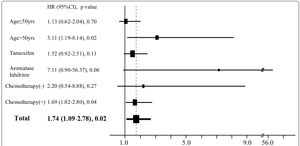
HRs for recurrence according to dichotomized MDR (≥5% vs. < 5%) in various subgroups are shown in a for-est plot (Figure 2). Subgroup analysis showed that the association between low MDR and high risk of
recur-rence differed by age group (> 50 vs.≤50 years), with
MDR significantly associated with risk of recurrence only in the postmenopausal group. MDR also significantly predicted recurrence in patients taking AIs, and was strongly correlated with the factor of age. When adjusted by age and ET regimen the findings were consistent, showing low MDR as a significant risk factor for recur-rence in patients who had undergone chemotherapy (HR 1.70, 95% CI 1.04 to 2.77,P= 0.033).
We also calculated the MDRR in this group of patients. We found that MDRR was significantly asso-ciated with risk of recurrence when analyzed as a con-tinuous and as a binary variable. Compared with
patients with MDRR ≥25%, the HRs for recurrence for
patients with MDRR of 0 to 10% and MDRR < 0% were
2.09 and 2.17, respectively (P < 0.05 each; Table 5,
Figure 1B). After adjusting for confounding factors,
patients with MDRR≥ 15% had a higher risk of
recur-rence than patients with MDRR < 15% (HR 1.60, 95% CI 1.02 to 2.50,P= 0.041) (Additional file 1, Table S5).
Discussion
[image:4.595.57.290.121.426.2]We have shown here that short-term MDR is predictive of long-term outcomes following endocrine therapy in patients with ER-positive breast cancer. Patients who experienced < 5% absolute MDR and those with increased MD after about 1 year of endocrine therapy were at 1.92- and 2.26-fold greater risk of recurrence Table 2 Distribution of mammographic density before
and after treatment and change in mammographic density
Variable Mean (range) Number %
PreMD, %* 35.77 ± 13.94 (5.42-82.18)
< 10% 26 2.4
10% - 25% 223 20.9
25% - 50% 641 60.2
≥50% 175 16.4
PostMD, % 29.84 ± 12.12 (3.90-72.31)
< 10% 35 3.3
10% - 25% 364 34.2
25% - 50% 611 57.4
≥50% 55 5.2
MDR, % 5.92 ± 7.08 (-17.2-36.9)
< 5% 505 47.4
≥5% 560 52.6
< 0% (increased) 190 17.8
0%-5% 314 29.5
5%-10% 276 25.9
≥10% 285 26.8
MDRR, %
< 15% 486 45.6
≥15% 579 54.4
< 0% (increased) 190 17.8
0%-10% 198 18.6
10%-25% 356 33.4
≥25% 321 30.1
Mammographic density reduction (MDR) and MDR ratio (MDRR) ((preMD -postMD) × 100/preMD) were initially evaluated as continuous variables then as dichotomized, quartered variables. Patients were divided into two groups
(MDR < 5%, 5%≤MDR), and four groups (MDR≥10%, 5%≤MDR < 10%, 0≤
[image:4.595.57.539.627.702.2]MDR < 5%, and MDR < 0% (increased MD)). PreMD: initial preoperative mammographic density; PostMD: density of follow up mammography after 8 to 20 months of hormone therapy.
Table 3 Factors associated with mammographic density reduction (MDR)
Variable Odds ratio 95% Confidence interval P-value
Age≤50 yr 1.84 1.30, 2.61 0.001
Interval to follow-up mammography, months* 1.07 1.02, 1.12 0.006
Initial tamoxifen (vs. AI) 0.99 0.65, 1.51 0.958
PreMD, % 1.06 1.04, 1.07 < 0.001
Adjuvant chemotherapy 1.41 1.00, 2.00 0.052
respectively, than patients with MDR ≥10%. This asso-ciation was also observed when absolute MDR was ana-lyzed as a continuous variable, and when MDRR rather than absolute MDR was assessed.
Regardless of the evolution of anti-estrogen therapy, a substantial proportion of patients with ER-positive breast cancer experience disease recurrence during fol-low-up. Effective biomarkers are needed to predict endocrine resistance despite ER expression. Many pre-vious investigations have focused on tumor factors asso-ciated with endocrine resistance [6,7]. The level of MDR resulting from endocrine treatment is a host factor indi-cating individual susceptibility to endocrine agents. These findings support the hypothesis that host response to adjuvant endocrine therapy may be as important and should be considered in addition to the clinicopathological characteristics of the primary tumor.
Breast density is one of the strongest risk factors for breast cancer development [8]. A recent study showed that the magnitude of the association of exogenous or endogenous hormone exposure and mammographic density change is related to future risk of breast cancer
[9]. Cuzicket al. conducted a nested case-control study
within the IBIS-I study, a randomized prevention trial of tamoxifen versus placebo to determine the association between tamoxifen-induced density change and breast cancer risk. They showed that the 12- to 18-month change in mammographic breast density is an excellent predictor of tamoxifen efficacy in the preventive setting [5].
[image:5.595.58.539.100.247.2]Our findings raise the question of whether treatment strategy should be altered based on change in MD after only one year of endocrine therapy. For the clinical application of individualized therapy, studies are needed Table 4 Predictive impact of mammographic density reduction (MDR) on recurrence-free survival
Variable Hazard ratio 95% Confidence interval P-value
Age, yr (continuous) 0.99 0.96, 1.02 0.393
MDR
≥10% (reference) 1.00 0.101
5-10% 1.33 0.67, 2.65 0.413
0-5% 1.92 1.01, 3.64 0.048
< 0% (increased) 2.26 1.10, 4.64 0.027
Size, cm (continuous) 1.19 1.05, 1.35 0.006
Lymph node positive 2.02 1.20, 3.40 0.008
High histologic grade 1.29 0.78, 2.16 0.323
Chemotherapy 0.79 0.39, 1.60 0.520
Ki-67≥10% 1.77 1.05, 3.00 0.033
Cox proportional hazard regression model for recurrence-free survival. Risk of recurrence was analyzed using the group with the greatest reduction (MDR≥10%)
as the reference. MDR: absolute mammographic density reduction (pre-treatment - post-treatment).
G
˙‹… fiL fi0‹ _months
†
¨ ¨t
U„‘
0.70
‹ 5˙t X 5-10 ‚\‹ ` 10 ‚\˙t `‹ `
1.00
0.95
0.90
0.85
0.80
0.75
0 12 24 36 48 60 72
Time (Months)
Recurrence free survival
1.00 (referent) 1.33 (0.67-2.65) 1.92 (1.01-3.64) 2.26 (1.10-4.64)
G
Mammographic density reduction(MDR,%)
MDR≥10%
MDR<0% 0≤MDR<5% 5≤MDR<10%
Hazard Ratio (95%CI)
1.00
0.95
0.90
0.85
0.80
0.75
0 12 24 36 48 60 72
Hazard Ratio (95%CI)
1.00 (referent) 1.37 (0.73-2.58) 2.09 (1.07-4.06) 2.17 (1.08-4.35)
Relative mammographic density reduction(MDRR,%)
MDRR≥25%
MDRR<0% 0≤MDRR<10% 10≤MDRR<25%
Recurrence free survival
Time (Months)
(A) (B)
[image:5.595.61.540.500.679.2]to evaluate the ability of shorter-term changes in MD, such as after < 6 months, to predict risk of recurrence, and new treatment strategies also should be tested according to the predicted result.
We also found that factors such as age < 50 years, high preoperative MD, and long interval between the start of endocrine therapy and the second mammogram were significantly associated with high MDR, indicating
that a dense breast per se is not a sign of endocrine
resistance, and that MD decreases more with prolonged endocrine treatment.
Although MDR was greater in patients < 50 years of
age than in those aged≥ 50 years, the degree of MDR
was not associated with recurrence in younger patients.
The reason for this is not clear, although it may be due to the complicated hormonal milieu and factors other than endocrine therapy affecting breast density in younger women. The relatively small number of events in patients aged < 50 years (43/680, 7.1%) in our dataset could have affected the statistical significance and further evaluation within a larger dataset is required.
The degree of MDR may differ according to the type of endocrine therapy. Tamoxifen was reported to be associated with an 8% mean absolute reduction in breast density at 1.5 years and a reduction of 14% after 4.5 years [10,11]. Raloxifene has been reported to decrease absolute breast density by 1.5% per year [12]. In a small study involving 54 patients, adjuvant anastrozole had no
Age≤50yrs
Age>50yrs
Tamoxifen
Aromatase Inhibitor
Chemotherapy(-)
Chemotherapy(+)
Total
1.13 (0.62-2.04), 0.70 HR (95%CI), p value
3.11 (1.19-8.14), 0.02
1.52 (0.92-2.51), 0.11
7.11 (0.90-56.37), 0.06
2.20 (0.54-8.88), 0.27
1.69 (1.02-2.80), 0.04
1.74 (1.09-2.78), 0.02
[image:6.595.54.540.88.325.2]1.0 5.0 9.0 56.0
Figure 2Subgroup analysis of association between mammographic density reduction (MDR) and disease recurrence. Forest plot shows hazard ratios (HRs) for recurrence in patients with MDR < 5% vs. those with MDR≥5% in the different patient subgroups.
Table 5 Predictive impact of mammographic density reduction ratio (MDRR) on recurrence-free survival
Variable Hazard ratio 95% Confidence interval P-value
Age, yr (continuous) 0.99 0.97-1.02 0.600
MDRR (%)*
≥25% 1.00 Referent 0.078
10%-25% 1.37 0.73-2.58 0.327
0%-10% 2.09 1.07-4.06 0.030
< 0% (increased) 2.17 1.08-4.35 0.030
Size, cm (continuous) 1.19 1.05-1.35 0.007
Lymph node positive 1.98 1.18-3.34 0.010
High histologic grade 1.34 0.80-2.24 0.261
Chemotherapy done 0.79 0.39-1.60 0.519
Ki-67≥10% 0.60 0.33-0.94 0.029
Cox proportional hazard regression model for recurrence-free survival. Risk of recurrence was analyzed using the group with the greatest reduction (MDRR≥
[image:6.595.58.539.565.713.2]effect on breast density in the contralateral breast after 6
months and resulted in a 16% relative reduction (P =
0.08) after 12 months [13]. Letrozole and exemestane also did not reduce mammographic breast density [14]. Addition of an aromatase inhibitor to hormone replace-ment therapy resulted in a significant reduction in breast density [1]. In a univariate analysis we found that tamoxifen treatment was associated with a higher MDR
than treatment with aromatase inhibitors (P < 0.001).
However, an association between MDR and recurrence-free survival was observed in our aromatase inhibitor group.
There is no definite evidence-based mechanism for an association between anti-estrogen therapy, reduced breast density and a better outcome. With aromatase inhibitors, it is possible that reduced density reflects effective circulating estrogen deprivation, and as a result, also affects micro-metastases. Another explanation is the difference in the drug metabolism efficiency of the host. Patients with adequate serum drug concentrations should have a better response and outcome. However, the metabolic mechanism of tamoxifen and aromatase inhibitors must be different. Adherence to a prescribed drug can partly explain the correlation between density change and patient outcome. In premenopausal and perimenopausal women, a good chemotherapy response could cause chemotherapy-induced ovarian failure, and as a result, could reduce breast density and improve dis-ease-free survival. It is unknown, but is less likely that tumor-associated fibroblasts or other stromal cells in the breast might directly affect distant micro-metastatic cancer cells.
We also found that 17.8% of our study subjects had increased MD after endocrine therapy. Similarly, in the preventive tamoxifen study IBIS-1, 11% of patients trea-ted with tamoxifen and 24% given placebo had an increased MD. The mechanism of this increase in MD is unknown. Investigations are needed to determine whether in these women, tamoxifen acts as an estrogen agonist in the breast.
The reproducibility of MD measurement is important, and studies indicate good intraobserver reproducibility with correlation coefficients of 0.92 to 0.96 [15]. We used Cumulus software, which has been the most widely used for MD measurements in previous studies. Using digital mammographic images, we found that the Pear-son correlation coefficient was 0.93.
The major limitation of the study was the absence of data on factors that may be closely associated with breast density, such as body mass index. Because this study was retrospective in design, the timing of follow-up mammography was not uniform, varying from 8 to 20 months after initiation of endocrine therapy. Another limitation was that the study subjects received
heterogeneous adjuvant therapy regimens. We also could not determine the degree of ER expression in tumors, a potential major factor affecting resistance to endocrine treatment.
To our knowledge, this study was the first to assess the value of MD change as a predictive surrogate in breast cancer patients receiving adjuvant endocrine ther-apy. The positive result we obtained warrants larger-scale prospective studies. Basic research to identify the molecular pathways related to endocrine resistance and mammographic density is also needed.
Conclusion
In conclusion, low MDR or increased MD during short-term endocrine therapy was independently associated with poor recurrence-free survival in patients with ER-positive breast cancer. Change in MD may be predictive of response to adjuvant endocrine therapy. Studies should be designed to investigate how to use this valu-able information in routine clinical practice.
Additional material
Additional file 1: Table S1: Univariate analysis for mammographic density reduction (MDR). Analysis of factors associated with MDR divided into two group (MDR < 5% vs. MDR≥5%). Younger age, tamoxifen use, longer interval from initial endocrine therapy, higher PreMD, adjuvant chemotherapy were likely to have higher MDR (≥5%). Table S2:Stepwise regression analysis (forward selection) of factors for MDR*≥5%. After adjusting for the confounding factors, age, interval to follow up, and preoperative mammographic density, adjuvant chemotherapy was not independently associated with MDR.Table S3: Cox proportional analysis for recurrence-free survival (RFS): MDR as a continuous variable. MDR analyzed as a continuous variable was an independent risk factor for recurrence, along with size, lymph node (LN) status, and Ki-67 level.Table S4:Cox proportional hazard regression (forward selection) analysis for RFS. After adjusting for confounding factors, patients with MDR < 5% had 1.67 times significantly higher risk of recurrence than the MDR≥5% group. Tumor size, lymph node (LN) positivity, and Ki-67 (cut-off 10%) were independent prognostic factors as known.Table S5:Cox proportional hazard regression (forward selection) analysis for RFS. After adjusting for confounding factors, patients with MDRR < 15% had 1.60 times significantly higher risk of recurrence than the MDRR≥15% group (P= 0.041). Tumor size, lymph node (LN) positivity, and Ki-67 (cut-off 10%) were independent prognostic factors as known.
Abbreviations
AI: aromatase inhibitor; CC: cranio-caudal; ER: estrogen receptor; DCIS: ductal carcinomain situ; ET: endocrine therapy; MD: mammographic density; MDR: mammographic density reduction; preMD: initial preoperative
mammographic density; postMD: mammographic density after endocrine therapy; MDRR: mammographic density reduction ratio.
Acknowledgements
This work was supported by a National Research Foundation of Korea (NRF) Grant funded by the Korean Government (20110005753 and 20110031417).
Author details 1
University, 101 Daehakro, Seoul, 110-744, Korea.3Department of Surgery, Chung-Ang University College of Medicine, 102 Heuksukro, Seoul, 156-755, Korea.4Department of Surgery, Sun General Hospital, 29 Mokjungro, Daejeon, 301-725, Korea.5Department of Internal Medicine, Seoul National University College of Medicine, 101 Daehakro, Seoul, 110-744, Korea. 6Department of Radiology, Seoul National University College of Medicine, 101 Daehakro, Seoul, 110-744, Korea.
Authors’contributions
All the authors have made substantial contributions to conception and design, acquisition of data, or analysis and interpretation of data. HRK, JMC, NC, and WKM carried out analysis of the imaging profiles of each patient including the Cumulus density measurement. SWH, SAI, and TYK confirmed patients’outcomes of recurrence and the adequacy of endocrine therapy. SKA, HCS, and JMY directly participated in the whole process throughout the research and statistical analysis. HGM and DYN participated in the study design and helped to draft the manuscript. JK measured the percent mammographic density and performed the research. As corresponding author, WSH designed and coordinated the research and provided close guidance throughout the process. All authors read and approved the final manuscript. The authors have been involved in drafting the manuscript or revising it critically for important intellectual content and have all given final approval of the version to be published.
Competing interests
The authors declare that they have no competing interests.
Received: 9 April 2012 Revised: 30 May 2012 Accepted: 6 July 2012 Published: 6 July 2012
References
1. Mousa NA, Crystal P, Wolfman WL, Bedaiwy MA, Casper RF:Aromatase inhibitors and mammographic breast density in postmenopausal women receiving hormone therapy.Menopause2008,15:875-884. 2. Jeon JH, Kang JH, Kim Y, Lee HY, Choi KS, Jun JK, Oh DK, Lee CY, Ko K,
Park EC:Reproductive and hormonal factors associated with fatty or dense breast patterns among korean women.Cancer Res Treat2011, 43:42-48.
3. Ghosh K, Vachon CM, Pankratz VS, Vierkant RA, Anderson SS, Brandt KR, Visscher DW, Reynolds C, Frost MH, Hartmann LC:Independent association of lobular involution and mammographic breast density with breast cancer risk.J Natl Cancer Inst2010,102:1716-1723.
4. Boyd NF:Tamoxifen, mammographic density, and breast cancer prevention.J Natl Cancer Inst2011,103:704-705.
5. Cuzick J, Warwick J, Pinney E, Duffy SW, Cawthorn S, Howell A, Forbes JF, Warren RM:Tamoxifen-induced reduction in mammographic density and breast cancer risk reduction: a nested case-control study.J Natl Cancer Inst2011,103:744-752.
6. Ali S, Coombes RC:Endocrine-responsive breast cancer and strategies for combating resistance.Nat Rev Cancer2002,2:101-112.
7. Musgrove EA, Sutherland RL:Biological determinants of endocrine resistance in breast cancer.Nat Rev Cancer2009,9:631-643. 8. Boyd NF, Guo H, Martin LJ, Sun L, Stone J, Fishell E, Jong RA, Hislop G,
Chiarelli A, Minkin S, Yaffe MJ:Mammographic density and the risk and detection of breast cancer.N Engl J Med2007,356:227-236.
9. Boyd NF, Melnichouk O, Martin LJ, Hislop G, Chiarelli AM, Yaffe MJ, Minkin S:Mammographic density, response to hormones, and breast cancer risk.J Clin Oncol2011,29:2985-2992.
10. Atkinson C, Warren R, Bingham SA, Day NE:Mammographic patterns as a predictive biomarker of breast cancer risk: effect of tamoxifen.Cancer Epidemiol Biomarkers Prev1999,8:863-866.
11. Cuzick J, Warwick J, Pinney E, Warren RM, Duffy SW:Tamoxifen and breast density in women at increased risk of breast cancer.J Natl Cancer Inst 2004,96:621-628.
12. Freedman M, San Martin J, O’Gorman J, Eckert S, Lippman ME, Lo SC, Walls EL, Zeng J:Digitized mammography: a clinical trial of postmenopausal women randomly assigned to receive raloxifene, estrogen, or placebo.J Natl Cancer Inst2001,93:51-56.
13. Prowell TM, Blackford AL, Byrne C, Khouri NF, Dowsett M, Folkerd E, Tarpinian KS, Powers PP, Wright LA, Donehower MG, Jeter SC, Armstrong DK, Emens LA, Fetting JH, Wolff AC, Garrett-Mayer E, Skaar TC,
Davidson NE, Stearns V:Changes in breast density and circulating estrogens in postmenopausal women receiving adjuvant anastrozole. Cancer Prev Res (Phila)2011,4:1993-2001.
14. Cigler T, Richardson H, Yaffe MJ, Fabian CJ, Johnston D, Ingle JN, Nassif E, Brunner RL, Wood ME, Pater JL, Hu H, Qi S, Tu D, Goss PE:A randomized, placebo-controlled trial (NCIC CTG MAP.2) examining the effects of exemestane on mammographic breast density, bone density, markers of bone metabolism and serum lipid levels in postmenopausal women. Breast Cancer Res Treat2011,126:453-461.
15. Byng JW, Boyd NF, Fishell E, Jong RA, Yaffe MJ:The quantitative analysis of mammographic densities.Phys Med Biol1994,39:1629-1638.
doi:10.1186/bcr3221
Cite this article as:Kimet al.:Breast density change as a predictive surrogate for response to adjuvant endocrine therapy in hormone
receptor positive breast cancer.Breast Cancer Research201214:R102.
Submit your next manuscript to BioMed Central and take full advantage of:
• Convenient online submission
• Thorough peer review
• No space constraints or color figure charges
• Immediate publication on acceptance
• Inclusion in PubMed, CAS, Scopus and Google Scholar
• Research which is freely available for redistribution