R E S E A R C H A R T I C L E
Open Access
MicroRNA-18a inhibits hypoxia-inducible factor 1
α
activity and lung metastasis in basal breast cancers
Raisa Krutilina
1,2, Wenlin Sun
3, Aarti Sethuraman
1,2, Martin Brown
2, Tiffany N Seagroves
1,2, Lawrence M Pfeffer
1,2,
Tatyana Ignatova
4and Meiyun Fan
1,2*Abstract
Introduction:In breast cancer, distinct expression profiles of microRNAs (miRNAs) have been associated with molecular subgroups and clinicopathological characteristics, implicating a diagnostic and prognostic role of miRNAs. However, the biological functions of deregulated miRNAs in tumor progression are not yet completely defined. In this study, we investigated the function of miR-18a in regulating breast cancer metastasis through the hypoxia-inducible factor 1α(HIF1A)–dependent hypoxic response.
Methods:An orthotopic metastatic breast cancer xenograft model (MDA-MB-231 cells) was used to identify miRNAs associated with spontaneous lung metastasis. The function of miR-18a in regulating HIF1A expression, as well as cellular responses to hypoxia and metastasis, were then studiedin vitroandin vivoby assessing ectopic miR-18a expression or miR-18a inhibition. miRNA–mRNA interactions (AGO2 immunoprecipitation and 3′untranslated region Luciferase reporter assays), gene expression (quantitative PCR and microarray), cell migration and invasion, and cell growth were assessed under normoxic or hypoxic conditions, complemented by orthotopic xenograft of tumor cells to the mammary fat pad to investigate the effect of modulating miR-18a expression on primary tumor growth and lung metastasis. Last, clinically relevant correlations between miR-18a, HIF1A, hypoxia-responsive gene expression and distant metastasis–free survival (DMFS) were assessed using published expression array breast tumors data sets. Results:miRNAs encoded by theMIR17HGgene were downregulated in lung metastases compared to primary tumors. Ectopic expression of miR-18a, aMIR17HGfamily member, in a metastatic variant of MDA-MB-231 cells reduced primary tumor growth and lung metastasis, whereas miR-18a inhibition in the parental cells promoted tumor growth and lung metastasis. We identified HIF1A as a direct target of miR-18a. Modulating miR-18a expression significantly affected hypoxic gene expression, cell invasiveness and sensitivity to anoikis and hypoxiain vitroin a HIF1A-dependent manner. Analysis of previously published data revealed that higher expression of HIF1A and a panel of hypoxic genes is associated with shorter DMFS interval in patients with basal-like breast tumors, and that, within this subtype, miR-18a expression is inversely correlated with hypoxic gene expression. Together, these data support a role of miR-18a in repressing distant metastasis through a HIF1A-dependent pathway.
Conclusions:The results of this study reveal a novel role for miR-18a in targeting HIF1A and repressing metastasis of basal-like breast tumors.
* Correspondence:mfan2@uthsc.edu
1Departments of Pathology and Laboratory Medicine, University of
Tennessee Health Science Center, 19 South Manassas Street, Memphis, TN 38163, USA
2
Center for Cancer Research, University of Tennessee Health Science Center, 19 South Manassas Street, Memphis, TN 38163, USA
Full list of author information is available at the end of the article
Introduction
MicroRNAs (miRNAs) play an important role in co-ordinating spatial and temporal expression of proteins by regulating mRNA translation and stability [1]. Deregulation of miRNAs has been linked to tumor development and progression, and a growing number of miRNAs have been described as candidate oncogenes or tumor suppressors [2]. In breast cancer, distinct expression profiles of miRNAs have been associated with specific molecular subtypes and clinicopathological characteristics, implicating a diagnostic and prognostic role of miRNAs [3-5]. However, the bio-logical functions of the deregulated miRNAs in tumor progression have not yet been completely defined.
One of the most frequently deregulated miRNA-encoding genes in human cancer is the polycistronicMIR17HGgene, which encodes six miRNAs (miR-17, miR-20a, miR-18a,
miR-19a, miR-19b and miR-92a) [6].MIR17HG was
ori-ginally described as an oncomir because of its oncogenic function in the hematological system, thyroid and lung [7]. However, emerging evidence suggests that loss of function ofMIR17HGmight contribute to the development and progression of other types of cancers, implicating a tumor suppressor function. For example, loss of
heterozy-gosity at chromosome 13q31, where the humanMIR17HG
gene is located, was detected in approximately 25% of
human breast tumors [8]. In addition, MIR17HG
over-expression was found to inhibit proliferation of luminal breast cancer cells by targeting a steroid receptor coactiva-tor (NCOA3), cyclin D1 (CCND1) and estrogen receptorα (ESR1) [9-12]. Furthermore, distinct physiological func-tions have been linked to the different miRNA members encoded by the MIR17HG gene [10,13,14]. Therefore, to gain a more complete understanding of the physiological impact of MIR17HG deregulation in cancer, a detailed investigation of each individualMIR17HGfamily member in multiple types of tumor cells is required.
In this study, we discovered that, compared to parental cells (MB231RN) or a subline derived from the primary
tumors (MB231RN-MFP), miRNAs encoded byMIR17HG
were downregulated in a MDA-MB-231 subline isolated from spontaneous lung metastases (MB231RN-LM) and generated from tumor cells orthotopically implanted in the mammary fat pad. Functional studies of miR-18a, a relatively understudiedMIR17HGfamily member, revealed a major role in limiting continuous tumor growth and suppressing tumor metastasis, in part by direct regulation of hypoxia-inducible factor 1α (HIF1A) activity. Analysis of previously published expression data revealed that higher expression of HIF1A and a panel of hypoxic genes is associated with a shorter interval of distant metastasis– free survival (DMFS) only in basal-like breast tumors. Additionally, a significant inverse correlation between miR-18a expression and hypoxic gene expression was discovered in basal-like tumors. These data suggest that
downregulation of miR-18a and concomitant upregulation of HIF1A activity may be essential to promoting basal breast cancer metastasis to distant organs, including the lungs.
Material and methods
Cell culture and stable transfection
MDA-MB-231, MCF7 and MDA-MB-436 cells (American Type Culture Collection, Manassas, VA, USA) were main-tained in minimal essential medium supplemented with 100 U/ml penicillin, 100μg/ml streptomycin and 10% fetal bovine serum. To facilitate tumor imaging and tumor cell isolation in vitro, MDA-MB-231 cells were stably transfected with pEF-mRFP [15] and pReceiver-Lv151 (GeneCopoeia, Rockville, MD, USA). The transfected cells that express red fluorescent protein (RFP) and neomycin-resistant marker were designated MB231RN and were used to derive other sublines used in this study. To establish cells that constitutively express miR-18a, lung metastatic variants of MDA-MB-231 (LM) cells were transduced with pEZX-MR06 (GeneCopoeia), a lenti-viral vector that encodes a miR-18a stem loop and green fluorescent protein (GFP), and were sorted to obtain GFP-positive cells (LM-miR-18a). Control cells were transduced with pEZX-MR03 (GeneCopoeia) expressing GFP only (LM-GFP). The miArrest miR-18a inhibitor encoded by a lentiviral vector that coexpresses mCherry (HmiR-AN0255-AM03; GeneCopoeia) was used to es-tablish MDA-MB-231 sublines with low miR-18a activ-ity (MB231-18aIN). Control cells were transduced with pEZX-AM03 (GeneCopoeia) expressing mCherry only (MB231-C). HIF1A knockdown was achieved by trans-ducing MB231-C or MB231-18aIN cells with lentiviruses
expressing a short-hairpin RNA (shRNA) to HIF1A
(pLKO.1-HIF1AshRNA: NM_001530.x-1048s1c1; Open Biosystems/GE Dharmacon, Lafayette, CO, USA).
Generating cell sublines from spontaneous lung metastases and orthotopic primary tumors
MB231RN cells (5 × 10⁵ cells/10 μl phosphate-buffered saline) were surgically inoculated into the right inguinal
mammary glands of 4-week-old female NODscidγmice
washed four times with Dulbecco’s modified Eagle’s medium (DMEM) and plated in gelatin-coated flasks containing DMEM/F-12 medium (Life Technologies, Carlsbad, CA, USA) supplemented with 10% fetal bo-vine serum, 30% human foreskin fibroblast conditioned medium, 100 U/ml penicillin, 100μg/ml streptomycin, 10 mg/ml fungin (InvivoGen, San Diego, CA, USA) and
400 μg/ml neomycin. Cells obtained from three lungs
were pooled and designated the MB231RN-LM subline. Primary tumors grown in mammary gland fat pads were digested and subjected to culture to obtain the MB231RN-MFP subline. The same experimental procedures were used to generate MB231RN-CE-LM and MB231RN-CE-MFP sublines derived from mice inoculated with a clonal ex-pansion (CE) of a single colony from MB231RN cells (MB231RN-CE). All animal studies adhered to proto-cols approved by the Institutional Animal Care and Use Committee of the University of Tennessee Health Science Center.
Primary tumor growth and lung metastasis
To monitor primary tumor growth, mice were inspected weekly by manual palpation for tumor appearance. Tumor growth was monitored twice weekly using digital calipers. Tumor volume was calculated as follows: Volume = (Width2× Length)/2. To examine lung metastases, pri-mary tumors were either surgically removed 4 weeks after inoculation or left intact as indicated, and the lungs were extracted as described per experiment. Metastatic foci of RFP-expressing cells located on the dorsal surface of the left lung lobe were imaged and counted using a fluorescence microscope at × 10 mag-nification (CFI60; Nikon Instruments). The presence of tumor cells in the left lobes was further confirmed by hematoxylin and eosin (H&E) staining of formalin-fixed lung sections (10μm thick). In addition, the right lobes of the lungs were digested to prepare DNA, which was then subjected to quantitative PCR (qPCR) analysis using primers specific for the human Alu sequences (hAlu-qPCR), as described previously [16].
Quantitation of mRNA and miRNA expression by qPCR
Total RNA was prepared using TRIzol reagent (Life Technologies). mRNAs and miRNAs were converted to cDNA using iScript cDNA synthesis kits (Bio-Rad Laboratories, Hercules, CA, USA) and the NCode™miRNA First-Strand cDNA Synthesis Kit (Life Technologies),
re-spectively. qPCR was performed on the CFX96™Touch
Real-Time PCR Detection System using SYBR Green Supermix (Bio-Rad Laboratories). mRNA and miRNA expression data were normalized toβ-actin (ACTB) or a small nuclear ribonucleic acid (U6), respectively, using
the 2−ΔΔCt method. Primer sequences for mRNAs were
obtained from PrimerBank [17], and for miRNAs they
were designed according to the manufacturer’s
instruc-tions included with the NCode™ miRNA First-Strand
cDNA Synthesis Kit. The expression levels of unprocessed
transcript of MIR17HG (pri-MIR17HG) were examined
using the TaqMan Pri-miRNA assay (Life Technologies) as described previously [16].
HIF1α3′-UTR luciferase reporter assay
Luciferase reporter containing the 3′untranslated region (3′-UTR) ofHIF1AmRNA (HIF1A-3′-UTR-Luc, S809216;
SwitchGear Genomics, Carlsbad, CA, USA) and pSV-β
-galactosidase (Promega, Madison, WI, USA), along with pEZX-MR06 (expressing miR-18a and GFP) or control vector pEZX-MR03 (expressing GFP only), were trans-fected to MDA-MB-231/DROSHA-shRNA cells [16] using Lipofectamine 2000 reagent (Life Technologies) according to the manufacturer’s instructions. The lucifer-ase and β-galactosidase activities were determined at 48 hours posttransfection using the Luciferase andΒ-Glo Assay Systems (Promega), respectively. Reported luciferase activities were normalized toβ-galactosidase activities. The Phusion Site-Directed Mutagenesis Kit (Thermo Scientific, Asheville, NC, USA) and the following 5′-phosphorylated
primers were used to generate HIF1A-3′-UTR-Luc
re-porters with mutated or deleted miR-18a binding sites:
mutagenic primer, forward 5′-CATTTTAAAAAATGC
ggCgTTTTATTTATTTATT-3′and reverse 5′-ATG CTA
CTGCAATGCAATGGTTTAAATAC-3′; deletion primer
forward 5′-TTTATTTATTTTTGGCTAGGGAGTTTAT
CC-3′ and reverse 5′-TTTTTAAAATGATGCTACTGC
AATGC-3′.
AGO2 immunoprecipitation, immunoblotting and enzyme-linked immunosorbent assay
Migration and invasion assays
Cells (2 × 10⁴cells/0.5 ml/well) were plated onto control membrane inserts with 8-μm pores or onto Matrigel-coated membrane inserts (BD Biosciences, San Diego, CA, USA) that were placed in 24-well chambers filled with 0.6 ml of growth medium. Twenty-four hours after plating, cells that remained on the upper surface of the membrane were removed using cotton-tipped swabs, and cells that had mi-grated to or invaded the lower surface of the membrane were fixed with methanol, stained with 0.5% crystal violet and counted under the microscope. The migration per-centage was calculated as follows: Migration (%) = (Mean number of cells invading through uncoated insert × 100)/ Mean number of cells seeded onto the regulator culture surface. The invasion percentage was calculated as follows: Invasion (%) = (Mean number of cells invading through Matrigel insert membrane × 100)/Mean number of cells migrating through control insert membrane.
Cell growth and viability assays
To examine cell growth, cells were plated at a density of 3 × 10⁴cells/2 ml/well (six-well dish) and counted daily. To culture cells under lactic acidosis and hypoxic condi-tions, cells were seeded into medium with 25 mM lactic acid (pH 6.7) and incubated in a Galaxy 14S multigas
incubator supplemented with 2% O2 and 5% CO2. To
induce anoikis, cells (5 × 10⁴/well) were seeded into six-well dishes coated with polyhydroxyethylmethacrylate (Sigma-Aldrich, St Louis, MO, USA) to prevent cell attachment. Viable cells were counted using a trypan blue exclusion assay.
Microarray analysis
To induce chemical hypoxia, cells were treated with 200 μM cobalt(II) chloride (CoCl2) for 4 hours. Total
RNA from two sets of independent experiments was prepared using the RNeasy Mini Kit (QIAGEN, Valencia, CA, USA) and submitted to the Center of Genomics and Bioinformatics at the University of Tennessee Health Science Center (Memphis, TN, USA) for label-ing and hybridization to HT-12 expression BeadChips (Illumina, Chicago, IL, USA). Hybridization signals were processed using Illumina Genome Studio software for an-notation, background subtraction, quantile normalization and presence call filtering. The normalized hybridization signals with intensity <40 were set to the flooring value of 40. GeneSpring GX software (Agilent Technologies, Santa Clara, CA, USA) was used for statistical computing. The cutoff parameters for differentially expressed genes were false discovery rate (FDR) = 0.1 and fold change (FC)≥1.5. The differentially expressed genes were further subjected to functional annotation and signaling pathway
mapping using QIAGEN’S Ingenuity Pathway Analysis
(IPA) (Redwood City, CA, USA). The expression array data
have been deposited in the Gene Expression Omnibus (GEO) database [GEO:GSE45362].
Statistical analysis of correlation between miR-18a and HIF1αexpression in breast tumors
The expression data of breast tumor samples were re-trieved from the GEO database [GEO:GSE19783, GEO: GSE22220, GEO:GSE28884] or the Cancer Genome Atlas (TCGA). Tumor samples were assigned to five subtypes (luminal A, luminal B, Her2-enriched, basal-like and normal-like) using the PAM50 classifier [19]. The cor-relation between miR-18a and HIF1A expression was analyzed using GraphPad Prism 5 software (GraphPad Software, La Jolla, CA, USA). The association of HIF1A and hypoxia-responsive genes with distant metastasis was analyzed with the GOBO program (Gene expression-based Outcome for Breast cancer Online) [20].
Results
Generation of MDA-MB-231 subline with high metastatic potential
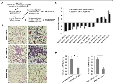
To identify miRNAs involved in breast cancer metastasis, we generated a lung metastatic subline by using an ortho-tropic metastatic mouse model of basal-like breast cancer MDA-MB-231 cells. MDA-MB-231 cells that express RFP and the neomycin-resistant marker (MB231RN) were surgically inoculated into the right inguinal mammary gland fat pad of 4-week-old female NSG mice, followed by primary tumor resection 4 weeks later, and lung tissue was extracted at 9 weeks after cell inoculation as shown in Figure 1A. The primary tumor and lung tis-sues were digested and subjected to in vitroculture to obtain the sublines MB231RN-MFP and MB231RN-LM, representing tumor cells grown at the orthotopic site of the mammary fat pad (MFP) or that spontaneously metas-tasized to lung (LM), respectively. To compare the meta-static potential of the sublines, cells (2.5 × 10⁵/injection) were inoculated into the right inguinal mammary gland fat pad of 4-week-old female NSG mice, and the presence of spontaneous metastatic foci in the lungs was examined by histological analysis at 7 weeks posttransplant. Small lung metastases (micrometastases) were observed in mice inoculated with MB231RN-MFP cells (Figure 1B, top panel). In contrast, large areas of lung parenchyma were occupied by tumor cells (macrometaststases) in all mice
(n= 5) that received MB231RN-LM cells (Figure 1B,
middle panel). These results validate the enhanced lung metastatic potential of the MB231RN-LM subline relative to MB231RN parental cells or MB231RN-MFP cells.
Downregulation of microRNAs in MB231RN-LM cells
human miRNAs examined, 10 were found to be signifi-cantly deregulated in lung metastatic MB231RN-LM cells in comparison to MB231RN or MB231RN-MFP cells (six downregulated and four upregulated). The differential ex-pression of these miRNAs was independently confirmed by qPCR (Figure 1C). Notably, all of the downregulated miRNAs in the MB231RN-LM cells are encoded by MIR17HG. Examination of unprocessed transcripts showed that the expressed level ofpri-MIR17HGwas sig-nificantly reduced in MB231RN-LM cells compared to MB231RN-MFP cells (the number of experimental re-peats = 3, FC = 1.54,P< 0.05). This result suggests that
de-regulation of miRNAs encoded by MIR17HG in
metastatic cells is due to reduced gene transcription rather than to impaired miRNA processing.
To rule out the possibility that the downregulation of MIR17HGin lung metastatic cells resulted from selec-tion of a clonal variant present in the pooled MB231RN population that may intrinsically express low levels of MIR17HG, we repeated the samein vivo experiment using a subline derived from a single MB231RN colony (MB231RN-CE). Tumor cells iso-lated from spontaneous lung metastases CE-LM) and orthotopic primary tumors (MB231RN-CE-MFP) using this clonally expanded line were subjected to miRNA expression analysis. The expres-sion of miR-18a, along with miR-17, was significantly decreased in cells derived from lung metastases relative to those derived from primary tumors (Figure 1D). This
[image:5.595.58.539.89.440.2]result confirmed that MIR17HG downregulation is an
acquired trait associated with spontaneous lung metastasis rather than clonal selection.
miR-18a suppresses tumor growth and lung metastasis in vivo
Compared to otherMIR17HGfamily members, miR-18a
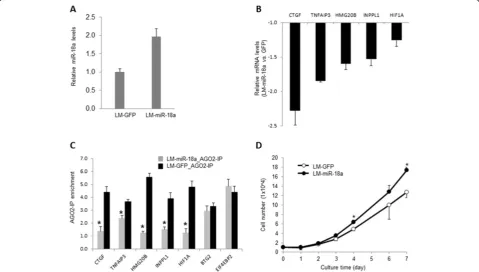
is understudied and its function in tumor cells is largely undefined. The maturation of miR-18a, but not other MIR17HGfamily members, has been reported to be spe-cifically regulated by heterogeneous nuclear riboprotein A1 (HNRNPA1), implicating tight control of miR-18a abundance in human cells [21]. Therefore, we speculated that miR-18a deregulation might directly impact cell metastatic potential. To examine whether miR-18a regu-lates primary tumor growth and metastasis, we used a len-tiviral construct that encodes a miR-18a stem loop and GFP to ectopically express miR-18a in MB231RN-LM cells (LM-miR-18a). An approximately twofold increase of miR-18a expression was detected by qPCR in the trans-duced cells, in comparison to cells that were transtrans-duced with a control vector expressing GFP only (LM-GFP) (Figure 2A). Because translation inhibition mediated by
miRNAs is usually coupled with target mRNA degradation [22], we performed qPCR to examine the mRNA levels of a panel of predicted miR-18a targets to confirm that the ectopically expressed miR-18a functions properly in the transfected cells. Five predicted targets of miR-18a were identified according to TargetScan [23], including con-nective tissue growth factor (CTGF), tumor necrosis factor
α–induced protein 3 (TNFAIP3), high-mobility group 20B (HMG20B), inositol polyphosphate phosphatase-like 1 (INPPL1) andHIF1A. As shown in Figure 2B, the expres-sion levels of these five mRNAs that harbor conserved binding sites with an exact match to positions 2 through 8 of miRNA-18a in 3′-UTR were significantly decreased (the number of experimental repeats = 3;P< 0.05) in response to ectopic miR-18a expression. We next examined the inter-action of miR-18a targets with RNA-induced silencing com-plex (RISC) by using AGO2 immunoprecipitation followed by qPCR analysis. As shown in Figure 2C, ectopic miR-18a expression significantly increased the amount of the target mRNAs in AGO2 immunocomplexes. In contrast, AGO2
binding of B-cell translocation gene 2 (BTG2) and
[image:6.595.58.538.363.635.2]eukaryotic translation initiation factor 4E binding protein 2
(EIF4EBP2), which are validated targets of miR-21 [16], was not affected by ectopic expression of miR-18a. Notably, ec-topic miR-18a expression slightly increased the cell growth ratein vitro(Figure 2D), which is in agreement with a previ-ous report showing that miR-18a overexpression enhances proliferation of K562 and hepatoma cells [24,25].
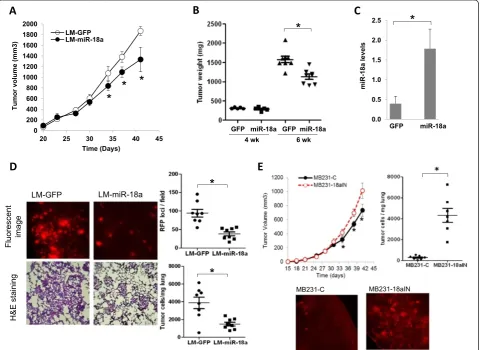
We next examined the effect of ectopic miR-18a ex-pression on primary tumor growthin vivo. LM-miR-18a and LM-GFP cells (2.5 × 10⁵) were inoculated into the fourth inguinal mammary gland fat pads of 4-week-old NSG mice. All females developed palpable tumors within
[image:7.595.59.541.234.584.2]2 weeks. Both the LM-miR-18a and LM-GFP tumors exhibited similar growth rates until grown to a volume of approximately 500 mm3(Figure 3A and 3B). At this point of tumor progression (day 35 posttransplant), we ob-served slower growth of the tumors derived from LM-miR-18a cells. The differences in tumor growth were con-firmed by comparing end-stage tumor wet weight (6 weeks after inoculation; Figure 3B). Sustained expres-sion of miR-18a in end-stage LM-miR-18a tumors was confirmed by qPCR (Figure 3C). These results suggest that ectopic miR-18a expression does not impact tumor
Figure 3Effects of ectopic microRNA-18a expression on primary tumor growth and spontaneous lung metastasis. (A)Growth curves of primary tumors generated from lung metastatic and green fluorescent protein microRNA-18a cells (LM-miR-18a and LM-GFP, respectively). The results are presented as mean ± SE (n= 10). *P< 0.05 by Kruskal-Wallis analysis of variance followed by Dunn’s multiple-comparisons test. (B)Wet weights of primary tumors that were surgically removed at either 4 weeks or 6 weeks postinoculation. *P< 0.05 by Kruskal-Wallis analysis of variance followed by Dunn’s multiple-comparisons test.(C)Expression levels of miR-18a in end-stage primary tumors. Total RNA was prepared from primary tumor tissues harvested 6 weeks after inoculation and subjected to quantitative PCR (qPCR) analysis for miRNA expression. The results are presented as mean ± SD (n= 3). *P< 0.05 by Student’st-test.(D)Spontaneous lung metastasis from orthotopic sites. Metastatic foci of red fluorescent protein (RFP)–expressing cells on the dorsal surface of the left lung lobe were imaged and counted using a fluorescence microscope at × 10 magnification. *P< 0.05 by Mann-WhitneyUtest (n= 8). The presence of tumor cells in the lungs was visualized by hematoxylin and eosin (H&E) staining of formalin-fixed lung sections (10μM) and quantified by qPCR using the human-specific primers of Alu repeats (hAlu-qPCR). *P< 0.05 by Mann-Whitney
initiation, but instead limits continuous expansion of the tumor mass.
To examine the effect of ectopic miR-18a expression on spontaneous lung metastasis, primary tumors were surgically removed at 4 weeks after inoculation, when the tumor sizes were equivalent (LM-GFP: 235 ± 75 mm3vs LM-miR-18a: 231 ± 84 mm3). Lung metastases were eval-uated 5 weeks after primary tumor resection via gross examination of the dorsal surface of the left lung lobes under a fluorescence microscope, followed by examination of H&E-stained tissue sections. The number of lung meta-static foci in mice inoculated with LM-miR-18a cells was significantly reduced compared to that of mice inoculated with LM-GFP cells (Figure 3D). Likewise, the number of tumor cells detected by qPCR using primers specific for the human Alu sequence in the lungs of mice inoculated with LM-miR-18a was reduced by about 60% compared to lungs of mice inoculated with LM-GFP (3,877 ± 641.6 cells/mg vs 1,496.0 ± 208.8 cells/mg lung tissue, respect-ively (n= 8) (Figure 3D). When inspecting tissue sections
(10 μm thick) from mice inoculated with LM-miR-18a
cells, we observed similar intensities of GFP (coexpressed with miR-18a) in lung metastatic cells and primary tumor cells, suggesting that the ectopically overexpressed miR-18a was retained in lung metastases. Taken together, these re-sults demonstrate that ectopic miR-18a expression reduced spontaneous lung metastasis from the MFPs.
We next examined the effect of inhibiting miR-18a ex-pression on spontaneous lung metastasis in MDA-MB-231 cells. A lentiviral vector expressing miArrest inhibitor of miR-18a and mCherry (HmiR-AN0255-AM03) was used to establish the subline MB231-18aIN. In contrast to ectopic miR-18a expression, which reduced late-stage tumor growth and lung metastasis, miR-18a inhibition enhanced late-stage tumor growth and substantially in-creased spontaneous lung metastasis (Figure 3E). Spe-cifically, miR-18a inhibition increased final tumor wet weight by about 20% (MB231-18aIN: 1,012 ± 278 mg (n= 14) vs MB231-C: 852 ± 230 mg (n= 14),P= 0.0743 by unpaired t-test with Welch’s correction, 6 weeks posttransplant) and increased cell numbers in lung tissues more than 10-flold as measured by hAlu-qPCR (MB231-18aIN: 4,274 ± 1,978 cells/mg lung tissue (n= 7) vs MB231-C: 294 ± 183 cells/mg lung tissue (n= 7);P< 0.001 by unpairedt-test with Welch’s correction).
miR-18a regulates HIF1αactivity and cell response to hypoxia
HIF1A activation by hypoxia has been identified as a key event for continuous expansion of tumor mass and me-tastasis by stimulating angiogenesis and activating the expression of metastatic signature genes in various types of tumor cells [18,26-28]. We identified an evolutionarily conserved miR-18a target site located in the 3′-UTR of
HIF1AmRNA, consistent with observations that ectopic miR-18a expression significantly increasedHIF1AmRNA interaction with RISC (Figure 2B). Therefore, we hypothe-sized that miR-18a might affect tumor growth and metas-tasis by targeting HIF1A.
First, we examined whether miR-18a targets HIF1A
mRNA through the conserved binding site located in
the 3′-UTR using a MDA-MB-231/DROSHA-shRNA
subline that is defective in miRNA synthesis [16] and a
HIF1A-3′-UTR-Luc reporter. As shown in Figure 4A
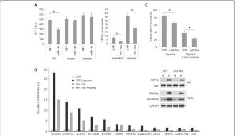
(left panel), HIF1A-3′-UTR-Luc activity in cells cotrans-fected with pEZX-MR06 (expressing miR-18a and GFP) was significantly decreased in comparison to cells cotransfected with a control vector (expressing GFP only). Point mutations or deletion of the miR-18a target site (GCACCUU, positions 409 to 415 of the HIF1A 3′-UTR) abolished the inhibitory effect of miR-18a on reporter luciferase expression.
We next compared HIF1A protein expression in cells cultured under normoxia or hypoxia (2% O2for 16 hours)
using a cell-based ELISA that measures total HIF1A pro-tein levels in the context of whole cells (Figure 4B). HIF1A protein levels in LM-GFP cells were increased about six-fold in response to hypoxia. LM-miR-18a cells expressed lower levels of HIF1A protein than LM-GFP cells at either normoxia or hypoxia. In agreement with the reduced HIF1A expression, reduced hypoxia induction of a panel of known hypoxia responsive genes was reduced in
LM-miR-18a cells, including PFKBP3 and BHLHE40,
which were also confirmed at the protein level by Western blotting (Figure 4B).
To examine the consequence of HIF1A reduction by miR-18a on cell growth under hypoxia, alone or in com-bination with acidosis, environments that are frequently encountered by cells within solid tumors, we measured viable cell numbers after growing cells under hypoxia (2% O2) with or without exposure to a high concentration
of lactic acid (25 mM) for 48 hours. Ectopic miR-18a ex-pression significantly decreased viable cell numbers when cells were exposed to hypoxia alone or to hypoxia in com-bination with lactic acidosis (Figure 4C).
To obtain a more global and unbiased characterization of the effect of miR-18a on HIF1A activity, we compared gene expression profiles of LM-GFP and LM-miR-18a cells treated with CoCl2, a hypoxia mimetic that rapidly
stabilizes HIF1A protein by inhibiting its prolyl hydrox-ylation [29,30]. After exposure to CoCl2 (200 μM for
4 hours), 219 or 132 genes were found to be significantly upregulated or downregulated, respectively, in LM-GFP cells (FDR = 0.1, FC > 1.5). All data are reported in the GEO database [GEO:GSE45362]. Consistent with miR-18a-mediated suppression of HIF1A, a smaller number
of genes were found to change in response to CoCl2
upregulated or downregulated, respectively (Figure 5A). Ingenuity Pathway Analysis revealed that several signal-ing pathways were differentially affected by CoCl2 in
LM-GFP and LM-miR18a cells (Figure 5B). For example, CoCl2-induced genes in LM-GFP cells were significantly
enriched for HIF1A, granulocyte-macrophage colony-stimulating factor and hepatocyte growth factor signaling pathways, indicating activation of HIF1A and downstream angiogenic signaling pathways. CoCl2-downregulated genes
in LM-GFP cells were significantly enriched in the integrin, paxillin and integrin-linked kinase signaling pathways involved in cell adhesion and migration. In contrast, in LM-miR-18a cells, none of these known HIF1A regulated path-ways were significantly affected by CoCl2exposure. Taken
together, these observations provide additional evidence in support of an essential role for miR-18a in regulating HIF1A activity in breast tumors. Notably, according to three target
prediction programs (TargetScan, PicTar and miRBase), most of the CoCl2-responsive genes affected by miR-18a lack target
sites of miR-18a, suggesting that miR-18a regulates CoCl2
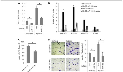
[image:9.595.57.539.89.368.2]-responsive genes primarily through indirect mechanisms. To investigate whether miR-18a-mediated HIF1A inhib-ition can be extended to other basal-like breast cancer cell lines, we examined the impact of ectopic miR-18a expres-sion in MDA-MB-436 cells. As shown in Figure 6, miR-18a expression downregulated the expression of HIF1A protein as well as the induction of HIF1A target genes at hypoxia. In addition, ectopic miR-18a expression in MDA-MB-436 cells significantly decreased cell growth under hypoxia (Figure 6C) and abolished hypoxia-induced cell invasion (Figure 6D). Taken together, these observations provide additional evidence that supports a conserved role for miR-18a in limiting HIF1A activity in basal-like breast can-cer cells.
Figure 4Effects of microRNA-18a on hypoxia-inducible factor 1αexpression and hypoxic response. (A)MicroRNA-18a (miR-18a) directly targeted the 3′untranslated region (3′-UTR) of hypoxia-inducible factor 1α(HIF1A) mRNA and ectopic miR-18a expression decreased HIF1A protein levels in MB231RN-LM cells cultured at either normoxia or hypoxia (2% O2for 16 hours). HIF1A-3′-UTR-Luciferase reporter activities were measured in MDA-MB-231/DROSHA-shRNA cells cotransfected with either pEZX-MR06 (expressing miR-18a) or control vector pEZX-GFP. GFP, Green fluorescent protein; shRNA, Short-hairpin RNA. Luciferase activities were normalized to internal control pSV-β-galactosidase and are presented as mean ± SD. *P< 0.05 by Student’st-test (n= 4). WT, mutant and deletion represent wild type, miR-18a binding site mutation and miR-18a binding site deletion of HIF1A-3′-UTR-Luciferase reporter, respectively. HIF1A protein levels in MB231RN-LM cells transduced with miR-18a or GFP were measured by a cell-based enzyme-linked immunosorbent assay, normalized to cytochrome c and presented as mean ± SD (n= 4). *P< 0.05 by Student’st-test. (B)miR-18a attenuated the induction of HIF1A target genes by hypoxia in MB231RN-LM cells. Cells were exposed to hypoxia (2% O2for 16 hours). mRNA levels were measured by quantitative PCR and normalized toACTB, and data are presented as mean ± SD (n= 3). The inset shows the expression levels of indicated proteins in nuclear extracts (NEs) or whole-cell lysates (WCEs) detected by immunoblotting. TATA-binding protein (TBP) and glyceraldehyde 3-phosphate dehydrogenase (GAPDH) were used as loading controls for NEs and WCEs, respectively.(C)Ectopic miR-18a expression in MB231RN-LM cells reduced viable cell numbers after cells were exposure to hypoxia (2% O2) and lactic acidosis (25 mM lactic acid, pH 6.7) for 48 hours. *P< 0.05 by Student’s
miR-18a represses tumor cell metastatic properties primarily through hypoxia-inducible factor 1α
In cultured cells, miR-18a inhibition enhanced gene
expression induced by hypoxia (2% O2 for 6 hours)
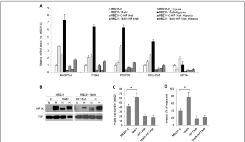
(Figure 7A). Immunoblotting assay results showed that miR-18a inhibition increased baseline protein levels of HIF1A (Figure 7B, left panel). Consistent with the obser-vation that miR-18a inhibition promoted tumor growth and metastasis in vivo (Figure 3E), MB231-18aIN cells were more resistant to anoikis (Figure 7C) and more in-vasive (Figure 7D) in comparison to control MB231-C cells. To confirm that these cellular changes induced by miR-18a inhibition were mediated by HIF1A, we examined the impact of HIF1A knockdown on the efficacy of a miR-18a inhibitor. Expression of HIF1A shRNA in
MB231-C or MB231-18aIN cells reducedHIF1AmRNA
levels by approximately 70% compared to control cells transduced with empty vector (Figure 7A). The reduced HIF1A expression in MB231-18aIN-HIF1AshRNA cells compared to MB231-18aIN-Vec cells was also confirmed by Western blotting (Figure 7B, right panel). HIF1A knockdown substantially decreased hypoxia-induced gene expression in both MB231-C and MB231-18aIN cells (Figure 7A), including PFKFB3, BHLHE40, angiopoietin-like 4 (ANGPTL4) and integrin α5 (ITGA5). Consistent with the known function of HIF1A to protect cells against
anoikis and promote cell invasion, HIF1A knockdown substantially reduced the viability of cells cultured in suspension as well as cell invasive activity (Figure 7C and 7D). Importantly, no difference in cell viability was observed in suspension culture or in cell invasion be-tween MB231-C-shHIF1A and MB231-18aIN-shHIF1A cells, demonstrating that the predominant target of miR-18a that mediates phenotypes in MDA-MB-231 cells is HIF1A.
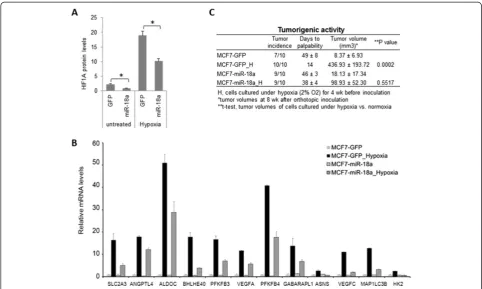
Ectopic HIF1A expression in MDA-MB-231 cells was previously reported to suppress tumor growth in a xenograft model [29]. Therefore, it is difficult to rescue a miR-18a-mediated suppressive effect on tumor growth and metastasis by ectopic HIF1A expression in LM-miR-18a cells. To provide further evidence that miR-LM-miR-18a regu-lates HIF1A activity, we examined the effect of ectopic miR-18a-5p (miR-18a) expression on hypoxia-induced tumorigenic activity of MCF7 cells. In an orthotopic xenograft model, MCF7 cells (1 × 10⁶ cells/injection) generated small tumors with a latency period of ap-proximately 7 weeks. Prolonged exposure of MCF7 cells to hypoxia (2% O2for 4 weeks in vitro) prior to
[image:10.595.66.537.89.334.2]inoculation substantially reduced the latency period of tumor initiation and enhanced tumor growth. Ectopic miR-18a expression in MCF7 cells reduced HIF1A protein expression (Figure 8A), dampened hypoxia-induced gene
expression (Figure 8B) and completely abolished hypoxia-induced tumorigenic activity (Figure 8C). These results provide additional evidence supporting a role for miR-18a in regulating hypoxic response in breast cancer cells.
Reverse correlation of miR-18a expression with expression of HIF1A and panel of hypoxia-responsive genes in basal-like breast tumors
To examine the clinical relevance of miR-18a mediated HIF1A inhibition, we investigated the relationship
be-tween miR-18a and HIF1A mRNA levels in human
breast tumor specimens by analyzing four data sets that contain both mRNA and miRNA expression data, which are available from TCGA or in the GEO database [GEO: GSE19783, GEO:GSE22220, GEO:GSE28884] [30-33].
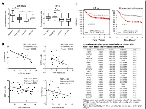
The expression of miR-18a, along with other MIR17HG
family members, was higher in basal-like tumors than in the other molecular subtypes defined by the PAM50 classifier [19] (Figure 9A, left panel). HIF1A mRNA is highly expressed in both basal and ERBB2 subtypes com-pared to luminal subtypes (Figure 9A, right panel), in
agreement with the observation that HIF1A protein is expressed at higher levels in basal cancers relative to lu-minal cancers [34]. Within the basal-like breast tumors, we found a significant inverse correlation between the ex-pression levels of miR-18a andHIF1A mRNA in all four data sets (Figure 9B). This correlation was not detected for any other subtype of breast cancer. These results im-plicate a role for miR-18a in restrainingHIF1Aactivity in basal-like breast tumors.
[image:11.595.57.538.88.369.2]We further examined the correlation of the expression levels of miR-18a and hypoxia-responsive genes that were associated with distant metastasis of breast tumors. The hypoxia-responsive genes in basal-like breast cancer cells were retrieved from GEO data sets [GEO:GSE17188, GEO:GSE18494] [28,35]. The correlation of hypoxia-re-sponsive gene expression with DMFS interval was assessed using the GOBO algorithm, which analyzes the correlation of gene expression and clinicopathological characteristics using expression data of 1,881 breast tumor specimens annotated with clinical information [33]. A panel of hypoxia-responsive genes was found to be
associated with reduced DMFS interval of patients with basal-like tumors (P< 0.05 by Kaplan-Meier analysis with 10-year censoring). We examined their correlation with miR-18a expression in the above-mentioned four data sets, which contain both mRNA and miRNA ex-pression data. This analysis led to the identification of a panel of metastasis-associated hypoxic genes that nega-tively correlated with miR-18a (Figure 9C). The individ-ual or collective roles of these genes in mediating the miR-18a-dependent suppression of lung metastasis in basal breast cancers warrant further studies.
Discussion
Deregulation of MIR17HG has been linked to breast
cancer progression, but the function of each individual
miRNA encoded byMIR17HGin breast tumor cells has
not yet been fully defined [9,12,36-38]. Compared to other
family members of MIR17HG, miR-18a is less studied,
and only limited information is available regarding its function in human cancer cells. Regarding luminal breast cancer cells, miR-18 has been shown to inhibit cell prolif-eration by targeting ESR1 [11]. In glioblastomas, miR-18a has been reported to repress tumor progression through
targeting SMAD3 and CTGF [39]. Here we report evi-dence that miR-18a plays an important role in restricting tumor growth and pulmonary metastasis of basal-like breast cancer by regulating HIF1A expression and hypoxic
response. HIF1A was reported as a target of MIR17HG
in lung cancer cells, but the specific role of miR-18a in the control of HIF1A expression and activity was not tested [40].
We found that miR-18a, along with other family mem-bers encoded byMIR17HG, was downregulated in breast cancer cells that spontaneously metastasized to lungs in an orthotopic xenograft model. We further confirmed that MIR17HG downregulation is an acquired trait associated with spontaneous lung metastasis by using orthotopic xenograft models derived from cell colony expansion. This finding is in line with the previous observation that MIR17HGexpression is downregulated in human primary breast tumors with positive lymph node metastasis com-pared to those with no metastasis [12].MIR17HG down-regulation during metastatic progression is also congruent with the reported inverse correlation between expression
of miR-92a, another member encoded byMIR17HG, and
[image:12.595.56.539.88.367.2]the tumor grade and recurrence-free survival of breast
cancer patients [38]. However, loss ofMIR17HG expres-sion was not recognized in a previous study on lung metastatic derivatives of MDA-MB-231 cells [41]. The discrepancy could be caused by differences in experi-mental approach, including the use of different mouse strains and different inoculation routes of tumor cells than those we employed in our present study.
The results of our in vivo studies with orthotopic
xenograft models of the metastatic MDA-MB-231 variant demonstrate that ectopic miR-18a expression significantly attenuated continuous expansion of primary tumor mass and decreased spontaneous lung metastasis. In contrast, miR-18a inhibition increased primary tumor growth and lung metastasis. These observations support a role of miR-18a in repressing tumor mass expansion and distant metastasis. Cells in solid tumors commonly encounter fluctuations in nutrient and oxygen availability caused by unbalanced cell proliferation and blood vessel devel-opment [26]. Continuous tumor growth is dependent on adaptation of tumor cells to hypoxic stresses and angiogenesis, two processes that are primarily regulated by HIF1A [42]. In addition, mounting evidence suggests a critical role of hypoxia in promoting tumor metastatic progression by rendering cells resistant to apoptosis and
invasive features. The results of our mechanistic studies provide evidence that miR-18a directly regulates HIF1A expression and consequently the expression of hypoxia-responsive genes and cell response to hypoxia.
[image:13.595.56.539.88.377.2]Expression data analysis using a metadata set composed of data from 1,881 breast tumors [20] revealed that higher expression of HIF1A and a panel of hypoxia-responsive genes is associated with shorter DMFS interval of patients with basal-like tumors, but not other subtypes. This find-ing is in agreement with previous reports that enhanced hypoxic response is associated with poor prognosis and chemotherapy resistance of basal-like tumors [43,44] and supports the notion that HIF1A activation drives metastatic progression of basal-like tumors. Importantly, on the basis of four independent data sets containing both miRNA and mRNA expression data, we found that miR-18a expression is inversely correlated with the expression of HIF1A and hypoxia-responsive genes in basal-like tu-mors. However, the available samples of basal-like tumors with both miRNA expression and DMFS data are not sufficient to establish the prognostic value of miR-18a for distant metastasis. Taken together, our observations suggest a role of miR-18a in suppressing metastasis by restricting hypoxic response in basal-like breast tumors.
Basal-like tumors are characterized by high prolifera-tion rates and rapid mass expansion, which lead to cyclic hypoxia and reoxygenation. Fine-tuned HIF1A activity by miR-18a may represent an essential mechanism to balance cell proliferation, survival and metastasis. In general, activation of HIF1A and hypoxia-responsive genes puts tumor cells in a survival and invasive state with reduced proliferation [45-48]. High expression of miR-18a in basal-like tumors (Figure 9A) may play a role in promoting cell growth when oxygen supply becomes abundant by restricting the antiproliferative activity of the HIF1A signaling pathway. This notion is consistent with previously report findings that tumors derived from transformed HIF1A−/− astrocytes grew faster than its HIF1A+/+ counterpart when placed in the vascular-rich brain parenchyma [49]. However, cells under hypoxic
stress rely on enhanced HIF1A activation to survive and obtain invasive features. Constitutive high miR-18a expres-sion might be detrimental to tumor mass expanexpres-sion and metastatic progression by restraining hypoxic response and consequently deterring hypoxia-induced apoptotic re-sistance and invasion.
The outcome of hypoxia-induced HIF1A activation was shown to be cell context–and tumor microenvironment– dependent [47,49]. As an upstream regulator of HIF1A, miR-18a function is likely tumor type–dependent. This notion is in line with previously reported findings that
miRNAs encoded byMIR17HG promote development of
[image:14.595.58.540.90.450.2]B-cell lymphomas and lung cancer, but repress glioblast-oma progression [38]. On the basis of our study results, we cannot exclude the possibility that other targets may contribute to miR-18a-mediated suppression on tumor
growth and metastasis. By integrated analysis of genes reg-ulated by miR-18a under normoxia using a data set from this study [GEO:GSE45362], miRNA target sites, expres-sion correlation of mRNA and miRNA, and correlation with DMFS of patients with basal-like breast tumors, we discovered that miR-18a potentially targets the prometa-static gene matrix metallopeptidase 3 (MMP3). Since MMP3is a hypoxia-responsive gene, miR-18a may modify the outcome of hypoxia by modulating the expression of specific hypoxia-responsive genes in addition to a general attenuation of hypoxic response by directly targeting HIF1A. It remains to be determined whether MMP3 downregulation contributes to miR-18a-mediated me-tastasis suppression.
Conclusions
The results of this study reveal a novel role for miR-18a in repressing metastasis of basal-like breast tumors by restricting HIF1A activity and regulating cell responses to hypoxia. The inverse correlation between miR-18a and HIF1A and hypoxia signature genes in basal-like breast tumors is significant in clinical samples and is consistent across multiple data sets. However, understanding how fluctuating miR-18a and HIF1A levels ultimately drive metastasis from the primary tumor to reduce survival requires further investigation.
Abbreviations
CoCl2:Cobalt(II) chloride; DMFS: Distant metastasis-free survival; GFP: Green fluorescent protein; HIF1A: Hypoxia-inducible factor 1α; microRNA-18a: MiR-18a; miRNA: MicroRNA; RFP: Red fluorescent protein; RISC: RNA-induced silence complex.
Competing interests
The authors declare that they have no competing interests.
Authors’contributions
MF and TI designed the experiments and wrote the main manuscript text. RK, AS and MB carried out most of the experiments. WS performed the statistical analysis. TNS and LMP provided reagents, participated in data interpretation and helped to draft the manuscript. All authors read and approved the final manuscript.
Acknowledgments
This work was supported by grants CA140346 (to MF) and CA138488 (to TS) from the National Cancer Institute, National Institutes of Health.
Author details
1Departments of Pathology and Laboratory Medicine, University of
Tennessee Health Science Center, 19 South Manassas Street, Memphis, TN 38163, USA.2Center for Cancer Research, University of Tennessee Health
Science Center, 19 South Manassas Street, Memphis, TN 38163, USA.
3Department of Pharmacology, University of Tennessee Health Science
Center, 424 Crowe research building, 874 Union Avenue, Memphis, TN 38163, USA.4Department of Neurosurgery, University of Tennessee Health
Science Center, 847 Monroe Avenue, Memphis, TN 38163, USA.
Received: 10 September 2013 Accepted: 1 July 2014 Published: 28 July 2014
References
1. Bartel DP:MicroRNAs: target recognition and regulatory functions.Cell
2009,136:215–233.
2. Lujambio A, Lowe SW:The microcosmos of cancer.Nature2012,
482:347–355.
3. Iorio MV, Ferracin M, Liu CG, Veronese A, Spizzo R, Sabbioni S, Magri E, Pedriali M, Fabbri M, Campiglio M, Ménard S, Palazzo JP, Rosenberg A, Musiani P, Volinia S, Nenci I, Calin GA, Querzoli P, Negrini M, Croce CM:
MicroRNA gene expression deregulation in human breast cancer.Cancer Res2005,65:7065–7070.
4. Persson H, Kvist A, Rego N, Staaf J, Vallon-Christersson J, Luts L, Loman N, Jonsson G, Naya H, Hoglund M, Borg A, Rovira C:Identification of new microRNAs in paired normal and tumor breast tissue suggests a dual role for theERBB2/Her2gene.Cancer Res2011,71:78–86.
5. Foekens JA, Sieuwerts AM, Smid M, Look MP, de Weerd V, Boersma AWM, Klijn JGM, Wiemer EAC, Martens JWM:Four miRNAs associated with aggressiveness of lymph node-negative, estrogen receptor-positive human breast cancer.Proc Natl Acad Sci U S A2008,105:13021–13026. 6. Volinia S, Calin GA, Liu CG, Ambs S, Cimmino A, Petrocca F, Visone R, Iorio M,
Roldo C, Ferracin M, Prueitt RL, Yanaihara N, Lanza G, Scarpa A, Vecchione A, Negrini M, Harris CC, Croce CM:A microRNA expression signature of human solid tumors defines cancer gene targets.Proc Natl Acad Sci U S A2006,
103:2257–2261.
7. Mendell JT:miRiad roles for the miR-17-92 cluster in development and disease.Cell2008,133:217–222.
8. Eiriksdottir G, Johannesdottir G, Ingvarsson S, Bjornsdottir IB, Jonasson JG, Agnarsson BA, Hallgrimsson J, Gudmundsson J, Egilsson V, Sigurdsson H, Barkardottir RB:Mapping loss of heterozygosity at chromosome 13q: loss at 13q12-q13 is associated with breast tumour progression and poor prognosis.Eur J Cancer1998,34:2076–2081.
9. Hossain A, Kuo MT, Saunders GF:Mir-17-5pregulates breast cancer cell proliferation by inhibiting translation ofAIB1mRNA.Mol Cell Biol2006,
26:8191–8201.
10. Yu Z, Wang C, Wang M, Li Z, Casimiro MC, Liu M, Wu K, Whittle J, Ju X, Hyslop T, McCue P, Pestell RG:A cyclin D1/microRNA 17/20 regulatory feedback loop in control of breast cancer cell proliferation.J Cell Biol
2008,182:509–517.
11. Castellano L, Giamas G, Jacob J, Coombes RC, Lucchesi W, Thiruchelvam P, Barton G, Jiao LR, Wait R, Waxman J, Hannon GJ, Stebbing J:The estrogen receptor-α-induced microRNA signature regulates itself and its transcriptional response.Proc Natl Acad Sci U S A2009,106:15732–15737.
12. Yu Z, Willmarth NE, Zhou J, Katiyar S, Wang M, Liu Y, McCue PA, Quong AA, Lisanti MP, Pestell RG:microRNA 17/20 inhibits cellular invasion and tumor metastasis in breast cancer by heterotypic signaling.Proc Natl Acad Sci U S A2010,107:8231–8236.
13. Olive V, Bennett MJ, Walker JC, Ma C, Jiang I, Cordon-Cardo C, Li QJ, Lowe SW, Hannon GJ, He L:miR-19is a key oncogenic component of
mir-17-92.Genes Dev2009,23:2839–2849.
14. Hong L, Lai M, Chen M, Xie C, Liao R, Kang YJ, Xiao C, Hu WY, Han J, Sun P:
ThemiR-17-92cluster of microRNAs confers tumorigenicity by inhibiting oncogene-induced senescence.Cancer Res2010,70:8547–8557. 15. Campbell RE, Tour O, Palmer AE, Steinbach PA, Baird GS, Zacharias DA,
Tsien RY:A monomeric red fluorescent protein.Proc Natl Acad Sci U S A
2002,99:7877–7882.
16. Fan M, Krutilina R, Sun J, Sethuraman A, Yang CH, Wu Z, Yue J, Pfeffer LM:
Comprehensive analysis of microRNA (miRNA) targets in breast cancer cells.J Biol Chem2013,288:27480–27493.
17. Spandidos A, Wang X, Wang H, Seed B:PrimerBank: a resource of human and mouse PCR primer pairs for gene expression detection and quantification.Nucleic Acids Res2010,38:D792–D799.
18. Schwab LP, Peacock DL, Majumdar D, Ingels JF, Jensen LC, Smith KD, Cushing RC, Seagroves TN:Hypoxia-inducible factor 1αpromotes primary tumor growth and tumor-initiating cell activity in breast cancer.Breast Cancer Res2012,14:R6.
19. Parker JS, Mullins M, Cheang MC, Leung S, Voduc D, Vickery T, Davies S, Fauron C, He X, Hu Z, Quackenbush JF, Stijleman IJ, Palazzo J, Marron JS, Nobel AB, Mardis E, Nielsen TO, Ellis MJ, Perou CM, Bernard PS:Supervised risk predictor of breast cancer based on intrinsic subtypes.J Clin Oncol
2009,27:1160–1167.
20. Ringnér M, Fredlund E, Häkkinen J, Borg Å, Staaf J:GOBO: gene expression-based outcome for breast cancer online.PLoS One2011,6:e17911. 21. Guil S, Cáceres JF:The multifunctional RNA-binding protein hnRNP A1
is required for processing of miR-18a.Nat Struct Mol Biol2007,
22. Omer AD, Janas MM, Novina CD:The chicken or the egg: microRNA-mediated regulation of mRNA translation or mRNA stability.Mol Cell2009,35:739–740. 23. Friedman RC, Farh KK, Burge CB, Bartel DP:Most mammalian mRNAs are
conserved targets of microRNAs.Genome Res2009,19:92–105. 24. Scherr M, Venturini L, Battmer K, Schaller-Schoenitz M, Schaefer D, Dallmann I,
Ganser A, Eder M:Lentivirus-mediated antagomir expression for specific inhibition of miRNA function.Nucleic Acids Res2007,35:e149. 25. Liu WH, Yeh SH, Lu CC, Yu SL, Chen HY, Lin CY, Chen DS, Chen PJ:
MicroRNA-18a prevents estrogen receptor-αexpression, promoting proliferation of hepatocellular carcinoma cells.Gastroenterology2009,
136:683–693.
26. Dewhirst MW, Cao Y, Moeller B:Cycling hypoxia and free radicals regulate angiogenesis and radiotherapy response.Nat Rev Cancer2008,8:425–437. 27. Liao D, Johnson RS:Hypoxia: a key regulator of angiogenesis in cancer.
Cancer Metastasis Rev2007,26:281–290.
28. Lu X, Yan CH, Yuan M, Wei Y, Hu G, Kang Y:In vivodynamics and distinct functions of hypoxia in primary tumor growth and organotropic metastasis of breast cancer.Cancer Res2010,70:3905–3914. 29. Chiavarina B, Whitaker-Menezes D, Migneco G, Martinez-Outschoorn UE,
Pavlides S, Howell A, Tanowitz HB, Casimiro MC, Wang C, Pestell RG, Grieshaber P, Caro J, Sotgia F, Lisanti MP:HIF1-αfunctions as a tumor promoter in cancer associated fibroblasts, and as a tumor suppressor in breast cancer cells: autophagy drives compartment-specific oncogenesis.
Cell Cycle2010,9:3534–3551.
30. Network CGA:Comprehensive molecular portraits of human breast tumours.Nature2012,490:61–70.
31. Enerly E, Steinfeld I, Kleivi K, Leivonen SK, Aure MR, Russnes HG, Rønneberg JA, Johnsen H, Navon R, Rødland E, Mäkelä R, Naume B, Perälä M, Kallioniemi O, Kristensen VN, Yakhini Z, Børresen-Dale AL:
miRNA-mRNA integrated analysis reveals roles for miRNAs in primary breast tumors.PLoS One2011,6:e16915.
32. Buffa FM, Camps C, Winchester L, Snell CE, Gee HE, Sheldon H, Taylor M, Harris AL, Ragoussis J:microRNA-associated progression pathways and potential therapeutic targets identified by integrated mRNA and microRNA expression profiling in breast cancer.Cancer Res2011,
71:5635–5645.
33. Farazi TA, Horlings HM, Ten Hoeve JJ, Mihailovic A, Halfwerk H, Morozov P, Brown M, Hafner M, Reyal F, van Kouwenhove M, Kreike B, Sie D, Hovestadt V, Wessels L, van de Vijver MJ, Tuschl T:MicroRNA sequence and expression analysis in breast tumors by deep sequencing.Cancer Res2011,71:4443–4453. 34. Regan Anderson TM, Peacock DL, Daniel AR, Hubbard GK, Lofgren KA,
Girard BJ, Schorg A, Hoogewijs D, Wenger RH, Seagroves TN, Lange CA:
Breast tumor kinase (Brk/PTK6) is a mediator of hypoxia-associated breast cancer progression.Cancer Res2013,73:5810–5820.
35. Xia X, Kung AL:Preferential binding of HIF-1 to transcriptionally active loci determines cell-type specific response to hypoxia.Genome Biol2009,
10:R113.
36. Li H, Bian C, Liao L, Li J, Zhao RC:miR-17-5p promotes human breast cancer cell migration and invasion through suppression of HBP1.Breast Cancer Res Treat2011,126:565–575.
37. Kim K, Chadalapaka G, Lee SO, Yamada D, Sastre-Garau X, Defossez PA, Park YY, Lee JS, Safe S:Identification of oncogenic microRNA-17-92/ZBTB4/ specificity protein axis in breast cancer.Oncogene2012,31:1034–1044. 38. Nilsson S, Möller C, Jirström K, Lee A, Busch S, Lamb R, Landberg G:
Downregulation of miR-92a is associated with aggressive breast cancer features and increased tumour macrophage infiltration.PLoS One2012,
7:e36051.
39. Fox JL, Dews M, Minn AJ, Thomas-Tikhonenko A:Targeting of TGFβsignature and its essential component CTGF by miR-18 correlates with improved survival in glioblastoma.RNA2013,19:177–190.
40. Taguchi A, Yanagisawa K, Tanaka M, Cao K, Matsuyama Y, Goto H, Takahashi T:
Identification of hypoxia-inducible factor-1αas a novel target formiR-17-92 microRNA cluster.Cancer Res2008,68:5540–5545.
41. Tavazoie SF, Alarcón C, Oskarsson T, Padua D, Wang Q, Bos PD, Gerald WL, Massagué J:Endogenous human microRNAs that suppress breast cancer metastasis.Nature2008,451:147–152.
42. Pouyssegúr J, Dayan F, Mazure NM:Hypoxia signalling in cancer and approaches to enforce tumour regression.Nature2006,441:437–443. 43. Tan EY, Yan M, Campo L, Han C, Takano E, Turley H, Candiloro I, Pezzella F,
Gatter KC, Millar EK, O'Toole SA, McNeil CM, Crea PD, Segara D, Sutherland RL, Harris AL, Fox SB:The key hypoxia regulated geneCAIXis upregulated in
basal-like breast tumours and is associated with resistance to chemotherapy.Br J Cancer2009,100:405–411.
44. Choo JR, Nielsen TO:Biomarkers for basal-like breast cancer.Cancers (Basel)2010,2:1040–1065.
45. Hubbi ME, Kshitiz, Gilkes DM, Rey S, Wong CC, Luo W, Kim DH, Dang CV, Levchenko A, Semenza GL:A nontranscriptional role for HIF-1αas a direct inhibitor of DNA replication.Sci Signal2013,6:ra10.
46. De Bock K, Mazzone M, Carmeliet P:Antiangiogenic therapy, hypoxia, and metastasis: risky liaisons, or not?Nat Rev Clin Oncol2011,8:393–404. 47. Carmeliet P, Dor Y, Herbert JM, Fukumura D, Brusselmans K, Dewerchin M,
Neeman M, Bono F, Abramovitch R, Maxwell P, Koch CJ, Ratcliffe P, Moons L, Jain RK, Collen D, Keshert E:Role of HIF-1αin hypoxia-mediated apoptosis, cell proliferation and tumour angiogenesis.Nature1998,
394:485–490.
48. Koshiji M, Kageyama Y, Pete EA, Horikawa I, Barrett JC, Huang LE:HIF-1α induces cell cycle arrest by functionally counteracting Myc.EMBO J2004,
23:1949–1956.
49. Blouw B, Song H, Tihan T, Bosze J, Ferrara N, Gerber HP, Johnson RS, Bergers G:The hypoxic response of tumors is dependent on their microenvironment.Cancer Cell2003,4:133–146.
doi:10.1186/bcr3693
Cite this article as:Krutilinaet al.:MicroRNA-18a inhibits hypoxia-inducible factor 1αactivity and lung metastasis in basal breast cancers.Breast Cancer Research201416:R78.
Submit your next manuscript to BioMed Central and take full advantage of:
• Convenient online submission
• Thorough peer review
• No space constraints or color figure charges
• Immediate publication on acceptance
• Inclusion in PubMed, CAS, Scopus and Google Scholar
• Research which is freely available for redistribution