Report
Stomatal Function Requires Pectin
De-methyl-esterification of the Guard Cell Wall
Graphical Abstract
Highlights
d
The guard cell wall is distinguished by a relatively low level of
methylated pectin
d
Increased methyl pectin leads to stomata with a smaller
dynamic range of movement
d
These plants show increased evaporative cooling and
decreased growth under drought
d
Elevated CO2
restores mutant plant growth to normal
Authors
Sam Amsbury, Lee Hunt,
Nagat Elhaddad, ..., J. Paul Knox,
Andrew J. Fleming, Julie E. Gray
Correspondence
[email protected] (A.J.F.),
[email protected] (J.E.G.)
In Brief
Guard cell wall mechanics must play a
role in setting the dynamics of stomatal
movement. Amsbury et al. show that the
degree of pectin methylation in the wall
sets the range of cell swelling, with
consequences for plant water use and
growth being dependent on CO2
level.
Stomatal mechanics are likely to
influence plant response to climate
change.
Amsbury et al., 2016, Current Biology26, 1–8
Stomatal Function Requires Pectin
De-methyl-esterification of the Guard Cell Wall
Sam Amsbury,1Lee Hunt,2Nagat Elhaddad,2,3Alice Baillie,1Marjorie Lundgren,1Yves Verhertbruggen,4 Henrik V. Scheller,4J. Paul Knox,5Andrew J. Fleming,1,6,*and Julie E. Gray2,*
1Department of Animal and Plant Sciences, University of Sheffield, Sheffield S10 2TN, UK
2Department of Molecular Biology and Biotechnology, University of Sheffield, Sheffield S10 2TN, UK 3Department of Botany, University of Omar Al-Mukhtar, Al-Baida, Libya
4Biological Systems and Engineering Division and Joint BioEnergy Institute, Lawrence Berkeley National Laboratory, Berkeley,
CA 94720, USA
5Centre for Plant Sciences, Faculty of Biological Sciences, University of Leeds, Leeds LS2 9JT, UK 6Lead Contact
*Correspondence:[email protected](A.J.F.),[email protected](J.E.G.)
http://dx.doi.org/10.1016/j.cub.2016.08.021
SUMMARY
Stomatal
opening
and
closure
depends
on
changes in turgor pressure acting within guard
cells to alter cell shape [
1
]. The extent of these
shape changes is limited by the mechanical
properties of the cells, which will be largely
dependent on the structure of the cell walls.
Although it has long been observed that guard
cells are anisotropic due to differential thickening
and the orientation of cellulose microfibrils [
2
],
our understanding of the composition of the cell
wall that allows them to undergo repeated swelling
and deflation remains surprisingly poor. Here, we
show that the walls of guard cells are rich in
un-esterified pectins. We identify a pectin
methyles-terase gene,
PME6
, which is highly expressed in
guard cells and required for stomatal function.
pme6-1
mutant guard cells have walls enriched
in methyl-esterified pectin and show a decreased
dynamic range in response to triggers of stomatal
opening/closure, including elevated osmoticum,
suggesting that abrogation of stomatal function
reflects a mechanical change in the guard cell
wall. Altered stomatal function leads to increased
conductance and evaporative cooling, as well
as decreased plant growth. The growth defect
of the
pme6-1
mutant is rescued by maintaining
the plants in elevated CO2, substantiating gas
exchange analyses, indicating that the mutant
stomata can bestow an improved assimilation
rate. Restoration of
PME6
rescues guard cell
wall pectin methyl-esterification status, stomatal
function, and plant growth. Our results establish
a link between gene expression in guard cells
and their cell wall properties, with a
corre-sponding effect on stomatal function and plant
physiology.
RESULTS AND DISCUSSION
Analysis of Guard Cell Wall Composition by an Antibody Screen
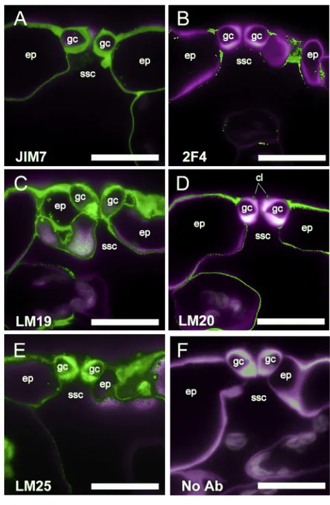
Probing Arabidopsis thaliana leaf sections with a panel of 36 monoclonal antibodies by fluorescence microscopy revealed a range of antibody-binding patterns, including clear differences in the composition of guard cell walls compared to epidermal or mesophyll cells (Figure 1;Figure S1;Table S1). Homogalactur-onan (HGA) is a polysaccharide of a-1,4-linked galacturonic acid (GalA) residues and is the predominant form of pectin in A. thaliana [3, 4]. It is synthesized at the Golgi apparatus and secreted from cells in a highly methyl-esterified form. These methyl ester groups can subsequently be removed by enzymatic activity in the cell wall, allowing for a range of methyl-esterifica-tion states. A broad range of HGA methyl-esterificamethyl-esterifica-tion patterns are recognized by the JIM7 antibody [5], andFigure 1A shows uniform JIM7 binding within the walls of guard cells, epidermal pavement, and mesophyll cells, indicating a wide distribution of HGA. HGA can form pectate calcium cross-links when contin-uous stretches of GalA residues are blockwise de-esterified, and this can be detected using the 2F4 antibody [6]. De-esterified, calcium cross-linked HGA was not detected in the guard cell walls or in neighboring cell walls but, instead, was limited to the junction regions between cells of the epidermis (Figure 1B). In contrast, relatively unesterified HGA (detected by antibody LM19) showed strong labeling in guard cell and epidermal cell walls (Figure 1C). Highly methyl-esterified HGA, as indicated by antibody LM20, was excluded from guard cells but was abun-dant in the junctions between guard cells and neighboring epidermal cells (Figure 1D). The LM20 signal was observed in the cell walls of epidermal pavement and mesophyll cells, as well as the cuticular ledges of guard cells. This specific pattern of pectins contrasted with the more uniform signal observed following immunolocalization with antibodies against other cell wall components, such as xyloglucan (LM25) (Figure 1E), while signal was absent when the primary antibody was not included (Figure 1F). Out of 36 cell wall antibodies tested, LM20 (against high methyl-esterified HGA) revealed a clear guard-cell-specific pattern (Figure S1;Table S1) being absent from guard cell walls but detected strongly in neighboring cell walls.
These results can be compared with previous investigations of guard cell wall composition [7, 8]. For example, analysis of Commelina communishas shown that guard cells are rich in pectin but did not report on the differential methyl-esterification patterns described here. Our data show that both highly methyl-esterified and calcium cross-linked blockwise de-methyl-esterified HGA
are excluded from the guard cells and that un-esterified HGA is the predominant form of pectin in the guard cell wall. Pectic ara-binans have previously been implicated as being important for guard cell movements via ectopic application of carbohydrate-modifying enzymes [8]. Thus, our data support the idea that the structural properties of the pectin network are important for guard cell function. We took a molecular genetic approach to test this hypothesis.
Identification of a Mutant,pme6-1, with Altered Guard Cell Wall Pectin Distribution
The enzymes that modify plant cell wall pectins are typically en-coded by large gene families. For example 66, 35, and 89 pectin methylesterase genes have been annotated inA. thaliana,Oryza sativa, andPopulus trichocarpa,respectively [9]. The encoded proteins contain pectin methylesterase (PME), or both PME and pectin methylesterase inhibitor domains (proPME proteins). The PME and proPME enzymes control the methyl-esterification status of HGA by removing methyl ester groups from HGA [9]. This large number of genes has made it difficult to attribute spe-cific pectin modifications of cell walls to particular physiological properties. As our experiments identified guard cells as having a distinct pectin methyl-esterification status, we sought to identify genes encoding pectin-modifying enzymes with a guard-cell-specific expression pattern. We focused on a proPME gene, PME6(TAIR:AT1G23200), which is expressed at >36-fold higher levels in guard cell protoplasts relative to mesophyll cell proto-plasts [10] and is also expressed during seed coat develop-ment [11].PME6expression has previously been shown to be repressed in scap1, a mutant with altered expression of cell wall modification genes and a resultant change in methyl-ester-ification state [12]. Negi et al. [12] proposed thatPME6might act downstream ofSCAP1to elicit at least part of the stomatal phenotype observed.
[image:3.603.58.299.99.467.2]PME6encodes a single PMEI domain and a PME domain that contains the two conserved active-site aspartic acid residues necessary for PME activity [9], as well as an N-terminal signal peptide, suggesting that it is a secreted protein. A PME6 promoter b-glucuronidase (GUS) fusion construct containing approximately 1,400 bp upstream of the start codon (proPME6:: GUS) was stably introduced into A. thaliana, and GUS histo-chemical localization indicated that this DNA region directs expression predominantly in mature guard cells (Figure S2A). Analysis of transcriptome data indicated thatPME6mRNA accu-mulates to a high level in the scrm-D mutant, which has an excess of mature guard cells [13] and to a lower level in mutants in which epidermal cell differentiation is blocked at the pavement cell stage (spch) [14] or at the stage of meristemoid formation (scrm-D mute) [13, 15] (Figure S1B). These data suggest that PME6 is expressed in guard cells at a relatively late stage of differentiation. Transcriptome data indicated thatPME6is also expressed in siliques [16], and analysis of the proPME6::GUS lines confirmed this. Analysis of apme6mutant (described later) did not reveal any change in seed germination or seed weight, so our further investigation focused on stomatal function. To inves-tigate the function ofPME6, we obtained anA. thalianaline with a Ds transposon inserted within the PME6 gene (hereinafter referred to aspme6-1) from the Nottingham Arabidopsis Stock Centre. PCR and RT-PCR analyses showed that pme6-1
Figure 1. Guard Cells Show Specific Patterns of Wall Epitopes (A) Ubiquitous presence of pectin in cell walls. The JIM7 antibody binds to HGA with a broad range of methyl-esterification and shows labeling in all cell walls in a cross-section through the epidermis (ep) encompassing guard cells (gc) above a sub-stomatal cavity (ssc).
(B) Calcium cross-linked HGA is restricted to cell interstices. The 2F4 antibody indicates cell walls containing calcium cross-linked HGA characterized by stretches of unesterified HGA residues.
(C) Unesterified HGA is present in GC walls. Binding of the LM19 antibody indicates that HGA with no or little esterification is prevalent in all cell walls of the epidermis.
(D) Highly methyl-esterified pectin is absent from the guard cell wall, as indi-cated by the lack of binding of the LM20 antibody.
(E) Binding of the LM25 antibody indicates that xyloglucan is present in all cell walls of the epidermis.
(F) A control with no primary antibody (Ab) showing low levels of auto-fluorescence against the Calcofluor White staining of the cell wall.
In all panels, the green signal shows binding of the specific primary antibody indicated, and the magenta signal (false color) indicates Calcofluor White fluorescence of cell walls. Scale bars, 20mm. See alsoFigure S1andTable S1.
2 Current Biology26, 1–8, November 7, 2016
homozygous plants harbor an insertion in the single intron of the PME6 gene (Figure S2C) and have no detectable ex-pression of thePME6mRNA transcript (Figure S2D). Comple-mentation of the pme6-1 line by introducing a native PME6 gene construct under the control of the proPME6 promoter restored the levels ofPME6 mRNA in two independent lines (proPME6::pme6) (Figure S2D).
Immunolocalization analyses of pme6-1 revealed a major change in the methyl-esterification status of guard cell wall pec-tins. We first confirmed that the guard cells ofL. erectawild-type (WT) plants (thepme6-1background) showed strong binding of LM19 (Figures 2A and 2B), indicating an abundance of relatively unesterified pectin. There was an absence of LM20 binding ( Fig-ures 2E and 2F), indicating that highly methyl-esterified pectin is absent from the guard cells. In contrast,pme6-1plants had a reduction in the levels of de-esterified pectin in the guard cell, as indicated by the weaker binding of LM19 (Figures 2C and 2D) and abundant highly esterified pectin, as indicated by LM20 binding (Figures 2G and 2H). These data indicate that the structure of the HGA component of the pectin network has been altered in the pme6-1 knockout line and, in particular, that the pectin of guard cells is more highly methyl-esterified in plants lackingPME6. JIM7-binding patterns remained consis-tent between the WT and pme6-1 (Figures 2I–2L), indicating that the differences observed inFigures 2A–2H were due to an alteration in the methyl-esterification status of the guard cells rather than a change in the overall distribution of HGA in the cell wall. The pattern of methyl-esterification in guard cells was highly reproducible, as shown inFigures 2Q–2S.pme6-1lines complemented with a proPME6::PME6 construct showed a restoration of the WT methyl-esterification pattern (Figure S3). Analysis of controls (Figures 2M–2P) indicated that the patterns observed inFigures 2A–2L did not simply reflect patterns of cell wall thickness or overall distribution of cellulose. These data indi-cate that PME6 is crucial for the de-methyl-esterification of guard cell wall HGA.
A Mutant with Altered Guard Cell Wall Pectin Methylation Has Impaired Stomatal Function
To study the functional significance of the changing pectin methyl-esterification status of the guard cells, we investigated the stomatal opening and closure responses of pme6-1.
Figure 3A shows the stomatal aperture response in isolated epidermal strips exposed to buffers supplied with elevated (1,000 ppm) or decreased (0 ppm) levels of CO2. Exposure to
elevated CO2caused WT stomatal apertures to decrease, and CO2-free air caused WT apertures to increase, as previously re-ported [17]. In contrast,pme6-1stomatal apertures were rela-tively insensitive to CO2,with the responses to both elevated and decreased CO2being lost. The stomatal aperture response to CO2was restored in the complemented lines. A restricted abil-ity ofpme6-1stomata to respond to abscisic acid, a classical regulator of stomatal function [17], was also observed ( Fig-ure S4E), suggesting that the altered pectin methyl-esterification status of the guard cells was affecting a fundamental property of the stomata.
Since stomata play a major role in controlling the water rela-tions of the plant, thermal imaging was used to investigate the effects of thepme6-1-altered guard cell wall properties at the whole-plant level by gauging leaf temperature as a measure of evaporative cooling (which is tightly linked to stomatal function [18]). Under well-watered growth conditions, there were mini-mal differences in temperature between thepme6-1, the WT, and complemented mutant lines (Figure 3B). However, under drought conditions, pme6-1 plants were significantly cooler than the WT and complemented lines (Figures 3B and 3C), prob-ably due to a higher rate of transpiration through their more open stomata. These results are consistent with the data in Fig-ure 3A, indicating that the pme6-1 mutant stomata have a more restricted range of stomatal opening/closure as a result of altered guard cell wall properties and a more restricted response to ABA (Figure S4). To further investigate the physio-logical outcome of altered stomatal performance in thepme6-1 mutant, we conducted infrared gas exchange analysis to assess stomatal conductance (gs), in response to shifts in CO2 condi-tions. Under ambient CO2conditions, thepme6-1leaves had a higher gscompared to the WT (Figure 4A). When exposed to elevated CO2, the pme6-1 gs value decreased slightly but remained higher than the WT value. However, when the CO2 level was decreased to sub-ambient, bothpme6-1and WTgs increased but the maximal level achieved by thepme6-1leaves plateaued at a lower level than that of the WT; thus, the overall dynamic range ofgsshown by thepme6-1 leaves was lower than for the WT. Unlike stomatal aperture measurements in epidermal peels, gs is influenced not only by the guard cells but also by a variety of physiological processes in the whole leaf and plant, which may counteract the actions of individual stomata, leading to an amelioration of the response seen in iso-lated peels [19]. The data inFigure 4A indicate thatpme6-1 sto-mata have a more restricted dynamic range of opening/closure
Figure 2. Guard Cell Wall Pectin Composition Is Altered inpme6-1Plants
(A–D) The high level of unesterified HGA in WT guard cells indicated by LM19 antibody binding in both cross-sections (A) and paradermal sections (B) is greatly diminished inpme6-1(C and D). ep, epidermis; gc, guard cells.
(E–H) Highly methyl-esterified HGA is absent in WT guard cell walls (E and F) but accumulates in the guard cell walls of thepme6-1mutant, as revealed by binding of the LM20 antibody (G and H).
(I–L) The general distribution of HGA (indicated by the JIM7 antibody) is similar in the WT (I and J) and thepme6-1mutant (K and L).
(M–P) Control sections not hybridized with primary antibody but stained with Calcofluor White indicate the signal specificity of the immunolabeling experiments in (A)–(L) and the general distribution of the cell wall material. In all panels, the green signal shows binding of the specific primary antibody indicated, and the magenta signal (false color) indicates Calcofluor White fluorescence of cell walls.
(Q–S) Counting of stomata showing the patterns of labeling with each antibody indicate the switch in LM20/LM19 labeling pattern between WT and thepme6-1
mutant guard cells. Localization of fluorescence in transverse sections after antibody binding was scored as fully covering guard cells (as in I), partially covering guard cells (as in C), or limited to guard cell-epidermal cell junctions (as in E). Data are shown for LM19 (Q), LM20 (R), and Jim7 (S) immunolabeling. Quantification was based on scoring patterns from 50 stomata, with five stomata scored from each of ten plants.
Scale bars, 20mm. See alsoFigures S1–S3.
4 Current Biology26, 1–8, November 7, 2016
that cannot be completely compensated for at the whole plant level, thus leading to altered water relationships.
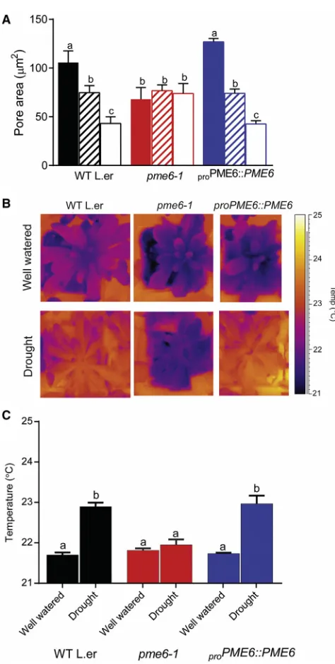
An altered dynamic range of guard cell swelling and deflation was also indicated by direct observation of stomatal pore area after immersion in an osmoticum expected to decrease turgor pressure [8]. Despite being subjected to a similar decrease in os-motic potential (1.23 MPa), thepme6-1stomatal pores remained significantly larger than those of the WT (Figure 4B). Analysis of guard cells by electron microscopy did not reveal any overt difference in surface shape or size (Figures S2E and S2F), and overall cell ultrastructure appeared similar in the two genotypes (Figures S2G and S2H), suggesting that the different behavior of the stomata was not due to large-scale change in cell structure but rather to some alteration in the mechanical properties of the cell wall. The differential response to a similarly imposed bio-physical challenge via mannitol treatment supports the idea that the pectin structure in the guard cells sets the mechanics of the cellular complex, thus limiting the range of size change possible. It has been postulated that the methyl-esterification status of pectin influences the ability of HGA domains to associate via Ca2+cross-links and that the degree of Ca2+cross-linking has a major effect on the mechanical stiffness of the cell wall matrix. Indeed, previous work on guard cell walls suggested that ara-binan side chains of the rhamnogalacturonan-I pectic domain associated with HGA might play a role in physically separating HGA domains, thus influencing Ca2+ cross-linking [8]. Since the formation of pectic network structures will be strongly dependent on the methyl-esterification status of HGA, PMEs can be predicted to play a major role in determining the overall mechanical properties of the cell wall. However, although recent evidence strongly supports the role of pectin methyl-esterifica-tion status in cell wall mechanics [20–22], simple inference of mechanics based on pectin methyl-esterification has proven problematic, since it appears to be highly dependent on cellular context [23, 24]. Our data suggest a situation in which a reduc-tion in pectin de-esterificareduc-tion leads to guard cells that are rela-tively stiffer, limiting potential changes in cell size.
The Influence of Altered Guard Cell Wall Structure on Plant Growth Is Environment Dependent
[image:6.603.53.290.93.563.2]From our gas exchange analysis, we derived A/Ci (photosyn-thesis rate/internal CO2 concentration) curves relating instan-taneous carbon assimilation rate to CO2 level (Figure 4D). At ambient CO2, both pme6-1 and WT leaves showed similar assimilation rates; however, at elevated CO2, a much higher rate was measured in pme6-1. As CO2 level rises relative to O2, it is expected that photorespiration in C3 plants (such as Arabidopsis) will decrease [25], thus leading to a higher net assimilation rate. Leaves with stomata that show a decreased closure response to CO2, as observed here forpme6-1, might be expected to have higher internal CO2 levels and, thus, a greater increase in assimilation rate than the WT, as indicated
Figure 3.pme6-1Plants Have Altered Guard Cell Physiology and
Water Relationships
(A) Guard cell opening/closure response to changing CO2concentration is lost in thepme6-1mutant. Pore area was measured from stomata in epidermal peels taken from the genotypes indicated (WT,pme6-1, andpme6-1 com-plemented with aproPME6::PME6 construct) after incubation of the peels with either CO2-free air (0 ppm CO2; solid bars), ambient CO2(hatched bars) or high CO2(1,000 ppm; open bars). Each column shows the mean and SEM. (n = 6), with statistical differences determined by ANOVA with a post hoc Tukey test. Columns indicated with identical letters cannot be distinguished from each other (p < 0.01).
(B)pme6-1plants are less able to adjust leaf temperature under drought conditions. Thermal images are shown of well-watered plants of the genotypes indicated (top images) taken at day 0 post-drought. Images of equivalent plants at day 5 post-drought (lower panel) show that thepme6-1plants have a lower leaf temperature than the WT or the complementedpme6-1mutant. (C) Quantification of thermal image data shows thatpme6-1leaf temperature does not change significantly under drought conditions, while the WT and the
complemented mutant leaf temperature increases. Each bar represents the mean temperature for the rosette with error bars indicating SEM (n = 6). Statistical differences were determined by ANOVA with a post hoc Tukey test. Columns indicated with identical letters cannot be distinguished from each other (p < 0.05).
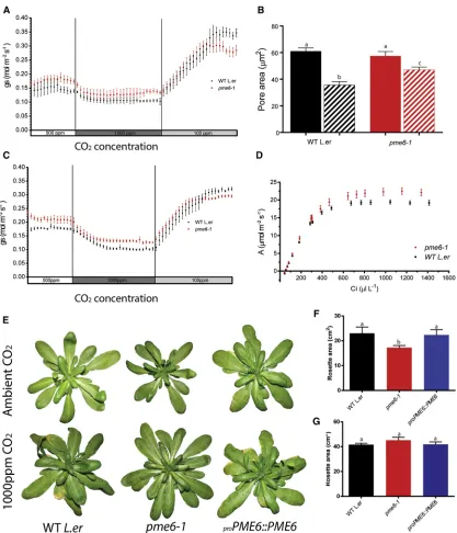
Figure 4.pme6-1Plants Show a Limited Dynamic Range of Stomatal Movement and Decreased Growth under Ambient CO2, which Is
Rescued by Elevated CO2
(A)pme6-1leaves show a limited dynamic range in stomatal conductance (gs) in response to changing CO2level. Gas exchange data for WT andpme6-1leaves show that, under ambient CO2conditions, thepme6-1leaves have highergsthan the WT. Following exposure to elevated (1,000 ppm) CO2,gsin both mutants and WTs fall. Exposure to a low (100-ppm) CO2regime induces increasedgs, but thepme6-1 gstrace plateaus to a lower value than for WT leaves. Error bars indicate the SEM (n = 8). (B)pme6-1stomata show a differential pore size response after incubation in high osmoticum. Stomatal pore areas were measured in epidermal peels from either WT orpme6-1leaves incubated either in resting buffer (solid bars) or resting buffer with addition of mannitol to 0.5 M (hatched bars). Statistical differences were determined by ANOVA and a post hoc Tukey test. Columns indicated with identical letters cannot be distinguished from each other (p < 0.01, n = 3, with 40 stomata counted from a total of four plants, repeated on 3 consecutive days). Error bars indicate the SEM.
(C) The more limited dynamic range ingsexhibited bypme6-1leaves is maintained after the growth of plants at elevated CO2. Gas exchange data for WT and
pme6-1leaves taken from plants grown continually under elevated CO2. The traces for the WT andpme6-1as the CO2level is altered during gas exchange analysis are comparable to those shown in (A), with thepme6-1trace again reaching a lower plateau after exposure to sub-ambient CO2level (n = 8). (D) At elevated CO2levels, thepme6-1leaves have a greater potential to assimilate CO2than WT leaves. A/Ci curve analysis of WT andpme6-1leaves indicates that the instantaneous C assimilation rate at ambient CO2levels is comparable but that as Ciincreases, thepme6-1leaves show a greater maximum potential assimilation rate (n = 5 for WT; n = 6 forpme6-1; error bars indicate the SEM).
(legend continued on next page)
6 Current Biology26, 1–8, November 7, 2016
inFigure 4D. To investigate the outcome of this at a whole-plant level, we compared the growth of WT andpme6-1mutant plants under ambient and elevated CO2.pme6-1plants were smaller than WTs when grown under ambient CO2(Figures 4E and 4F). When grown under elevated CO2, thepme6-1and WT plants were larger than the equivalent plants grown under ambient CO2(as expected) [26], but there was no difference in size be-tween mutants and WTs (Figures 4E and 4G). Gas exchange analysis confirmed that the more limited dynamic range ings observed in thepme6-1leaves under ambient CO2conditions was maintained when the plants were grown under elevated CO2(Figure 4C), indicating that the underlying, more limited, dy-namic range of stomatal function was also present in the mutant under these growth conditions, as expected for a genetically determined change in cell wall structure.
Stomatal size and density are well-characterized parameters known to influence leaf performance under various environ-ments [26]. In the wake of rising atmospheric CO2levels, there has been much interest in understanding how stomatal parame-ters provide an insight into past evolutionary events linked to earlier environments [27] and into the potential modification of stomata to create crop plants better attuned to present and pre-dicted climates [28, 29]. The potential role of the guard cell wall in setting and modulating the response dynamics of stomata has been underexplored. Since our analysis did not indicate any dif-ferences in stomatal density or stomatal index betweenpme6-1 and WT leaves (Figures S4A–S4D), the most plausible interpreta-tion of our data is that that modulainterpreta-tion of the pectin matrix of guard cells leads to altered wall properties so that the stomata are mechanically limited in their responses to exogenous cues, thus altering plant-water relations.
Despite a firm theoretical basis for the importance of differen-tial guard cell wall stiffness in the mechanism of stomata opening and closure in response to altered turgor pressure [1, 30], exper-imental evidence has often been correlative, e.g., measurements of cell wall thickening, observations of cellulose microfibril orien-tation [2]. Indeed, despite a wealth of physiological data on turgor pressure and ion fluxes [31], and intricate details of molec-ular signaling in guard cells [17], the causal relationship between guard cell wall structure/composition and stomatal function has been surprisingly underexplored. The most insightful data have come from experiments in which the exogenous supply of cell-wall-modifying enzymes suggested an important role for pectic arabinans in stomatal function [8]. However, the nature of the genes involved and, indeed, formal genetic evidence to support this hypothesis, are lacking. In this paper, we provide molecular data to show that not only doArabidopsisguard cells have a spe-cific HGA methyl-esterification status (unesterified), but also that this status is required for normal stomatal function. The simplest interpretation of our data is that abnormal pectin methyl-esterifi-cation alters mechanical properties of the guard cells, leading to an inability to show appropriate opening and closure in response to environmental signals. Due to the importance of the
mechan-ical properties of guard cells in setting the dynamics of stomatal opening and, thus, whole-plant/water relations, this alteration in guard-cell-specific cell wall gene expression leads to a poorer ability of the leaf to control water loss under drought conditions and poorer growth of the plants under ambient CO2levels. Inter-estingly, this growth defect was overcome when the mutant plants were grown at elevated CO2, indicating that, under certain conditions, altered guard cell wall mechanics are not detri-mental. In summary, our data indicate that, in addition to the well-explored regulation of stomatal dynamics via signal trans-duction signals acting on ion transport to vary turgor pressure, cell wall modification plays an important role in setting the overall limits of the system. Although this study focused on the role of pectins, it is clear that cell wall matrix components function together to influence mechanical properties [32], and exploring the roles of both structural carbohydrates, such as xyloglucans and cellulose [33], and modulating protein factors, such as expansins [34], will allow a deeper understanding of the system. Allied to this, targeted modulation of guard cell wall structure provides a novel avenue for the future manipulation of stomatal function.
EXPERIMENTAL PROCEDURES
Details of the experimental procedures are available in theSupplemental Information.
SUPPLEMENTAL INFORMATION
Supplemental Information includes Supplemental Experimental Procedures, four figures, and one table and can be found with this article online athttp:// dx.doi.org/10.1016/j.cub.2016.08.021.
AUTHOR CONTRIBUTIONS
Conceptualization, A.J.F. and J.E.G.; Investigation, S.A., L.H., N.E., A.B., M.L., Y.V, and A.J.F.; Writing – Original Draft, A.J.F., J.E.G., and S.A.; Writing – Re-view & Editing, A.J.F., J.E.G., S.A., L.H., J.P.K., Y.V., and H.V.S.; Resources, J.P.K.; Supervision, A.J.F. and J.E.G.; Funding Acquisition, A.J.F., J.E.G., and H.V.S.
ACKNOWLEDGMENTS
The work reported here was funded by a White Rose BBSRC-DTP award (to S.A. and A.J.F.); BBSRC grant BB/I002154/1 (to L.H. and J.E.G.); the Gatsby Foundation (to A.B.), the U.S. Department of Energy, Office of Science, Office of Biological and Environmental Research, through Contract DE-AC02-05CH11231 between the Lawrence Berkeley National Lab and the U.S. Department of Energy (to Y.V. and H.V.S.); and a Leverhulme Research Fellow-ship (to A.J.F.). Ray Wightman (SLCU, Cambridge) assisted with SEM. The Microscopy Facility at the Sainsbury Laboratory is supported by the Gatsby Charitable Foundation.
Received: March 15, 2016 Revised: July 6, 2016 Accepted: August 5, 2016 Published: October 6, 2016
(E–G)pme6-1plants are smaller than WTs under ambient CO2, but growth at elevated CO2leads to plants attaining a similar size. Images of plants (genotypes as indicated) under ambient CO2are shown in (E, top row) and under elevated (1,000 ppm) CO2in (E, bottom row). Quantitation of total rosette area of plants grown under ambient CO2(F) shows thatpme6-1plants achieve a smaller final size, whereas growth in elevated CO2(G) leads to all plants reaching a similar mean size. In (F and G), error bars indicate the SEM, n = 8.
REFERENCES
1.Franks, P.J., Cowan, I.R., and Farquhar, G.D. (1998). A study of stomatal mechanics using the cell pressure probe. Plant Cell Environ.21, 94–100.
2.Palevitz, B.A., and Hepler, P.K. (1976). Cellulose microfibril orientation and cell shaping in developing guard cells of Allium: The role of microtubules and ion accumulation. Planta132, 71–93.
3.Zablackis, E., Huang, J., Mu¨ller, B., Darvill, A.G., and Albersheim, P. (1995). Characterization of the cell-wall polysaccharides of Arabidopsis thaliana leaves. Plant Physiol.107, 1129–1138.
4.Caffall, K.H., and Mohnen, D. (2009). The structure, function, and biosyn-thesis of plant cell wall pectic polysaccharides. Carbohydr. Res.344, 1879–1900.
5.Verhertbruggen, Y., Marcus, S.E., Haeger, A., Ordaz-Ortiz, J.J., and Knox, J.P. (2009). An extended set of monoclonal antibodies to pectic homoga-lacturonan. Carbohydr. Res.344, 1858–1862.
6.Liners, F., Letesson, J.J., Didembourg, C., and Van Cutsem, P. (1989). Monoclonal antibodies against pectin: recognition of a conformation induced by calcium. Plant Physiol.91, 1419–1424.
7.Majewska-Sawka, A., Mu¨nster, A., and Rodrı´guez-Garcı´a, M.I. (2002). Guard cell wall: immunocytochemical detection of polysaccharide com-ponents. J. Exp. Bot.53, 1067–1079.
8.Jones, L., Milne, J.L., Ashford, D., and McQueen-Mason, S.J. (2003). Cell wall arabinan is essential for guard cell function. Proc. Natl. Acad. Sci. USA 100, 11783–11788.
9.Pelloux, J., Ruste´rucci, C., and Mellerowicz, E.J. (2007). New insights into pectin methylesterase structure and function. Trends Plant Sci.12, 267–277.
10.Yang, Y., Costa, A., Leonhardt, N., Siegel, R.S., and Schroeder, J.I. (2008). Isolation of a strong Arabidopsis guard cell promoter and its potential as a research tool. Plant Methods4, 6.
11.Levesque-Tremblay, G., Mu¨ller, K., Mansfield, S.D., and Haughn, G.W. (2015). HIGHLY METHYL ESTERIFIED SEEDS is a pectin methyl esterase involved in embryo development. Plant Physiol.167, 725–737.
12.Negi, J., Moriwaki, K., Konishi, M., Yokoyama, R., Nakano, T., Kusumi, K., Hashimoto-Sugimoto, M., Schroeder, J.I., Nishitani, K., Yanagisawa, S., and Iba, K. (2013). A Dof transcription factor, SCAP1, is essential for the development of functional stomata in Arabidopsis. Curr. Biol.23, 479–484.
13.Kanaoka, M.M., Pillitteri, L.J., Fujii, H., Yoshida, Y., Bogenschutz, N.L., Takabayashi, J., Zhu, J.-K., and Torii, K.U. (2008). SCREAM/ICE1 and SCREAM2 specify three cell-state transitional steps leading to arabidopsis stomatal differentiation. Plant Cell20, 1775–1785.
14.MacAlister, C.A., Ohashi-Ito, K., and Bergmann, D.C. (2007). Transcription factor control of asymmetric cell divisions that establish the stomatal line-age. Nature445, 537–540.
15.Pillitteri, L.J., Sloan, D.B., Bogenschutz, N.L., and Torii, K.U. (2007). Termination of asymmetric cell division and differentiation of stomata. Nature445, 501–505.
16.Schmid, M., Davison, T.S., Henz, S.R., Pape, U.J., Demar, M., Vingron, M., Scho¨lkopf, B., Weigel, D., and Lohmann, J.U. (2005). A gene expression map of Arabidopsis thaliana development. Nat. Genet.37, 501–506.
17.Kim, T.-H., Bo¨hmer, M., Hu, H., Nishimura, N., and Schroeder, J.I. (2010). Guard cell signal transduction network: advances in understanding absci-sic acid, CO2, and Ca2+ signaling. Annu. Rev. Plant Biol.61, 561–591.
18.Wang, Y., Holroyd, G., Hetherington, A.M., and Ng, C.K.Y. (2004). Seeing ‘cool’ and ‘hot’–infrared thermography as a tool for non-invasive, high-throughput screening of Arabidopsis guard cell signalling mutants. J. Exp. Bot.55, 1187–1193.
19.Pantin, F., Monnet, F., Jannaud, D., Costa, J.M., Renaud, J., Muller, B., Simonneau, T., and Genty, B. (2013). The dual effect of abscisic acid on stomata. New Phytol.197, 65–72.
20.Peaucelle, A., Braybrook, S.A., Le Guillou, L., Bron, E., Kuhlemeier, C., and Ho¨fte, H. (2011). Pectin-induced changes in cell wall mechanics underlie organ initiation in Arabidopsis. Curr. Biol.21, 1720–1726.
21.Peaucelle, A., Louvet, R., Johansen, J.N., Ho¨fte, H., Laufs, P., Pelloux, J., and Mouille, G. (2008). Arabidopsis phyllotaxis is controlled by the methyl-esterification status of cell-wall pectins. Curr. Biol.18, 1943–1948.
22.Peaucelle, A., Braybrook, S., and Ho¨fte, H. (2012). Cell wall mechanics and growth control in plants: the role of pectins revisited. Front. Plant Sci.3, 121.
23.Hongo, S., Sato, K., Yokoyama, R., and Nishitani, K. (2012). Demethylesterification of the primary wall by PECTIN METHYLESTERASE35 provides mechanical support to the Arabidopsis stem. Plant Cell24, 2624–2634.
24.Liu, Q., Talbot, M., and Llewellyn, D.J. (2013). Pectin methylesterase and pectin remodelling differ in the fibre walls of two gossypium species with very different fibre properties. PLoS ONE8, e65131.
25.Long, S.P., Ainsworth, E.A., Rogers, A., and Ort, D.R. (2004). Rising atmo-spheric carbon dioxide: plants FACE the future. Annu. Rev. Plant Biol.55, 591–628.
26.Doheny-Adams, T., Hunt, L., Franks, P.J., Beerling, D.J., and Gray, J.E. (2012). Genetic manipulation of stomatal density influences stomatal size, plant growth and tolerance to restricted water supply across a growth carbon dioxide gradient. Philos. Trans. R. Soc. Lond. B Biol. Sci.367, 547–555.
27.Chater, C., Gray, J.E., and Beerling, D.J. (2013). Early evolutionary acqui-sition of stomatal control and development gene signalling networks. Curr. Opin. Plant Biol.16, 638–646.
28.Hepworth, C., Doheny-Adams, T., Hunt, L., Cameron, D.D., and Gray, J.E. (2015). Manipulating stomatal density enhances drought tolerance without deleterious effect on nutrient uptake. New Phytol.208, 336–341.
29.Franks, P.J.W., Doheny-Adams, T.W., Britton-Harper, Z.J., and Gray, J.E. (2015). Increasing water-use efficiency directly through genetic manipula-tion of stomatal density. New Phytol.207, 188–195.
30.Mott, K.A., and Franks, P.J. (2001). The role of epidermal turgor in stomatal interactions following a local perturbation in humidity. Plant Cell Environ. 24, 657–662.
31.Pandey, S., Zhang, W., and Assmann, S.M. (2007). Roles of ion channels and transporters in guard cell signal transduction. FEBS Lett.581, 2325– 2336.
32.Braybrook, S.A., and Jo¨nsson, H. (2016). Shifting foundations: the me-chanical cell wall and development. Curr. Opin. Plant Biol.29, 115–120.
33.Rui, Y., and Anderson, C.T. (2016). Functional analysis of cellulose and xy-loglucan in the walls of stomatal guard cells of Arabidopsis thaliana. Plant Physiol.170, 1398–1419.
34.Goh, H.-H., Sloan, J., Dorca-Fornell, C., and Fleming, A. (2012). Inducible repression of multiple expansin genes leads to growth suppression during leaf development. Plant Physiol.159, 1759–1770.
8 Current Biology26, 1–8, November 7, 2016