Advance Access publication February 14, 2006.
© The Author 2006. Published by Oxford University Press on behalf of the European Society of Human Reproduction and Embryology. All rights reserved. 1349
For Permissions, please email: journals.permissions@oxfordjournals.org
The online version of this article has been published under an open access model. Users are entitled to use, reproduce, disseminate, or display the open access version of this article for non-commercial purposes provided that: the original authorship is properly and fully attributed; the Journal and Oxford University Press are attributed as the original place of publication with the correct citation details given; if an article is subsequently reproduced or disseminated not in its entirety but only in part or as a derivative work this must be clearly indicated. For commercial re-use, please contact journals.permissions@oxfordjournals.org
Cytotrophoblast stem cell lines derived from human
embryonic stem cells and their capacity to mimic invasive
implantation events
R.Harun
1,4, L.Ruban
1, M.Matin
1,5, J.Draper
1, N.M.Jenkins
1, G.C.Liew
1, P.W.Andrews
1,
T.C.Li
2, S.M.Laird
3and H.D.M.Moore
1,61Centre for Stem Cell Biology, University of Sheffield, 2Royal Hallamshire Hospital, Central Sheffield University Teaching Hospital
Trust and 3Biomedical Research Centre, Sheffield Hallam University, Sheffield, UK
4Present address: National Population and Family Development Board, Ministry of Women, Family and Community Development,
Bangunan LPPKN, 12B Jalan Raja Laut, Peti Surat 10416, 50712 Kuala Lumpur, Malaysia
5Present address: Department of Biology, Institute of Biotechnology and Tissue Engineering, Ferdowsi University of Mashhad,
Mashhad, Iran
6To whom correspondence should be addressed at: Centre for Stem Cell Biology, University of Sheffield, Western Bank,
Sheffield S10 2TN, UK. E-mail: h.d.moore@shef.ac.uk
BACKGROUND: An effective embryonic–maternal interaction is crucial for successful human pregnancy. Failure of this process is a major cause of infertility and can lead to placental dysfunction resulting in recurrent miscarriage, fetal retardation and pre-eclampsia. Research is severely constrained by ethical and practical considerations; therefore, we aimed to generate cytotrophoblast stem (CTBS) cell lines from human embryonic stem cells (HESCs). METHOD: b -HCG was used as a marker of viable trophoblast cells. In defined culture, embryoid bodies were generated from HESCs and selected for trophoblast enrichment by rounds of cellular aggregation and disaggregation. Distinct CTBS cell lines were isolated and characterized. Spheroid cytotrophoblast bodies were generated and their interaction with luteal-phase endometrial stroma was analysed by real-time image analysis. RESULTS: Three CTBS cell lines were derived, which were maintained in the absence of residual HESCs, fibroblast feeder cells or extracellular matrix. CTBS cells dis-played typical cytotrophoblast and syncytiotrophoblast characteristics and exhibited further differentiation to invasive endovascular cell phenotype. One cell line was generated with constitutive expression of enhanced green fluorescent protein (eGFP). Spheroid trophoblast bodies mimicked closely the early invasive stages of implantation when incubated with human endometrial stromal preparations in vitro. CONCLUSION: These human CTBS cell lines are a significant new model for investigating human placentation and may have considerable potential in cell therapy applications.
Key words: cytotrophoblast/embryonic stem cells/implantation
Introduction
The continual proliferation of trophoblast stem cells (TS) by the blastocyst and post-implantation embryo is of critical importance for the maintenance of pregnancy (Paria et al., 2002; Red-Horse et al., 2004). Initially, the rapidly dividing trophoblast cells generate syncytiotrophoblast by cell fusion. This terminally differentiated and nondividing syncytium penetrates the endometrial epithelium to invade underlying endometrial stroma where it forms a contiguous layer at the interface with maternal tissue and provides a primary barrier to
1350
greater maternal blood flow for the continuation of fetal devel-opment and growth. Endovascular cells also express membrane and soluble HLA-G, the nonclassical histocompatibility antigen believed to be involved in immune tolerance of the fetus (Le Bouteiller et al., 2003). Aberrant development of trophoblast (including defective HLA-G function) is associated with serious complications, including recurrent miscarriage, pre-eclampsia and restricted fetal growth (Paria et al., 2002; Laird et al., 2003; Red-Horse et al., 2004). Recently, elevated HLA-G secretion from the preimplantation blastocyst has been corre-lated with successful pregnancy outcome after assisted repro-duction techniques (Noci et al., 2005; Yie et al., 2005).
The process of trophoblast differentiation is poorly under-stood in women because investigations are severely constrained by ethical and practical considerations.
Various cell lines have been generated from normal, malignant or transformed (immortalized) trophoblast and placental tis-sues, and these have provided useful in vitro models to investi-gate placental development and function (King et al., 2000). However, many of these cell lines originate from late stage or term placental tissues or have lost functional capacity after transformation and therefore fail to recapitulate the physiology of trophoblast during the peri-implantation stages of develop-ment. Hence, we sought to generate human cytotrophoblast stem (CTBS) cell lines as a renewable source of progenitor tro-phoblast cells to serve in model systems of implantation.
In the mouse, TS cells isolated from pre- and post-implantation embryos can be maintained indefinitely in culture and have the capacity to differentiate along the trophoblast lineage (Tanaka et al., 1998). However, so far the derivation of human TS cells from preimplantation blastocysts has not been achieved, high-lighting the differences in early embryo and placental develop-ment between these species (Henderson et al., 2002). Therefore, we investigated the use of human embryonic stem cells (HESCs) as a route to obtaining TS cell population. Although HESCs differentiate spontaneously to trophoblast-like cells in culture (Thomson et al., 1998; Draper et al., 2002), when supplemented with bone morphogenetic protein 4 (Xu et al., 2002) or when Oct-4 is down-regulated (Matin et al., 2004), these cells are terminally differentiated and display a limited proliferative capacity. Trophoblast differentiation can develop further when HESCs are aggregated to form embryoid bodies (EBs), but residual HESCs and other cell types in the culture resulting from spontaneous differentiation can con-found the findings from these preparations (Gerami-Naini et al., 2004). We surmised that the proportion of trophoblast cells that developed in human EBs in vitro would vary and sought to identify viable EBs containing trophoblast cells by measuring the secretion of the β-subunit of HCG (β-HCG) as secreted by syncytiotrophoblast. Such EBs might potentially contain a CTBS cell population for enrichment and purification.
Materials and methods
Materials
Human embryonic stem cell medium (HES medium) was prepared as described previously (Matin et al., 2004). Unless stated otherwise,
media and reagents were from Invitrogen (Paisley, UK) and antibod-ies from Sigma-Aldrich (Poole, UK).
HESC maintenance, differentiation and trophoblast enrichment
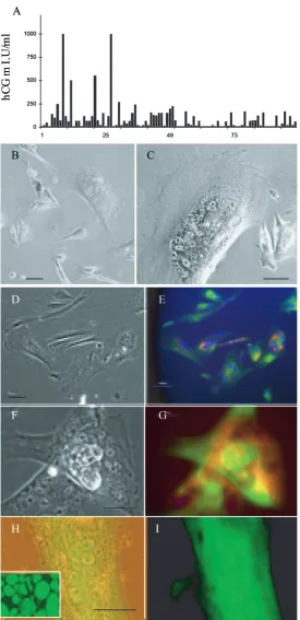
HESCs (H7 and H14, University of Wisconsin) of normal karyotype were maintained and passaged by standard protocols (Henderson et al., 2002; Matin et al., 2004). Batches of EBs were usually prepared from three T25 flasks of HESCs (dissociated with 1 mg/ml collagenase IV) and placed in HES medium (Thomson et al., 1998) without basic fibrob-last growth factor (FGF) in Petri dishes in 5% CO2 in air. HESCs did not adhere to the Petri dish surface and instead aggregated to form EBs. On day 5, well-formed spherical EBs of approximately the same diameter (approximately 200 μm) were transferred singly to wells of a 96-well culture plate and cultured for a further 3 days before aliquots of medium were subjected to enzyme-linked immunosorbent assay (ELISA) as described previously (Acevedo et al., 1997; Nishimura et al., 1989).
To select for CTBS cells, EBs exhibiting high levels of β-HCG secretion were subjected to several rounds of selective enrichment by growth in conditioned ‘TS’ medium as described by Tanaka et al. (1998) for differentiation of murine TS cells from extraembryonic ectoderm. TS medium comprised Roswell Park Memorial Institute (RPMI) 1640 medium supplemented with 20% fetal calf serum, 1 mm sodium pyruvate, 100 μm β-mercaptoethanol, 2 mM L-glutamine and supplemented with fibroblast growth factor 4 (FGF4) and heparin. The medium was conditioned by incubation with confluent T25 flasks of mouse embryonic fibroblast (density of 5 × 10⁶ per flask) for 72 h, filtered and stored at –20°C until used. This conditioned medium was diluted with fresh nonconditioned medium at a ratio of 70 : 30. Those EBs maintaining a high secretion of β-HCG were pooled, disaggre-gated and allowed to form new EBs, and this enrichment protocol was repeated consecutively for three rounds until all EBs displayed con-sistently high β-HCG secretion. EBs were disaggregated (0.25% trypsin-EDTA) and then single cells allowed to proliferate in adherent culture in TS medium without feeders. Medium was changed daily, and cells were passaged every 5 days by splitting 1 : 4 from T25 flasks. Density of cells was 1–2 × 10⁶ per flask.
Cell lines were cryopreserved using the freezing medium of fetal calf serum supplemented with 10% dimethylsulphoxide (DMSO). Briefly, the medium was removed from the flask and cells were washed with 5 ml PBS before the addition of trypsin-EDTA solution for 5 min. Complete medium was added to neutralize the enzyme, and cells were recovered by aspiration, washed by centrifugation and resuspended in ice-cold freezing medium in cryovials which were placed in a polystyrene container overnight at –70°C before transfer to liquid nitrogen. Vials of cells were thawed at 37°C in a water bath, and the cells were washed by centrifugation thrice before transfer to fresh TS medium in a T25 flask.
ELISA determination of b-HCG concentration in cell cultures
Concentrations of β-HCG were determined using a sandwich enzyme immunoassay kit (DRG Diagnostics, Marburg, Germany). The stan-dards and samples were incubated with 100 μl antiHCG enzyme con-jugate for 30 min at room temperature followed by a five-times washing procedure. A second incubation with 100 μl substrate solution for 10 min was terminated with the addition of 50 μl stop solution. Absorbance was read at 450 ± 10 nm with a microtitre plate reader. The concentration of β-HCG in the samples was determined from the standard curve as m IU/ml.
Analysis of CTBS cell lines
1351 marker cytokeratin 7 (Kam et al., 1999; Haigh et al., 1999),
stage-spe-cific embryonic antigen 1 (SSEA1; Henderson et al. 2002) and human placental lactogen (HPL; King et al. 2000). Additionally, RT–PCR was performed with primer sequences for marker genes of HESCs and trophoblast. Total RNA (2 μg) was reverse transcribed using 1 μg oligo-dT primer with Moloney murine leukaemia virus (MMLV) Reverse-Transcriptase (Promega, Cowley, UK) in a 40 μl reaction vol-ume containing 1.25 mM dNTPs at 37°C. PCR was performed using 1
μl of cDNA in 50 μl reaction volume containing 15 pmol of each primer, 0.2 mM dNTPs and 1 unit Taq polymerase (Promega). Primer sequences used and conditions of these reactions were as follows:
b-actin-F: 5´-ATCTGGCACCACACCTTCTACAATGAGCTGCG-3´; b-actin-R: 5´-CGTCATACTCCTGCTTGCTGATCCACATCTGC-3´
(60°C annealing, ×23 cycles).
Oct4-F: 5´-CGACCATCTGCCGCTTTGAG-3´;
Oct4-R: 5´-CCCCCTGTCCCCCATTCCTA-3´ (60°C annealing,
×23 cycles).
Sox2-F: 5´-CCCCCGGCGGCAATAGCA-3´;
Sox2-R: 5´-TCGGCGCCGGGGAGATACAT-3´ (60°C annealing,
×38 cycles).
FGF4-F: 5´-CTACAACGCCTACGAGTCCTACA-3´;
FGF4-R: 5´-GTTGCACCAGAAAAGTCAGAGTTG–3´ (56°C annealing, ×40 cycles).
Nanog-F: 5´-GCCTCAGCACCTACCTACCC-3´;
Nanog-R: 5´-GGTTGCATGTTCATGGAGTAG-3´ (60 annealing and ×30 cycles).
Eomes-F: 5´-TCACCCCAACAGAGCGAAGAGG-3´;
Eomes-R: 5´-AGAGATTTGATGGAAGGGGGTGTC-3´ (57°C annealing, ×35 cycles).
Cdx2-F: 5´-CCTCCGCTGGGCTTCATTCC-3´;
Cdx2-R: 5´-TGGGGGTTCTGCAGTCTTTGGTC-3´ (60°C annealing,
×35 cycles);
HLA-G-F: 5´-GCGGCTACTACAACCAGAGC-3´;
HLA-G-R: 5´-GCACATGGCACGTGTATCTC-3´ (55°C annealing,
×26 cycles).
CD9-F: 5´-TTGGACTATGGCTCCGATTC-3´;
CD9-R: 5´-GATGGCATCAGGACAGGACT-3´ (55°C annealing,
×26 cycles).
CK7-F: 5´-ACAGAGCTGCAGTCCCAGAT-3´;
CK7-R: 5´-GTAGGTGGCGATCTCGATGT-3´ (55°C annealing,
×26 cycles).
Fluorescence-activated cell sorter
Trophoblast cells were prepared for cell sorting by dissociating CTBS cells into single cells with trypsin-EDTA solution. To block nonspe-cific binding sites, the cells were resuspended at 5 × 10⁶/ml in fluores-cence-activated cell sorter (FACS) buffer with 40% normal goat serum on ice for 20 min. About 90 μl of the cell suspension was aliquoted into FACS tube, and 10 μl of G233 (marker for HLA-G) and W6/32 HLA class I control was added. G233 supernatant with NaN3 (mouse IgG2a) was kindly given by Dr Ashley King, University of Cambridge. The cells were incubated on ice for 30 min and washed twice by cen-trifugation before being labelled with anti-mouse polyvalent immu-noglobulin fluorescein isothiocyanate (FITC) conjugate for 30 min on ice. The cells were washed again and resuspended in 300 μl buffer.
Constitutive expression of enhanced green fluorescent protein in HESCs
A pCAG-GFP expression vector was constructed by excision of enhanced green fluorescent protein (eGFP) from pEGFP-1 (Clontech, Cowley, UK) with XhoI and NotI and insertion into the pCAG vector (Niwa et al., 2002). H7 cells were seeded at the equivalent of 2 × 10⁵
per well of a 6-well plate on Matrigel (Becton Dickinson, Cowley, UK). The next day they were transfected using ExGen 500 (Fermentas, Hanover, Germany) according to the manufacturer’s instructions. The DNA/NaCl ExGen mixture was then added directly to the normal growth medium in the well. The plate was centrifuged at 280 g for 5 min and placed back in the incubator. The medium was exchanged the next day with HES growth medium containing puromycin (at 1 μg/ ml). Viable colonies were picked after 2–3 weeks.
Endometrial–CTBS spheroid co-culture
The invasive implantation potential of the CTBS cell lines was exam-ined using spheroid culture. This technique has been shown to main-tain extravillous cytotrophoblast phenotype of first trimester placental tissue (Korff et al., 2004). Aggregates of CTBS cells were generated in a similar manner to EBs from confluent monolayers (three T25 flasks) following brief trypsinization and incubation in nonadherent (Petri dish) culture for 5–10 days, where they formed spheroid tro-phoblast bodies (TBs). These TBs were explanted to Matrigel culture in TS medium or to endometrial stromal co-culture. For the latter, luteal-phase endometrial biopsies were obtained from women under-going hysterectomy under full ethical approval and patient consent. Endometrial epithelial and stromal cells were isolated as described previously (Laird et al., 1993). Preparations were embedded in Matrigel on membrane inserts and primed with progesterone for 24 h before the start of co-culture with CTBS. In monolayer co-culture, TBs were cultured on a confluent layer of stromal or epithelial cells in culture wells. The co-cultures were maintained in 500 μl of either con-ditioned TS medium or serum-free HES medium up to 6 days.
Time-lapse microscopy
CTBS cultures or TB–endometrial co-cultures were continuously monitored under an inverted microscope in a humidified physiological chamber at 37°C in 5% CO2 in air for up to 3 days. Preliminary exper-iments indicated no difference in the viability of cells maintained under these conditions compared with a standard incubator. Regions of interest (ROI) were identified and programmed for analysis using Simple PCI software (Digital Pixel, Brighton, UK) with control over xyz scan, transmitted light and image capture. Routinely 20 ROIs were identified with image capture every 15 min.
Results
Derivation of CTBS cell lines
1352
failed to proliferate after passages 8 and 12. The remaining two lines have retained persistent proliferative capacity for more than 30 passages (CTBS1 from H7 HESC and CTBS2
[image:4.612.155.430.43.610.2]1353 methods but from H7 HESCs with constitutive expression of
eGFP (Figure 1H and I). Continuous proliferation of each cell line was related to the persistence of a mononuclear cytotro-phoblast population (relative to syncytium formation) as determined by immunostaining for cytokeratin and β-HCG (Figure 1D–G). Cell proliferation was maintained by regular cell passage every 5 days, because this inhibited terminal differentiation.
When CTBS cells (CTBS1 and 2) and TBs were returned to mouse embryonic fibroblast feeders with HES medium they failed to revert to or generate either HESC colonies or EBs with pluripotent developmental capacity other than trophoblast.
Characterization of CTBS cell lines
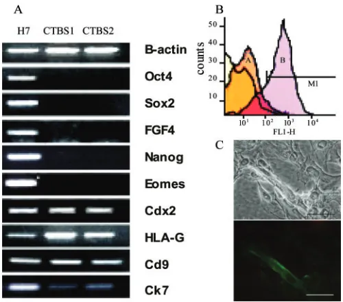
CTBS cells displayed dual immunolocalization of CK7 and HCG and also expressed HPL and SSEA1. Most (>80%) CTBS cells were single mononuclear cells which did not form colonies. However with time in culture before passage, cells fused to form syncytium (see below). They did not localize antibody markers to undifferentiated HESCs, namely Tra-1-60, SSEA3 or SSEA4. The mRNA expression for Oct-4, Sox2, FGF4 and Nanog in the derived cell lines was absent, but trophoblast-related mRNAs for Cdx2 (caudal-related homeobox), HLA-G and Cd9 were up-regulated or for Ck7 was maintained (Figure 2A). Expression of eomesodermin, a marker of murine post-implantation trophob-last, was absent in the CTBS cells but present in HESCs.
To further assess the subtype of trophoblast cells, the com-parative cell surface expression of histocompatibility HLA class I (pan HLA antibody W6/32) and HLA-G (antibody G233) antigens was determined by FACS and immunolocalization.
The majority of cells (approximately 90%) expressed HLA class I histocompatibility antigens (Loke and King, 1995), con-sistent with extravillous trophoblast (Figure 2B). The expres-sion of HLA-G was relatively weak in most cells, but a small proportion (approximately 10%) of cells displayed strong immunoreactivity (Figure 2B and C). Some cells expressed vimentin, a marker of interstitial cytotrophoblast (King et al., 2000).
CTBS cell differentiation to endovascular cell phenotype
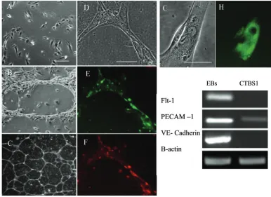
Following extended culture for one week or more in T25 flasks, the proportion of HLA-G+ cells increased considerably (>90%). These cells exhibited distinct endothelial cell mor-phology similar to cultures of differentiating cytotrophoblast from first trimester human placental tissue (Nagamatsu et al., 2004) and also resembling endothelial morphological differen-tiation from primate embryonic stem cells (Levenberg et al., 2002; Kaufman et al., 2004). Significantly, however, the cells co-expressed HLA-G and the platelet endothelial cell adhesion molecule-1 (PECAM-1), both markers of invasive endovascu-lar (endothelial-like) cytotrophoblast (Red-Horse et al., 2004) (Figure 3). Vascular endothelial (VE)-cadherin and E-cadherin immunolocalization were weak or absent on endovascular cells but strong on a relatively small proportion (<5%) of multinu-cleated cells also present at this stage and most likely equival-ent to the syncytial giant cells found in stroma of the developing placenta. As determined by RT–PCR, adherent endovascular trophoblast in culture exhibited PECAM-1 mRNA expression but neither vascular endothelial growth fac-tor recepfac-tor 1 (flt-1) nor VE cadherin exhibited the expression in comparison with EBs, again distinguishing these cells from a true endothelial phenotype (Figure 3I).
Functional analysis of CTB cells
To determine their functional capacity, CTBS cells were moni-tored by continuous time-lapse recording for cell–cell fusion and the formation of nonproliferative, syncytiotrophoblast (Paria et al., 2002). Adherent cells displayed progressive migration across the culture dish promoted by pseudopodia-like extension of cells. When cells occasionally converged they fused to form multicellular syncytiotrophoblast cells (Figure 1G) that were β-HCG- and Ck7-positive but HLA class I nega-tive. This cell fusion was captured unequivocally by digital time-lapse microscopy. Single cells fusing with each other, sin-gle cells fusing with multinucleated cells and multinucleated cells fusing with each other (Figure 4) were all observed.
[image:5.612.61.311.417.637.2]The invasive implantation potential of the CTBS cell lines was investigated by generating TBs. When cultured in extracel-lular matrix (Matrigel) drops, these spheroid aggregates developed characteristic outgrowths, which expressed β-HCG and cytokeratin (Figure 5Ai and ii). On further culture with primary human endometrial tissue (luteal phase), prepared using well-characterized protocols (Laird et al., 1993), TBs attached to both epithelial and stromal cells. qSignificantly, as shown by time-lapse microscopy (Figure 5B), TBs with stromal cell cul-tures displayed a characteristic circular migratory movement and exhibited polar outgrowths from which endovascular cells Figure 2. RT–PCR and fluorescence-activated cell sorter (FACS)
1354
streamed. After about 24–36 h in co-culture, these trophoblast outgrowths were the site of erosion of the extracellular matrix of the stroma. This was identified by the rapid retraction of the tro-phoblast vesicle owing to the dissolution of underlying extracel-lular matrix of the stromal cells (Figure 5B2–5). The erosion site was characterized by extravillous (HLA–G+) trophoblast that expressed matrix metalloproteinase (MMP)-2 (gelatinase A; Figure 5B4i and ii); single GFP-trophoblast cells with endome-trial stroma in culture displayed a similar response.
Discussion
As far as we are aware, these are the first human CTBS cell lines to be derived from HESCs and then maintained independ-ently without feeder cells. Although trophoblast cells can be derived directly from HESCs (Xu et al., 2000; Matin et al., 2004), they rapidly lose proliferative capacity after forming syncytium, and any self-renewing progenitor CTBS population is lost. In the mouse, trophoblast cells proliferate in response to FGF4 via their FGFR2 receptor (Tanaka et al., 1998), and a stem cell population can be generated. Therefore, we used TS medium containing FGF4 and heparin to maintain CTBS cells derived from HESCs, which could be subsequently enriched, and selected for, to create three cell lines. These CTBS cell lines differ from many of the immortalized placental lines (Pavan et al., 2003; Feng et al., 2005) in that they have
multipotent capacity to differentiate to various trophoblast phe-notypes including syncytiotrophoblast and extravillous cytotrophoblast. This differentiation process is initiated in the first trimester of human pregnancy by mechanisms that are largely unknown, although recent evidence supports the con-tention that early cytotrophoblast has a bipotential capacity. In the mouse, withdrawal of FGF4 from TS medium induced tro-phoblast differentiation, but for our human stem cell lines, FGF4-supplemented medium was required for cell prolifera-tion and subsequently allowed differentiaprolifera-tion to occur. This suggests a difference between mouse and human TS cells. The addition of FGF4 and heparin to early human villous explants inhibits syncytiotrophoblast regeneration in favour of clumps of FGF2R-positive cytotrophoblast which can be differentiated to extravillous cytotrophoblast (Baczyk et al., 2005). The CTBS cell lines exhibited a similar bipotential differentiation in culture, such as cell–cell fusion to syncytium, an invasive implantation into endometrial stroma and terminal differentia-tion to endovascular cells that expressed HLA-G. Moreover, following the constitutive expression of eGFP in HESCs, it was possible to derive a GFP–CTBS cell line to provide unam-biguous identification in co-culture with other cell types.
[image:6.612.97.483.47.328.2]Clonally derived HESCs maintain full pluripotency and proliferation (Amit et al., 2000) suggesting that CTBS cells develop from a homogeneous HESC population rather than multiple (i.e. ES and TS) precursors. This finding differs from Figure 3. Differentiation of cytotrophoblast stem (CTBS) cell line to endovascular cells in ‘trophoblast stem (TS) cells’ conditioned medium. (A) Phase-contrast micrograph of single adherent cytotrophoblast of CTBS1 cell line; bar = 20 μm. (B) The cells in (A) after 1–2 weeks in cul-ture. Long aggregates display typical endothelial-like morphology; bar = 15 μm. (C) Dark field, low-power micrograph of culture flask; bar = 100
1355 the mouse, where trophoblast cells may be derived from the early embryo and extraembryonic ectoderm (Beddington and Robertson, 1989) but not from murine ESCs without condi-tional gene expression (Tanaka et al., 1998; Niwa et al., 2000; Velkey and O’Shea, 2003). Our cell lines continued prolifera-tive capacity without HESCs or feeder cells, strongly indicating the presence of a subpopulation of CTBS. This view was rein-forced by complete failure of the cell line to revert to HESCs or pluripotency (other than trophoblast lineage) under permissive conditions, thereby confirming the absence of any residual HESCs in the lines and also their restricted developmental capacity as shown also for mouse TS cells (Tanaka et al., 1998). The latter can be derived from a cell ‘niche’ that spans the entire extraembryonic ectoderm and chorionic ectoderm (Uy et al., 2002), but it remains unclear whether a similar wide range of trophoblast development supports human TS cells. The direct developmental progression in vitro would possibly be the differentiation of HESCs to the immediate trophectoder-mal stem cell phenotype of the preimplantation blastocyst. However, the specific routes of derivation of our CTBS lines still require verification as initial EB formation may induce various trophoblast cell types from which the cell lines were ultimately selected. For practical purposes we adopted a rela-tively simple procedure for selecting viable EBs based on an appropriate secretory marker (β-HCG), followed by rounds of enrichment by cell disaggregation and EB regeneration. The HCG receptor is expressed on invasive cytotrophoblast as well as syncytiotrophoblast, and similar observations have been reported for EB differentiation to trophoblast (Gerami-Naini et al., 2004). The method was effective but did not allow pro-spective analysis of the differentiation process. Nevertheless, subsequent antibody and gene profile analyses of the cell lines using trophoblast-specific markers were consistent with the derivation of CTBS cells.
[image:7.612.88.285.45.648.2]Compared with HESCs, mRNA expression for Oct-4, Sox2, FGF4 and Nanog in CTBS cell lines was absent, but trophob-last-related mRNAs for Cdx2 (caudal-related homeobox; Strumpf et al., 2005), HLA-G and Cd9 were up-regulated, whereas CK7 expression was maintained. The expression together of the latter three genes is considered as a verification for extravillous cytotrophoblast (King et al., 2000) which invades the uterine decidua during placentation (Pijnenborg et al., 1980; Enders et al., 1997). Surprisingly, eomesodermin (eomes), a marker of mouse early post-implantation trophob-last (Russ et al., 2000), was expressed strongly in HESCs but was weak or absent in the CTBS cells. Several reports have highlighted differences between mouse and human ESCs (Henderson et al., 2002; Ginis et al., 2004) including eomeso-dermin expression in HESCs but not mouse ES cells (Ginis et al., 2004). Furthermore, the expression of some trophoblast markers in stock cultures of HESCs may relate to spontaneous differentiation to trophoblast lineage. We had previously shown that expression of trophectodermal markers in such cultures occurred predominantly in the SSEA(–) and SSEA1(+) subsets of cells, consistent with their expression in the differentiated derivatives of the HESCs rather than in the HESCs themselves (Henderson et al., 2002; Ginis et al., 2004).
1356
The differentiation of several trophoblast subtypes in the cell cultures probably accounts for the physiological attributes of the CTBS cell to mimic implantation processes in vitro. Syncytiotrophoblast was readily generated, and the formation of syncytium by cell–cell fusion was determined precisely using time-lapse cultures under the microscope.
[image:8.612.99.484.48.569.2]1357 is not clear. Regular passaging of adherent cells in culture
inhibited syncytium formation, but we are presently investi-gating culture medium that may inhibit syncytiotrophoblast while retaining cell proliferation. Such conditions may allow the rescue of an early trophectodermal stem cell phenotype.
Following extended culture without passaging, CTBS cells formed the endothelial phenotype of invasive endovascular cells as displayed by cytotrophoblast from first trimester pla-cental tissue (Nagamatsu et al., 2004). Previously, the exact origin of these cells has been in question, but their derivation from CTBS cell lines provides definitive evidence of their differentiation from cytotrophoblast. Significantly, although these cells expressed some markers of endothelial cells, they lacked expression of vascular endothelial growth factor receptor 1 (flt-1) and exhibited the nonclassical histocompatibility anti-gen, HLA-G on their surface. Thus, although their morphology closely resembled that of endothelial cells in culture, they were clearly a separate phenotype, as determined by biomarkers.
To gain an indication of the physiology of CTBS cells and their derivatives during implantation events, spheroidal tro-phoblast bodies, TBs (akin to EBs formed from HESCs) were generated to resemble the implanting embryo. Matrigel, an extracellular matrix preparation rich in laminin and collagen, was used to mimic the endometrial deciduas (Tarrade et al., 2002; Hemberger et al., 2004) along with human stromal cell preparations from late luteal phase endometrial biopsy tissue (Laird et al., 1993). In response to Matrigel culture, TBs exhib-ited extensive differentiation with projections of endothelial-like cells, which were cytokeratin 7-positive. Cells within the body of the TBs expressed HCG. A similar response to Matrigel has been observed with EBs where a proportion of the cells differentiate to trophoblast (Gerami-Naini et al., 2004). Significantly, TBs in co-culture with endometrial stromal cells exhibited a migration around the culture dish with cycles of erosion of extracellular matrix followed by reattachment. Because this co-culture system was essentially two dimen-sional, the migration of the TB most likely represents a regu-lated invasion process across the stromal culture rather than through it. Indeed, a proliferative column of cytotrophoblast was observed along with the faster migrating endovascular cells. A similar process of trophoblast invasion has been observed also for human blastocyst co-culture with stromal cells in vitro (Carver et al., 2003). During implantation, cytotrophoblast expresses various zinc-dependent proteolytic enzymes that cleave constituents of the extracellular matrix called matrix metalloproteinases (MMPs). A complex interac-tion with tissue inhibitors of MMPs (TIMPS) ensures a care-fully controlled regulation of invasion. MMP-2 (gelatinase A) was identified as a key enzyme correlated with first trimester invasive capacity of human cytotrophoblast (Huppertz et al., 1998; Xu et al., 2000; Wang et al., 2001; Staun-Ram et al., 2004) and activity alters in cytotrophoblast (Campbell et al., 2004) and plasma of women that develop pre-eclampsia (Myers et al., 2005). Therefore, immunolocalization of MMP-2 specifically at the edge of erosion site between TBs and stroma was good preliminary evidence that the CTBS cells were secreting physiologically relevant factors. A much more detailed examination of the mechanisms regulating gene
expression and enzyme activity in this model system is now in progress.
Overall, these cell lines therefore represent a significant advance for studies related to human implantation, for example cell cultures to mimic the early embryo invasion process, or investigations of placental dysfunctions such as pre-eclampsia. In the future, somatic nuclear replacement techniques (thera-peutic cloning) may enable HESCs and subsequently, trophob-last cell lines to be generated from patients with specific implantation and placental dysfunctions. Moreover, efficient generation of endovascular cytotrophoblast may have a wide utility for regenerative medicine. Their pseudo-vasculogenic and invasive characteristics might be utilized in a variety of cell therapies remote from the uterus but related to angiogen-esis and vessel remodelling, especially as expression of HLA-G (Kovats et al., 1990) and indoleamine 2,3-deoxygenase (Mellor et al., 2002) render the cells naturally refractory to immune rejection.
Acknowledgements
We are grateful to Liz Tuckerman for technical assistance with endometrial cultures. R.H. thanks the Malaysian Government for a studentship. J.D. are supported by the BBSRC. Centre for Stem Cell Biology is a MRC Resource Centre for Human Embryonic Stem Cells.
Competing interests statement
There are no competing interests.
References
Acevedo HF, Hartsock RJ and Maroon JC (1997) Detection of membrane-associated human chorionic gonadotrophins and its subunits on human cul-tured cancer cells and the nervous system. Cancer Detect Prev 21,295–303. Amit M, Carpenter M, Inokuma M, Chiu C-P, Harris C, Waknitz M,
Itskovitz-Eldor J and Thomson J (2000) Clonally derived human embryonic stem cell lines maintain pluripotency and proliferative potential for prolonged periods of culture. Dev Biol 227,271–278.
Baczyk D, Dunk C, Huppertz B, Maxwell C, Reister F, Giannoulias D and Kingdom JC (2005) Bi-potential behavior of cytotrophoblasts in first trimes-ter chorionic villi. Placenta June 9. (epub ahead of print)
Beddington R and Robertson E (1989) An assessment of the developmental potential of embryonic stem cells in the midgestation mouse embryo. Devel-opment 105,733–737.
Campbell S, Rowe J, Jackson CJ and Gallery ED (2004) Interaction of cocul-tured decidual endothelial cells and cytotrophoblasts in preeclampsia. Biol Reprod 71,244–252.
Draper J, Pigott C, Thomson J and Andrews PW (2002) Surface antigens of human embryonic stem cells: changes on differentiation in culture. J Anat 200,249–258.
Carver J, Martin K, Spyropoulou I, Barlow D, Sargent I and Mardon H (2003) An in vitro model for stromal invasion during implantation of the human blastocyst. Hum Reprod 18,283–290.
Enders A, Lantz K, Peterson P and Hendrickx A (1997) From blastocyst to pla-centa: the morphology of implantation in the baboon. Hum Reprod Update 3,561–573.
Feng HC, Choy MY, Deng W, Wong HL, Lau WM, Cheung AN, Ngan HY and Tsao SW (2005) Establishment and characterization of a human first-trimester extravillous trophoblast cell line (TEV-1). J Soc Gynecol Investig 12,21–32.
Gerami-Naini B, Dovzhenko O, Durning M, Wegner F, Thomson J and Golos. T (2004) Trophoblast differentiation in embryoid bodies derived from human embryonic stem cells. Endocrinology 145,1517–1524.
1358
Haigh T, Chen C, Jones C and Aplin J (1999) Studies of mesenchymal cells from 1st trimester human placenta: expression of cytokeratin outside the tro-phoblast lineage. Placenta 20,615–625.
Hemberger M, Hughes M and Cross JC (2004) Trophoblast stem cells dif-ferentiate in vitro into invasive trophoblast giant cells. Dev Biol 271,362–371.
Henderson JK, Draper JS, Baillie HS, Fishel S, Thomson JA, Moore HD and Andrews PW (2002) Preimplantation human embryos and embryonic stem cells show comparable expression of stage-specific embryonic antigens. Stem Cells 20,329–337.
Huppertz B, Kertschanska S, Demir AY, Frank HG and Kaufmann P (1998) Immunohistochemistry of matrix metalloproteinases (MMP), their sub-strates, and their inhibitors (TIMP) during trophoblast invasion in the human placenta. Cell Tissue Res 291,133–148.
Kam E, Gardner L, Loke Y and King A (1999) The role of trophoblast in the physiological change in decidual spiral arteries. Hum Reprod 14,2131–2138. Kaufman DS, Lewis RL, Hanson ET, Auerbach R, Plendl J and Thomson JA (2004) Functional endothelial cells derived from rhesus monkey embryonic stem cells. Blood 103,325–332.
King A, Thomas L and Bischof P (2000) Cell culture models of trophoblast II: trophoblast cell lines—a workshop report. Placenta 21 (Suppl. A), S113–S119.
Korff T, Krauss T and Augustin HG (2004) Three-dimensional spheroidal cul-ture of cytotrophoblast cells mimics the phenotype and differentiation of cytotrophoblasts from normal and preeclamptic pregnancies. Exp Cell Res 297,415–423.
Kovats S, Main E, Librach C, Stubblebine M, Fisher S and Demars R (1990) A class I antigen, HLA-G expressed in human trophoblasts. Science 248,220–223.
Laird SM, Li TC and Bolton AE (1993) The production of placental protein 14 and interleukin 6 by human endometrial cells in culture. Hum Reprod 8,795–798.
Laird SM, Tuckerman EM, Cork BA, Linjawi S, Blakemore AI and Li T-C (2003) A review of immune cells and molecules in women with recurrent miscarriage. Hum Reprod Update 9,163–174.
Le Bouteiller P, Pizzato N, Barakonyi A and Solier C (2003) HLA-G, pre-eclampsia, immunity and vascular events. J Reprod Immunol 59,219–234. Levenberg S, Golub JS, Amit M, Itskovitz-Eldor J and Langer R (2002)
Endothelial cells derived from human embryonic stem cells. Proc Natl Acad Sci USA 99,4391–4396.
Loke Y and King A (1995) Trophoblast expression of major histocompatibility complex class I antigens. In Human Implantation, Cell Biology and Immu-nology. Cambridge University Press, Cambridge, pp. 82–101.
Matin MM, Walsh JR, Gokhale PJ, Draper JS, Bahrami AR, Morton I, Moore HD and Andrews PW (2004) Specific knockdown of Oct4 and β2-microglobulin expression by RNA interference in human embryonic stem cells and embry-onal carcinoma. Stem Cells 22,659–668.
Mellor AL, Chandler P, Lee GK, Johnson T, Keskin DB, Lee J and Munn DH (2002) Indoleamine 2,3-dioxygenase, immunosuppression and pregnancy. J Reprod Immunol 57,143–150.
Myers JE, Merchant SJ, Macleod M, Mires GJ, Baker PN and Davidge ST (2005) MMP-2 levels are elevated in the plasma of women who subse-quently develop preeclampsia. Hypertens Pregnancy 24,103–115.
Nagamatsu T, Fujii T, Ishikawa T, Kanai T, Hyodo H, Yamashita T, Osuga Y, Momoeda M, Kozuma S and Taketani Y (2004) A primary cell culture sys-tem for human cytotrophoblasts of proximal cytotrophoblast cell columns enabling in vitro acquisition of the extra-villous phenotype. Placenta 25,153–165.
Nishimura R, Kitajima T, Hasegawa K, Takeuchi K and Mochizuki M (1989) Molecular forms of human chorionic gonadotrophin in choriocarcinoma serum and urine. Jpn J Cancer Res 80,968–974.
Niwa T, Masui S, Chambers I, Smith A and Miyazaki J-A (2002) Phenotypic complementation establishes requirement for specific POU domain and
generic transactivation function (of) ct-3/4 in embryonic stem cells. Mol Cell Biol 22,1526–1536.
Niwa H, Miayazaki J and Smith AG (2000) Quantitative expression of Oct3/4 defines differentiation, dedifferentiation or self-renewal of ES cells. Nat Genet 24,372–376.
Noci I, Fuzzi B, Rizzo R, Melchiorri L, Criscuoli L, Dabizzi S, Biagiotti R, Pellegrini S, Menicicci A and Baricordi O (2005) Embryonic soluble HLA-G as a marker of developmental potential in embryos. Hum Reprod 20,138–146.
Paria BC, Reese J, Das SK and Dey SK (2002) Deciphering the cross-talk of implantation: advances and challenges. Science 296,2185–2188.
Pavan L, Tarrade A, Hermouet A, Delouis C, Titeux M, Vidaud M, Therond P, Evain-Brion D and Fournier T (2003) Human invasive trophoblasts trans-formed with simian virus 40 provide a new tool to study the role of PPAR-gamma in cell invasion process. Carcinogenesis 24,1325–1336.
Pijnenborg R, Dixon G, Robertson WB and Brosens A (1980) Trophoblastic invasion of human deciduas from 8 to 18 weeks of pregnancy Placenta 1,3–19.
Red-Horse K, Zhou Y, Genbacev O, Prakobphol A, Foulk R, McMaster M and Fisher SJ (2004) Trophoblast differentiation during embryo implantation and formation of the maternal–fetal interface. J Clin Invest 114,744–754. Russ AP, Wattler S, Colledge WH, Aparicio SA, Carlton MB, Pearce JJ,
Barton SC, Surani MA, Ryan K, Nehls MC, Wilson V and Evans MJ (2000) Eomesodermin is required for mouse trophoblast development and meso-derm formation. Nature 404,95–99.
Staun-Ram E, Goldman S, Gabarin D and Shalev E (2004) Expression and importance of matrix metalloproteinase 2 and 9 (MMP-2 and -9) in human trophoblast invasion. Reprod Biol Endocrinol 2,59.
Strumpf D, Mao CA, Yamanaka Y, Ralston A, Chawengsaksophak K, Beck F and Rossant J (2005) Cdx2 is required for correct cell fate specification and differentiation of trophectoderm in the mouse blastocyst. Development 132,2093–2102.
Tanaka S, Kunath T, Hadjantonakis A-K, Nagy A and Rossant J (1998) Pro-motion of trophoblast cell proliferation by FGF4. Science 282,2072–2075. Tarrade A, Goffin F, Munaut C, Lai-Kuen R, Tricottet V, Foidart JM, Vidaud M,
Frankenne F and Evain-Brion D (2002) Effect of matrigel on human extravillous trophoblasts differentiation: modulation of protease pattern gene expression. Biol Reprod 67,1628–1637.
Thomson J, Iskovitz-Eldor J, Shapiro S, Waknitz M, Swiergiel J, Marshall V and Jones J (1998) Embryonic stem cell lines derived from human blasto-cysts. Science 282,1145–1147.
Uy G, Downs K and Gardner R (2002) Inhibition of trophoblast stem cell potential in chorionic ectoderm coincides with occlusion of the ectoplacen-tal cavity in the mouse. Development 129,3913–3924.
Velkey JM and O’Shea S (2003) Oct4 RNA interference induces trophecto-derm differentiation in mouse embryonic stem cells. Genesis 37,18–24. Wang M, Li Q, Shao L and Zhu C (2001) Expression of matrix
metalloproteinase-2, -9, -14, and tissue inhibitors of metalloproteinase-1, -metalloproteinase-2, -3 in the endometrium and placenta of rhesus monkey (Macaca mulatta) during early pregnancy. Biol Reprod 65,31–40.
Xu P, Wang Y-L, Zhu S-J, Luo S-Y, Piao Y-S and Zhang L-Z (2000) Expres-sion of matrix metalloproteinase-2, -9, -14, and tissue inhibitors of metallo-proteinase-1 and matrix proteins in human placenta during the first trimester. Biol Reprod 62,988–994.
Xu R, Chen X, Li D, Addicks G, Glennon C, Zwaka T and Thomson J (2002) BMP4 initiates human embryonic stem cell differentiation to trophoblast. Nat Biotechnol 20,1261–1264.
Yie SM, Balakier H, Motamedi G and Librach CL (2005) Secretion of human leukocyte antigen-G by human embryos is associated with a higher in vitro fertilization pregnancy rate. Fertil Steril 83,30–36.