ISOLATION AND STRUCTURAL DETERMINATION OF
POLYPHENOLS IN TASMANIAN EUCALYPTS.
by
RICHARD BARRY BROWN
A thesis submitted in partial fulfilment of the requirements for the degree of
-MASTER OF SCIENCE
UNIVERSITY OF TASMANIA,
Signed:
Except as stated herein, this thesis contains no Material which has been accepted for the award of
any other degree or diploma in any university and, to the best of my knowledge, this thesis contains no copy nor paraphrase of material previously published or written, except when due reference is made in the text of the thesis.
iii
TABLE OF CONTENTS
Page.
ABSTRACT.
ACKNOWLEDGEMENTS. vii
CHAPTER 1. INTRODUCTION.
1
3
4
7
12 1.0. EUCALYPT CLASSIFICATION AND CHEMOTAXONOMY.
1.1. HILLIS' SURVEY OF EUCALYPT POLYPHENOLS. 1.2. HILLIS' KNOWN AGLYCONES AND GLYCOSIDES. 1.3. HILLIS' UNKNOWNS.
CHAPTER 2. ISOLATION - EXPERIMENTAL. 17 2.0. ISOLATION - GENERAL PROCEDURES.
19
2.0.1. Sources of leaves.
19
2.0.2. Liquid-liquid extraction.
19
2.0.3. Chromatography. •
19
2.1. ISOLATION - SPECIAL PROCEDURES.
27
2.1.1. Liquid-liquid extraction. 272.1.2. Chromatography.
31
CHAPTER
3.
IDENTIFICATION - EXPERIMENTAL.48
3.0. SPECTRAL DETERMINATIONS. 50
3.1. SPECTRA. 52
3.2. DERIVATIVES.
100
3.3.
ROTATIONS, ANALYSES, MELTING POINTS. 103iv
TABLE OF CONTENTS (continued)
Page. CHAPTER 4. IDENTIFICATION - DISCUSSION. 122
4.0. POLYPHENOLS OF KNOWN STRUCTURE. 123 4.0.1. Hydroxybenzoic acids. 123
4.0.2. Flavonols. 126
4.0.3. Flavonol glycosides.
128
4.0.4.
Ellagic acid. 1324.0.3. (+)-Catechin.
13
64.1. POLYPHENOLS OF UNKNOWN STRUCTURE 139 (HILLIS' UNKNOWNS).
4.1.1. Unknown D - Flavanones. 139 4.1.2. Unknown D Flavones. 143 4.1.3. Unknown A (4-0-methylellagic acid)
. 153
4.1.4.
Unknown B (1,5-dimethy1-2,6-bis- 160(trihydroxyphenyl) furo [1,5-c] - furan).
4.1.3. Unknown F (a tetrahydroxydibenzofuran- 166 dicarboxylic acid.isomer).
APPENDIX. CHROMATOGRAPHIC BEHAVIOUR - SUMMARY.
. 1.71
1. POLYPHENOLS OF KNOWN STRUCTURE.172
2. POLYPHENOLS OF UNKNOWN STRUCTURE.173
REFERENCES.
174
ABSTRACT.
Chapter 1 contains a brief review of
chemotaxonomy as it relates to the classification of eucalypts.
Particular reference is made to a chromatographic survey by Hillis of the low molecular weight
polyphenols on eucalypt leaves.
That survey provisionally identified a number of polyphenols of known structure but labelled other
compounds "Unknowns", because of the lack of evidence for the identification of their structure.
The present work recounts the isolation and examination of the polyphenols in the leaves of three Eucalypt species important to Tasmania: E. delegatensis, E. sieberi and E. coccifera.
Chapter 2 gives experimental details of the
isolation of polyphenols from these species especially the isolation of some of Hillis' unknowns.
Cha ter 3 describes the experimental techniques by which evidence for the identity of the isolated polyphenols has been gathered.
Chapter 4 then discusses the identity of these polyphenols.
vi
protocatechuic acids; quercetin, myricetin,
kaempferol; the flavonol glycosides, afzelin, rutin, quercitrin, isoquercitrin, hyperin and cannabiscitrin; ellagic acid and (+)-catechin.
Secondly, some of Hillis' unknowns have been isolated and identified.
Unknown D has been isolated
(1) from E. sieberi and is identified as a mixture of flavanones: pinocembrin, alpinetin and a new natural product, 0,0-.dimethylpinocembrin; (2) from E. coccifera and is identified as a mixture
of flavones: apigenin and two C-methylflavones, sideroxylin and 4',5-dihydroxy-7-methoxy-6-
methylflavone, the last identified provisionally. Unknown A is identified as 4-0-methylellagic acid, a new ellagic acid ether.
Unknown B is provisionally identified as a new lignan, 1,5dimethy12,6bis (trihydroxyphenyl) furo
-[1,5-c] furan.
Unknown F is tentatively given the structure of a tetrahydroxydibenzofuran-dicarboxylic acid isomer.
Throughout the discussion the use of chromatography of mixtures to provide evidence for ch,emotaxonomy
ACKNOWLEDGEMENTS.
The author wishes to thank his supervisor, Dr. I.R.C. Bitk, Chemistry Department, University of Tasmania, for continued advice and encouragement during the course of this work.
Gratitude is also extended to Associate Professor J.B. Polya and Dr. J.B. Bremner of the same Department and University for the supervision of the final draft . of this thesis.
Dr. W.E. Hillis, Senior Principal Research
Scientist, C.S.I.R.O., Division of Applied Chemistry, Officer-in-Charge, Wood and Forest Science Section, suggested the project, gave continual encouragement and advice and supplied many authentic samples; to him special thanks are given.
Dr. T.J. Batterham, John Curtin School of Medical Research, Australian National University, gave the use of his laboratory and facilities during a two-week
stay in that laboratory, together with his interest and advice.
Finally, thanks are due to Mr. L. Brasch, Librarian, Associated Pulp and Paper Mills Ltds
Library, Burnie and to the Librarian, Forest Products Laboratory Library, Melbourne, for the use of the facilities of their respective libraries.
POLYPHENOLS OF TASMANIAN EUCALYPTS.
CHAPTER 1.
1
CONTENTS.
Page.
100. EUCALYPT CLASSIFICATION AND CHEMOTAXONOMY.
3
1.1. HILLIS' SURVEY OF EUCALYPT POLYPHENOLS.
4
1.2. HILLIS' KNOWN POLYPHENOL AGLYCONES AND
7
GLYCOSIDES.3
CHAPTER 1.
1.0. EUCALYPT CLASSIFICATION AND CHEMOTAXONOMY.
The classification of eucalypt species in use 'today is Blakely's "A Key to the Eucalypts" 16 based
on the so-called antheral system of Bentham. A second classification used by Blakely was based on general morphology but in some cases it
contradicted the first.
While the original key has anomalies it remains the only complete system evolved and.is the key
upon which any classification used in this study is based.
The reliable classification of this difficult genus requires as many taxonomic criteria as
possible.
• In recent years increasing study has been made of wood anatomy, bark bark anatomy, 23 cytology, 101 pollen grains,91 seed coat anatomy, 34 and floral morphology2021 as taxonomic criteria.
Further it has been proposed that the chemical composition of various plant tissues could also
4
11classification.. Bate-Smith, Hasegawa, Horn Horn et al. and and 47
more recently and extensively, Hillis have studied the chemical composition of a variety of plants and plant tissues.
7
Baker and Smith - first brought the focus of chemotaxonomy to bear on the Eucalypt genus in 1890 concentrating on the essential oil composition in eucalypt leaves.
Various plant tissues have been studied to decide which Would provide the most reliable chemotaxonomic guide.
Wood and phloem extractives have proved of limited usefulness although heartwood extractives have shown
43
more promise, while the greatest attention has been given to leaf extractives which seem to provide a more reliable guide to phenotype than extractives from other plant tissues.
Different groups of compounds found in leaf tissue have been studied from the chemotaxonomic viewpoint.
In addition to the study of essential oils by
Baker and Smith and their successors, leaf 7 Waxes have been - examined by Horn and his co-workers 59 who found them to
be composed mainly of long-chain /3-diketones with little promise of taxonomic significance.
1.1. HILLIS 1 SURVEY OF EUCALYPT POLYPHENOLS.
Hillis' extensive survey of over 300 eucalypt species used the pattern of low Molecular
weight-polyphenols in conjunction with other criteria to draw conclusions about eucalypt classification.
This examination was largely chromatographic, as far as possible using standard markers, and Hillis pointed out the need of extending the work by employing isolation and standard procedures of identification. 48 He proposed thereby to verify the identity of key
polyphenols as well as to identify other polyphenols to which not even provisional identity was ascribed but which were simply labelled "Unknowns".
The present work takes the results Of Hillis'
survey as they apply to Tasmanian eucalypts, in particular to three species, and by isolation and techniques of
structural analysis confirms the provisional identity of many of Hillis.' polyphenols. It establishes the identity of some unknowns and isolates and identifies an additional polyphenol hitherto unrecorded by Hillis.
Further, the most abundant glycosides in each species are characterised to complete the chemotaxonomic picture.
As a result of this work a question is raised about the advisability of using chromatography alone as a guide even to provisional identity of compounds where overlying spots on the chromatogram are common.
to such provisional identity in classification.
This is not to deny the validity of the use of chemical criteria in taxonomy in conjunction with other criteria but to suggest that the identity of compounds which are put forward as playing a significant role in classification must be accurately known if the
conclusions based on their identity are to be reliable. Two species of the Fraxinales or Ash series were examined for comparison of their polyphenolic content, E. dele atensis and E. sieberi. While not endemic both species have particular significance for Tasmania.
16a
E. delegatensis (R.T. Baker) or E. gigantea 1.6a
(Hook,t.) is particularly important to the State as a Source of paper pulp and as a timber, Tasmanian Oak.
16a
E. sieberi (L. Johnson) or Tasmanian Ironbark grows locally in the north-east from Coles Bay to
Georges River between the coast and the headwaters of the South Esk River. Attempts to grow E. sieberi in
large stands in other parts
of
the State for paper-making purposes have failed.Although E. sieberi grows also in the sub-alpine regions of Victoria and southern New South Wales, Hillis found in his survey that the chemical composition of the Tasmanian samples was significantly different from that of the samples gathered on the mainland.
16b
coccifera (Hook f.) or Tasmanian Snow Gum grows in sub-alpine conditions at altitudes of 2000-4500
6
feet. A Tasmanian endemic, it is widespread except in the north-east of the State.
17.
Time permitted examination of only one of the ' Piperitales or Peppermint series, E. coccifera, although comparison of the polyphenol content of a second Species of the series would have proved useful for chemotaxonomy. 1.2. HILLIS' KNOWN POLYPHENOL AGLYCONES AND GLYCOSIDES.
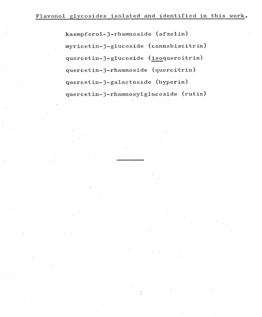
Table i lists the aglycones chromatographically identified by Hillis together with those isolated and identified during the course of this current work. The flavonol glycosides isolated are listed in Table ?.
Other polyphenols, provisionally called caffeic, chlorogenic and sinapic acids and Hillis' unknown.H;
were observed in chromatograms in the course of this study but were not isolated, so they are not listed in Table 1.
The structures corresponding to Hillis' known aglydones listed in Table 1 are given in Chart 1, while the structures of the known aglycones and glycosides actually isolated here are given in. Chart 2 on
page'll. _
As Charts (1) and (2) show, there is substantial agreement between the known polyphenols identified by Hillis and those isolated in. this work. But one
should note the isolation of a third hydroxybenzoic
0,0-dimethylpinocembrin
pinocembrin )
)
.alpinetin )
) 0,0-dimethylpinocembrin )
E. delegatensis.
E. sieberi. unknown D
TABLE 1.
1. Polyphenol • aglycones chromatographically identified by Hillis (H) and identified after isolation by
Brown B . kaempferol quercetin Myricetin
gallic acid (H & B) gentisic acid (H & B)
protocatechuic acid (B)
a catechin (H) (+)-catechin (B) chlorogenic acid (H)
ellagic acid (H & B)
2. Poly henel a•1 ones called unknowns by Hillis and structurally identified by Brown.
HILLIS unknOwn A unknown B
BROWN SPECIES
4-0-methylellagic acid E. sieberi. 1,5-dimethy1-2,6-bis(3,4,5-
trihydroxyphenyi
furo-[1,5-c] furan E.•sieberi. yc
unknown F
) ) sideroxylin (4',5-dihydroxy , )
7-methoxy-6,8-dimethylflavoneD E. coccifera. )
4',5-dihydroxy-7-methoxy- )
6-methylflavone )
TABLE 2 .
Flavonol glycosides isolated and identified in this work.
kaempferol-3-rhamnoside (afzelin)
myricetin-3-glucoside (cannabiscitrin) quercetin-3-glucoside (isoquercitrin) quercetin-3-rhamnoside (quercitrin) quercetin-3-galactoside (hyperin)
quercetin-3-rhamnosylglucoside (rutin)
Ho
HO
OH 0
10
R
1'R2= H/R3= Rhamnosyl; Afzelin R1' R 2 = H ; Kaerripf erol
Ri =OH,R2 =H I R3 =Rhamnosylgtucosyl, Rutin R
1 =OH,R2=H ; Quercetin
R
2=0H ; Myricetin
R3 = Rhamnosyl ; Quercitrin
R
3= Glucosyl: Isoquercitrin
R3 =Galactosyl, Hype rin
R
11R = OH R3 = GI ucosyl; Cannabiscitrin
OH
OH
Ellagic acid
COOH
R11 R2 = OH,R3 = H ; Gallic acid R1 ,R3 = OH,R 2 =H ; Gent isic acid
R2 = OH,Rv R3 H ; Protocatechuic acid
OH OH
HO
OH
OH
(4-)-Catechin (2R: 3S)
11
HO
OH
RI = OH,R2=H: Cyanidin
R
1 ,R2 =OH; Oelphinidin
OH
OH 0
R1' R 2 =H: Kaempferol
- R 1 =OH, R 2 =H: Quercetin
R1 R 2 = OH: Myricetin
Chlorogenic acid
OH
OH
Cat ec-hin
HO
OH
CHART 2. HILLIS' MAIN PHENOLIC COMPONENTS IN SOLUTION AFTER ACID' TREATMENT OF EUCALYPT LEAVES.
HO
COOH
2
R
1.= OH,R2=H; Gallic acid
In addition to the identification of
protocatechuic acid ( 1 ), a more detailed identification has been made of the catechin isolated.
COOH
12
OH
1.3. HILLIS' UNKNOWN POLYPHENOLS.
The major concern of the present study has been the isolation and identification of unknown compounds in . these species. The structures of the compounds isolated
from the ashes with chromatographic behaviour similar to that of Hillis' unknown Dare given in Chart 3 and are seen to be flavanones, one of which is a new compound. Whereas the compounds isolated from the peppermint and also called unknown D by Hillis are shown in the same chart to be flavones, of which two are rather rare C-methylflavones.
Hillis stresses the importance of unknown D in chemotaxonomy, states that it is found only in the
1
3
uses the compound to indicate association between numerous renantherous species. 49 He also states this unknown is present in the peppermint, E. coccifera, although he points out the anomalous chemical
composition of this species. '
In proposing that unknown D is not a single
component but a mixture and indeed a different mixture in the ashes and the peppermint examined, the present work raises doubts as to the taxonomic significance of unknown D.
Unknown A has been identified as a monomethyl ether of ellagic acid (Chart 3), previously
unreported in the literature. This is in substantial agreement with the tentative identification made by Hillis who referred to it as an "ellagic acid-like"
compound.48 It was found in his survey that about half of the species examined contained the compound in varying amounts but the species were spread throughout the genus in such a random manner that the presence of this
compound was apparently not significant to classification. Unknown B was also described by Hillis as
"ellagic acid-like" because of its-chromatographic behaviour.48 A compound from E. sieberi isolated and reported upon in this thesis has properties on paper
R 0 1
UNKNOWN D
14E.detegatensis E.sieberi
Me0
o
R 02
o
0 0- Dimethylpinocembrin / Ft17R2 =H ; Pinocembrin
II / =1-11R2 =Me ; Alpinetin
R1 = R2= Me; 010-Dimethylpinocembrin
E.coccif era
R 0 4 0
RR2,R3IR4 = H: Apigenin
R1,R21R3 = Me, R4=H ; Sideroxylin
R27R3= Me, R1,R4 = H; 5/4'-Dihydroxy-7-methoxy-6-methytflavone
UNKNOWN A
E.sieberi
4- 0 -Methytellagic acid
Finally, unknown F is tentatively identified now as a dibenzofuran shown in Chart 4. Hillis found that this compound was spread erratically throughout the Eucalypt genus and he based no chemotaxonomic
conclusions upon its presence or absence.48
It was a particular difficulty in the isolation of this polyphenol that ellagic acid •tended to remain as an impurity. Frequently unknown F spots on
chromatograms showed not as an authentic yellow but as an ambiguous brown colour, highlighting the problem -of provisional chromatographic identification.
G.1.c. methods of trimethylsilyl-ether separation 44 '
45199
f this unknown were not attempted, as explained later.
HO
HO
HO
3
2 4
CH CH2 \ 1 5
H C-C—C CH3 3
CH2 HC 8 zy 6
0 7
OH
OH
OH
UNKNOWN B
1 ,5 -dimethyl- 2,6-bis(3,4,5-frihydroxyphenyl)furo[1,5-c]furan
OH COOH
HO
OH
COOH OH
UNKNOWN F
' 3,4,7,8 -tetrahydroxydibenzofurem-1,5-dicarboxylic acid
CHAPTER 2 .
17
CONTENTS.
2.0. ISOLATION - GENERAL PROCEDURES. 2.0.1. SOURCES OF LEAVES.
2.0.2. LIQUID-LIQUID EXTRACTION. 2.0.3. CHROMATOGRAPHY.
2.0.3.1. Paper Chromatography.
2.0.3.2. Thin Layer Chromatography. 2.0.3.3. Column Chromatography. 2.0.3.4. Gel Filtration.
2.0.3.5. Gas-Liquid Chromatography.
2.1. ISOLATION - SPECIAL PROCEDURES. 2.1.1. LIQUID-LIQUID EXTRACTION.
2.1.1.1. E. delegatensis. 2.1.1.2. E. sieberi.
2.1.1.3. E. coccifera. 2.1.2. CHROMATOGRAPHY.
2.1.2.1. E. delegatensis. 2.1.2.2. E. sieberi.
2.1.2.3. E. coccifera.
Page. 19 19 19 19 23 25
26 26 26
CHAPTER 2.
2.0. ISOLATION - GENERAL PROCEDURES.
Since the isolation techniques of liquid-liquid extraction and chromatography followed for all three species were similar, these techniques are given first as general procedures.
Small departures from these general processes as well as additions to them for a particular species are then given separately as special procedures, : -
. (2.1, page 27 ).
2.0.1. SOURCES OF LEAVES.
Samples of leaves of E. delegatensis examined
were .obtained from Associated Pulp and Paper Mills Ltdis forests at Meander. Leaves of E. sieberi were collected at Fingal and those of E. cOccifera on Mt. Fenton,
Mt. Field National Park, Hobart. 2.0.2. LIQUID-LIQUID EXTRACTION.
Dried milled leaves of each eucalypt (2 kg)
were exhaustively extracted with methanol and the extract, concentrated in vacuo to 2 1, was poured in a thin
streaminto 10 1 of vigorously stirred water. The green waxy material which precipitated was filtered,
re-dissolved in methanol and re-precipitated as before.
The process was repeated until paper chromatograms showed the precipitate showed no polyphenols.
The combined aqueous methanolic extracts, concentrated in vacuo to
3
1, were extracted withpetroleum ether (40 0 -60 0 ) until chromatograms showed that all chlorophylls, waxes and oils had been
removed.
The aqueous methanolic concentrate was carried through a series of liquid-liquid extractions,
summarised in Chart
5,
to be read with the legend in Chart 6 9 pages 21 9 22.2.0.3. CHROMATOGRAPHY.
Many of the chromatographic techniques used in this work have found application not only in the isolation but also the identification of polyphenols. The materials used are grouped here for convenience but their use is detailed in Chapter 2.1, Special Procedures.
References to this section will also be made in Chapter
3
which concerns identification ofpolyphenols.
21 Legend overleaf.
I or IH
(II) Ether extraction.
(III) Bicarbonate extraction.
(VII) Ethyl acetate extraction.
(vi) (IV)
Acidification plus ether
extraction.
Not examined
further.
(v)
(VIII) Bicarbonateextraction. (XII) n-Butanol
extraction.
•
(IX) Acidification
plus ethyl acetate extraction.
Bicarbonate (XIII)
extraction.
Not
examined further.
(x
v
i)(XVII) Not
examined further.
(XIV) Acidification plus butanol
extraction
Not examined further.
CHART 6 LEGEND CHART 6. LEGEND TO ACCOMPANY FLOW CHART 5.
22 (I) An aqueous methanolic extract of eucalypt leaves
was concentrated under vacuum and extracted with
petroleum ether to remove chlorophylls, waxes and oils. (IH) Portion of the aqueous methanolic extract from
(I) was hydrolysed with 2M hydrochloric acid. ETHER EXTRACTION.
(II) The extract from (I) or (Da was extracted continuously with ether and the ether extract concentrated to 500 ml.
(III) The ether concentrate from (II) was extracted with
5%
aqueous sodium bicarbonate (6 x 100 ml. portions (IV) The aqueous bicarbonate extract from (III) wasacidified, re-extracted with ether and this ether extract concentrated, becoming (V).
(VI) The ether extract from (III), i.e. after bicarbonate extraction, was concentrated. ETHYL ACETATE EXTRACTION.
(VII) The aqueous methanolic extract from (II), i.e. after ether extraction, was extracted continuously with ethyl acetate and the ethyl acetate extract concentrated.
(VIII) The ethyl acetate extract from (VII) was extracted with
5%
aqueous sodium bicarbonate.(IX) The aqueous bicarbonate extract from (VIII) was acidified, re—extracted with ethyl acetate and
the ethyl acetate extract concentrated, becoming (X). (XI) The ethyl acetate extract from (VIII), i.e. after
bicarbonate extraction was concentrated. n-BUTANOL EXTRACTION.
(XII) The aqueous rnethanolic extract from (VII) was hand extracted with n-butanol and the butanol
extract concentrated.
(XIII) The butanol extract from (XII) was extracted with
5
%
aqueous sodium bicarbonate.(XIV) The aqueous bicarbonate extract from (XIII) was acidified, re-extracted with n-butanol and the butanol extract concentrated, becoming (XV). (XVI) The butanol extract from (XIII), i.e. after
bicarbonate extraction, was concentrated, becoming
(xvT)
23
2.0.3.1. PAPER CHROMATOQRAPHY.
Paper chromatography was carried out on either Whatman No 1 or Whatman No 3MM paper using the following
solvent systems:
Volume Abbreviation Solvent system.
ratio. adopted. 1. _ n-butanol- acetic acid- water48 (6:1:2) BAW (612) 2. n-butanol- acetic acid- water39
(4:1:5
BAW (415)upper phase)
3. t-butanol- acetic acid- water75a (3:1:1) TBA 4. benzene- acetic acid- water54 (6:3:7) BeAW 5. acetic acid- water48 (6:94)
6H0Ac.
6. acetic acid- water 75a (15:85) 15H0Ac, 7. hydrochloric p.cid- acetic
acid- water
5
1
(3:30:10) Forestal = F 8. phenol- water3957
ethyl acetate- formic acid- water (10:2:3) EFW Systems 1-9 were used in the development of either two-dimensional analytical chromatograms or one-dimensional preparative chromatograms of aglycones and glycosides.
(saturated,
24
Solvent system. Volume Abbreviation ratio, adopted. 10. ethyl acetate- pyridine- wateri0 (12:5:4) EPW 11. n-butanol- pyridine- water i0 (6:4:3) BPW 12. acetone- water39 (80:20) AcW Systems 10-12 were used in the one-dimensional analytical chromatography of sugars.
The chromatograms were examined under u.v. light at wavelengths of 365 nm and 254 nm. before and after exposure to concentrated ammonia vapour.
Polyphenol spots and bands on paper were visualised by spraying with:
Class of 'Spray'. Abbreviation, compound
detected, 1. ferric chloride (1%)- potassium
ferricyanide (1%) 10 FeFe 2. diazotised 2.-nitroaniline (0.05%)
103 in aqueous 20% sodium acetate pNA 3. vanillin in conc. hydrochloric
acid
(1%)
13
4. ethanolic 2-toluenesulphonic acid
(3%)
96
sodium borohydride in isopropyl alcohol (1%) 3 followed by acid fuming
85
6. diazotised sulphanilic acid 7. chlorine vapour followed by 10%aqueous sodium sulphite 80
all phenols
all phenols
flavan-3-ols
flavan-3-ols
flavanones lignans
25
Sugar spots were visualised with:
8. aniline hydrogen phthalate73 (1% aqueous) 9. aniline phosphate3 (1% aqueous)
10. 2.-anisidine hydrochloride in butanol (1%) 17,75b followed by heat (13e)
2.0.3.2. THIN LAYER CHROMATOGRAPHY.
Chromatoplates were prepared both in analytical (0.1 mm) and preparative thickness (0.3 mm) using the following adsorbents:
1. Camag silica, KieselgeI DSF-5, and Merck G.F. 254 Kieselgel.
2. Camag Cellulosepulver DF.
1.
2. 3.
4.
3. Polyamide (Woelm) - silica- rice Solvent systems used in thin
82 starch. layer
Volume Abbreviation chromatography were:
Solvent system.
ratio. adopted. methanol- chloroform- petroleum
ether (60°-80°)54
54 chloroform- acetic acid chloroform- ethyl acetate-
54 formic acid
toluene- ethyl formate- r4
formic acid)
58 benzene- pyridine- formic acid
(2:4:7)
(6:1)
(5:4:1)
(5:4:1) (36:9:5)
MCP CA
CEF
TEF
26 Spots and bands on chromatoplates were
visualised by spraying with:
1. antimony (III) chloride (10% in CHC1 3 ) followed by heating. 58
2. diazotised E-nitroaniline (0.05%) in 20% aqueous sodium acetate. 36
3. basic lead acetate (25% aqueous). 2.0.3.3. COLUMN CHROMATOGRAPHY.
The following adsorbents. and eluting solvents - have been used in one or other isolation:
Adsorbent. Eluent.
1. Polyamide (Grisamid, Type TPU„
Knapsack, Grisheim). ethanol-water
2. Cellulose (Whatman). aqueous acetic acid 3. Silica (Kieselgel) 200 mesh. chloroform: ethyl
acetate: formic acid (7:4:0:5, V:V:V) 2.0.3.4. GEL FILTRATION.
The gel used as the stationary phase was Sephadex LH 20 with ethanol as organic eluent. 2.0.3.5. GAS-LIQUID CHROMATOGRAPHY.
Gas-liquid chromatography of trimethysilyl ether (TMS) derivatives of plant polyphenols showed g.l.c. to be a very useful addition to paper chromatography and t.l.c. in the analysis of flavonoids. 44
27 2.1. ISOLATION - SPECIAL PROCEDURES.
The following isolation methods for each species were followed in conjunction with, or as departures from,
the General Procedures in Charts 5,6, pages 21,22. 2.1.1. LIQUID-LIQUID EXTRACTION.
2.1.1.1. E. delegatensis.
Hydroxybenzoic acids, quercetin, ellagic acid.
The concentrated ether extract (VI) chromatographed two-dimensionally showed the presence of mainly
hydroxybenzoic acids, quercetin and ellagic acid. Upon further concentration, of (VI) in vacuo impure ellagic acid precipitated which, after filtering, was crystallized from aqueous ethanol (75 mg).
Rutin.
The unhydrolysed aqueous methanolic extract (I) after concentration and standing yielded a yellow
precipitate of impure rutin (200 mg) which was crystallized from aqueous ethanol.
The filtrate (I) was extracted immediately with ethyl acetate (VII) and the strong acids removed
as outlined • (VIII).: 2.1.1.2. E. sieberi.
Unknown D.48.
An off-white.precipitate settled in the
28
Unknown F.48
The ether extract remaining after removal of the above flavanones was extracted repeatedly with 5% aqueous sodium bicarbonate until no appreciable colour showed in the bicarbonate layer. After neutralisation and re-extraction with ether, this extract showed the presence of Hillis' unknown F48 together with other components.
After 'several weeks of standing in the cool room a yellow-white precipitate (500 mg) settled in the
ether extract of the bicarbonate solubles. A paper chromatogram of this precipitate in two dimensions showed that it contained traces of Hillis' unknown D, later identified as a flavanone mixture, some ellagic acid and Hillis' unknown F.
Trituration of the yellow-white precipitate with ether removed the traces of unknown D. Subsequent repeated trituration with ethyl acetate monitored on two-dimensional chromatograms showed that unknown F was more soluble in ethyl acetate than was ellagic acid
so that the triturates gradually concentrated the proportion of unknown F.
However after five triturations and
29 Unknown A48
The aqueous methanolic extract (I) after
continuous extraction with ether (II) was then hydrolysed with 2M hydrochloric acid until paper chromatograms
showed all glycosides had been ruptured.
This acid hydrolysate was then extracted continuously with ethyl acetate (VII). and ributanol
as in the General Procedure previously given on pages 21,22. An off-white precipitate in the ethyl acetate
extract of the hydrolysed solution. after Step (VII) later proved to be a mixture of two components, the minor
component being ellagic acid and the major one4 Hillis' unknown A.
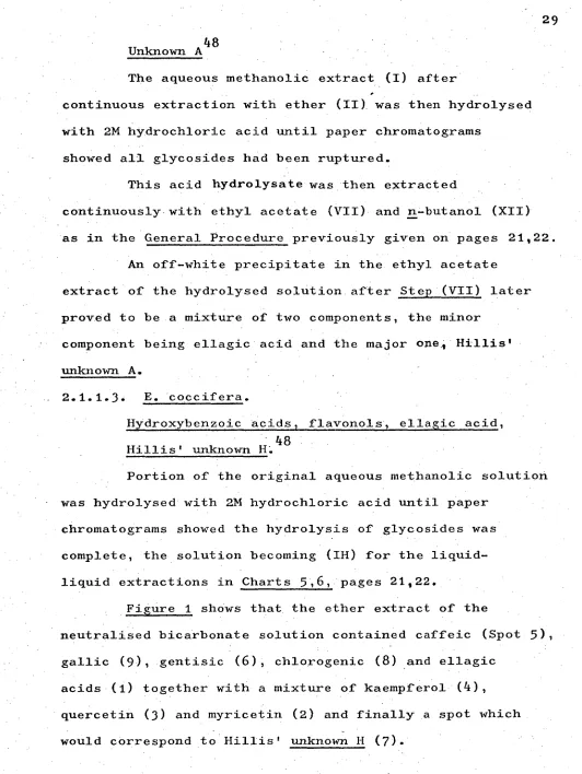
2.1.1.3. E. coccifera.
Hydroxybenzoic Acids, flavonols . , ellagic acid, Hillis' unknown H:48
Portion of the original aqueous methanolic solution was hydrolysed with 2M hydrochloric acid until paper
[image:36.559.27.559.12.720.2]chromatograms showed the hydrolysis of glycosides was complete, the solution becoming (IH) for the liquid- liquid extractions in Charts 5 .,6, pages 21 22.
Figure 1 shows that the ether extract of the
neutralised bicarbonate solution contained caffeic (Spot 5), gallic (9), gentisic
(6),
chlorogenic(8)
and ellagiccm
Fig.l. POLYPHENOLS of E.coccif era extracted by 5% NaHCO3 30
</\\\\\\
PDLYPHENOL 1.
Z.
3.
5. blue-wh. blue-white 6. blus-wh bl-gr-white
7. pink orange
8. blue intense green
4.
365nrri MH
3365nm
rrauve dull yellow orange intense or.
yellow intense y.
p-gr, . intense y-gr.
CM
9. mauve dark mauve (254-nm) (254m)
I I
2 4 6 8 cm 15% HOAc
Abbreviations: or, = orange y. . yellow
yellow-green blue-who = blue-white bl-gr-white . blue-
green,-white. 12 10 8 BAW 4:1:5 6
F1g.2. POLYPHENOLS in E.coccifera ETHER EXTRACT after NaHCO3 extraction.
4
15% HOAc
POLYPHENOL 365nm NH 365nm
3
1. - mauve dull yellow
2. orange intense or.
3. yellow intense y,
4. Y400 . intense y-gr.
10. purple purple-blue
Abbreviations: or. = orange y, = yellow
y-gr, . yellow-green
cm
8
48
Unknown D.
The ether solution from (III), i.e. after bicarbonate extraction of it, was shown on paper
chromatograms (Figure 2) to contain a mixture of compounds, mainly unknown D (Spot 10), a flavonol mixture (4,3,2)
and ellagic acid (1). Glycosides.
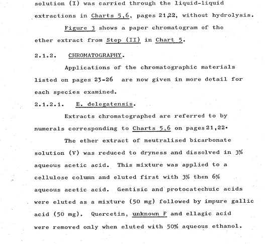
A second portion of the original aqueous methanolic solution (I) was carried through the liquid-liquid
[image:38.559.16.543.230.714.2]_extractions in Charts 5,6, pages 21,22, without hydrolysis. Figure
3
shows a paper chromatogram of theether extract from Step (II) in Chart
5.
2.1.2. CHROMATOGRAPHY.Applications of the chromatographic materials listed on pages 23-26 are now given in more detail for each species examined.
2.1.2.1. E. delegatensis.
Extracts chromatographed are referred to by numerals corresponding to Charts 5,6 on pages21,22. The ether extract of neutralised bicarbonate solution (V) was reduced to dryness and dissolved in 3% aqueous acetic acid. This mixture was applied to a cellulose column and eluted first with 3% then 6%
aqueous acetic acid. Gentisic and protocatechuic acids • were eluted as a mixture (50 mg) followed by impure gallic
32 Fig.3. POLYPHENOLS in ETHER EXTRACT of E.coccif era oci:Me0H solution.
6 BAW 4:15
PO LYPH EN OL 1. ella.gic acid
2. myricetin
3. quercetin.
4. kaempferol
5. caffeic acid
6. gentisic acid.
7. Hillis' 'Unknown H
8. chlorogenic acid.
9. gallic acid.
10. Hillis' Unknown, D
it. afzelin.
12.qu.ercitrio
13.
isoQuercitrin14.hyperin
15, sinapic acid. (trace)
4 6 15% HOAc colour (365nm) 'mauve orange yellow yellow-green blue-white blue-white pink blue
dark nauve (254m)
purple-blue purple purple purple-purple blue-green. colour/NH.3(365nm) dull yellow intense orange intense yellow intense yellow-green. white-blue blue-green-white orange intense green_
intense, dark mauve (251414
Column eluates were monitored on paper chromatograms which showed that while gentisic and protocatechuic acids had approximately identical R
f values when developed with 6% acetic acid,, they had different R
f _values in BAW (415). .
Repeated preparative paper chromatography on
Whatman No
3
paper using BAW (415) as developing solvent, with drying between developments, isolated sufficientgentisic (35 mg) and protocatechuic acids (20 mg) for their identification.
The ether extract remaining after extraction with bicarbonate (VI) was concentrated and a portion
(2 ml) applied to a preparative thickness cellulose thin layer plate and the plate developed with CEF solvent yielding impure quercetin (100 mg), which crystallized as yellow needles from dilute alcohol (85 mg), and a compound (7 mg) corresponding in chromatographic properties to Hillis' unknown D48 crystallized as white needles from methanol (3 mg).
The unhydrolysed methanolic aqueous extract (I) after concentration and standing yielded a yellow
precipitate of impure rutin (200 mg) which crystallized from dilute alcohol.
The ethyl acetate solution after bicarbonate extraction (XI) was taken to dryness onto polyamide powder, which was applied to a polyamide column eluting first with water and then with ethanol-water mixtures
31j
of increasing ethanol concentration. Bands were
observed in u.v. light and the ethanol concentration increased until a satisfactory movement of bands was achieved.
The 30-70% ethanol eluates were combined, evaporated to dryness and portion of the residue • dissolved in aqueous acetic acid after which it was streaked onto six sheets of Whatman No 3 paper and
developed three times with 6% aqueous acetic acid drying the papers after each development. The papers yielded impure quercitrin (30 mg) which crystallized as yellow needles from water (25 mg).
A small quantity of a second quercetin glycoside (5 mg) was also eluted from these papers from a band having a smaller Rf than quercitrin and later
identified as isoquercitrin, after crystallization from water.
The 80-100% ethanol eluates were evaporated to dryness and the residue chromatographed two-dimensionally showing the presence of rutin and a spot initially
interpreted as gallic acid but soon detected as a
catechin isomer by its colours when sprayed with vanillin in hydrochloric13. and ethanolic 2.-to1uene sulphonic
acids•. 96
35
concentration and standing the buffer extract yielded a precipitate of impure catechin isomer (90 mg) which crystallized from dilute acetic acid (70 mg).
2.1.2.2. E. sieberi.
Preparative paper chromatography of the
bicarbonate extractives (III) using the descending method in 6% aqueous acetic acid separated the glycosides
(20 mg) from other polyphenols with a minimum of hydrolysis. Dissolved in methanol the mixture of glycosides
was spotted onto sheets of Whatman No 1 paper and the sheets developed in BAW (415) in the ascending direction with the chromatograms being dried and re-developed in the same solvent a second time. The same papers were then developed in the second dimension using 15% aqueous acetic acid as developing solvent and again the
development was repeated.
In this way the separation of the glycosides shown in .Chart
7
was achieved.Spots with corresponding R f values were eluted with ethanol containing 30% water and the eluate
evaporated and spotted on Whatman No 1 paper for final purification by two-dimensional development in the
same solvents as above.
Final elution and evaporation yielded glycosides which after crystallization were identified as quercitrin
0
quercitrin isoquercitrincan nabiscitrin TBA
15HOAc
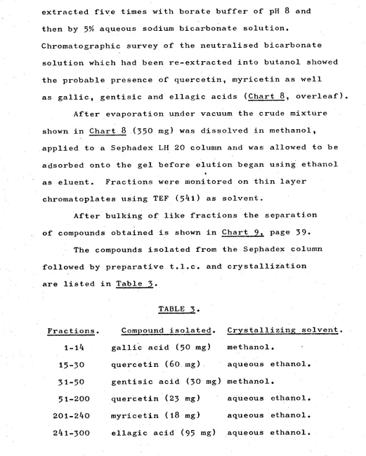
The butanol extract obtained from Ste (XII) was extracted five times with borate buffer of pH
8
and then by 5% aqueous sodium bicarbonate solution.Chromatographic survey of the neutralised bicarbonate solution which had been re-extracted into butanol showed the probable presence of quercetin, myricetin as well
as gallic, gentisic and ellagic acids (Chart
8,
overleaf). After evaporation under vacuum the crude mixture shown in Chart8
(350 mg) was dissolved in methanol, applied to a Sephadex LH 20 column and was allowed to be adsorbed onto the gel before elution began using ethanol as eluent. Fractions were monitored on thin layerchromatoplates using TEF (541) as solvent.
After bulking of like fractions the separation of compounds obtained is shown in Chart_24, page
39.
- The compounds isolated from the Sephadex column followed by preparative t.l.c. and crystallization are listed in Table
3.
Fractions.
TABLE
3.
Cry stallizing solvent. Compound isolated.
1-14 galliC acid (50 mg) methanol.
15-30 quercetin (60 mg) aqueous ethanol. 31-50 gentisic acid (30 mg) methanol.
51-200 quercetin (23 mg) aqueous ethanol. 201-240 myricetin (18 mg) aqueous ethanol. 241-300 ellagic acid (95 mg) aqueous ethanol.
[image:44.559.23.544.61.711.2]gent isic acid
quercetin
gallic acid
SAW
4:1:5
myricetin
ellagic acid
A
15H0Ac
SEPHADEX LH 20 COLUMN
TLC
toluene :ethyl acetate: formic acid
gentisic acid
SILICA
5
1
gall ic acid
quercet in
0 ° 0 0
0 0 0
myricetin el lagj.c acid.
ellagic acid and polymetic material
.
P P 0
11)
CI)
TEF
R
f
fractions
1
-14
15
30
37-50
51 200 201 240 241-300
An off-white precipitate (350 mg) settled in the ethyl acetate extract (VII) of the hydrolysed solution which had been left to stand in the refrigerator.
Paper chromatography showed this precipitate to be in the main a mixture of two components, the minor component being ellagic acid and the major component having colours in u.v. light similar to those of Hillis' unknown A48 although not having identical R
f values, possibly because of slightly different solvent
systems.
A chromatoplate of silica gel to which the
mixture (125 mg) had been applied was developed with the 15HOAC solvent system. In this way unknown A was
separated from ellagic acid and crystallized from aqueous ethanol to give a pure compound (60 mg) which has been identified as 4-0-monomethya ellagic acid.
The ethyl acetate extract remaining after removal of the above precipitate was paper chromatographed and showed in addition to the above . compounds the presence of hydroxybenzoic acids and a spot with properties similar to that of Hillis' unknown B.48
Repeated extraction of the ethyl acetate solution with 5% aqueous sodium bicarbonate with subsequent
neutralisation of the bicarbonate solution showed by
two-dimensional chromatograms that the unknown B compound had been extracted by the bicarbonate extraction.
Re-extraction of the neutralised bicarbonate
solution with ethyl acetate and. evaporation was
followed by application of the mixture (150 mg) to a. silica plate developed With BPF (.3:9 - :5). The top band of the plate contained impure unknown B.
- After elution and taking to dryness, the
component in the top band (k5 mg) was applied .to sheets of Whatman.No 3 MM paper which were developed with 15% aqueous acetic acid, dried and re-developed with the
same solvent until the characteristic diffuse mauve bands of Unknown B were resolved.
Elution of these bands with aqueous ethanol and evaporation gave a residue which crystallized from aqueous ethanol, to give a compound (28 mg) with
chromatographic properties similar to those of unknown B. Whatman cellulose powder was formed into a small column,the unknown F mixture obtained by trituration of an ether precipitate was, applied in 15%'
aqueous acetic acid and this solvent used in column development. Fractions collected showed on paper
chromatograms that separation had still not been achieved. Finally a.portion (150 mg) of the unknown F.
4
2 In this way sufficient unknown F (16 mg)was obtained after three re-crystallizations to obtain spectral analyses.
2.1.2.3. E. coccifera.
Roman numerals used in the fifillOwini-pararaPhs refer to Charts 5,6:onpages 21,22.
The ether solution remaining after extraction with bicarbonate (VI) was shown by paper chromatography to contain a mixture of unknown D, flavonols and
ellagic acid.
The unknown D mixture spots on paper did not
give red colours when sprayed with sodium borohydride and subsequently exposed to acid vapour33 as the E. sieberi flavanones had done. This prompted the isolation of the E. coccifera unknown D mixture.
Analytical thin layer chromatography using
Camag silica gel as adsorbent, and chloroform; ethyl acetate; formic acid (7:4:1, CEF) as solvent suggested that
column chromatography using the same adsorbent and
developing solvent might separate the unknown D mixture. The ether after bicarbonate extraction was taken to dryness (3 g) and this was dissolved in CEF (7:4:0.5), placed on a silica column and developed and eluted with the same solvent system. Fractions collected were
Figure 4 summarises the separation after fractions of similar composition had been joined.
Fraction A contained a mixture of apparently two compounds fluorescing purple under u.v. light
(365 urn) and a minor component of higher R
f value
fluorescing blue under u.v. light. Preparative t.l.c. (silica, CEF) then separated the blue component from the purple components mentioned above.
The compounds in Fraction A which fluoresced purple were separated into two spots on analytical
silica plates using . CEF as solvent.
Preparative t.l.c. using the same adsorbent and solvent separated the purple-fluorescent materials into ( two bands which when eluted gave apigenin as the band
of lower R
f and what appeared to be another single -compound of higher R f .
In all, six solvent systems were used before one of them, BPF, showed that in fact the apparently single compound was a mixture of a major and minor component.
Figure 5 shows the analytical t.1. c. separation of this mixture using BPF, into components later
identified as
(4), (5)
and(6).
It now appeared that the purple fluorescing spot in Fraction A was a mixture of three compounds, one of lower R
f whose isolation has already been
Fig.4. SILICA COLUMN SEPARAHON of E.coccifera POLYPHENOLS.
10
SOLVENT C E F
8
0
c
3)
f i I I I I
A B C D E F 0
FRACTIONS
A. Unidentified compound and. Unknown D mixture.
B. Unknown D mixture and kaempferol
C. Kaempferol and quercetin
D. Kaempferol, quercetin and. myricet5_n_
E. Querceti-n, nwricetin
F. Quercetin, myricetin
G. Traces of quercetin, myricetim and. ellagic acid.
HO
(5)
01-1 [image:52.559.23.548.33.717.2]( ) E. coccifera FLAVONES.
Fig. 5. TLC SEPARATION of FLAVONE MIXTURE USING CEF and BPF SOLVENTS.
Rf 0.74 0.72 0.67
R f
0.73 0.69
4
7
CEF BPF
CHCI:EtOACHCOOH C
6 H IC5 H5 N:HCOOH
3 6
closely related compounds of higher R f .
Repeated preparative t.l.c. on silica using BPF and involving the procedure of developing, drying and re-developing, separated the major component of
the mixture. Several crystallizations from ethanol gave a chromatographically pure compound, identified on
evidence .recorded in the discussion section as a rare C-methylflavone, found only once before in nature, sideroxylin or 4',5-dihydroxy-7-methoxy-6,8-dimethyl flavone (25 mg) (6).
The minor component (5) in this mixture was present in too small an amount to enable isolation in sufficient quantity for complete spectral analysis. However enough material (0.1 mg) was obtained by
eluting spots from six analytical silica chromatoplates developed three times with BPF for u.v. spectral
analysis, discussed in Chapter 4 1 page 150.
Fraction C from the column was shown by
two-dimensional paper chromatography to be a mixture mainly of kaempferol and quercetin. Kaempferol was isolated using preparative silica t.l.c. plates with MCP as developing solvent; kaempferol (20 mg) crystallized from aqueous ethanol.
An ether extract of the original unhydrolysed
aqueous methanolic solution (I) contained four glycosides. Quercitrin, isoquercitrin and hyperin were
identified using known markers on paper chromatograms.
The remaining glycoside, different from others previously found in this work, was isolated by
preparative paper chromatography by repeated
two-dimensional development using first 6H0Ac and then BAW (612) as developing solvents.
In this way sufficient of tliis glycoside was obtained (2 mg) to identify it as afzelin.
CHAPTER
3.
48
CONTENTS.
3.0. SPECTRAL DETERMINATIONS. 3.0.1. U.v. SPECTRA. 3.0.2. N.m.r. SPECTRA. 3.0.3. I.r. SPECTRA. 3.0.4. MASS SPECTRA.
3.1. SPECTRA.
3.1.1. POLYPHENOLS OF KNOWN STRUCTURE. 3.1.2. POLYPHENOLS OF UNKNOWN STRUCTURE
(HILLIS' UNKNOWNS).
3.2. DERIVATIVES.
3.2.1. TRIMETHYLSILYLATION. 3.2.2. METHYLATION.
3.2.3. TRIDEUTERIOMETHYLATION. 3.2.4. ACETYLATION.
3.2.5. PEROXIDE OXIDATION OF FLAVONOL-3 GLYCOSIDES.
3.2.6. ACID HYDROLYSIS OF GLYCOSIDES.
3.3. ROTATIONS, ANALYSES, MELTING POINTS. 3.4. EXPERIMENTAL DATA.
POLYPHENOLS OF KNOWN STRUCTURE. 3.4.2. POLYPHENOLS OF UNKNOWN STRUCTURE
(HILLIS' UNKNOWNS).
49
Paze.
50 50 51 51 52 52
52
66
100 100 101 101 102 102
103
103
104
104
50
CHAPTER
3.
3.0. SPECTRAL DETERMINATIONS. 3.0.1. U.v. SPECTRA.
3.0.1.1. Stock diagnostic reagents used throughout this 75b work and prepared according to the methods of Mabry et al. were sodium methoxide (Na0Me), aluminium (III) chloride
(A1C1
3), hydrochloric• acid (HC1), sodium acetate (Na0Ac) and boric acid (H
3B03)0 The reagents above were applied and spectra run according to the procedures given in
detail by the same authors.
3.0.1.2. U.v. spectra were run on a Hitachi Perkin-Elmer 124 double beam spectrophotometer or on a Perkin-Elmer 400 spectracord in methanol alone or in methanol with the
reagents given above; stock solutions of the polyphenols being prepared in either of the following ways: 75b
3.0.1.3. The solutions used were made from re-crystallized compound (0.1 mg) in Analar methanol (10 ml) and the
concentration adjusted so that maximum absorbance
between 240-420 nm occurred between 0.6 - 0.8. When log E values were obtained the concentration was accurately
determined.
3.0.1.4. Alternatively, the polyphenols were purified by one-or two-dimensional paper chromatography. Spots, when sufficiently discrete, were viewed under u.v. light,
• 51
re-dissolved in a minimum of methanol and its spectrum
was recorded as above.
Since methanol may elute u.v.-absorbing
compounds from the paper itself, when spectra were run in this way a reference solution was made by extracting a piece of blank chromatogram which had been developed in the same way as the chromatogram containing the polyphenol(s).
3.0.2. N.m.r. SPECTRA.
N.m.r. spectra were recorded on a Jeolco JNM4H-100 spectrometer.at 100 MHz or on a Varian A60 spectrometer at 60 MHz for 5% solutions in DMSO-d 6 with HMDS or TMS as external standard or in the case of the 60 MHz spectra, as internal standard, or using C5D5N or CDC1
3 or mixtures of the first two and the last, with TMS as internal standard. In the case of spectra run to
analyse trimethylsilyl ethers of polyphenols a drop each
of hexamethyldisilazane and trimethylchlorosilane was
75c
added to ensure anhydrous conditions. It was • standard procedure to run additional spectra after
addition of deuterium oxide for detection of hydroxyl groups.
3.0.3. I.r. SPECTRA.
These were determined in Nujol and
52
3.0.4. MASS SPECTRA.
Mass spectra were measured on an Ae.I.MS9
instrument at 70 eV. or on an EA1 QUAD 300 quadrupole mass spectrometer.
3.1. SPECTRA.
Spectra are presented in two sections, first (3.2.1.), those referring to polyphenols of known
structure and the second (3.2.2.), those of polyphenols
•
of unknown structure (Hillis' unknowns). 48
The spectra given are referred to in discussion in Chapter
4.
3.1.1. SPECTRA OF POLYPHENOLS OF KNOWN STRUCTURE. 3.1.1.1. Hydroxybenzoic acids.
Chart 10. N.m.r. spectra of gallic, gentisic and protocatechuic acids.
11. Mass spectra. 3.1.1.2. Flavonols.
Chart 12. N.m.r. spectrum of quercetin. 13. N.m.r. spectrum of quercetin
compared with that of myricetin. 14. Mass spectra.
53
3.1.1.3. Ellagic acid.
Chart 16. Mass spectrum and fragmentation. 17. N.m.r. spectrum (C
55N/Et0H). 18. N.m.r. spectrum of TMS ether
(). CDC1 3 3.1.1.40 (+)-Catechin.
Chart 19. N.m.r. spectrum (DMSO-d6 20. N.m.r. spectrum (C
47- 6
HO 2H
l 11 TUT-17111 111111111T-111T11111111- 1111
6
COOH 1•72
6 OH H-Z
*\\ HO 3
2H
GENTISIC ACID
11111111111 H-3
1H
sir
11 1 1 11 li1111
8 7
ilit
2H
H-2,6
7 6 5
CHART 10. N.M.R. SPECTRA of HYDROXYBENZOIC ACIDS .
7
( p.p.m.) 5 PROTOCATECHUIC ACID
111111111111 ■ 11 7.29
OH
GALLIC ACID
COOH 2 x OH
GENTISIC ACID
PROTOCATECHUIC ACID GALLIC ACID
100
110
137
136 125
108
(m+)
.153 170 110
rn/e
(Scale 6}
9 8
. I ....
6 P.P.m.
6
tIli tit I. I
IIII.t.,,,,,„.
7
QUERCETIN
6
S.
DMSOd (int.TMS) 6
8
H
6
OH 0
H6' H2'
OH
5
2
'
QUERCETIN
H2'
H6'
MYRICETIN
OH
<-0H
HO.
°
0H
0
1
WOH
OH 0
H8 H6
S 8
7
318
302
100—
80
60
oh
40
209
(base peak)
MYRICETIN
153
iS1 137
125
20
0
100-
80
60
Oh
40
1
20
.145
(base peak)
.137
.128
.142
.302
.153
256
274
284QUERCETIN
100 140 180 220 260
OH 0
HO
OH
OH 0
OH
OH
R 1 R 2
m/e 137,153
HO
OH 0+
m/e 137
R1 =1-1
7
QUERCETIN
R?:OH, MYRICETIN
HO
R1R2
HO
OH
OH
64.
OH
R
1
R
2
m/e 153
0+
m/e 137,153
1
7
-CO
M -18 142
7
150
2
-CO
-CO
m/e 109,125 m/e 109
m/e 109,125
100
( base peak )
202(M
4
)
ELLAGIC ACID
80
-0 -0
CHO
—200
—
CO —CO —CO
— CHO
—
CHO
60
40
20
27o 266 162 189
190
218
2f7 228 .246 273
0
me
100 120 140 160 180 200 220 240 260 280
•CHART
16.MASS SPECTRUM and FRAGMENTATION of ELLAGIC ACID
1111 11•111 1
1II I111111i I
r\vPi\vw,pl,tytivAftchtv\■vjLeAra.",
8 7 6 1. 3 2
OH
ELLAGIC ACID
P..C
5D5N
OH
cHa
8-01
P A
Yi Vp,AA) '1.'44014.0.,0\vni-N,yvovi\ANNI\AA,^v
P.P.rn.)
Et0H
7.63
2H
1 III I Il I I I IlfIlI1
ELLAGIC ACID TMS ETHER
CDCC
NAMe )401))1 feltOlverNtgYs.thrtAithiltellAWV,14/\AVVW.15A- leVAAA''\'11\i"PAm.N"ii
(.6p.P.rni
9 7 6 5 . 4 3 1
(-0—Catechin
21-1
SOH
701-1
4
.
0H
30H 3.0H
H2
/
H5
/
DMSOd6
H8 H6
H2 H3 •
2,xH4
9
CHART 19.
6 5
7
6.76 6.64 8+1 6-H
113 7 6
illirliiiii.rf,
a-ax
-4
-
"
L
t-J
• 9 3.4 ax OH-44
'leg
P .Ct %N
3-H _L._ 22Hz
i3.4eq
1" -13.4q-ax
AB
3.70 3.31
Heq 4Hq.ax
5.20 241
1H
2H
( p.p.m.) OH
(4)-CATECHIN
8 7 6 5 4 3 2
OH •>
OH
OH
W
.
= m/e 290
— CHO
OH
m/e 123
rDA
HO
HO
HO OH
CH
2
OH
m/e
139
CH
2
OH
m/e 138
CH
OH +
2
m/e 139
66
3.1.2. SPECTRA OF POLYPHENOLS OF UNKNOWN STRUCTURE. 3.1.2.1. Unknown D48 - Flavanones.
Chart 22. N.m.r. spectra and configuration. 23. I.r. spectral data.
N.m.r. spectral data.
25. N.m.r. spectrum of pinocembrin. 26. N.m.r. spectrum of alpinetin. 27. N.m.r. spectrum of 0,0-dimethyl-
pinocembrin. 28. Mass spectra.
29. Mass fragmentation. 3.1.2.2. Unknown D48 - Flavones.
Chart 30. N.m.r. spectrum of apigenin. 31. Mass spectrum of apigenin.
32. . Mass fragmentation of apigenin. 33. Mass spectrum of sideroxylin.
34. Mass fragmentation of sideroxylin. 35. N.m.r. spectrum of sideroxylin
(Dmsod
6/CDC13 ).
36. N.m.r. spectrum of sideroxylin
(c
5D5N/cDc13 ).
37. N.m.r. spectrum of sideroxylin mixture. 38. N.m.r. spectrum of TMS ether of
sideroxylin mixture.
39. Mass fragmentation of second C-methyl flavone.
3.1.2.3. Unknown A48 - 4-0-methyl ellagic acid.
67
Chart 41. N.m.r. spectrum.42. N.m.r. spectrum of methylated unknown A (CDC1
3
).
43. N.m.r. spectrum of methylated unknown A (C
6D6 .
44. U.v. spectra of unknown A and ellagic acid in methanol.
45. U.v. spectra of the same in methanol plus solid sodium acetate.
46. U.v. spectra of the same in methanol plus sodium methoxide solution.
47. Mass spectrum and fragmentation of unknown A.
3.1.2.4. Unknown B48 - (1,5-dimethy1-2,6-bis(3,4 9 5-
Chart 48.
119.
trihydroxyphenyl) furo ,[1 1 5-c] furan.). U.v. spectrum of unknown B in
methanol.
U.v. spectrum in methanol plus sodium methoxide solution. Mass spectrum.
50. N.m.r. spectrum.
3.1.2.5. Unknown F48 - (a tetrahydroxydibenzofuran- dicarboxylic acid).
Chart 51. N.m.r. spectrum.
52. I.r. spectra of ellagic •acid and unknown F.
H
3ax
H
3eq
3eq
H
3ax
NMR SPECTRA and CONFIGURATION of
E.sieberi
FLAVANONES.
4
2-30
. -1.- 16 Cos2 154 ° 4.. 13 .0 Hz
J
H2-3eq
1000S257° 3.0Hz
-J 4 12 Hz
J 2.5 Hz
OBSERVED ..F.13 Hz
CHART 22.N.M.R. SPECTRA of E.sieberi .FLAVANONES and
CONFIGURATION.
Ph
eq
2 eq
2S
H
2ax
H — H
aX a XH H
eq. ax
DIHEDRAL ANGLES
H
2a x
57 °
H
3eq
154 °
MET HOXYL CARBONYL
3000 1700 1600 1500 1300 1200 1100 , 1000 GOO 700 600 cm PHENYL
Ph
Ph
3400 3200 HYDROXYL, 3600
H2ax
ALPINETIN
PINOCEMBRIN
appm
5-90
11-20
7.79
HO
00
\
H--"
12-48
H 3•07
1-1
3-60
H3 ) 2 SO4
Acetone
CH2N2
(CH3)2SO
NMR SPECTRAL DATA for E.sieberi FLAVANONES.
3.78
Me0
7-40
310
Me()
5,7-DIMETHOXYFLAVANONE
11
1 1
11 T -1 11111 1 1111- 11-1- 1 I I I 11
HO
8 7 6 5 4
Soo 00 1300 too 400 J700 ?OD 200
Ti 111 11
1120
7.'0H
vovr,r0401
tiltlit111111i tilt] 111111111111111111111111111111111111 1 1
3
iiiimimil 12.48
5-0H
5H
P1NOCEMBRIN
7-0H
11.20
• P.Prn .
4 3 2 1
lit
8 7
I 1 1 1 1 1 till
So goo doo • foo Woo .foo • .20o 100
1 111111j111111T-1111 III 1- 11IIT•11-11-11-11111 -11111-TATT1- 111111{1-1-TTi111111. IITI11 1 T1 111111 1 11i
ALPINETIN
X 1i
I
)\
I \
I
I
j
u
t
'reittiMpvPherek
y
(
1
1
/
1
4
1
`!
144°41/MANIVAlftir4413*1 *410
4
01(
0
/
1 N
t
i
V\MoWel'
I
N
*A1
3H
HS H6 5H
OMe 5
Ph H
DMS0d6
2H
X 1
wirkfi
rwov
4,4
H2
A
0\441444* ;
‘fli
14
,04
rrvh
Y
T
Y
E-1!T'600 foe
1111111 400 Joe I
,
200 II III
Joe IlIjil I r Il l I
OMe7 0.0-DIMETHYLPINOCEMBRIN
+ 5 H ‘,
\2H OMe5
3H 3H
2H
C RING A RING
H
X 1
CDC/
tkrov
o4Osoryle"*.'$0wHei
H 1 H6 •
•
PI
?
Ilk) Veff4,1' NItrA4k1 4‘1V4) v1 i i
H2 1 H3trans H3cis
III
1 i! ilV1 1
41,Wkr-ivs**446A11400V) '"N;mtniwnrAti 104/0,ymmA1/41v4^v.,44nivekAfry kh
YTY
B RING
C RING
P P m
8 7 6 5 4 3 2 1
124 152 266'
.256
179
PINOCEMBRIN
104
100
0
166
270
ALPINETtN
(M
+
)
193
138 •
100
0
/0
-104
100
269
X8o
0,0-DIMETHYL-PINOCEMBRIN
2.94.
162
233
0
rve
'100 120 140 160 180 200 220 240 260 280 300
M+.= 256 ,R R
2
= H =A
M4-= 270 , =H, R
2
=Me =B
M+.= 284. R
1
= Me , R
2
= Me C
a
R
I
R
2
0
R
2
0 OH R
2
0 OH
A , m/e =152 m/e=104 m/e =
B, 166
..c., 180 N.00
). 124,138,152
CHART29. FRAGMENTATION of E.sieberi FLAVANONES,
179 ,
m/e = 255
193
269
2.H DMSOd
6 / int.TMS
H
1
OH
H-6 H-2',6'
H-3
. H-8
H-35'
J =9 Hz J =3Hz
SPPm 8 7 6
D'
A':Nro ll°.1