A STUDY OF EXPRESSION OF EPIDERMAL GROWTH
FACTOR RECEPTOR(EGFR) AND VASCULAR
ENDOTHELIAL GROWTH FACTOR (VEGF) IN
EPITHELIAL OVARIAN NEOPLASMS.
Dissertation submitted in
partial fulfilment of the requirements for the degree of
M.D. (PATHOLOGY) BRANCH - III
GOSCHEN INSTITUTE OF PATHOLOGY AND ELECTRON
MICROSCOPY
MADRAS MEDICAL COLLEGE
CHENNAI – 600 003
THE TAMIL NADU
DR. M.G.R. MEDICAL UNIVERSITY CHENNAI
CERTIFICATE
This is to certify that this Dissertation entitled “A STUDY OF
EXPRESSION OF EPIDERMAL GROWTH FACTOR RECEPTOR
(EGFR) AND VASCULAR ENDOTHELIAL GROWTH FACTOR
(VEGF) IN EPITHELIAL OVARIAN NEOPLASMS” is the bonafide
original work of Dr. D.KANMANI, in partial fulfillment of the requirement
for M.D., (Branch III) in Pathology examination of the Tamilnadu Dr.M.G.R
Medical University to be held in April 2016.
Prof. Dr K. RAMA, M.D., Prof. Dr. M.SARASWATHY,M.D.,
Professor of Pathology, Director & Professor, Institute of social obstetrics and, Institute of Pathology , Govt KasturbaGandhi hospital, Madras Medical College, Madras Medical College, Chennai – 600003. Chennai – 600003.
Prof. Dr. R.VIMALA, M.D.,
DEAN,
DECLARATION
I, Dr.D.KANMANI, solemnly declare that the dissertation titled
“A STUDY OF EXPRESSION OF EPIDERMAL GROWTH FACTOR
RECEPTOR(EGFR) AND VASCULAR ENDOTHELIAL GROWTH
FACTOR (VEGF) IN EPITHELIAL OVARIAN NEOPLASMS” is the
bonafide work done by me at the Institute of pathology, Madras Medical College
under the expert guidance and supervision of Prof. Dr.K.RAMA, M.D.,
Professor of Pathology,Institute of social obstetrics and Govt. Kasturba Gandhi
hospital , Madras Medical College. The dissertation is submitted to the
Tamilnadu Dr. M.G.R Medical University towards partial fulfillment of
requirement for the award of M.D., Degree (Branch III) in Pathology.
Place: Chennai
ACKNOWLEDGEMENT
I express my sincere thanks to Prof. Dr.R.VIMALA , M.D., Dean,
Madras Medical College and Government General Hospital, for permitting me
to utilize the facilities of the Institution.
I take the opportunity to express my thanks to Prof.
Dr.M.SARASWATHY, M.D., Director and Professor, Institute of Pathology,
Madras Medical College, Chennai for her keen interest, constant
encouragement and valuable suggestions throughout the study.
I am extremely thankful to Prof .Dr.K.RAMA, M.D., Professor of
Pathology, Institute of social obstetrics and Govt Kasturba Gandhi Hospital ,
Madras Medical College, for her valuable suggestions, constant support, advice
and encouragements throughout the study.
I am truly thankful to Prof.Dr.Padmavathi M.D.,
Prof. Dr.Ramamurthy M.D Prof.Dr.Geetha Devadas M.D., D.C.P.. Prof.
Dr. Sudha Venkatesh M.D., Prof. Dr. M.P Kanchana M.D.,
Prof.Dr.Rajavelu Indira M.D, Prof. Dr. S. Pappathi M.D.(PATH), D.C.H.,
I thank the Director of Institute of Social Obstetrics and Govt Kasturba
Gandhi hospital for permitting me to utilize the materials of the institution.
I express my heartfelt sincere thanks to all my Assistant Professors for
their help and suggestions during the study.
I would like to thank the Institutional Ethics Committee for
approving my study.
On a personal level, I extend my gratitude to all the members of my
family for their constant support.
I am thankful to the statistician , for helping me in statistical analysis.
I thank my Friends, Colleagues, Senior Postgratuates, Junior
Postgraduates, Technicians and the Staffs for their continuing support and
ABBREVIATIONS
EGFR : Epidermal Growth Factor Receptor
VEGF : Vascular Endothelial Growth Factor
NCRP : National Cancer Registry Programme
WHO : World Health Organisation
ICMR : Indian Council Of Medical Research
HNPCC : Hereditary Non Polyposis Colorectal Cancer
STIC : Serous Tubal Intraepithelial Carcinoma
CK : CytoKeratin
IHC : ImmunoHistoChemistry
CEA : CarcinoEmbryonic Antigen
EDTA : Ethylene Diamine Tetra Acetic acid
MAPK : Mitogen Activated Protein Kinase
H & E : Hematoxylin & Eosin
FIGO : International Federation of Gynecology and
CONTENTS
S. NO. TITLE PAGE
NUMBER
1 INTRODUCTION 1
2 AIMS AND OBJECTIVES 5
3 REVIEW OF LITERATURE 6
4 MATERIALS AND METHODS 28
5 OBSERVATION AND RESULTS 31
6 DISCUSSION 63
7 SUMMARY 75
8 CONCLUSION 78
ANNEXURES BIBLIOGRAPHY
A STUDY OF EXPRESSION OF EPIDERMAL GROWTH FACTOR
RECEPTOR(EGFR) AND VASCULAR ENDOTHELIAL GROWTH
FACTOR (VEGF) IN EPITHELIAL OVARIAN NEOPLASMS
ABSTRACT
INTRODUCTION
Ovarian carcinoma is the 6th most common carcinoma among women in
the world and forms 1.7 to 8.7% of female cancers in India.It is the most
common cause of gynecological cancer death in women. Surface epithelial
ovarian carcinoma accounts for 90 to 95% of ovarian malignancies.
EGFR (Epidermal Growth Factor Receptor)
Among various prognostic indicators, EGFR a 170 Kd glycoprotein
maintained its independent prognostic value,and brings about increased DNA
synthesis, cell proliferation and differentiation. With the availability of EGFR –
inhibitors, selection of patients for EGFR – targeted therapy becomes more
important.
VEGF (Vascular Endothelial Growth Factor)
VEGF is a dimeric glycoprotein functioning as a tumour angiogenesis
factor.
Bevacizumab – Anti VEGF, antibody shows promise in the treatment of
This study is an attempt to determine the expression of the above two
markers -EGFR and VEGF in epithelial ovarian neoplasms.
AIMS AND OBJECTIVES:
To study the expression of EGFR (Epidermal Growth Factor Receptor)
and VEGF (Vascular Endothelial Growth Factor) in epithelial ovarian
neoplasms, which could thence be, used as therapeutic targets in future.
MATERIALS AND METHODS:
30 cases paraffin sections of ovarian specimen diagnosed as borderline
and malignant epithelial ovarian neoplasms were subjected to staining with
ImmunoHistoChemical markers-EGFR and VEGF.
RESULTS:
Out of 4 borderline ovarian neoplasms, 50% showed positivity for EGFR
while 75% of them showed positivity for VEGF.
Among malignancies, 80.76% of them showed EGFR positivity while
84.02% showed VEGF positivity.
CONCLUSION:
With Immunohistochemical analysis, the percentage of EGFR and VEGF
expression showed a significant increase in malignant compared to borderline
correlation with tumour grade and FIGO stage. High grade and advanced stage
tumours showed EGFR and VEGF overexpression compared to low grade and
early stage carcinomas.
KEYWORDS:
Surface epithelial ovarian carcinoma, EGFR, VEGF,
1
INTRODUCTION
Ovarian carcinoma is the 6th most common carcinoma among women in
the world [1] and it ranks fifth in cancer deaths among women. [2]. Surface
epithelial ovarian carcinoma accounts for 90 to 95% of ovarian malignancies [3]
Surface epithelial tumours, statistically the most important group of
neoplasms are derived from surface coelomic or germinal epithelium that is
continuous with the mesothelium that covers the peritoneal cavity, sharing with
it a common origin and many morphological features. The ovarian surface
epithelium involved in metaplastic or neoplastic conditions often undergo
‘mullerian differentiation’ and may produce any of the adult structures formed
by the mullerian ducts including tubal, endometrial and endocervical mucosa,
singly or in combination [5]. It has also been noted that many of the surface
epithelial tumors arise from the invaginated portion of the epithelium that
forms surface epithelial glands and cysts [6]. Another proposed origin of some
ovarian epithelial tumours (especially serous type) is the epithelium of the tubal
fimbriae and fimbriae are the most common sites of early serous carcinoma in
2
The parameters based on which the surface epithelial ovarian tumors are
classified are:
1. Cell histological type: Serous, mucinous, endometroid etc
2. Growth pattern: cystic, solid etc
3. Proportion of fibrous stroma.
4. Degree of atypia and invasiveness: benign, borderline and malignant [6]
A new model divides surface epithelial tumours into 2 major categories:
Type 1 and Type 2, based on their clinicopathological features and
characteristic molecular genetic changes [8].
Type 1 tumors are slow growing, generally confined to the ovary at the
time of diagnosis and developing from well-established precursor lesions [9].
Type 2 tumors are rapidly growing, highly aggressive neoplasms for
which well-defined precursor lesions have not been identified. More than 75%
of them have TP53 mutations [10].
ROLE OF BIOMARKERS:
Ovarian carcinoma is comparatively asymptomatic in early stage and is
aptly called a “silent killer disease”.70% of patients present in stage III and IV
underscoring the need for early biomarkers since the survival rates vary
3
TABLE 1: 5 YEAR SURVIVAL RATES FOR EPITHELIAL OVARIAN CANCERS:
STAGE OF THE EPITHELIAL OVARIAN CARCINOMA
5 YEAR SURVIVAL RATE
STAGE I 90%
STAGE II 70%
STAGE III 39%
STAGE IV 17%
“Survival rate for ovarian cancer by stage”- AMERICAN CANCER SOCIETY-retrieved on 29 oct 2014.
BIOMARKERS IN DIAGNOSIS:
The long used CA-125 is raised in only 50% of early stage ovarian
cancers [11]. It is also highly non-specific. The need of the hour are other
complimentary biomarkers in early diagnosis and prognostication. Two amidst
these novel biomarkers are EGFR (Epidermal Growth Factor Receptor) and
VEGF (Vascular Endothelial Growth Factor).A multivariate cox analysis
regression model showed that high serum VEGF expression in stage I patients
is correlated with 8 fold increase in cancer mortality[12].Compared to benign
ovarian lesions,early stage ovarian cancer patients showed raised levels of
VEGF.Hence when used in combination with CA-125,the sensitivity was
increased upto 96% and specificity up to 77%.[13]
BIOMARKERS IN PROGNOSTICATION:
Higher levels of EGFR and VEGF are associated with metastases,
4 BIOMARKERS IN THERAPEUTICS:
It has been predicted that simultaneous inhibition of 2 key tumor
dependent growth factor pathways EGFR and VEGF, causes Receptor Tyrosine
Kinase (RTK) pathway disruption and consequently tumor growth
arrest/inhibition. [13]
EGFR (Epidermal Growth Factor Receptor)
Among various prognostic indicators, EGFR maintained its independent
prognostic value. EGFR is a 170KD transmembrane glycoprotein. Ligand
binding triggers intrinsic tyrosine kinase activity of the receptor activating
numerous cellular responses like increased DNA synthesis, cell proliferation
and cell differentiation. With the availability of EGFR inhibitors, selection of
patients for EGFR – targeted therapy becomes more important.
VEGF (Vascular Endothelial Growth Factor)
The dimeric glycoprotein VEGF is structurally similar to platelet
derived growth factor and may function as a tumour angiogenesis factor.
Bevacizumab – anti VEGF, antibody shows promise in the treatment of ovarian
cancer. VEGF has been known to have crucial role in neovascular formation in
tumors, providing nourishment for the highly metabolic tumor cells and
5
AIMS AND OBJECTIVES
1. To study the expression of EGFR (Epidermal Growth Factor Receptor)
and VEGF (Vascular Endothelial Growth Factor) in epithelial ovarian
6
REVIEW OF LITERATURE
NORMAL ANATOMY AND HISTOLOGY
The ovaries are a pair of female reproductive organs ,lying in the pelvis
on either side of the uterus close to lateral pelvic wall, behind broad ligament
and anterior to rectum. The mesovarium attaches it to posterior aspect of broad
ligament along its anterior margin. The ovarian ligament attaches it to the
ipsilateral uterine cornua and infundibulopelvic ligament attaches it to the
lateral pelvic wall [14]. Adult ovary has an ovoid shape and measures (3 to 5
cm) x (1.5 to 3 cm) x (0.6-1.5 cm) and weighs 5 to 8 grams during the
reproductive period. After menopause, they shrink to one half of this size [15]
LYMPHATICS
The majority of the ovarian lymph vessels drain to large trunks that
form a plexus at the hilus and finally drain into Para aortic nodes. Few of them
also drain into internal and external iliac, common iliac and inguinal nodes [16]
BLOOD VESSELS
The ovarian artery, a direct branch of the aorta, courses along the
infundibulopelvic ligament, anastomoses with the ovarian branch of uterine
artery and forms an arcade from which about 10 arterial branches arise and
penetrate the ovarian hilus and medulla. These form a plexus at the cortico
medullary junction from which the radial cortical arterioles arise [15]. The veins
7
vein drains into the left renal vein and the right ovarian vein drains into the
inferior vena cava [18]
NERVE SUPPLY
Nerve supply to the ovaries is through ovarian, hypogastric and aortic
plexuses.
HISTOLOGY
A single layer of cuboidal cells that constitute the germinal epithelium
covers the ovarian free surface. The ovarian substance is divisible into cortex
and medulla. Immediately deep to the germinal epithelium, the cortex is
covered by a condensed connective tissue called the tunica albuginea, which is
much thinner and less dense than that of testis. Deep to this, the ovarian stroma
is made of slender spindle shaped cells, fine collagen fibres and ground
substance. Scattered in this stroma are ovarian follicles at various stages of
development – each containing a developing ovum.
The inner medulla consists of connective tissue in which numerous
blood vessels are seen.It also contains elastic fibres and some smooth muscle
fibres. The ovarian hilus cells are similar to the interstitial cells of the testis.
OOGENESIS
Oogonia are the stem cells from which ova are derived. An oogonium
enlarges to form a primary oocyte with diploid number of chromosomes. It
8
number of chromosomes. However the cytoplasm is not equally divided and
most of it goes to one daughter cell which is large. The second daughter cell
with hardly any cytoplasm forms the first polar body. The secondary oocyte
undergoes second meiotic division to give rise 2 unequal cells – the larger one
is the mature ovum and the smaller one is the second polar body.
FORMATION OF OVARIAN FOLLICLES
The ovum with the surrounding flat stromal cells forms a primordial
follicle. These form majority of follicles in the ovary. The flat stromal cells or
the follicular cells become columnar and form the primary follicle. The
follicular cells proliferate to form several layers of granulosa cells. The
homogenous membrane – the ‘zona pellucida’ appears between the follicular
cells and the developing ovum. This is a ‘secondary follicle’. A follicular
cavity – the antrum appears, filled with a fluid – the liquor folliculi. The oocyte
lies eccentrically in the follicle surrounded by some granulosa cells, the
cumulus oophorus. As the follicle expands – the stromal cells surrounding the
granulosa become condensed to form a covering called “theca interna” outside
which some fibrous tissue becomes condensed to form another covering for the
follicle – “the theca externa”. The first meiotic division is completed just
before ovulation to form the secondary oocyte. Follicular antrum enlarges
markedly. The follicle reaches the size of 1.5 cm to 2.5 cm and bulges under
9 CORPUS LUTEUM
When the graafian follicle ruptures, it collapses and becomes folded and
fills with blood. The granulous cells are enlarged with abundant pale cytoplasm
and round nuclei, abundant smooth endoplasmic reticulum and mitochondria
and numerous lipid droplets giving a yellow tinge and hence the name“corpus
luteum” which secretes progesterone.
EMBRYOLOGY OVARY
Formed essentially from the gonadal ridge.
OVA
In early embryonic phase, the primordial germ cells are formed from the
dorsal endoderm of the yolk sac and migrate along the hindgut to the gonadal
ridge [24].
DESCENT OF THE OVARIES
From the lumbar region, ovaries descend to the pelvic cavity by the pull
of gubernaculum ovarii which stretches from ovary to the skin of labium
majus. Their descent is arrested at the pelvis by the developing uterus and the
broad ligament.
FUNCTIONS OF THE OVARY
1. Gamete production associated with periodical release of ova.
2. Endocrine functions – Ovaries secrete estrogen, progesterone and small
10 TUMOURS
WHO classification of Ovarian tumours given under Annexure 1
SURFACE EPITHELIAL TUMORS
Form two-thirds of all ovarian neoplasms [23]. They are further classified
according to the following parameters
a. Histological Cell type – serous, mucinous, endometrioid etc
b. Growth pattern – cystic, solid etc
c. Proportion of fibrous stroma.
d. Degree of atypia and invasiness – benign, borderline and malignant
EPIDEMIOLOGY
In western countries, ovarian carcinoma is the most common cause of
gynaecological cancer death. It constitutes 4% of total carcinomas in women
[93]. The approximate risk of American women developing ovarian carcinoma
in their lifetime is 1.4%. Generally, we can say that the disease is seen more
commonly in industrialized western countries because of their low parity, an
important exception being Japan, because though the parity is lower, they have
relatively lower incidence of ovarian carcinoma.On the other hand,
Scandinavia shows one of the highest annual incidence rates of more than 16
per 1 lakh females [94]
The incidence of ovarian carcinoma in India, ranged from 1.7% to 8.7%
11
working under the network of National Cancer Registry Programme (NCRP) of
ICMR (Indian Council of Medical Research) [107]. The total number of new
cancer patients in India is well on the rise partly due to the increase in
population and also due to the relative rise in the proportion of elderly
population due to improved life expectancy. Ovarian carcinoma ranks
third/fourth among the cancers occurring in women in India. In the national
cancer registries from Ahmedabad / Bengaluru and Chennai, an increase in the
mean annual percentage change was noted in age group of 55-64 years [108].
Lifestyle changes towards industrialization and urbanization in India ,
especially rise in age at marriage, delay in age at first birth, reduced parity,
increase in incidence of obesity, diabetes, hypertension, cancer corpus uterus-,
diet rich in saturated/animal fats have all contributed to the increased incidence
of ovarian carcinoma in India. Maximum increase over the last 10 year period
was observed in Nagpur with the mean annual percentage increase of 2.4%.
Some of this increase is also attributed to improved certification and
registration of the disease in the recent years.
In the recent years, one significant change noted is that there is a
relative fall in the incidence of ovarian carcinoma, as tubal carcinoma and
peritoneal carcinomas have started showing an increasing trend [95]
Migration studies show that, the rate of ovarian carcinoma is determined
by the immigration place rather than the emigration place - indicating a
12
The incidence of ovarian carcinoma also shows a distinct variation
according to the ethnicity. White women have increased incidence compared to
African-American and Asians. Asian women have a 48% lower death rate
compared to that of white women. Jews have eight times increased risk of
developing ovarian carcinoma compared to non-Jewish women because 1 in 40
of them have a BRCA1 mutation [96]
ETIOLOGY AND RISK FACTORS
1. Age: Risk increases with age. Mean age in India is 40-59 years. The
average age of women affected in hereditary syndromes (like Lynch
syndrome) is much lower than others.
2. Reproductive factors: Early menarche and late menopause are
significant risk factors. Increase in number of pregnancies and consistent
oral contraceptive pill usage are proved to be protective against ovarian
carcinoma. Increase in number of pregnancies appear to be relatively
more protective against endometroid and clear cell carcinoma subtypes.
3. Ovulation and Hormonal factors: “Incessant ovulation” predisposes to
malignant transformation of the actively proliferating surface
epithelium. The occurrence of ovarian carcinoma is directly linked to the
total duration of reproductive years without interruption by pregnancies
(or) oral contraceptive pill usage. Recent studies support the fact that
consistent oral contraceptive pill usage reduces the risk of ovarian
13
duration of usage. Another theory says that increased levels of
circulating gonadotrophins increases the chance of incidence of ovarian
cancer either directly (or) by increasing the circulating levels of
oestrogen. Another theory proposes that the levels of androgens are also
important in the causation of ovarian carcinoma.
4. Inflammation: High grade serous carcinomas are associated with
chronic salpingitis in 53% of cases [24]
5. Others: Include Body Mass Index, diet, talc, smoking, ionizing
radiation, surface epithelial dysplasia, surface epithelial inclusions,
endometriosis, serous tubal intraepithelial carcinoma. Other important
protective factors include hysterectomy, fallopian tube ligation, and
bilateral salpingo – oophorectomy, -the protective mechanism being
prevention of retrograde passage of endometrial tissue and,
endometriosis. Hence the incidence of clear cell carcinoma varies
inversely with tubal ligation [97]
6. Genetic Factors: At least 10% of ovarian carcinomas arise in the setting
of highly penetrant, autosomal dominant genetic predisposition. These
include BRCA1 and BRCA2, HNPCC (Hereditary Nonpolyposis
14 CLINICAL FEATURES
The patient often presents with vague, nonspecific symptoms like
bloating, abdominal distension, dyspepsia, lower abdominal pain, loss of
appetite and loss of weight, nausea, vomiting, increased frequency and urgency
to urinate [26].
PROBABLE HISTOPATHOLOGICAL PRECURSOR LESIONS
1. Surface epithelial Dysplasia
Recent investigations indicate that subtle nuclear changes were seen in
ovaries removed prophylactically from high risk women compared to normal
controls [98].
2. Surface epithelial inclusions
Several studies have shown that ovaries of prophylactic oophorectomy
specimens from high risk women showed invaginations of cortical epithelium
(clefts) and papillomatosis more commonly than in controls.
3. Endometriosis
The best studied and most easily recognized precursor lesion is
“endometriosis”. Endometriosis is a common lesion found in about 10% of
reproductive age women. A series of studies support the fact that endometriosis
was at least as common as serous cystadenoma and hence would be the most
15
Extensive studies also show that the incidence of carcinoma in a known
case of endometriosis is just 0.3 to 3% [99]. In a Sweden based study of more
than 20,000 hospitalized women with endometriosis, a 11.4 year follow-up
showed that the relative risk of carcinoma in ovarian endometriosis is 1.9. The
mean age of occurrence of carcinoma in these cases was 51 – showing that the
incidence of carcinoma in endometriosis occurs in a relatively younger age
group.
4. Benign and atypical proliferating neoplasms
Molecular analysis studies, strongly suggest that borderline tumours are
forerunner lesions of low grade serous, endometroid and mucinous carcinomas.
5. Serous Tubal Intraepithelial carcinoma (STIC) and p53 signature
There is a recent proposal that fallopian tubal fimbriae are the origin of
some of serous carcinomas. Serous tubal Intraepithelial Carcinomas (STICs)
have been found to be associated with greater proportion of high grade serous
carcinomas [100]. These STIC lesions harbour TP53 mutations. Though they
are cytologically malignant lesions, they are confined to the tubal epithelium. A
minimum of 12, p53 positive, fallopian tubal secretory epithelial cells define a
case of “p53 signature”. This p53 signature being a candidate for STIC
precursor.
The junctions between different types of epithelium have long been
16
junction (TPJ) or the meeting zone of peritoneum with fimbrial epithelium has
been evaluated as the source of serous carcinomas.
PREVENTION
A study involving more than 80,000 women has shown that there exists
an inverse relation between caffeine intake and ovarian carcinoma risk.
Smoking has been found to increase the risk of mucinous carcinoma.Avoidance
of smoking and all other possible risk factors may play a role in prevention, to
a certain extent.
STAGE AND PATTERN OF SPREAD
Grading and FIGO staging of ovarian carcinoma has been given in the
annexure.
FIGO stage appears to be the most powerful predictor of outcome in
ovarian carcinoma compared to most other prognostic factors. Histological type
of ovarian carcinoma determines the stage of presentation. Most of the
mucinous subtypes presented in stage I while only about 3% of serous
carcinomas presented in stage I. A series of numerous studies show that only
14% of ovarian carcinomas presented in stage I. Most common presentation of
carcinoma ovary is in stage III and 84% of stage III carcinomas were stage III
C, involving spread to the abdominal (or) extra pelvic peritoneum [101]. Two
17
The incidence of lymph node metastasis varies with the stage. Stage I
tumours show lymph node metastasis in about 9% of cases, stage II – 36%,
stage III 55%, and stage IV tumours show lymph node metastasis in about 88%
of cases. Volume of residual disease forms an important prognostic factor for
stage III and IV carcinomas.
Stage IV tumours include those showing distant metastasis and includes
patients with liver parenchymal metastasis and extra abdominal metastasis.
Lung and pleural metastasis are seen in up to 45% of patients with ovarian
carcinoma, one of the most common causes of death among ovarian carcinoma
patients being respiratory failure. Metastasis to liver- seen in up to 50% of
ovarian carcinoma patients at autopsy. The average period of survival of
patients with liver metastasis is about 1 year. Skin and subcutis of periumblical
region have been the most frequent site of anterior abdominal wall metastasis.
Only 0.1% of patients show brain metastasis at presentation. 1-2% of patients
develop bone metastasis during the disease course.
SEROUS TUMORS
Constitute one fourth of all ovarian tumors of which 30% to 50% are
bilateral, 75% are benign or borderline while 25% of these are malignant [27].
The serous cystadenocarcinomas are the most common of all malignant ovarian
tumors. Common age group affected is between 20 to 50 years [22]. Grossly
they are solid and cystic with often papillary excrescences, areas of
18
columnar cells and filled with clear serous fluid. Borderline tumors may have
cellular atypia and stratification but there is no evidence of invasion.
‘Psammoma bodies’ if present are pathognomonic of papillary serous
cystadenocarcinomas. Micropapillary serous carcinomas are characterized by a
pattern of highly complex micropapillae arising from large bulbous papillary
structures and are characterized by higher rates of recurrence.
Immunohistochemically, they are typically, CK7, WT1 and CA125 positive.
The 5 year survival rate of borderline and malignant tumors are 90% and 25%
respectively.
MUCINOUS TUMORS
These are less common and are bilateral in only 10-20% of cases [29].
Microscopically divided into 2 major types the intestinal type wherein the
epithelial lining shows ‘picket fence’ appearance, goblet cells, paneth cells etc
[30]
. The endocervical or the mullerian type shows endocervical type lining
epithelium [31]. Stromal invasion differentiates borderline from malignant
tumors. 10 year survival rate for borderline and malignant mucinous tumors are
90% and 65% respectively. IHC – positive for CDX2, CEA, CK20, CA125
19 ENDOMETRIOID TUMORS
Comprise 10-25% of all primary ovarian carcinomas. Endometriotic
findings noted in 10 to 20% of cases [33]. 15 to 30% of cases show concomitant
endometrial hyperplasia or carcinoma. Microscopically made of endometrial
tubular glands. 40% of these tumors are bilateral tumors. Borderline tumors
have a complex branching pattern without stromal invasion. 5 year survival rate
for tumors confined to the ovary is 75%.
CLEAR CELL TUMORS
Microscopically grow in tubulocystic, papillary pattern and solid sheets
[36]
. The tumor cells are large with clear cytoplasm and nuclear hobnailing. [35].
IHC – CK7, CA125 positive and negative for CK20. Bilateral in less than 10%
of cases. They are aggressive tumors showing less response to chemotherapy
than other ovarian carcinomas. They have a very high association with pelvic
endometriosis.
BRENNER TUMOR AND TRANSITIONAL CELL CARCINOMA
Constitute 1-2% of all ovarian neoplasms [36]. Some are accompanied by
signs of hyperestrinism. Microscopically consist of nests of urothelium-like
cells surrounded by abundant fibroblastic stroma. The nuclei may exhibit
longitudinal grooves. Transitional cell carcinomas are those without the
accompanying benign component. They all originate from surface ovarian
20 SQUAMOUS CELL TUMORS:
Primary squamous cell carcinoma ovary is exceedingly rare.They
usually occur in ovaries as part of mature teratoma with malignant
transformation of the squamous elements,or as metastasis from non-ovarian
sources[119].Squamous elements may sometimes occur rarely as part of a
metaplastic process in an endometrioid carcinoma ovary.Squamous cell
carcinoma ovary is an aggressive ovarian tumor.CA-125 is either normal or is
only mildly elevated in case of primary squamous cell carcinoma
ovary.Presents radiologically as a heterogeneously echoic solid and cystic
mass.Microscopically identified with obvious invasion into the stroma.Keratin
formation and intercellular bridges seen in well-differentiated forms.Treatment
is surgery with adjuvant chemotherapy,but the prognosis is poor.[120].
MIXED EPITHELIAL TUMORS:
As per WHO classification,mixed epithelial tumors are those in which
the minor component is easily recognizable and should constitute atleast 10%
of the tumor on microscopic examination.Mixed epithelial tumors ovary
constitute <4% of all epithelial ovarian
neoplasms.Serous-endometrioid,serous-transitional,endometrioid-clear cell carcinoma types are the most frequent
combinations seen[116].The dominant cell type determines the biological
behaviour of the tumor.These tumors pose a diagnostic dilemma.Hence study
of multiple sections of a tumor is recommended to exclude a mixed epithelial
21
a representation of high grade serous carcinoma with areas that mimic
endometrioid and clear cell carcinoma.Clear cell carcinoma and endometrioid
carcinoma usually arise in the setting of endometriosis and hence may occur in
combination.In case of endometrioid and undifferentiated carcinoma occurring
together,we need to exclude the possibility of dedifferentiated endometrioid
carcinoma rather than a mixed epithelial tumor.[118].
MALIGNANT MIXED MULLERIAN TUMORS
More common in the uterus than in the ovary. The carcinomatous
component maybe of serous, endometrioid, squamous or clear cells. The most
common sarcomatous component is chondrosarcoma. Prognosis is extremely
poor.
ADENOSQUAMOUS CARCINOMA AND OTHER EPITHELIAL
TUMORS
Primary adenosquamous carcinoma of ovary is an extremely rare
malignancy occurring in <1% of all malignant ovarian tumors [38].
Microscopically cells are arranged in sheets, glandular and focal papillary
pattern. The cells show high grade pleomorphic vasicular nuclei. Also seen are
cells showing malignant squamous differentiation with keratin pearls
[39.]
Because of the rarity,the optimal management of primary adenosquamous
carcinoma ovary is unclear.Expression and immunohistochemical staining
intensity of EGFR and VEGF has been noted to be stronger and more prevalent
22
UNDIFFERENTIATED CARCINOMA:
These are a diagnosis of exclusion when the characteristic or diagnostic
histological differentiating feature is absent.Before diagnosing this we need to
exclude metastatic carcinomas and other non-epithelial neoplasms.It is
sometimes hard to elicit the epithelial differentiation of these tumors even with
immunohistochemistry.
PROGNOSTIC FACTORS
1. Age – Younger patients show better outcome
2. BRCA1 mutations and family history
3. Tumour stage and grade
4. Ascites – Unfavourable prognostic sign.
5. Psammoma bodies indicate better prognosis
6. DNA ploidy – aneuploid tumors show higher grade.
7. CA-125 levels.
8. P53 – overexpression associated with poor prognosis
9. Tumor angiogenesis
10.Histological type.
11.Intratumoral T cells
12.Other markers including EGFR, VEGF etc are all associated with
aggressive behaviour
23 TREATMENT
Depends on tumour stage and grade. Surgery is the initial treatment of
choice followed by chemotherapy with taxane or a platinum compound. Even
in late stages, debulking surgery reduces the tumour burden. Primary
cytoreductive surgery (at presentation) and secondary cytoreductive surgery
(on recurrence) prolong the survival and progression free interval.
Patients with advanced stage (FIGO III and IV) disease benefit from
chemotherapy utilising platinum based compounds with or without a taxane.
Platinum-based compounds have been shown to prevent extra abdominal
metastasis. Paclitaxel has been found to be useful in many patients with
platinum resistance.
Newer Modalities: Gefitinib – an EGFR (Epidermal Growth Factor
Receptor) inhibitor has shown promise in the treatment of ovarian carcinoma
since EGFR amplification is noted in about 20% to 80% of ovarian carcinomas.
Bevacizumab – a VEGF (Vascular Endothelial Growth Factor) inhibitor is
another drug showing promise in the ovarian carcinoma management [102]
IMMUNOHISTOCHEMISTRY
IHC refers to the process of detecting antigens in cells, by using specific
antibodies [40, 41]. The procedure was first initialized by Dr. Albert Coons in
1941. A number of ways are present to visualize the antigen-antibody
24
Some of the methods, are where the antibody is conjugated to an
enzyme, like peroxidase which catalyzes a color producing reaction [43, 44]
Sometimes the antibody is tagged to a fluorophane like fluoroscein or
rhodamine [45, 46, 47]
STEPS IN IMMUNOHISTO CHEMISTRY
1. Tissue processing and antigen or epitope retrieval.
a. 10% neutral buffered formalin is the preferred fixative.
b. These fixatives cause certain reversible changes in tertiary
and quarternary structure of proteins [48, 49]
c. Formalin fixed paraffin embedded tissue sections are cut 3 to
4 microns thick and mounted on glass slides.
d. Trypsin or protease enzyme digestion or
e. Heating in buffered solutions example- citrate or EDTA
buffer in either a microwave oven or pressure cooker”
retrieves “or “unmasks “the antigens that have been altered by
formalin fixation [50, 51, 52].
2. Antigen – antibody interaction
Either the direct or indirect method can be used.
3. Visualizing with detection systems
Antibody molecules can be labelled with either fluorescent
25
commonly used. The chromogens added thereafter are oxidized by
horseradish peroxidase enzyme – giving a resultant brown/red
colored IHC staining [53, 54, 55]
EGFR
The epidermal Growth Factor Receptor structure- wise has an
extracellular ligand binding domain, a transmembrane spanning region and an
intracellular kinase containing domain [56, 57]. Activation of EGFR causes
transmission of signals via intracellular MAPKS – Mitogen Activated Protein
Kinases and protein kinase B causing a multitude of cellular responses like
proliferation, cell motility and survival [58, 59, 60]. The EGFR gene is located on
chromosome 7p12 [61, 62]. It is overexpressed in 9-62% of human ovarian
cancers [63, 64]. Increased expression is linked with higher tumor grade, high
proliferation index and poor patient outcome [65].
The normal epithelial lining of ovary has got weak EGFR expression.
Epithelial ovarian carcinomas show overexpression of EGFR in 4-100% of
cases [103].
Therapeutic implications of EGFR: Small molecule Tyrosine Kinase
Inhibitors (TKIs) and monoclonal antibodies have been used currently in
blocking EGFR activity. Erlotinib is the most common TKI. It is orally active,
potent and also selectively inhibits EGFR Tyrosine Kinase. It binds reversibly
26
EGFR tyrosine kinase. This causes blockade of all subsequent EGFR signal
transduction pathways producing cell cycle arrest. Next to EGFR TKIs, anti
EGFR monoclonal antibodies like Cetuximab are the ones to be studied most
extensively. [104]
VEGF
A specific mitogen for vascular endothelial cells – the VEGF is a
heparin binding dimeric polypeptide [66, 67]. For VEGF epithelial expression,
about 5% of benign cystadenomas, 30% of borderline tumors and 80% of
epithelial carcinomas showed positive staining [68, 69]. The expression of VEGF
is increased in response to hypoxia, oncogenes and numerous cytokines. VEGF
causes endothelial cell proliferation, cell migration and apoptosis inhibition. It
also regulates angiogenesis [70].
Though there are several angiogenic factors, VEGF (Vascular
Endometrial Growth Factor) happens to be the single most robust molecule in
the process of angiogenesis. There is a direct correlation of VEGF with
intratumoral microvessel density. It heralds a poor prognosis in cancer patients.
VEGF inhibition has been shown to reduce the tumour vessel density and
tumour growth.
VEGF-A gene is located on chromosome – 6p12. Hyoxia Responsive
Elements (HREs) are present in this gene. Hence Hypoxic conditions including
27
responsible for malignant ascites production and eventual disease progression
[105]
. Even for patients with early stage disease, elevated VEGF levels were
associated with significant risk of recurrence.
VEGF targeting therapies
Two primary strategies to inhibit the VEGF pathway are
1. Inhibiting binding of VEGF ligand with antibodies
2. Inhibiting binding of VEGF with tyrosine kinase inhibitors
Bevacizumab
It is a 149 KDa recombinant humanized monoclonal anti-VEGF
antibody. Two pivotal phase II trials have evaluated the efficacy of
bevacizumab for the treatment of recurrent epithelial ovarian, peritoneal or
tubal carcinoma. These trials showed a tremendous response rate of 20 – 60% ,
in achieving stable phase [106].
Other VEGF receptor Tyrosine Kinase inhibitors that have been
evaluated are Ramncirumab, Cediranib, Semoxanib, Sunitinib, Sorafenib,
Vatalanib, Vandetanib, Intedanib, pazopanib etc…
Hence we find that VEGF is an attractive target for therapeutics and
28
MATERIALS AND METHODS
This study is a retrospective one conducted at Institute of Social
Obstetrics and Govt Kasturba Gandhi Hospital for Women and Children,
Madras Medical College, Chennai for a 3 year study period from 2013 to 2015.
Out of the total 9313 cases of histopathological specimens received, 171 were
ovarian neoplasms,out of which 92 were surface epithelial ovarian
neoplasms.Out of these 92 suface epithelial ovarian neoplasms,62 were benign,
4 were borderline and 26 were malignant.
DATA COLLECTION
Case details especially age, complaints, procedure done, grade and
stage of tumors were obtained from pathology registers. Hematoxylin and
Eosin sections of the paraffin tissue blocks were reviewed. Out of the 92
surface epithelial ovarian neoplasms, 26 ovarian malignancies and 4 borderline
tumors selected and their corresponding paraffin tissue blocks obtained for
immunohistochemical analysis of EGFR and VEGF.
TABLE 2: PROCEDURE OF IMMUNOHISTOCHEMISTRY
Antigen Vendor species (clone) Positive Control
EGFR PathnSitu Rabbit monoclonal Squamous cell Carcinoma
29
1. 4 micron thick sections were cut from formalin fixed paraffin embedded
tissue blocks and transferred onto gelatin –chrome-alum coated glass
slides
2. The glass slides were kept in an incubator at 58 degree Celsius overnight.
3. Deparaffinisation in xylene for 15 minutes x 2 changes
4. Dehydration with absolute alcohol for 5 minutes x 2 changes
5. Washing of sections done in tap water for 10 minutes
6. Then in distilled water for 5 minutes
7. Retrieval of antigen done with microwave oven with sections immersed
in Tris EDTA buffer for 20 minutes
a. 800 watts – 5 minutes
b. 600 watts – 10 minutes
c. 400 watts – 5 minutes
8. Cool the slides to room temperature and then washed with distilled water
for 10 minutes.
9. Then washed in phosphate buffer for 5 minutes x 2 changes
10. Application of peroxidase block over the sections for 10 minutes
11. Slides washed with phosphate buffer for 5 minutes.
12. Appropriate primary antibody was applied over the sections and
incubated for half an hour.
13. After washing with wash buffer, polyexcel target binder reagent applied
for 15 minutes.
30
15. Sections were covered with HRP micropolymer for 15 minutes
16. Washed with phosphate buffer for 2 minutes
17. 1 drop of DAB chromogen (prepared by diluting 1 drop of DAB
chromogen to 1 mL of DAB buffer) was applied for 2-5 minutes
18. Counterstaining was done with hematoxylin, washed in running tap
water, air dried, cleared with xylene and mounted.
INTERPRETATION AND SCORING
The IHC slides were analysed for the presence of the reaction, cellular
localization of the staining – EGFR shows membrane and/or cytoplasmic
staining. VEGF also shows cytoplasm and /or membrane staining. Percentage
of tumor cells taking up the stain and the intensity with which they stain were
also analysed.
STATISTICAL ANALYSIS
Performed with package for social science software version 11.5. The
expression of EGFR, VEGF were correlated and studied using student t-test
31
OBSERVATION AND RESULTS
In the 36-month study performed from June 2012 to June 2015, total of
9313 specimen were received at the Department of Pathology, Institute of
social obstetrics and Govt. Kasturba Gandhi Hospital for women and children
for histopathological examination. Out of the total 9313 cases, Ovarian
specimen were 2435, of which, 171 were neoplastic, 1418 were normal and
846 were non-neoplastic.
Thus ovarian specimen received constituted (26.15%) of the total
histopathological specimen (Table 3, Chart 1)
TABLE 3: FREQUENCY OF OVARIAN SPECIMEN AMONG TOTAL
HISTOPATHOLOGICAL SPECIMEN
Count Percentage
Ovarian Specimen 2435 26.15%
Others 6878 73.85%
CHART 1: FREQUENCY OF OVARIAN NEOPLASMS AMONG
TOTAL HISTOPATHOLOGICAL SPECIMEN:
26.15%
73.85%
32
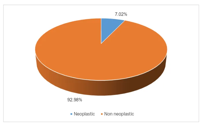
Amidst ovarian lesions, 846 were non-neoplastic and 171 were neoplastic
[image:49.612.156.504.334.551.2](Table 4, Chart 2).
TABLE 4: FREQUENCY OF NONNEOPLASTIC
AND NEOPLASTIC LESIONS OVARY
Count Percentage
Neoplastic 171 7.02%
Non neoplastic 846 92.98%
CHART 2: FREQUENCY OF NON-NEOPLASTIC AND
NEOPLASTIC LESIONS OVARY
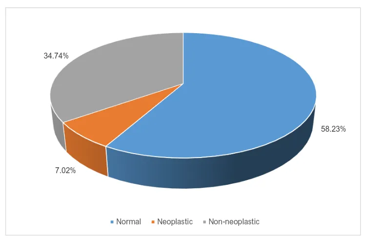
Hence amidst total ovarian specimen of 2435, normal ovaries were 1418
constituting 58.23%, non-neoplastic ovaries were 846 constituting 34.74%
and neoplastic ovaries were 171 constituting 7.02%(Table 5,chart 3).
7.02%
92.98%
33
TABLE 5:FREQUENCY OF NORMAL, NEOPLASTIC AND
NON-NEOPLASTIC OVARIES
Count Percentage
Normal 1418 58.23%
Neoplastic 171 7.02%
Non neoplastic 846 34.74%
CHART 3: FREQUENCY OF NORMAL, NEOPLASTIC AND
NON-NEOPLASTIC OVARIES
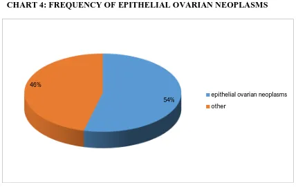
Amidst 171 ovarian neoplasms, 92 were surface epithelial ovarian
neoplasms that constituted 53.801% of total ovarian neoplasms, and hence
topped the list of total ovarian neoplasms and were statistically significant
(Table 6, Chart 4).
58.23% 7.02%
34.74%
34
TABLE 6: FREQUENCY OF EPITHELIAL OVARIAN NEOPLASMS
Count Percentage
Epithelial-Ovarian
Neoplasms 92 53.8%
Others 79 46.2%
CHART 4: FREQUENCY OF EPITHELIAL OVARIAN NEOPLASMS
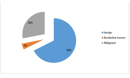
Amidst 92 surface epithelial ovarian neoplasms, 62 were benign, 4 were
borderline tumours and 26 were malignant (Table 7, Chart 5). 54%
46%
epithelial ovarian neoplasms
35
TABLE 7: FREQUENCY OF BENIGN ,BORDERLINE AND
MALIGNANT EPITHELIAL OVARIAN NEOPLASMS.
Count Percentage
Benign 62 68%
Borderline 4 4%
Malignant 26 28%
CHART 5: FREQUENCY OF BENIGN,BORDERLINE AND
MALIGNANT EPITHELIAL OVARIAN NEOPLASMS.
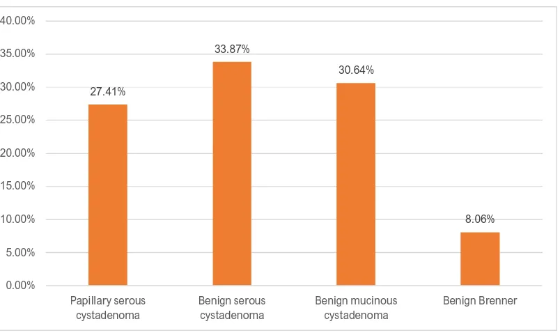
Amidst the 62 benign ovarian surface epithelial tumors, the frequency of
distribution of different histopathological types were-(Table 8, Chart 6) 68%
4% 28%
benign
Borderline tumors
36
TABLE 8: HISTOMORPHOLOGICAL DISTRIBUTION OF BENIGN
SURFACE EPITHELIAL OVARIAN NEOPLASMS:
Count Percentage
Papillary serous cystadenoma 17 27.41%
Benign serous cystadenoma 21 33.87%
Benign mucinous cystadenoma 19 30.64%
Benign Brenner 5 8.06%
CHART 6: HISTOMORPHOLOGICAL DISTRIBUTION OF BENIGN
SURFACE EPITHELIAL OVARIAN NEOPLASMS:
Amidst the 4 borderline tumours, 2 were atypical proliferating serous
tumours (50%), 2 were atypical proliferating mucinous tumours (50%)
(Table 9)
27.41%
33.87%
30.64%
8.06%
0.00% 5.00% 10.00% 15.00% 20.00% 25.00% 30.00% 35.00% 40.00%
Papillary serous cystadenoma
Benign serous cystadenoma
Benign mucinous cystadenoma
37
TABLE 9: HISTOMORPHOLOGICAL
DISTRIBUTION OF BORDERLINE TUMORS.
Count Percentage
Atypical proliferating serous
tumour 2 50%
Atypical proliferating
mucinous tumours 2 50%
Hence Benign serous tumours top the list constituting about 61.3% of
total benign epithelial ovarian neoplasms, closely followed by benign mucinous
cystadenomas that constituted about 30.64% of total benign epithelial ovarian
neoplasms.
Amidst the 26 surface epithelial ovarian malignancies, the different
histopathological types were as in (Table 10, Chart 7).
TABLE 10: HISTOMORPHOLOGICAL DISTRIBUTION OF
SURFACE EPITHELIAL OVARIAN MALIGNANCIES
Count Percentage
Papillary serous
cystadenocarcinoma 9 34.61%
Mucinous adenocarcinoma 4 15.38%
Endometroid adenocarcinoma 8 30.76%
Clear cell carcinoma 4 15.38%
38
CHART 7: HISTOMORPHOLOGICAL DISTRIBUTION OF
SURFACE EPITHELIAL OVARIAN MALIGNANCIES
Benign epithelial ovarian neoplasms had a peak incidence at age group
of 31 – 40 years that constitutes about 40.24% followed by the age group of 41
– 50 years that formed about 24.38%. Mean age is about 33.33 years( Table
11,Chart 8). 0% 5% 10% 15% 20% 25% 30% 35%
35%
15%
31%
15%
4%
HISTOMORPHOLOGICAL DISTRIBUTION OF
SURFACE EPITHELIAL OVARIAN
MALIGNANCIES:
Papillary serous cystadenocarcinoma
Mucinous adenocarcinoma
Endometroid adenocarcinoma
Clear cell carcinoma
39
TABLE 11: AGE WISE DISTRIBUTION OF BENIGN EPITHELIAL
OVARIAN NEOPLASMS
Age group Number of cases Percentage
21-30 years 5 10.97%
31-40 years 29 40.24%
41-50 years 16 24.38%
51-60 years 9 15.86%
>60 years 3 8.53%
Total cases 62 100%
CHART 8: AGE WISE DISTRIBUTION OF BENIGN EPITHELIAL
OVARIAN NEOPLASMS
0% 5% 10% 15% 20% 25% 30% 35% 40% 45%
21-30 years 31-40 years 41-50 years 51-60 years >60 years 11%
40%
24%
16%
9%
21-30 years
31-40 years
41-50 years
51-60 years
40
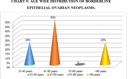
TABLE 12: AGE WISE DISTRIBUTION OF BORDERLINE
EPITHELIAL OVARIAN NEOPLASMS
Age group Number of cases Percentage
31-40 years 1 25%
41-50 years 2 50%
51-60 years - -
>60 years 1 25%
Total cases 4 100%
Maximum incidence of borderline epithelial ovarian neoplasms was
found in the age group of 41-50 years. Mean age affected was found to be
47.21 years (Table 12,Chart 9).
CHART 9: AGE WISE DISTRIBUTION OF BORDERLINE
EPITHELIAL OVARIAN NEOPLASMS.
0% 5% 10% 15% 20% 25% 30% 35% 40% 45% 50%
31-40 years 41-50 years 51-60 years >60 years
25%
50%
0
25%
41
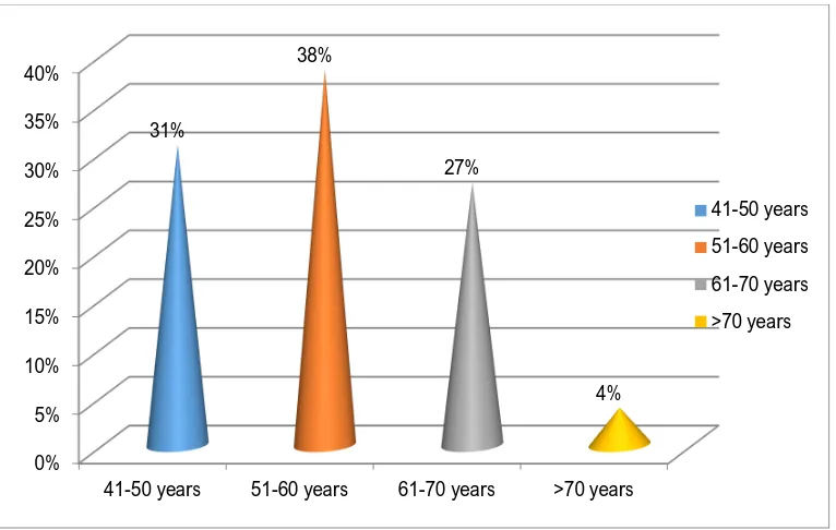
TABLE 13: AGE WISE DISTRIBUTION OF MALIGNANT
EPITHELIAL OVARIAN NEOPLASMS
Age group Number of cases Percentage
41-50 years 8 30.76%
51-60 years 10 38.46%
61-70 years 7 26.92%
>70 years 1 3.8%
Total cases 26 100%
Maximum incidence of malignant epithelial ovarian tumours was found
in the age group of 51 to 60 years followed by 41 to 50 years. Mean age
affected was found to be 54.5 years. (Chart 10).
CHART 10: AGE WISE DISTRIBUTION OF MALIGNANT
EPITHELIAL OVARIAN NEOPLASMS
0% 5% 10% 15% 20% 25% 30% 35% 40%
41-50 years 51-60 years 61-70 years >70 years 31%
38%
27%
4%
41-50 years
51-60 years
61-70 years
42
TABLE 14: GRADE WISE DISTRIBUTION OF MALIGNANT
EPITHELIAL OVARIAN NEOPLASMS
Grade Number of cases Percentage
I 5 19.23%
II 9 34.61%
III 12 46.16%
Total cases 26 100%
We can see that maximum tumours were in grade III (Chart 11).
CHART 11: GRADE WISE DISTRIBUTION OF MALIGNANT
EPITHELIAL OVARIAN NEOPLASMS
19%
35% 46%
I
II
43
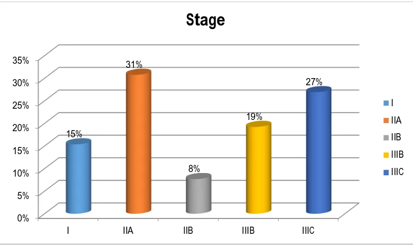
TABLE 15: DISTRIBUTION OF MALIGNANT EPITHELIAL OVARIAN NEOPLASMS ACCORDING TO THE FIGO (INTERNATIONAL FEDERATION OF GYNAECOLOGY AND OBSTETRICS) STAGE.
Stage Number of cases Percentage
I 4 15.38%
IIA 8 30.76%
IIB 2 7.69%
IIIB 5 19.23%
IIIC 7 26.92%
Total cases 26 100%
CHART 12: DISTRIBUTION OF MALIGNANT EPITHELIAL OVARIAN NEOPLASMS ACCORDING TO THE FIGO (INTERNATIONAL FEDERATION OF GYNAECOLOGY AND OBSTETRICS) STAGE.
0% 5% 10% 15% 20% 25% 30% 35%
I IIA IIB IIIB IIIC
44
CHART 13: STAGE DISTRIBUTION AMONG MALIGNANT
EPITHELIAL OVARIAN NEOPLASMS
Hence, maximum presentation was in stage III (Chart 13).
Results of ImmunohistoChemical Analysis
All the 26 malignant epithelial ovarian neoplasms and 4 borderline
epithelial tumors were subjected to a panel of 2 immunohistochemical markers-
EGFR(Epidermal Growth Factor Receptor) and VEGF(Vascular Endothelial
Growth Factor)
15%
39% 46%
Stage
I
II
45
TABLE 16: PERCENTAGE OF POSITIVE EXPRESSION OF EGFR,
VEGF AMONG BORDERLINE OVARIAN NEOPLASMS
IHC marker Positive Cases Negative Cases
EGFR 2 (50%) 2 (50%)
VEGF 3 (75%) 1 (25%)
Out of the four borderline ovarian neoplasms, 50% showed positivity for
EGFR and another 50% showed negativity for EGFR.
Out of four borderline epithelial ovarian neoplasms, 75% showed
positivity for VEGF.
TABLE 17: DISTRIBUTION OF POSITIVITY OF EGFR AND VEGF
AMONG TYPES OF BORDERLINE EPITHELIAL OVARIAN
NEOPLASMS.
IHC marker APST Positive
(%)
APMT Positive
(%) Total
EGFR 2 (100%) Nil positive 4
46
TABLE 18: DISTRIBUTION OF POSITIVITY AMONG MALIGNANT
EPITHELIAL OVARIAN NEOPLASMS
IHC marker Positive cases (%)
Negative cases
(%) Total
EGFR 21 (80.76%) 5 (19.23%) 26 (100%)
VEGF 22 (84.62%) 4 (15.38%) 26 (100%)
Out of the total 26 malignant epithelial ovarian neoplasms, 21 (80.76%)
of them showed positivity for EGFR and 19.23% of them were negative for
EGFR (Table 18, Chart 14).
CHART 14: DISTRIBUTION OF POSITIVITY AMONG MALIGNANT
EPITHELIAL OVARIAN NEOPLASMS
Out of the total 26 malignant epithelial ovarian neoplasms, 22 (84.62%)
of them showed positivity for VEGF while only 4 (15.38%) of them were
negative for VEGF.
81% 19%
Postive
47
TABLE 19: DISTRIBUTION OF POSITIVITY OF EGFR AND VEGF
AMONG TYPES OF MALIGNANT EPITHELIAL OVARIAN
NEOPLASMS.
Histopathological type of malignant
ovarian neoplasm EGFR Positive EGFR Negative VEGF Positive VEGF
Negative Total
Papillary serous cystadeno carcinoma 8 (88.89%) 1 (11.11%) 8 (88.89%) 1 (11.11%) 9 (100%) Endometroid
adenocarcinoma 7 (87.5%)
1
(12.5%) 7 (87.5%)
1 (12.5%)
8 (100%) Mucinous
adenocarcinoma 2 (50%) 2 (50%) 3 (75%) 1 (25%)
4 (100%) Clear cell
carcinoma 4 (100%) Nil 4 (100%) Nil
4 (100%) Adenosquamous
carcinoma Nil positive Nil positive
1 (100%)
Thus, we can infer that –
88.89% of papillary serous cystadenocarcinoma ovary showed positivity for
both EGFR and VEGF.
87.5% of endometroid adenocarcinoma ovary showed positivity for both
EGFR and VEGF.
Only 50% of mucinous adenocarcinoma showed positivity for EGFR while
75% of them showed positivity for VEGF
All the clear cell carcinomas – (100% of them) showed positivity for both
EGFR and VEGF
The adenosquamous carcinoma that was evaluated did not show positivity
48
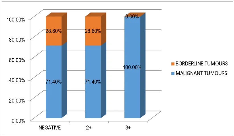
TABLE 20: TABLE FOR COMPARISON OF INTENSITY OF EXPRESSION OF EGFR AND VEGF AMONG BORDERLINE TUMORS AND
MALIGNANT EPITHELIAL OVARIAN TUMORS.
EGFR
Total
NEGATIVE 2+ 3+
MALIGNANT TUMOURS
Count 5 5 16 26
% within
EGFR 71.4% 71.4% 100.0% 86.7%
BORDERLINE TUMOURS
Count 2 2 0 4
% within
EGFR 28.6% 28.6% 0.0% 13.3%
Total
Count 7 7 16 30
% within
EGFR 100.0% 100.0% 100.0% 100.0%
P=0.042
CHART 15: TABLE FOR COMPARISON OF INTENSITY OF EXPRESSION OF EGFR AND VEGF AMONG BORDERLINE TUMORS AND
MALIGNANT EPITHELIAL OVARIAN TUMORS.
0.00% 20.00% 40.00% 60.00% 80.00% 100.00%
NEGATIVE 2+ 3+
49
CHART 16: PERCENTAGE OF EGFR POSITIVITY AMONG
BORDERLINE AND MALIGNANT EPITHELIAL OVARIAN NEOPLASMS.
From this, we infer that 86.7% of malignant epithelial ovarian tumours
showed varying degrees of positivity for EGFR while only 13.3% of borderline
epithelial tumours showed positivity. The P value was calculated as 0.042 and
hence this correlation was found statistically significant (Table 20,Chart 16.). 87%
13%
malignant epithelial ovarian tumors
50
TABLE 21: PERCENTAGE OF EXPRESSION OF EGFR IN
MALIGNANT EPITHELIAL OVARIAN NEOPLASMS.
HPE EGFR Total
NEGATIVE 2+ 3+
Papillary Serous Cystadenocarcinoma
Count 1 3 5 9
% 11.11% 33.33% 55.56% 100.00%
Endometrioid adenocarcinoma of
ovary
Count 1 2 5 8
% 12.50% 25.00% 62.50% 100.00%
Mucinous adenocarcinoma ovary
Count 2 0 2 4
% 50.00% 0.00% 50.00% 100.00%
Clear cell carcinoma ovary
Count 0 0 4 4
% 0.00% 0.00% 100.00% 100.00%
Adenosquamous carcinoma ovary
Count 1 0 0 1
% 100.00% 0.00% 0.00% 0.00%
Borderline tumors Count 2 2 0 4
% 50.00% 50.00% 0.00% 13.30%
Total Count 7 7 16 30
% 23.33% 23.33% 53.33% 100.00%
From this table we infer that nearly 100% of clear cell carcinomas
studied, 62.5% of endometroid carcinomas studied, 55.56% of papillary serous
carcinomas studied and 50% of mucinous carcinomas studied showed EGFR
51
[image:68.612.127.533.121.357.2]CHART 17: PERCENTAGE OF EXPRESSION OF EGFR IN BORDERLINE TUMORS AND MALIGNANT EPITHELIAL OVARIAN NEOPLASMS.
TABLE 22 : CORRELATION OF TUMOR GRADE
WITH EGFR EXPRESSION
EGFR
Total
NEGATIVE 2+ 3+
Tumor grade
1.00
Count 4 2 0 6
% within
EGFR 57.1% 28.6% 0.0% 20.0%
2.00
Count 2 5 3 10
% within
EGFR 28.6% 71.4% 18.8% 33.3%
3.00
Count 1 0 13 14
% within
EGFR 14.3% 0.0% 81.2% 46.7%
Total
Count 7 7 16 30
% within
EGFR 100.0% 100.0% 100.0% 100.0%
P<0.001 0% 20% 40% 60% 80% 100% P ap illa ry S er o u s C ys ta d e n o ca rc in o m a En d o m e tr io id ad e n o ca rc in o m a o f o va ry M u cin o u s ad e n o ca rc in o m a o va ry Cle ar c e ll ca rc in o m a o va ry A d e n o sq u am o u s ca rc in o m a o va ry B o rd e rlin e t u m o rs 11% 13% 50% 0% 100% 50% 33% 25% 0% 0% 0% 50%
56% 63% 50%
100%
0% 0%
3+
2+
52
In this study, 81.2% of grade III tumours showed 3+ EGFR positivity.
Higher the grade, higher was the expression of EGFR and this correlation was
statistically highly significant since the P value was less than 0.001(Table
22,Chart 18).
CHART 18: CORRELATION OF TUMOR GRADE WITH EGFR
EXPRESSION
0% 10% 20% 30% 40% 50% 60% 70% 80% 90%
Grade 1 Grade 2 Grade 3
57%
29%
14% 29%
71%
0% 0%
19%
81%
NEGATIVE
2+
53
TABLE 23: CORRELATION OF TUMOR STAGE WITH EGFR
EXPRESSION
EGFR
Total
NEGATIVE 2+ 3+
Stage
Count 2 2 0 4
% within
EGFR 28.6% 28.6% 0.0% 13.3%
II A
Count 3 3 4 10
% within
EGFR 42.9% 42.9% 25.0% 33.3%
II B
Count 1 1 0 2
% within
EGFR 14.3% 14.3% 0.0% 6.7%
III B
Count 0 1 5 6
% within
EGFR 0.0% 14.3% 31.2% 20.0%
III C
Count 1 0 7 8
% within
EGFR 14.3% 0.0% 43.8% 26.7%
Total
Count 7 7 16 30
% within
EGFR 100.0% 100.0% 100.0% 100.0%
P= 0.039
In this study 75% of stage III tumors showed 3+ positivity. Higher the
stage, higher was the expression of EGFR and this correlation was statistically
54
CHART 19 CORRELATION OF TUMOR STAGE
WITH EGFR EXPRESSION
0% 5% 10% 15% 20% 25% 30% 35% 40% 45%
IIA IIB IIIB
29%
43%
14%
0% 29%
43%
14% 14%
0%
25%
0%
31%
NEGATIVE
2+
55
TABLE 24: TABLE FOR COMPARISON OF INTENSITY OF VEGF
EXPRESSION AMONG BORDERLINE AND MALIGNANT
EPITHELIAL OVARIAN TUMORS
VEGF
Total NEGA
TIVE 1+ 2+ 3+
MALIGNANT TUMOURS
Count 4 0 4 18 26
% within VEGF
80.0% 0.0% 66.7% 100.0% 86.7%
BORDERLINE TUMOURS
Count 1 1 2 0 4
% within VEGF
20.0% 100.
0% 33.3% 0.0% 13.3%
Total
Count 5 1 6 18 30
% within VEGF
100.0% 100.
0% 100.0% 100.0% 100.0%
P=0.009
From this we infer that 86.7% of malignant epithelial ovarian tumors
showed varying degrees of positivity for VEGF while only 13.3% of borderline
tumors showed VEGF positivity. The P value 0.009 shows that this correlation