JOURNAL OFVIROLOGY, May 2004, p. 5068–5078 Vol. 78, No. 10
0022-538X/04/$08.00⫹0 DOI: 10.1128/JVI.78.10.5068–5078.2004
Copyright © 2004, American Society for Microbiology. All Rights Reserved.
Conserved Cysteine-Rich Domain of Paramyxovirus Simian Virus 5
V Protein Plays an Important Role in Blocking Apoptosis
Minghao Sun,
1Terri A. Rothermel,
1Laurie Shuman,
1,2Jason A. Aligo,
1,3Shibo Xu, Yuan Lin,
1Robert A. Lamb,
4and Biao He
1,2,3*
Department of Veterinary Science,1Graduate Program in Immunobiology, The Huck Institute for Life Sciences,2and
Intercollege Graduate Program in Genetics,3Pennsylvania State University, University Park, Pennsylvania 16802,
and Howard Hughes Medical Institute, Department of Biochemistry, Molecular Biology and Cell Biology, Northwestern University, Evanston, Illinois 602084
Received 9 September 2003/Accepted 26 January 2004
The paramyxovirus family includes many well-known human and animal pathogens as well as emerging viruses such as Hendra virus and Nipah virus. The V protein of simian virus 5 (SV5), a prototype of the paramyxoviruses, contains a cysteine-rich C-terminal domain which is conserved among all paramyxovirus V proteins. The V protein can block both interferon (IFN) signaling by causing degradation of STAT1 and IFN production by blocking IRF-3 nuclear import. Previously, it was reported that recombinant SV5 lacking the C terminus of the V protein (rSV5V⌬C) induces a severe cytopathic effect (CPE) in tissue culture whereas wild-type (wt) SV5 infection does not induce CPE. In this study, the nature of the CPE and the mechanism of the induction of CPE were investigated. Through the use of DNA fragmentation, terminal deoxynucleotidyl-transferase-mediated dUTP-biotin nick end labeling, and propidium iodide staining assays, it was shown that rSV5V⌬C induced apoptosis. Expression of wt V protein prevented apoptosis induced by rSV5V⌬C, suggesting that the V protein has an antiapoptotic function. Interestingly, rSV5V⌬C induced apoptosis in U3A cells (a STAT1-deficient cell line) and in the presence of neutralizing antibody against IFN, suggesting that the induction of apoptosis by rSV5V⌬C was independent of IFN and IFN-signaling pathways. Apoptosis induced by rSV5V⌬C was blocked by a general caspase inhibitor, Z-VAD-FMK, but not by specific inhibitors against caspases 1, 2, 3, 4, 5, 6, 7, 8, 9, 10, 11, and 13, suggesting that rSV5V⌬C-induced apoptosis can occur in a caspase 12-dependent manner. Endoplasmic reticulum stress can lead to activation of caspase 12; compared to the results seen with mock and wt SV5 infection, rSV5V⌬C infection induced ER stress, as demonstrated by increased expression levels of known ER stress indicators GRP 78, GRP 94, and GADD153. These data suggest that rSV5V⌬C can trigger cell death by inducing ER stress.
Apoptosis, or programmed cell death, is the physiological process by which unwanted cells undergo morphological changes, protease activation, chromosomal DNA fragmenta-tion, and (eventually) cell death. This process is important for normal development, tissue homeostasis, and immune modu-lation as well as for host defense against viral infection (12). Apoptosis can be initiated and executed through many differ-ent pathways, which can be categorized into two main groups: extrinsic and intrinsic pathways (2). Extrinsic pathways sense death signals from outside the cells and consist of caspases, death receptors (DR), and adapter proteins. The caspases (cys-teine aspartate-specific proteases) are critical players in regu-lation of different apoptotic pathways (8, 54). There are 14 known caspases that can be roughly divided into initiator and effector caspases. Initiator caspases are involved in upstream regulatory events resulting in activation of effector caspases that are directly responsible for proteolytic cleavages leading to cell death. Known initiator caspases include caspase 8 and 9; known effector caspases include caspase 3, 6, and 7. Some caspases (such as caspase 2) can be both initiator and effector caspases (54). Effector caspases, the executioners of apoptosis,
can be activated by death stimuli through activation of DR, normally a member of the tumor necrosis factor (TNF) recep-tor superfamily. Upon activation, DR associate with adapter proteins such as the TNF receptor-associated death domain through their death domains. This complex can activate initi-ator caspases (which in turn can activate effector caspases to trigger cell death) (49). Intrinsic pathways sense death signals such as stress from inside the cells and act mainly through mitochondria. Most intrinsic apoptotic pathways involve the Bcl-2 protein family, mitochondrion-released proteins, and cas-pases. In intrinsic pathways, death stimuli are sensed by the Bcl-2 protein family and cause damage to mitochondria result-ing in release of cytochrome c to the cytosol. The released cytochrome c activates Apaf1 (apoptotic protease-activating factor 1), which activates caspase 9 to trigger cell death. How-ever, knockout of the gene for cytochromec, Apaf1, or caspase 9 gene does not prevent the cell from undergoing stress-in-duced apoptosis in some cells, suggesting there might be ad-ditional intrinsic apoptotic pathways (reviewed in reference 7). It is known that damage to mitochondria can also cause release of apoptosis-inducing factor, which triggers apoptosis in a cas-pase-independent manner (5).
SV5 is a member of the Rubulavirus genus of the family Paramyxoviridae. The Paramyxoviridae family includes many well-known human and animal pathogens, such as mumps vi-rus, Newcastle disease virus (NDV), measles vivi-rus, and respi-* Corresponding author. Mailing address: Department of
Veteri-nary Science, Pennsylvania State University, 115 Henning Bldg., Uni-versity Park, PA 16802. Phone: (814) 863-8533. Fax: (814) 863-6140. E-mail: bxh40@psu.edu.
5068
on November 8, 2019 by guest
http://jvi.asm.org/
ratory syncytial (RS) virus, as well as important emerging vi-ruses such as Hendra virus and Nipah virus (26). The negative-stranded RNA genome of SV5 is 15,246 nucleotides long and encodes eight known viral proteins (26). The nucleocapsid protein (NP), phosphoprotein (P), and large RNA polymerase (L) protein are important for transcription and replication of the viral RNA genome. The fusion (F) protein, a glycoprotein, mediates virus entry into cells by virus-cell fusion and causes syncytial formation. The hemagglutinin-neuraminidase protein (HN), a viral glycoprotein, mediates virus-cell attachment and also cleaves sialic acid from complex carbohydrate chain of glycoprotein (necessary for virus release). The matrix (M) pro-tein plays an important role in virus assembly (45, 46). The small hydrophobic (SH) protein is a 44-residue hydrophobic integral membrane protein and is oriented in membranes with its N terminus in the cytoplasm (18). Recombinant SV5 (rSV5) lacking the SH gene (rSV5⌬SH) induces apoptosis in L929 cells through a TNF alpha (TNF-␣)-mediated extrinsic apo-ptotic pathway (14, 15, 30).
The V/P gene of SV5 is transcribed into both the V mRNA and the P mRNA through a process (commonly called “RNA editing”) of pseudo-templated addition of nucleotides (53). The V mRNA is transcribed when the viral RNA polymerase faithfully transcribes the V/P gene. On some occasion during transcription, however, the viral RNA polymerase complex recognizes a specific RNA sequence in the V/P gene and in-serts two non-template G residues at the editing site to gen-erate the P mRNA. As a result, the V/P gene is transcribed into two mRNAs that accumulate at about the same abundance level and the two mRNAs are translated into two proteins, V and P, which share identical N termini but have different C termini. The RNA editing at a specific site occurs for almost all members of the subfamilyParamyxoviridae(26). The V mRNA is faithfully transcribed from the genome RNA only for rubu-laviruses, however, whereas for the respiroviruses and the mor-billiviruses the P mRNA is faithfully transcribed from the ge-nome RNA and the V mRNA is the result of the presence of the additional pseudo-templated G nucleotide(s) (21). The se-quences of the C-terminal domain of the V proteins are highly conserved among the paramyxoviruses (56).
The V protein of the paramyxovirus simian virus 5 (SV5) is a multifunctional protein containing an N-terminal 164-resi-due domain that is shared with the P protein and a distinct C-terminal domain that is cysteine rich. The V protein C-ter-minal domain contains seven cysteine residues (resembling a zinc finger domain) and binds atomic zinc (31, 40, 48, 53). The V protein of SV5 interacts with soluble nucleocapsid protein (41), and the N-terminal domain of V binds RNA through a basic region (29). The SV5 V protein interacts with a cellular protein, the 127-kDa subunit of the damage-specific DNA-binding protein (DDB1) that is known to be involved in dam-aged DNA repair. The interaction of the V protein with DDB1 requires the presence of the C-terminal domain of V protein (28). Expression of the SV5 V protein slows down the cell cycle; this effect on the cell cycle is mediated via the V protein C-terminal domain (28). Coexpression of DDB1 can partially restore the changes in cell cycle caused by V (28). The V protein of SV5 also causes degradation of STAT1 protein, an essential regulator of interferon (IFN) signaling, through a proteasome-mediated pathway in human cells but not in
mouse cells (10). STAT2 also plays a role in V’s ability to cause degradation of STAT1 (37). It seems likely that the degrada-tion of STAT1 in SV5-infected cells and the alteradegrada-tions to the cell cycle are interrelated, as the V, DDB1, Cul4A, STAT1, and STAT2 proteins form a complex which is essential for V-mediated STAT1 degradation (1, 58). The analysis of prop-erties of an rSV5 that lacks the V protein C-terminal-specific domain (rSV5V⌬C) shows that the C terminus of the V pro-tein plays an essential role in causing degradation of STAT1 and preventing IFN- production. Whereas rSV5 grows in many cell types with minimal cytopathic effect (CPE), rSV5V⌬C causes extensive CPE in the same cell types. In this study, the nature of the CPE and the mechanism of rSV5V⌬C-induced CPE were investigated.
MATERIALS AND METHODS
Viruses and cells. Generation of rSV5V⌬C was described previously.
rSV5V⌬C were grown in Vero cells and harvested 5 to 7 days postinfection (dpi)
as described previously (16, 17, 39). Wild-type (wt) SV5 was grown in MDBK cells. Virus titers were determined by plaque assays using BHK 21F and Vero cells (39). To infect cells, monolayers were washed with phosphate-buffered saline (PBS) and then inoculated with viruses in Dulbecco’s modified Eagle’s medium (DMEM)–1% bovine serum albumin (BSA) at a multiplicity of infec-tion (MOI) of 5 for 1 to 2 h at 37°C. The monolayers were washed and incubated
with DMEM containing 2% fetal calf serum (FCS) at 37°C with 5% CO2. HeLa
cells, U3A cells, and Vero cells were maintained in DMEM–10% FCS. BHK 21F cells were maintained in DMEM–10% tryptose phosphate broth–10% FCS. Virus-infected cells were grown in DMEM–2% FCS.
Transfections were carried using Lipofectamine-PLUS (Invitrogen, Carlsbad, Calif.) following the manufacturer’s instructions. HeLa cells in 6-cm-diameter plates at about 70% confluency were washed once with Opti-MEM medium and
incubated in 2 ml of Opti-MEM. A total of 2g of plasmid DNA (vector or
pBH361 [which encodes V]) was mixed with 0.25 ml of Opti-MEM and 8l of
PLUS for 15 min at room temperature. Opti-MEM (0.25 ml) with 12l of
Lipofectamine was added to the DNA-PLUS mixture and incubated for 15 min at room temperature. The mixture of DNA, PLUS, and Lipofectamine was added to the cells and incubated for 16 h. The transfected cells were infected with
rSV5V⌬C. The transfected-infected cells were analyzed for V expression and
apoptosis by flow cytometry (described below).
DNA fragmentation assay, propidium iodide staining, terminal deoxynucle-otidyltransferase-mediated dUTP-biotin nick end labeling (TUNEL) assay, and trypan blue staining.Fragmented DNAs were purified as described in reference 64. Confluent HeLa cells in 6-cm-diameter plates were mock infected or infected
with SV5 or rSV5V⌬C at an MOI of 5. At 24 and 48 h postinfection (hpi), similar
numbers of HeLa cells in 60-mm-diameter dishes were washed twice with PBS
(⫺) and then incubated in 0.5 ml of TTE buffer (0.2% Triton X-100, 10 mM Tris,
15 mM EDTA, pH 8.0) at room temperature for 15 min. Cell lysates were
harvested into microtubes and subjected to centrifugation at 20,000⫻gfor
20 min. Supernatants were digested with 100g of RNase A/ml at 37°C for 1 h.
Samples were purified with phenol-chloroform extraction, precipitated, and then
washed with 70% ethanol. Pellets were air dried and redissolved in 10l of
Tris-EDTA buffer each. Electrophoresis was performed on 2% agarose gels with DNA size markers.
For propidium iodide (PI) staining, confluent HeLa cells in 6-cm-diameter
plates were mock infected or infected with SV5 or rSV5V⌬C at an MOI of 5.
Cells in monolayers were trypsinized and combined with the floating cells in the medium at different time points. The harvested cells were then centrifuged at 250
⫻gfor 8 min at 4°C and washed with PBS between each subsequent pair of steps.
The cells were fixed with 0.25% formaldehyde for 2 h at 4°C. The fixed cells were resuspended in 0.5 ml of 50% DMEM–50% FCS and permeabilized by adding 1.5 ml of 70% ethanol at 4°C for at least 2 h and for up to 3 days. To monitor expression of viral proteins, the permeabilized cells were incubated with 0.5 ml of anti-V monoclonal antibody 11C6 (40) at 1:500 in PBS–1% BSA at 4°C for 1 h and then with 0.5 ml of fluorescein isothiocyanate-labeled anti-mouse secondary antibody (Organon-Teknika Corp., Charlotte, N.C.) at 1:1,000 in PBS–1% BSA
for 1 h at 4°C. For PI staining, the cells were incubated with 500l of 50g of
propidium iodide (Sigma-Aldrich)/ml for 1 h at 4°C. The cells were then analyzed using a flow cytometer (EPICS XL; Beckman-Coulter). Single cells were selected
on November 8, 2019 by guest
http://jvi.asm.org/
on FL2-W (cell width) versus FL2-A (DNA content) plots. Infected cells were selected on FL2-A (DNA content) versus FL1-H (V expression) plots.
For TUNEL assays, the cells were fixed and permeabilized as described above.
The cells were then incubated with 25l of TUNEL reaction mixture (in situ
fluorescein cell death detection kit; Boehringer-Mannheim) for 2 to 3 h in the dark at 37°C. The cells were analyzed by flow cytometry.
To examine cell viability, the adherent cells were washed with PBS⫺and
trypsinized and were combined with the cells in the medium to obtain the entire
cell population. The cells were centrifuged 960⫻gwith a tabletop centrifuge and
resuspended in 0.08% trypan blue solution in DMEM. The cells were placed on a hemacytometer and examined under a microscope.
UV inactivation of virus.Confluent HeLa cells in 60-mm-diameter plates were
mock infected or infected with SV5 or rSV5V⌬C at an MOI of 5. The infected
cells were incubated in 5 ml of 2% FCS–DMEM for 2 days. Medium from infected cells was placed inside a Fisher Hamilton class II biological safety cabinet and UV treated for 30 min. The media were then filtered through a
0.22-m-pore-size filter to remove cell debris. The effectiveness of the UV
treatment with respect to inactivation of SV5 was confirmed by plaque assays.
Antibody treatment of infected cells.Confluent HeLa cells in 6-well plates
were mock infected or infected with SV5 or rSV5V⌬C at an MOI of 5 as
described before and incubated in 1 ml of DMEM–2% FCS with neutralizing
antibody against IFN-(CalBiochem, San Diego, Calif.) at 105kU/ml. At 2 dpi,
the cells were collected, stained with trypan blue, and counted.
Confluent L929 cells in 6-well plates were mock infected or infected with SV5,
rSV5⌬SH, or rSV5V⌬C at an MOI of 5 as described before and incubated in 1
ml of DMEM–2% FCS with neutralizing antibody against TNF-␣(BD
Pharm-ingen, San Diego, Calif.) at 20g/ml. At 2 dpi, the cells were photographed using
a light microscope equipped with a digital camera.
Caspase assays.For caspase 3 and 7 assays, triplicates of infected cells (⬃2⫻
106to 10⫻106cells/ml) were lysed with cell lysis buffer containing 10 mM
Tris-HCl, 10 mM NaH2PO4/NaHPO4 (pH 7.5), 130 mM NaCl, 1% Triton X-100,
and 10 mM NaPPi (sodium pyrophosphate). Protein concentrations of lysates were determined using a BCA protein assay kit (Pierce, Rockford, Ill.). For each
reaction in a 96-well microtiter plate, 30g of cell lysate was added to 200l of
reaction buffer (20 mM HEPES [pH 7.5], 20% glycerol, 4 mM dithiothreitol) and
20M caspase 3 substrate Ac-DEVD-AMC (BD Biosciences Pharmingen). For
caspase 2 assays, assay buffer containing 100 mM HEPES (pH 7.5), 10 mM
dithiothreitol, and 100M caspase 2 substrate Ac-LDESD-AMC (CalBiochem)
were used. Samples in the plates were mixed for 30 s and incubated for 1 h at 37°C. The plates were then read on a microtiter plate reader with an excitation wavelength of 380 nm and an emission wavelength of 430 to 460 nm. Similarly,
subsequent caspase assays were carried out using 20M Ac-YVAD-AMC
sub-strate (BD Bioscience) for caspase 1, 50M Ac-LEVD-AMC substrate (A.G.
Scientific Inc., San Diego, Calif.) for caspase 4, 50M Ac-LEHD-AMC
sub-strate (A.G. Scientific Inc.) for caspases 5, 9, 10, and 11, 50M Ac-VEID-AMC
substrate (A. G. Scientific Inc.) for caspase 6 assay, 20M Ac-LETD-AMC
substrate (BD Bioscience) for caspase 8, and 100M Ac-LEED-AFC substrate
(Enzymesys, Aurora, Ohio) for caspase 13.
Treatment of cells with caspase inhibitors.HeLa cells in 24-well plates were
mock infected or infected with SV5 or rSV5V⌬C and incubated with
DMEM–2% FCS containing dimethyl sulfoxide (DMSO) or 40M inhibitors
dissolved in DMSO. The cells were photographed at 2 dpi and counted after trypan blue staining. General caspase inhibitor Z-VAD-FMK, caspase 1 and 4 inhibitor Z YVAD-FMK, caspase 2 inhibitor Z-VAVAD-FMK, caspase 5, 9, 10, and 11 inhibitor Z-LEHD-FMK, caspase 8 inhibitor Z-LETD-FMK, and caspase 13 inhibitor Z-LEED-FMK were from Enzymesys. Caspase 3, 6, and 7 inhibitor Z-DEVD-FMK was from CalBiochem.
Immunoblotting.HeLa cells in 6-cm-diameter plates were mock infected or
infected with rSV5V⌬C or rSV5. At 1 or 2 dpi, cells were lysed in 0.5 ml of
protein lysis buffer (2% sodium dodecyl sulfate, 62.5 mM Tris-HCl [pH 6.8], 2%
dithiothreitol) and sonicated briefly to shear DNA. Up to 100l of the lysate was
subjected to sodium dodecyl sulfate-polyacrylamide gel electrophoresis using a 15% gel. Polypeptides were transferred (using a wet-gel transfer apparatus) to a polyvinylidene difluoride membrane. The membrane was first blocked with 5% dry fat-free milk and then incubated with primary antibodies against GRP 78 at 1: 200 dilution (Santa Cruz Biotechnology, Inc., Santa Cruz, Calif.), GRP 94 at
1g/ml (CalBiochem), GADD153 at 1:200 dilution (Santa Cruz Biotechnology),
or actin (Santa Cruz Biotechnology). Two caspase 12 antibodies were used. One (which recognizes residues 95 to 318 of mouse caspase 12) (Sigma) was used at 1:500; the other (which recognizes residues 100 to 116 of mouse caspase 12) (Oncogene) was used at 1:500. A mixture of anti-mouse and anti-rabbit second-ary antibodies conjugated to horseradish peroxidase was used to detect primsecond-ary
antibodies. The proteins on the membrane were detected using an ECL⫹kit
(Amersham Pharmacia, Piscataway, N.J.), and chemiluminescence was detected using a Storm System PhosphorImager (Molecular Dynamics Inc, Sunnyvale, Calif.).
RESULTS
Induction of apoptosis by rSV5V⌬C infection. Previously, we observed that rSV5V⌬C infection of several cell types (e.g., HeLa cells, CV1 cells, Vero cells, and L929 cells) caused a severe CPE by 30 hpi. Whereas rSV5 infection of HeLa cells produced small syncytial foci from 24 hpi onwards, rSV5V⌬ C-infected cells at times⬎30 hpi were detaching from the mono-layer. Although CPE induced by rSV5V⌬C in Vero and HeLa cells was evident by 30 hpi, protein synthesis did not appear to be affected at this time and neither was virus particle produc-tion (16) (excluding expression of viral proteins or producproduc-tion of virions as possible causes for increased CPE in rSV5V⌬ C-infected cells).
To examine whether the CPE was due to induction of apo-ptosis, the presence of fragmented chromosomal DNA in rSV5V⌬C-infected cells was examined. HeLa cell were mock infected or infected with SV5 or rSV5V⌬C viruses. DNAs were purified and resolved in agarose gel (Fig. 1A). Increasing amounts of fragmented DNA was detected in rSV5V⌬ C-in-fected cells in comparison with that found in mock- or SV5-infected cells, suggesting that rSV5V⌬C induced apoptosis in cells (Fig. 1A). To confirm that the CPE was due to induction of apoptosis in the rSV5V⌬C-infected cells and to quantify the number of apoptotic cells, the TUNEL assay (which directly measures the presence of nicked chromosomal DNA, a hall-mark of apoptotic cells) was employed. The percentages of TUNEL-positive cells, i.e., apoptotic cells, were quantified us-ing a flow cytometer as described previously (15). As shown in Fig. 1B, at 2 dpi about 45% of cells infected by rSV5V⌬C were TUNEL positive, indicating that the cells were apoptotic.
Deletion of the C-terminal domain of the V protein caused increased CPE and apoptosis in virus-infected HeLa cells, in-dicating that full-length V is required to prevent apoptosis in virus-infected cells. It is most likely that the phenotype of increased apoptosis was a result of loss of function of the C-terminal domain. However, it is possible that the N-terminal domain had acquired a novel function even though the half-life of the N-terminal domain is relative short (less than 45 min). To test whether intact V protein can inhibit apoptosis induced by rSV5V⌬C, SV5 V protein was expressed in rSV5V⌬ C-infected cells by coinfecting cells with wt SV5 or by transfect-ing plasmid DNA expresstransfect-ing the V protein. Coinfection of rSV5V⌬C with wt SV5 did not result in CPE (as shown in Fig. 1C), suggesting that V from a wt SV5 infection was able to block CPE induced by rSV5V⌬C infection and that the trun-cated form of the V protein was unlikely to be the reason for the increased apoptosis observed in rSV5V⌬C-infected cells. The ability of rSV5V⌬C to induce apoptosis in HeLa cells was further confirmed by PI staining, which detects cellular DNA content. DNA fragments due to apoptosis are lost during PI staining (in which lipid membranes of cells are extracted). As a result, the DNA content profile of apoptotic cells is the same as that of sub G0-G1cells. Increased populations of sub-G0and
-G1cells, i.e., apoptotic cells, were detected only in rSV5V⌬
C-infected cells at 2 dpi (Fig. 1D).
5070 SUN ET AL. J. VIROL.
on November 8, 2019 by guest
http://jvi.asm.org/
Furthermore, HeLa cells were transfected with a plasmid encoding V protein and then infected with rSV5V⌬C. V-ex-pressing cells were gated using a flow cytometer. The DNA content of the V-expressing cells was compared (using PI stain-ing) with that of the rSV5V⌬C-infected cells that did not express V intrans. In V-expressing cells, rSV5V⌬C infection did not induce apoptosis (Fig. 1D). Thus, V expressed alone can inhibit the apoptosis induced by rSV5V⌬C.
Activation of intrinsic apoptotic pathway by rSV5V⌬C in-fection.The major apoptotic pathways can be categorized into
[image:4.603.104.479.69.494.2]intrinsic and extrinsic pathways. IFN is thought to be an es-sential mediator of apoptosis in virus-infected cells, and in-creased expression of IFN- was detected in the media of rSV5V⌬C-infected cells (16). Thus, apoptosis induced by rSV5V⌬C infection could be caused by IFN via an extrinsic apoptotic pathway. To test this notion, U3A cells whose IFN signaling pathways are impaired due to STAT1 protein defi-ciency (47) were mock infected or infected with SV5 or rSV5V⌬C and apoptotic cell populations were examined using TUNEL assays. rSV5V⌬C induced apoptosis in U3A cells FIG. 1. Induction of apoptosis by rSV5V⌬C. HeLa cells were infected with SV5 or rSV5V⌬C or mock infected and were subjected to apoptosis assays. (A) DNA fragmentation. The infected cells were collected at 1 and 2 dpi, and DNAs were purified and resolved in agarose gel as described in Materials and Methods. The DNA marker wasDNA digested with BstEII enzyme. (B) TUNEL assay. The infected cells were collected at 2 dpi and subjected to TUNEL assays as described in Materials and Methods. (C) Inhibition of rSV5V⌬C induced CPE by SV5 coinfection. HeLa cells were infected with SV5, rSV5V⌬C, or SV5 plus rSV5V⌬C at an MOI of 5 or were mock infected. The cells were photographed at 2 dpi. (D) Inhibition of rSV5V⌬C induced apoptosis by V. HeLa cells were transfected with plasmid encoding the V protein and then infected with rSV5V⌬C at an MOI of 5. The cells were collected at 2 dpi, labeled with anti-V-specific antibody and stained with PI. rSV5V⌬C-infected cells expressing V were gated and analyzed using a flow cytometer. (E) Increased levels of apoptotic cells in rSV5V⌬C-infected U3A cell populations. Mock-, SV5-, or rSV5V⌬C-infected U3A cells were collected at 1, 2, and 3 dpi and examined using TUNEL assays as described before (15). (F) Treatment of infected cells with anti-IFN-. The cells were infected and incubated in the presence of anti-IFN-(105kU/ml) for 2 days. The
cells were then collected and counted after being stained with trypan blue as described in Materials and Methods.
on November 8, 2019 by guest
http://jvi.asm.org/
whereas wt SV5 did not induce apoptosis (Fig. 1E), suggesting that the induction of apoptosis by rSV5V⌬C does not require IFN signaling. Furthermore, neutralizing antibody against IFN-
did not block rSV5V⌬C-induced apoptosis (Fig. 1F), suggest-ing that the apoptosis induced by rSV5V⌬C did not require IFN signaling. This is consistent with the observation that rSV5V⌬C induced cell death in Vero cells, a cell line defective in IFN production.
The rSV5 lacking the SH gene grows as well as wt SV5, but this virus causes increased apoptosis in MDBK cells (a bovine cell line) and L929 cells (a mouse cell line) but not in human cell lines (such as HeLa and A549 cells) while growing as well as wt virus (15) (Fig. 2A). One possible explanation is that other SV5-encoded proteins also inhibit apoptotic pathways activated by SV5 infection in different cell types. Because rSV5V⌬C caused CPE and apoptosis in HeLa cells, the V
protein may block apoptosis in HeLa cells. SH and V could inhibit the same apoptotic pathway or different apoptotic path-ways in different cell types. To investigate whether the V pro-tein and the SH propro-tein block the same apoptotic pathway, we compared apoptotic pathways activated by rSV5V⌬C and rSV5⌬SH in L929 cells in which both viruses induced apoptosis (data not shown). rSV5⌬SH infection induces expression of TNF-␣, and TNF-␣is responsible for the induction of apopto-sis (30). Treating infected L929 cells with neutralizing antibody against TNF-␣blocked apoptosis induced by rSV5⌬SH but not that induced by rSV5V⌬C (Fig. 2B), suggesting that rSV5V⌬C induced apoptosis through a pathway different from that of rSV5⌬SH.
[image:5.603.128.454.73.453.2]To examine whether soluble death factors of extrinsic apo-ptotic pathways in the media of rSV5V⌬C-infected cells played a role in rSV5V⌬C-induced apoptosis, media from mock-, FIG. 2. Induction of apoptosis by rSV5V⌬C and rSV5⌬SH occurs by different mechanisms. (A) rSV5V⌬C but not rSV5⌬SH induces cell death in HeLa cells. HeLa cells were mock infected or infected with SV5, rSV5V⌬C, or rSV5⌬SH at an MOI of 5 and were stained with Hema 3 at 2 dpi. (B) rSV5V⌬C induced apoptosis in a TNF-independent manner. L929 cells were mock infected or infected with SV5, rSV5V⌬C, or rSV5⌬SH at an MOI of 5 and were treated with neutralizing antibody against TNF-␣(20g/ml). The cells were stained at 2 dpi and photographed. (C) Incubation of HeLa cells with conditioned medium from rSV5V⌬C-infected cells. Medium from mock-, SV5-, or rSV5V⌬C-infected cells was collected at 2 dpi and then UV irradiated to kill virus. (D) Mock- or SV5-infected cells were inoculated with the conditioned medium from rSV5V⌬C-infected cells. The percentages of dead cells were counted using trypan blue staining as described in Materials and Methods. Errors were standard deviations of means.
5072 SUN ET AL. J. VIROL.
on November 8, 2019 by guest
http://jvi.asm.org/
SV5-, or rSV5V⌬C-infected cells were collected at 2 dpi and UV irradiated. The conditioned media were applied to fresh HeLa cells. There was no increase of CPE in cells incubated with the conditioned media from rSV5V⌬C-infected cells com-pared to the results seen with conditioned media from mock-or SV5-infected cells, suggesting that soluble death factmock-ors in the conditioned media alone were not sufficient to induce cell death (Fig. 2C). Furthermore, the conditioned media from the rSV5V⌬C-infected cells were used to treat SV5-infected cells and there was no increase of CPE compared to the results seen with normal SV5 infection, suggesting that the conditioned media did not sensitize the infected cells for apoptosis induc-tion (Fig. 2D).
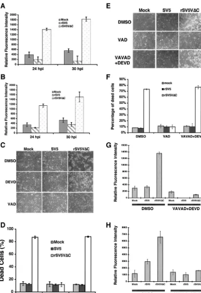
Activation of caspases in rSV5V⌬C-infected cells.Caspases are key players in the regulation of apoptotic pathways. Dif-ferent apoptotic pathways activate difDif-ferent caspases. An un-derstanding of which caspase is activated provides information about which apoptotic pathway is activated in apoptotic cells. The activity of caspase 3, a major effector caspase, in mock-, SV5-, or rSV5V⌬C-infected cells was examined. Increased cas-pase 3 activity was detected in rSV5V⌬C-infected HeLa and U3A cells but not in mock- or SV5-infected HeLa or U3A cells, suggesting that caspase 3 might play a role in apoptosis induced by rSV5V⌬C (Fig. 3A and B). Furthermore, activation of caspase 3 was confirmed by immunoblotting using anti-caspase 3 antibody (data not shown). DEVD, a specific in-hibitor against caspase 3, did not block apoptosis induced by rSV5V⌬C (Fig. 3C), however, suggesting that caspase 3 is not an essential regulator for rSV5V⌬C-induced apoptosis. Inter-estingly, a general caspase inhibitor was able to block apoptosis induced by rSV5V⌬C, suggesting that a caspase or caspases other than caspase 3 are responsible for rSV5V⌬C-induced apoptosis.
To identify the caspase that is activated in rSV5V⌬ C-in-fected HeLa cells, caspase activity assays were carried out; the results are shown in Table 1. There are 14 known caspases, and activity assays were carried out on 12 caspases. Caspase 14, a keratinocyte-specific caspase, is not expressed in differentiated adult cells (19), and there is no available activity assay for caspase 12. While increased caspase 2 and caspase 3 activities were detected in rSV5V⌬C-infected cells compared to the re-sults seen with mock- or SV5-infected cells, neither individual inhibitor against the caspases nor the combination of caspase 2 and 3 inhibitors inhibited rSV5V⌬C-induced apoptosis (Fig. 3E and F) (Table 2), suggesting that caspases 2 and 3 were not responsible for rSV5V⌬C-induced apoptosis. Activities of cas-pase 1, 4, 5, 6, 7, 8, 9, 10, 11, and 13 were not detected in the infected cells, although it is possible the assays were not suffi-ciently sensitive. To examine the involvement of these caspases in rSV5V⌬C-induced apoptosis, specific inhibitors against these caspases were applied to the infected cells; the results are in shown in Table 2. None of the specific caspase inhibitors blocked CPE caused by rSV5V⌬C infection. The levels of activity of caspase 2 and 3 inhibitors were measured, and the inhibitors were found to be effective (Fig. 3G and H).
Due to the lack of an enzymatic activity assay for caspase 12, the activation of caspase 12 was examined by immunoblotting using antibodies specific for caspase 12. The inactive form of caspase 12 (pro-caspase 12) is ⬃60 kDa and is activated by cleavage of its N-terminal prodomain. The active form of
cas-pase 12 is⬃50 kDa, and it can be further cleaved into a small subunit and a⬃35-kDa subunit (42). When two different an-tibodies against pro-caspase 12 were used, the inactive form of caspase 12 was not detected in rSV5V⌬C-infected cells whereas proteins of the expected mass of pro-caspase 12 were detected in mock or SV5-infected cells, suggesting that caspase 12 was activated in rSV5V⌬C-infected cells (Fig. 4A). A polypeptide species of⬃35 kDa, corresponding to the mass of the large subunit of active form of caspase 12, was observed in rSV5V⌬C-infected cells (Fig. 4B).
ER stress in rSV5V⌬C-infected cells.Activation of caspase 12 often associates with increased endoplasmic reticulum (ER) stress (35, 42). To examine ER stress of virus-infected cells, expression levels of major ER stress indicators such as GRP 78, GRP 94, and GADD153 (CHOP) were measured using immunoblotting (20, 32, 59). Whereas expression levels of GRP 78 and GRP 94 increased moderately in SV5-infected cells compared with the results seen with mock-infected cells, expression levels of these proteins were increased dramatically in rSV5V⌬C-infected cells, suggesting that rSV5V⌬C induced ER stress (Fig. 5A). Furthermore, expression of GADD153, which is often associated with cell death after ER stress, was examined. Expression of GADD153 was increased only in rSV5V⌬C-infected cells (Fig. 5B).
DISCUSSION
SV5 V is known to interrupt host IFN signaling and produc-tion. However, increased CPE caused by rSV5V⌬C in Vero cells, an IFN production-defective cell line, suggests that V has other functions in addition to circumventing the host IFN response. Recently, He et al. used four- to six-week-old BALB/c mutant mice homozygous for a targeted disruption of STAT1to establish a small animal model system for studying SV5 pathogenesis (16). InSTAT1⫺/⫺mice, rSV5 grew to a titer
of 0.44 ⫻107PFU/g of lung tissue whereas rSV5V⌬C grew
only to a titer of 0.14⫻106PFU/g. The higher titer of rSV5
compared to that seen with rSV5V⌬C in mice that lack a STAT1 signaling pathway suggests that the C terminus of the V protein has another function in the virus life cycle in addi-tion to mediating the anti-IFN activities. We hypothesize that the additional role of the V protein is that of preventing apo-ptosis in infected cells. rSV5V⌬C was less pathogenic in vivo than wt rSV5 (even though it caused vastly greater CPE in mouse and human cells), a finding consistent with the notion of virus clearance by apoptosis of infected cells in an infected animal (15). In this study, we found that HeLa cells infected with rSV5V⌬C underwent apoptosis and that the apoptosis was inhibited by V, suggesting that V had an anti-apoptosis function. Sendai virus, another paramyxovirus that lacks the V gene (rSeV⌬V), is viable and grows as well as wt SeV. How-ever, rSeV⌬V induces increased CPE in culture-grown cells and is attenuated in animal hosts (24). The phenotype of rSeV⌬V is very similar to that of rSV5V⌬C (i.e., increased CPE in culture and attenuated in vivo), suggesting that SeV V protein might have a function similar to that of SV5 V protein in preventing cell death. Recently, a recombinant NDV lacking its V gene (rNDV⌬V) has been recovered that induces in-creased apoptosis in CEF cells, suggesting that NDV V has an
on November 8, 2019 by guest
http://jvi.asm.org/
FIG. 3. Caspase activities in rSV5V⌬C-infected cells. HeLa or U3A cells were mock infected or infected with SV5 or rSV5V⌬C at an MOI of 5. The cells were collected at the times indicated, and caspase assays were performed as described in reference 30. (A) HeLa cells; (B) U3A cells. (C and D) Inhibition of CPE by caspase inhibitors. Mock-, SV5-, or rSV5V⌬C-infected HeLa cells were treated with DMSO (the solvent for caspase inhibitor), DEVD (a caspase 3 inhibitor) (100M), or VAD (a general caspase inhibitor) (50M) and photographed at 2 dpi. (E) Combinations of caspase 2 and caspase 3 inhibitors did not block rSV5V⌬C-induced cell death. Mock-, SV5-, or rSV5⌬C-infected HeLa cells were treated with DMSO, VAD (at 50M), or VAVAD (caspase 2 inhibitor at 80M) plus DEVD (caspase 3 inhibitor at 80M). Pictures were taken at 1 dpi. (F) The cells shown in panel E were stained with trypan blue and counted. (G) The effectiveness of caspase 2 inhibitor. The caspase 2 activities of the cells shown in panel E were measured using activity assays as described in Materials and Methods. (H) Caspase 3 activities of the cells shown in panel E.
5074
on November 8, 2019 by guest
http://jvi.asm.org/
anti-apoptosis function (38). The mechanism by which V pro-tein blocks virus-induced apoptosis is not clear.
Virus infection can activate a variety of cellular signaling pathways that lead to apoptosis. IFN produced in response to virus infection plays an essential role in inducing apoptosis in virus-infected cells (3, 9, 52). The infected host organisms are thought to inhibit and eliminate viral infection by sacrificing virus-infected cells through apoptosis. However, many viruses have developed means to delay and inhibit apoptosis to avoid being eliminated along with their host cells (4, 44). The exact mechanisms of IFN-promoted apoptosis in virus-infected cells are not clear. It is thought that IFN can activate caspases and up-regulate expression of genes with proapoptotic functions such as death factors (3, 9, 52). In rSV5V⌬C-infected HeLa cells, increased IFN production was detected and the IFN signaling pathway was intact. However, the induction of apo-ptosis by rSV5V⌬C also occurred in U3A cells and in Vero cells and in the presence of neutralizing antibody against IFN, suggesting that rSV5V⌬C induced apoptosis in an IFN-independent manner. As the apoptotic pathway activated by rSV5V⌬C was blocked by V expression (Fig. 1), we hypothe-size that V protein has an antiapoptotic function and that the antiapoptotic function is distinct from its anti-IFN function. Thus, SV5 (an RNA virus) can activate IFN-independent apo-ptotic pathways in virus-infected cells.
Examination of possible involvement of death factors showed that medium components from rSV5V⌬C-infected cells were not sufficient to induce cell death, suggesting that rSV5V⌬C induced apoptosis through an intrinsic apoptotic pathway. To identify the intrinsic apoptotic pathway activated by rSV5V⌬C, the involvement of caspases was examined. Whereas a caspase is undoubtedly involved in rSV5V⌬C-induced apoptosis (as demonstrated by inhibition of the apoptosis by the pan-caspase inhibitor Z-VAD-FMK) (Fig. 3 and Table 2), it is very intrigu-ing that caspase 3 and caspase 9, two major caspases in the intrinsic pathway, were not responsible for rSV5V⌬C-induced apoptosis. Examination (using immunoblotting) of caspase 12 indicated that caspase 12 is activated. Caspase 12 was first identified in the mouse and was found to be important for ER
stress-induced apoptosis (35). Although many reports indi-cated that caspase 12 has a similar function in human cells (6, 25, 57), there is one report indicating that (due to a mutation in the active site of caspase 12) there is no functional human caspase 12 (13). At present, unfortunately, there is no specific inhibitor available against caspase 12 to aid in resolving this uncertainty.
In our studies, we used immunoblotting analysis with two different antibodies to detect activation of caspase 12 in rSV5V⌬C-infected cells; inhibitors against all other known caspases did not inhibit rSV5V⌬C-induced apoptosis. We speculate that caspase 12 is responsible for rSV5V⌬C-induced apoptosis. However, we cannot rule out the possibility that an as-yet-unidentified mammalian caspase might be responsible for rSV5V⌬C-induced apoptosis. As activation of caspase 12 is often associated with ER stress, expression levels of ER stress-related proteins were examined and found to be up-regulated in rSV5V⌬C-infected cells, supporting the hypothesis that cas-pase 12 is activated in rSV5V⌬C-infected cells and that it might play an essential role in the induction of apoptosis. It is not surprising that viral infection can cause ER stress, as large amounts of viral glycoproteins are made in virus-infected cells (as reported previously for SV5 and for cells infected with other viruses) (22, 36, 62, 63). It is interesting that wt SV5 caused a mild increase of ER stress, as was evident in small increases in expression levels of GRP 78 and 94 (Fig. 5A) (two major host proteins involved in unfolded protein response [UPR]), and that wt SV5 did not induce cell death.
[image:8.603.45.284.89.250.2]It has been reported that increased expression of GRP 78 can prevent apoptosis by downregulation of Bax protein, a pro-apoptotic protein (43). It is also known that UPR can activate proapoptotic gene expression (such as that of GADD153), which in turn can induce infected cells to undergo apoptosis (23, 33, 61). Expression levels of GADD153 were increased only in rSV5V⌬C-infected cells (consistent with the induction of apoptosis seen in rSV5V⌬C-infected cells). It is not clear how cells regulate expression of GRP 78 and GRP 94 to have an antiapoptotic effect at moderate expression levels and to have a proapoptotic effect at higher expression levels. We speculate that expression levels of GRP 78 and GRP 94 might determine how cells respond to ER stress. Regardless of how TABLE 1. Caspase activities of mock-, SV5-, and
rSV5V⌬C-infected cellsa
Caspase sequenceTarget substrateTested
Result with indicated infection
Mock rSV5 rSV5V⌬C
1 YVAD Ac-YVAD-AMC ⫺ ⫺ ⫺
2 DESD Ac-LDESD-AMC ⫺ ⫺ ⫹
3 DEVD Ac-DEVD-AMC ⫺ ⫺ ⫹
4 LEVD Ac-LEVD-AMC ⫺ ⫺ ⫺
5 LEHD Ac-LEHD-AMC ⫺ ⫺ ⫺
6 VEID Ac-VEID-AMC ⫺ ⫺ ⫺
7 DEVD Ac-DEVD-AMC ⫺ ⫺ ⫺
8 LETD Ac-LETD-AMC ⫺ ⫺ ⫺
9 LEHD Ac-LEHD-AMC ⫺ ⫺ ⫺
10 LEHD Ac-LEHD-AMC ⫺ ⫺ ⫺
11 LEHD Ac-LEHD-AMC ⫺ ⫺ ⫺
12 NAb NA NA NA NA
13 LEED Ac-LEED-AFC ⫺ ⫺ ⫺
aThe caspase enzymatic activity assay was carried out as described in
Mate-rials and Methods.
bNA, not available.
TABLE 2. Effects of caspase inhibitors on the virus-infected cells
Inhibitor(s)a Targeted
caspase(s)
Result with
indicated infectionb
Mock rSV5 rSV5V⌬C
DMSO None ⫺ ⫺ ⫹
Z-VAD-FMK All caspases ⫺ ⫺ ⫺
Z-DEVD-FMK Caspases 3, 6, 7 ⫺ ⫺ ⫹
Z-VAVAD-FMK Caspase 2 ⫺ ⫺ ⫹
Z-LEHD-FMK Caspases 5, 9, 10, 11 ⫺ ⫺ ⫹
Z-YVAD-FMK Caspase 1, 4 ⫺ ⫺ ⫹
Z-LETD-FMK Caspase 8 ⫺ ⫺ ⫹
Z-VDVAD-FMK Caspase 2 ⫺ ⫺ ⫹
Z-LEED-FMK Caspase 13 ⫺ ⫺ ⫹
Z-VAVAD-FMK⫹
Z-DEVD-FMK Caspases 2, 3, 6, 7 ⫺ ⫺ ⫹
aZ-DEVD can inhibit caspases 3, 6, and 7. Z-LEHD-FMK can inhibit
cas-pases 5, 9, 10, and 11. Z-YVAD-FMK can inhibit cascas-pases 1 and 4 (27, 51, 55).
b⫺, without CPE;⫹, with CPE.
on November 8, 2019 by guest
http://jvi.asm.org/
[image:8.603.301.541.557.699.2]ER stress and cell death is regulated, it is conceivable that it is beneficial for viruses to prevent ER stress signaling to prevent host cells from undergoing apoptosis. The V protein of SV5 may play a role in eliminating ER stress signaling and enabling SV5 replication in nonapoptotic cells. Interestingly, it has been reported that human RS virus, which does not encode a V protein, induces expression of GRP 78 (a major indicator of ER stress in A549 cells) and causes cell death (6). However, there are other reports indicating that infection by RS virus does not induce apoptosis (11, 50).
It has been reported that mutant SV5 containing mutations in the shared N termini of V and P protein (known as CPI⫺
[image:9.603.57.530.72.280.2]virus) accelerates viral gene expression and causes increased cell death, with characteristics of apoptosis (60). Even though the mutant SV5 (CPI⫺virus) is different from rSV5V⌬C, they may induce cell death through similar pathways. CPI⫺ virus encodes two mutant proteins, whereas rSV5V⌬C encodes a truncated V protein with a very short half-life. Thus, the phe-notype of rSV5V⌬C is likely a result of loss of V function whereas phenotypes of CPI⫺virus may be due to gains as well as losses of functions of both V and P mutant proteins. It is not unexpected that mutations in the shared N termini of P and V proteins of CPI⫺virus affect viral gene expression, as SV5 P is essential for viral RNA replication and transcription. It is pos-FIG. 4. Activation of caspase 12 in rSV5V⌬C-infected cells. HeLa cells were mock (MK) infected or infected with SV5 or rSV5V⌬C. The infected cells were collected at 1 and 2 dpi and subjected to immunoblotting as described in Materials and Methods. (A) Antibody specific for pro-caspase 12 from Sigma (clone 14F7; catalog no. C 7611). (B) Antibody specific for pro-caspase 12 from Oncogene (catalog no. PC557T). Nonspecific proteins were also detected in mock-, SV5-, or rSV5V⌬C-infected cells by the antibodies.
FIG. 5. Increased expression levels of ER stress-related proteins. HeLa cells were mock (MK) infected or infected with SV5 or rSV5V⌬C. The infected cells were collected at 1 dpi and subjected to Western blotting as described in Materials and Methods. Numbers below the bands are protein expression levels relative to the value for mock-infected cells (defined as 1). (A) Anti-GRP 78 and anti-GRP 94; (B) anti-GADD153.
5076 SUN ET AL. J. VIROL.
on November 8, 2019 by guest
http://jvi.asm.org/
[image:9.603.87.493.498.692.2]sible that overexpression and early expression of viral proteins (including glycoproteins F and HN) in CPI⫺ virus-infected cells can cause ER stress and trigger cell death in a fashion similar to that seen with the induction of apoptosis by rSV5V⌬C infection, in which viral gene expressions are not affected in rSV5V⌬C-infected cells (since it still encodes a wt P protein). Alternatively, increased UPR response in rSV5V⌬C-infected cells may be a result of increased ER stress induced by overexpression of viral glycoproteins even though the increase was not obvious in rSV5V⌬C-infected cells.
Mitochondria play an essential role in intrinsic apoptotic pathways through caspase-dependent and caspase-indepen-dent pathways. In rSV5V⌬C-infected cells, apoptosis was cas-pase dependent but cascas-pase 9 activity was not detected and an inhibitor against caspase 9 did not affect rSV5V⌬C-induced apoptosis, suggesting that a previously unidentified caspase was activated through mitochondria or that mitochondria did not play a determining role in rSV5V⌬C-induced apoptosis. Caspase 12 localizes to ER, and there is no evidence of its as-sociation with mitochondria (34, 35). At present, it is not clear how ER stress induced by rSV5V⌬C infection affects mito-chondrial function. Studies of the relationship between ER stress and mitochondrion damage in virus-infected cells are in progress.
ACKNOWLEDGMENTS
We thank George Stark for providing U3A cells. We are grateful to Michael Teng for critically reading the manuscript. We appreciate other members of Biao He’s lab for discussion and technical help. The services provided by the General Clinical Research Center of the Pennsylvania State University are appreciated.
The General Clinical Research Center of the Pennsylvania State University was partially supported by National Institutes of Health grant M01 RR 10732. R.A.L. is an Investigator of the Howard Hughes Medical Institute. The work was supported by a seed grant from the College of Agricultural Sciences of the Pennsylvania State University to B.H. and grants from the National Institute of Allergy and Infec-tious Diseases to R.A.L. (R01 AI 23173) and to B.H. (R01 AI 051372).
REFERENCES
1. Andrejeva, J., D. F. Young, S. Goodbourn, and R. E. Randall.2002. Degra-dation of STAT1 and STAT2 by the V proteins of simian virus 5 and human parainfluenza virus type 2, respectively: consequences for virus replication in
the presence of alpha/beta and gamma interferons. J. Virol.76:2159–2167.
2. Ashkenazi, A., and V. M. Dixit.1998. Death receptors: signaling and
mod-ulation. Science281:1305–1308.
3. Barber, G. N.2001. Host defense, viruses and apoptosis. Cell Death Differ.
8:113–126.
4. Benedict, C. A., P. S. Norris, and C. F. Ware.2002. To kill or be killed: viral
evasion of apoptosis. Nat. Immunol.3:1013–1018.
5. Bernardi, P., V. Petronilli, F. Di Lisa, and M. Forte.2001. A mitochondrial
perspective on cell death. Trends Biochem. Sci.26:112–117.
6. Bitko, V., and S. Barik.2001. An endoplasmic reticulum-specific stress-activated caspase (caspase-12) is implicated in the apoptosis of A549
epi-thelial cells by respiratory syncytial virus. J. Cell. Biochem.80:441–454.
7. Cory, S., and J. M. Adams.2002. The Bcl2 family: regulators of the cellular
life-or-death switch. Nat. Rev. Cancer2:647–656.
8. Cryns, V., and J. Yuan.1998. Proteases to die for. Genes Dev.12:1551–1570.
(Erratum,13:371, 1999.)
9. Dai, C., and S. B. Krantz.1999. Interferon gamma induces upregulation and activation of caspases 1, 3, and 8 to produce apoptosis in human erythroid
progenitor cells. Blood93:3309–3316.
10. Didcock, L., D. F. Young, S. Goodbourn, and R. E. Randall.1999. The V protein of simian virus 5 inhibits interferon signalling by targeting STAT1 for
proteasome-mediated degradation. J. Virol.73:9928–9933.
11. Domachowske, J. B., C. A. Bonville, A. J. Mortelliti, C. B. Colella, U. Kim, and H. F. Rosenberg.2000. Respiratory syncytial virus infection induces expression of the anti-apoptosis gene IEX-1L in human respiratory epithelial
cells. J. Infect. Dis.181:824–830.
12. Evan, G., and T. Littlewood.1998. A matter of life and cell death. Science
281:1317–1322.
13. Fischer, H., U. Koenig, L. Eckhart, and E. Tschachler.2002. Human caspase 12 has acquired deleterious mutations. Biochem. Biophys. Res. Commun.
293:722–726.
14. He, B., G. P. Leser, R. G. Paterson, and R. A. Lamb.1998. The paramyxo-virus SV5 small hydrophobic (SH) protein is not essential for paramyxo-virus growth in
tissue culture cells. Virology250:30–40.
15. He, B., G. Y. Lin, J. E. Durbin, R. K. Durbin, and R. A. Lamb.2001. The SH integral membrane protein of the paramyxovirus simian virus 5 is required to
block apoptosis in MDBK cells. J. Virol.75:4068–4079.
16. He, B., R. G. Paterson, N. Stock, J. E. Durbin, R. K. Durbin, S. Goodbourn, R. E. Randall, and R. A. Lamb.2002. Recovery of paramyxovirus simian virus 5 with a V protein lacking the conserved cysteine-rich domain: the multifunctional V protein blocks both interferon-beta induction and
inter-feron signaling. Virology303:15–32.
17. He, B., R. G. Paterson, C. D. Ward, and R. A. Lamb.1997. Recovery of infectious SV5 from cloned DNA and expression of a foreign gene. Virology
237:249–260.
18. Hiebert, S. W., C. D. Richardson, and R. A. Lamb.1988. Cell surface expression and orientation in membranes of the 44 amino acid SH protein of
simian virus 5. J. Virol.62:2347–2357.
19. Hu, S., S. J. Snipas, C. Vincenz, G. Salvesen, and V. M. Dixit. 1998. Caspase-14 is a novel developmentally regulated protease. J. Biol. Chem.
273:29648–29653.
20. Hurtley, S. M., D. G. Bole, H. Hoover-Litty, A. Helenius, and C. S. Copeland.
1989. Interactions of misfolded influenza virus hemagglutinin with binding
protein (BiP). J. Cell Biol.108:2117–2126.
21. Jacques, J. P., and D. Kolakofsky.1991. Pseudo-templated transcription in
prokaryotic and eukaryotic organisms. Genes Dev.5:707–713.
22. Jordan, R., O. V. Nikolaeva, L. Wang, B. Conyers, A. Mehta, R. A. Dwek, and T. M. Block.2002. Inhibition of host ER glucosidase activity prevents Golgi processing of virion-associated bovine viral diarrhea virus E2 glycoproteins
and reduces infectivity of secreted virions. Virology295:10–19.
23. Jordan, R., L. Wang, T. M. Graczyk, T. M. Block, and P. R. Romano.2002. Replication of a cytopathic strain of bovine viral diarrhea virus activates PERK and induces endoplasmic reticulum stress-mediated apoptosis of
MDBK cells. J. Virol.76:9588–9599.
24. Kato, A., K. Kiyotani, Y. Sakai, T. Yoshida, and Y. Nagai.1997. The paramyxovirus, Sendai virus, V protein encodes a luxury function required
for viral pathogenesis. EMBO J.16:578–587.
25. Kitamura, Y., A. Miyamura, K. Takata, M. Inden, D. Tsuchiya, K. Naka-mura, and T. Taniguchi.2003. Possible involvement of both endoplasmic reticulum- and mitochondria-dependent pathways in thapsigargin-induced
apoptosis in human neuroblastoma SH-SY5Y cells. J. Pharm. Sci.92:228–
236.
26. Lamb, R. A., and D. Kolakofsky.2001.Paramyxoviridae: the viruses and their
replication.InD. M. Knipe and P. M. Howley (ed.), Fields virology, 4th ed.
Lippincott, Williams and Wilkins, Philadelphia, Pa.
27. Lee, P., J. Lee, S. Kim, M. S. Lee, H. Yagita, S. Y. Kim, H. Kim, and K. Suk.
2001. NO as an autocrine mediator in the apoptosis of activated microglial cells: correlation between activation and apoptosis of microglial cells. Brain
Res.892:380–385.
28. Lin, G. Y., and R. A. Lamb.2000. The paramyxovirus simian virus 5 V
protein slows progression of the cell cycle. J. Virol.74:9152–9166.
29. Lin, G. Y., R. G. Paterson, and R. A. Lamb.1997. The RNA binding region
of the paramyxovirus SV5 V and P proteins. Virology238:460–469.
30. Lin, Y., A. C. Bright, T. A. Rothermel, and B. He.2003. Induction of apoptosis by paramyxovirus simian virus 5 lacking a small hydrophobic gene.
J. Virol.77:3371–3383.
31. Liston, P., and D. J. Briedis.1994. Measles virus V protein binds zinc.
Virology198:399–404.
32. Little, E., M. Ramakrishnan, B. Roy, G. Gazit, and A. S. Lee.1994. The glucose-regulated proteins (GRP78 and GRP94): functions, gene regulation,
and applications. Crit. Rev. Eukaryot. Gene Expr.4:1–18.
33. McCullough, K. D., J. L. Martindale, L. O. Klotz, T. Y. Aw, and N. J. Holbrook.2001. Gadd153 sensitizes cells to endoplasmic reticulum stress by down-regulating Bcl2 and perturbing the cellular redox state. Mol. Cell. Biol.
21:1249–1259.
34. Nakagawa, T., and J. Yuan.2000. Cross-talk between two cysteine protease
families. Activation of caspase-12 by calpain in apoptosis. J. Cell Biol.150:
887–894.
35. Nakagawa, T., H. Zhu, N. Morishima, E. Li, J. Xu, B. A. Yankner, and J. Yuan.2000. Caspase-12 mediates endoplasmic-reticulum-specific apoptosis
and cytotoxicity by amyloid-beta. Nature403:98–103.
36. Ng, D. T., R. E. Randall, and R. A. Lamb.1989. Intracellular maturation and transport of the SV5 type II glycoprotein hemagglutinin-neuraminidase: specific and transient association with GRP78-BiP in the endoplasmic
retic-ulum and extensive internalization from the cell surface. J. Cell Biol.109:
3273–3289.
37. Parisien, J. P., J. F. Lau, J. J. Rodriguez, C. M. Ulane, and C. M. Horvath.
2002. Selective STAT protein degradation induced by paramyxoviruses re-quires both STAT1 and STAT2 but is independent of alpha/beta interferon
signal transduction. J. Virol.76:4190–4198.
on November 8, 2019 by guest
http://jvi.asm.org/
38. Park, M. S., A. Garcı´a-Sastre, J. F. Cros, C. F. Basler, and P. Palese.2003. Newcastle disease virus V protein is a determinant of host range restriction.
J. Virol.77:9522–9532.
39. Paterson, R. G., T. J. R. Harris, and R. A. Lamb.1984. Analysis and gene
assignment of mRNAs of a paramyxovirus, simian virus 5. Virology138:310–
323.
40. Paterson, R. G., G. P. Leser, M. A. Shaughnessy, and R. A. Lamb.1995. The paramyxovirus SV5 V protein binds two atoms of zinc and is a structural
component of virions. Virology208:121–131.
41. Randall, R. E., and A. Bermingham.1996. NP:P and NP:V interactions of the paramyxovirus simian virus 5 examined using a novel protein:protein
capture assay. Virology224:121–129.
42. Rao, R. V., E. Hermel, S. Castro-Obregon, G. del Rio, L. M. Ellerby, H. M. Ellerby, and D. E. Bredesen.2001. Coupling endoplasmic reticulum stress to the cell death program. Mechanism of caspase activation. J. Biol. Chem.
276:33869–33874.
43. Rao, R. V., A. Peel, A. Logvinova, G. del Rio, E. Hermel, T. Yokota, P. C. Goldsmith, L. M. Ellerby, H. M. Ellerby, and D. E. Bredesen.2002. Coupling endoplasmic reticulum stress to the cell death program: role of the ER
chaperone GRP78. FEBS Lett.514:122–128.
44. Roulston, A., R. C. Marcellus, and P. E. Branton.1999. Viruses and
apo-ptosis. Annu. Rev. Microbiol.53:577–628.
45. Schmitt, A. P., B. He, and R. A. Lamb.1999. Involvement of the cytoplasmic domain of the hemagglutinin-neuraminidase protein in assembly of the
paramyxovirus simian virus 5. J. Virol.73:8703–8712.
46. Schmitt, A. P., G. P. Leser, D. L. Waning, and R. A. Lamb.2002. Require-ments for budding of paramyxovirus simian virus 5 virus-like particles. J.
Vi-rol.76:3952–3964.
47. Stark, G. R.1997. Genetic analysis of interferon and other mammalian
signaling pathways. Harvey Lect.93:1–16.
48. Steward, M., A. C. R. Samson, W. Errington, and P. T. Emmerson.1995. The
Newcastle disease virus V protein binds zinc. Arch. Virol.140:1321–1328.
49. Strasser, A., L. O’Connor, and V. M. Dixit.2000. Apoptosis signaling. Annu.
Rev. Biochem.69:217–245.
50. Takeuchi, R., H. Tsutsumi, M. Osaki, K. Haseyama, N. Mizue, and S. Chiba.
1998. Respiratory syncytial virus infection of human alveolar epithelial cells
enhances interferon regulatory factor 1 and interleukin-1-converting
en-zyme gene expression but does not cause apoptosis. J. Virol.72:4498–4502.
51. Talanian, R. V., C. Quinlan, S. Trautz, M. C. Hackett, J. A. Mankovich, D. Banach, T. Ghayur, K. D. Brady, and W. W. Wong.1997. Substrate
speci-ficities of caspase family proteases. J. Biol. Chem.272:9677–9682.
52. Tanaka, N., M. Sato, M. S. Lamphier, H. Nozawa, E. Oda, S. Noguchi, R. D. Schreiber, Y. Tsujimoto, and T. Taniguchi.1998. Type I interferons are essential mediators of apoptotic death in virally infected cells. Genes Cells
3:29–37.
53. Thomas, S. M., R. A. Lamb, and R. G. Paterson.1988. Two mRNAs that differ by two nontemplated nucleotides encode the amino coterminal
pro-teins P and V of the paramyxovirus SV5. Cell54:891–902.
54. Thornberry, N. A., and Y. Lazebnik.1998. Caspases: enemies within. Science
281:1312–1316.
55. Thornberry, N. A., T. A. Rano, E. P. Peterson, D. M. Rasper, T. Timkey, M. Garcia-Calvo, V. M. Houtzager, P. A. Nordstrom, S. Roy, J. P. Vaillancourt, K. T. Chapman, and D. W. Nicholson.1997. A combinatorial approach defines specificities of members of the caspase family and granzyme B. Functional relationships established for key mediators of apoptosis. J. Biol.
Chem.272:17907–17911.
56. Tidona, C. A., H. W. Kurz, H. R. Gelderblom, and G. Darai.1999. Isolation and molecular characterization of a novel cytopathogenic paramyxovirus
from tree shrews. Virology258:425–434.
57. Tong, W. G., X. Z. Ding, and T. E. Adrian. 2002. The mechanisms of lipoxygenase inhibitor-induced apoptosis in human breast cancer cells.
Bio-chem. Biophys. Res. Commun.296:942–948.
58. Ulane, C. M., and C. M. Horvath.2002. Paramyxoviruses SV5 and HPIV2 assemble STAT protein ubiquitin ligase complexes from cellular
compo-nents. Virology304:160–166.
59. Wang, X. Z., B. Lawson, J. W. Brewer, H. Zinszner, A. Sanjay, L. J. Mi, R. Boorstein, G. Kreibich, L. M. Hendershot, and D. Ron.1996. Signals from the stressed endoplasmic reticulum induce C/EBP-homologous protein
(CHOP/GADD153). Mol. Cell. Biol.16:4273–4280.
60. Wansley, E. K., and G. D. Parks.2002. Naturally occurring substitutions in the P/V gene convert the noncytopathic paramyxovirus simian virus 5 into a virus that induces alpha/beta interferon synthesis and cell death. J. Virol.
76:10109–10121.
61. Watanabe, Y., O. Suzuki, T. Haruyama, and T. Akaike.2003. Interferon-gamma induces reactive oxygen species and endoplasmic reticulum stress at
the hepatic apoptosis. J. Cell. Biochem.89:244–253.
62. Watowich, S. S., R. I. Morimoto, and R. A. Lamb.1991. Flux of the paramyxovirus hemagglutinin-neuraminidase glycoprotein through the
en-doplasmic reticulum activates transcription of theGRP78-BiPgene. J. Virol.
65:3590–3597.
63. Wooden, S. K., L. J. Li, D. Navarro, I. Qadri, L. Pereira, and A. S. Lee.1991. Transactivation of the grp78 promoter by malfolded proteins, glycosylation block, and calcium ionophore is mediated through a proximal region con-taining a CCAAT motif which interacts with CTF/NF-I. Mol. Cell. Biol.
11:5612–5623.
64. Zhou, G., and B. Roizman.2002. Cation-independent mannose 6-phosphate receptor blocks apoptosis induced by herpes simplex virus 1 mutants lacking glycoprotein D and is likely the target of antiapoptotic activity of the
glyco-protein. J. Virol.76:6197–6204.
5078 SUN ET AL. J. VIROL.