A STUDY OF COGNITIVE EVOKED POTENTIAL
AND ARTERIAL OXYGEN SATURATION IN STABLE
COPD PATIENTS
Dissertation submitted
in partial fulfillment of the regulations for
the award of the degree of
M.D.PHYSIOLOGY
BRANCH V
REGISTER NUMBER : 201215051
DEPARTMENT OF PHYSIOLOGY
GOVERNMENT STANLEY MEDICAL COLLEGE
CHENNAI-1
THE TAMIL NADU
DR.M.G.R. MEDICAL UNIVERSITY
CHENNAI - 600 001
BONAFIDE CERTIFICATE
This is to certify that the dissertation titled “A STUDY OF COGNITIVE
EVOKED POTENTIAL AND ARTERIAL OXYGEN SATURATION IN
STABLE COPD PATIENTS’’ is a bonafide record work done by Dr. Rowena
Victor, under my direct supervision and guidance, submitted to The Tamil
Nadu Dr. M. G. R. Medical University in partial fulfillment of University
regulation for M.D., Branch-V (Physiology).
Dr.AL.Meenakshi Sundram,M.D., D.A., Dr.K.Balasubramanian,M.D.,
Dean Professor and HOD
Govt. Stanley Medical College Govt. Stanley Medical College
Chennai – 600 001. Chennai - 600001
DECLARATION
I, Dr. Rowena Victor, solemnly declare that the dissertation titled ‘‘A
STUDY OF COGNITIVE EVOKED POTENTIAL AND ARTERIAL
OXYGEN SATURATION IN STABLE COPD PATIENTS’’ has been
prepared by me. I also declare that this work was not submitted by me or any
other, for any award, degree, diploma to any other University board either in
India or abroad. This is submitted to The Tamil Nadu Dr.M.G.R. Medical
University, Chennai in partial fulfillment of the rules and regulation for the
award of M.D degree Branch-V (Physiology) to be held in April-2015.
Place : Chennai Dr. ROWENA VICTOR
ACKNOWLEDGEMENT
My sincere thanks to Dr. AL. Meenakshisundaram M.D., D.A., The
Dean, Govt. Stanley Medical College, Chennai, for allowing me to carry out
this study and I also thank The Institutional Ethical Committee, Govt. Stanley
Medical College, Chennai, for consenting to carry out the investigations in the
hospital
I am deeply indebted to Dr. K. Balasubramanian, M.D., The Professor
and Head of the Department, Govt. Stanley Medical College, Chennai, for the
valuable guidance, inspiration, support and encouragement he rendered
throughout this project.
I also thank
Dr.S.Ravichandran D.O, M.D.,
Professor, Department
of Physiology, Govt. Stanley Medical College, for his support and
guidance for doing this study.
I express my profound gratitude to Dr.Viji Devanand M.D., and
Dr.C.C.Umayal M.D., Additional Professors, Department of Physiology, Govt.
Stanley Medical College, for their support and guidance for doing this study.
I convey my gratefulness to Dr. Sridhar M.D., DTCD, Professor, Dept
of T.B and Chest diseases, Govt. Stanley Medical College Hospital, Chennai
with his team for guiding me and supporting me and Dr.Senthilkumar,
DTCD., in the Dept of T.B, and Chest diseases. Govt. Stanley Medical College
Hospital, Chennai.
I express my sincere thanks to The Professor of Microbiology and Nodal
Gastroenterology, and the team of lab Technicians under the leadership of
Mr.Karthick, Physician Asst and Lab In charge for their support to this project.
I express my profound thanks to all the Assistant Professors, Dept of
Physiology, Govt. Stanley Medical College for their inspiring guidance.
My heartfelt gratitude goes to all my colleagues and all the staff
members of this Dept of Physiology for their constant support and
encouragement.
I gratefully acknowledge all the subjects who co-operated to submit
themselves for this study. I also thank Statistician Mr.S.Venkatesan, ZIGMA
'statistical analysis' for his guidance in statistical analysis.
My wholehearted thanks to my dear husband who constantly encouraged
and supported me in all my endeavours especially this work without which I
would not have completed .
CONTENTS
Sl.No. Title Page No.
1. INTRODUCTION 1
2. REVIEW OF LITERATURE 7
3. AIM AND OBJECTIVES 65
4. MATERIALS AND METHODS 66
5. RESULTS 83
6. DISCUSSION 102
7. CONCLUSION 111
8. SUMMARY 112
BIBILIOGRAPHY
ANNEXURES
LIST OF TABLES
S.No. TABLE PAGE No.
1. Comparison of the demographic characteristics of
the two groups
84
2. Comparison of occupational status of the two
groups
87
3. Distribution of smoking Habits in the two groups 88
4. Comparison of smoking pack years between the
two groups
89
5. Classification of mean values of FEV1 based on
severity of COPD
90
6. Comparison of mean values of FEV1, FVC and
FEV1% indices between the two groups
91
7. Comparison of mean values of MMSE score (for
30) between the two groups
93
8. Comparison of mean values of Stroop test
between the two groups
94
9. Comparison of mean values of N100 wave latency
in (ms) between the two groups
95
10. Comparison of mean values of P300 wave latency
in (ms) between two groups
96
11. Comparison of mean values of P300 amplitude in
(µV) between the two groups
97
12.
Comparison of mean values of Arterial oxygen saturation values between the two groups
98
13. Mean values of Arterial oxygen saturation and
Arterial oxygen tension in COPD patients
99
14. Correlation between Arterial oxygen tension and
MMSE in COPD patients
100
15. Correlation between Arterial oxygen tension and
Stroop test in COPD patients
100
16. Correlation between Arterial oxygen tension and
P300 latency in COPD patients
101
17. Correlation between Arterial oxygen tension and
P300 amplitude in COPD patients
LIST OF
GRAPHS
S.No. GRAPHS PAGE No.
1. Comparison of (mean ± sd) values in weight
between the two groups. 85
2. Comparison of (mean ± sd) values in BMI
between the two groups. 86
3. Comparison of mean values of FEV1, FVC and
FEV1% indices between the two groups
92
4. Comparison of mean values of MMSE score
(for 30) between the two groups 93
5. Comparison of mean values of Stroop test
between the two groups 94
6. Comparison of mean values of N100 wave latency
in (ms) between the two groups 95
7. Comparison of mean values of P300 wave latency
in (ms) between two groups 96
8. Comparison of mean values of P300 amplitude in
(µV) between the two groups 97
9 Comparison of mean values of Arterial oxygen
LIST OF FIGURES
S.No. FIGURE PAGE
No.
1. Typical progression of symptoms in COPD
patients
14
2. Changes in Lung Parenchyma 18
3. Pathogenesis of COPD 20
4. Cognition domains and specific functions 37
5. Waves in Cognitive Evoked potential 50
6. Possible treatments and outcomes of cognitive
impairment
59
7. Computerized Recorder of the cognitive evoked
potential
[image:11.595.68.492.128.430.2]LIST OF PHOTOGRAPHS
PHOTO
No.
TITLE
PAGE
No.
1. PULSE OXIMETRY
70
2. ABG MACHINE
72
3.
RECORDING OF CEP IN RECORDERS MEDICARE SYSTEMS (RMS EMG EP MARK-II MACHINE)
81
ABBREVIATIONS
COPD CHRONIC OBSTRUCTIVE PULMONARY DISEASE
GOLD GLOBAL initiative for OBSTRUCTIVE LUNG DISEASE
ABG ARTERIAL BLOOD GAS analysis
FEV FORCED EXPIRATORY VOLUME
FEV1 FORCED EXPIRATORY VOLUME in 1sec
FVC FORCED VITAL CAPACITY
MMSE MINI MENTAL STATUS EXAMINATION
P300 POSITIVE wave in 300 ms
SaO2 ARTERIAL OXYGEN SATURATION
Pa O2 PARTIAL PRESSURE OF OXYGEN
EP EVOKED POTENTIAL
N100 NEGATIVE WAVE IN 100ms
PFT PULMONARY FUNCTION TEST
EEP EVENT EVOKED POTENTIAL
CEP COGNITIVE EVOKED POTENTIAL
BMR BASAL METABOLIC RATE
Ag/Ag Cl SILVER / SILVER CHLORIDE
SPSS STATISTICAL PACKAGE FOR SOCIAL SCIENCES
ANOVA ANALYSIS OF VARIANCE
m sec MILLISECONDS
ABSTRACT
A STUDY OF COGNITIVE EVOKED POTENTIAL AND ARTERIAL OXYGEN SATURATION IN STABLE COPD PATIENTS
DEGREE FOR WHICH SUBMITTED : DOCTOR OF MEDICINE (MD) IN PHYSIOLOGY
SUPERVISOR AND GUIDE : PROF.DR. K.BALASUBRAMANIAN.M.D. HEAD OF THE DEPARTMENT
DEPARTMENT : DEPARTMENT OF PHYSIOLOGY COLLEGE : GOVT.STANLEY MEDICAL
COLLEGE. CHENNAI – 600 001 UNIVERSITY : THE TAMILNADU DR. M.G.R. MEDICAL UNIVERSITY, CHENNAI – 600 032 YEAR : 2012-15
BACKGROUND:
Chronic obstructive pulmonary disease (COPD) is a complex multicomponent
disease which is associated with cognitive impairment due to neuronal damage mediated by
hypoxia. Cognitive impairment affects the self-management, clinical management and
pulmonary rehabilitation of COPD subjects. Progressive impairment of the auditory P300
evoked potential with prolonged latency occurs with increasing severity of COPD. So routine
screening of COPD patients for cognition is helpful in assessing the progression of the
disease.
AIM:
To assess the cognitive function and to correlate it with arterial oxygen saturation in
OBJECTIVE:
To assess cognitive functions in stable COPD patients and age-matched healthy
controls using Cognitive evoked potential (CEP) study, Mini mental status examination
(MMSE) and Stroop test to correlate them with Arterial oxygen saturation ( SpO2)using
Pulse oximetry and Arterial oxygen tension ( PaO2)by arterial blood gas analysis.
MATERIALS AND METHODS:
The study population were 50 Stable COPD patients and 50 controls. They were
subjected to MMSE, Stroop test, and CEP study and SpO2 measured by Pulse oximetry. In
addition, Arterial blood gas analysis was done for the patients to measure PaO2. The data
were analyzed using Mean, Standard deviation, Independent t-test, ANOVA and Tukey post
hoc test and pearson's correlation were used for analysis. The mean difference was
significant when p < 0.05 level.
RESULTS:
In stable COPD patients, MMSE score was significantly lower, interference score
was significantly increased in Stroop test and prolonged N100,P300 latency and reduced P300
amplitude in CEP study than in controls. SpO2 by pulse oximetry shows significant decrease
in patients than in the controls. There is positive correlation between P300 amplitude and PaO2
in the patients.
CONCLUSION:
There is significant cognitive impairment in Stable COPD patients as evidenced by
Stable COPD patients to detect early cognitive impairment so that timely intervention can be planned to improve the quality of their life.
Key words : Stable COPD patients, Cognitive evoked potential, P300, MMSE, Stroop
INTRODUCTION
The Objective of Global initiative for chronic Obstructive Lung Disease
(GOLD) is," to increase awareness of COPD among health related officials and
general community, to improve the diagnosis, management and prevention
of COPD, and to encourage exploration in COPD".
COPD is a major public health problem. It is a group of chronic lung
disorders, specifically encompassing Emphysema and Chronic Bronchitis and
about 600 million people are affected globally and it is now the fourth leading
cause of death and it is the only cause of mortality whose incidence continues
to rise and will be the third cause by 20201. In India, 53% of all deaths were
estimated to be due to Non communicable diseases and 44% of DALY
(disability adjusted life years) lost in 2005.Of these7% deaths and 3% DALYs
lost were due to Chronic Respiratory diseases 2.There are 30 million COPD
patients in India as estimated approximately3. In India burden of Chronic
Bronchitis was estimated as 14.84 million.
The reported prevalence is from 2 to 22% in men & from 1.2 to 19% in
women.4 Tobacco use is the main cause of global wave of smoking-related
illnesses resulting in 5.4 million deaths worldwide each year and it will increase
to 6 million by 2015. 30% of the tobacco-related deaths will be caused by
chronic respiratory diseases5. Cigarette smoking is the leading cause of COPD
Passive smoking, air pollution, and occupational chemical fumes or dust may
act synergistically with active smoking to increase the risk of COPD7.
COPD is a multicomponent non communicable disease with not only
primary pulmonary involvement but co morbidities like cardiovascular disease,
osteoporosis, anaemia depression and cognitive impairment is one among them.
The Airflow limitation in COPD is not reversible and the disease is progressive
but it is preventable and treatable8. It is a multisystem disorder with both
physical effects and impaired psychological and cognitive functioning. In
addition to the involvement of motor nerve, encephalopathy and cognitive
dysfunction have been noted in patients with chronic respiratory insufficiency.
Kayakan et al (2001) observed that cigarette smoking, limitation of airflow, and
COPD in the last stage causes hypoxemia and hypercapnia which involves the
pontomedullary portion of the brain9. Stable COPD patients are those who are
on regular medical intervention for the past 4 weeks.
Cognitive impairment is one of the extra pulmonary feature of COPD10
Cognition is the higher intellectual function of the brain. The cognitive decline
of the individual results in a great burden on the self, family and community.
The incidence of cognitive dysfunction in COPD patients varies in different
studies from 12% to 88%11. Domains of cognition are memory, learning ability
both visual and verbal, attention or vigilance, concentration, abstract thinking,
and problem solving. Neurocognitive defects such as slowed information
processing speed, poor learning, poor memory and attention deficit are well
domain is memory and attention, though speed, coordination and learning
abilities are also affected. Increasing age and low level of education are also
associated with cognitive impairment. J. W. Dodd et al in 2010 described a
specific pattern of cognitive impairment in patients with COPD.12 Patients find
very difficult to breath, and are much depressed, dysfunctional, disabled,
desperate, and are very difficult to deal with. They are encouraged to become
more knowledgeable about the disease, to actively involve in self management
and to become more independent in daily activities2. Cognitive dysfunction
reduces the level of functioning13,14, and it is associated with poor compliance
with both medication and oxygen therapy which increases the risk of acute
exacerbation.
Cognitive impairment affects functional, social, emotional, affective, and
communication skills. Also Impairment of Cognition predicts mortality in
hypoxemic COPD patients.15 A good cognitive status is essential for the
individual to understand his real state and to abstain from smoking, which
makes his life dejected by affecting his lung function. Level of cognitive
functioning of these patients must be taken into consideration before self-care
can be planned and it is modified toward the patient’s individual capability and
needs. Number of studies have been done in the past which says that patients
with COPD have poor quality of life and is again confirmed by Kaplan and
Ries16,17.
In most of the previous studies, they had used neuropsychological battery
literature which have employed electrophysiological tests to quantify cognitive
impairment in COPD. Also Pulmonary Rehabilitation which is an important
aspect of treatment for COPD patients should include initial evaluation of
patient's cognition, psychological state, social support etc which is very
important to improve the quality of life in COPD patients. Also cognitive
training should be included in the treatment schedule of COPD patients
especially in long term Pulmonary rehabilitation programs.
The term arterial oxygen saturation the percentage of Hb molecules
which are saturated with oxygen (SpO2) which is SaO2 measured by pulse
oximetry.SpO2 is related to partial pressure of oxygen in a complex way in
oxyhaemoglobin dissociation curve. Partial pressure of oxygen is the arterial
oxygen tension and it is blood oxygen level Hypoxemia produces varying
responses in different tissues greatest need is to the brain and heart.
In the present study, in addition to the Mini Mental State
Examination(MMSE) and Stroop test, Cognitive evoked potential study was
done to assess cognitive function in the Stable COPD patients. MMSE is a brief,
quick objective method of assessing global cognitive functioning. It is a reliable
and validated method commonly used to assess cognitive status. The Stroop
Colour Word Test is used to assess cognitive flexibility.18 The interference score
in Stroop test gives the measure of inhibition of a habitual response (reading)
which is part of the executive functioning. It measures focussed attention. CEP
analysis is to investigate specific types of information processing by the brain.
performing various cognitive tasks. It is produced when the person attends to
and discriminates between stimuli which differ from one another in some
dimension such as modality, intensity, frequency or duration. Cognitive evoked
potential study is recommended for detecting and quantifying early cognitive
impairment which is not usually detected by other traditional methods of
assessment of cognition.19
The present study was undertaken to assess the cognition in the Stable
COPD patients using Mini Mental State Examination and Stroop test and
Cognitive Evoked Potential study and compare it with that of normal
individuals, and to correlate with Arterial partial pressure of oxygen.
The possibility of using cognitive evoked potential as an investigatory
tool to detect early cognitive impairment in Stable COPD patients was being
explored in the present study along with neuropsychological tests.
I hypothesise that stable COPD patients have cognitive dysfunction
REVIEW OF LITERATURE
Chronic obstructive pulmonary disease (COPD) is a multi component
disease in which cognitive dysfunction is one of the components. Cognitive
function is essential for daily activities like attention, concentration, memory,
logical thinking, reasoning and execution of a motor act.
In this section, the literature relevant to COPD and COGNITION in terms
of the following side headings is being discussed.
1. Historical Aspects
2. COPD
3. Pulmonary Function Tests
4. Cognition
5. Neuropsychological test – MMSE and Stroop test
6. Neurophysiological test – Cognitive evoked potential study
7. Pulse Oximetry and Arterial Blood Gas Analysis.
In the early days, the term lung was not mentioned in the body except
for"pneuma" or vital spirits and air of universe. Respiratory system was studied
after circulatory system and this was done by Robert Boyle (1627-1691), an
Irish scholar. He proved that candle will stop burning when kept in an airless
jar. Joseph Priestly discovered oxygen, dephlogisticated air. Antonie
Lavoisier, (1743-1794) a French chemist, confirmed that oxygen is in inspired
air and carbon dioxide is in expired air.24
People regarded life as one and the same with breathing and life starts
and finishes with breathing. The BIBLE says that God breathed the breath of
life into the nostrils of ADAM and then took a rib from ADAM'S chest, to give
life to Eve. In 5th and 6th B.C., Hippocrates said that the function of lung is to
cool the heart. In 18th Century, the true role of breathing was established after
the study of chemistry of gases.25
The word Emphysema has its origin from the Greek meaning "to blow
into" and was described by Bonet in 1676 and Morgagni in 1769. Ruysh in
1721, described emphysema in humans with illustrations.
Matthew Baillie in 1807, documented, illustrated and denoted the real
destructive character of emphysema.
In 1892, William Osler in his "Text book of Medicine" described
hypertrophic emphysema as," a well marked clinical affection, with
enlargement of lungs due to distension of airways and atrophy of their walls,
J. Gough in 1952, described centrilobar emphysema. Gough &
Wentworth developed paper section technique.
McLean gave a comprehensive microscopic description of emphysema
and demonstrated the relationship of destruction to inflammatory alterations of
the bronchioles and vessels.
Laennec, who invented stethoscope in 1819 first characterised
emphysema by making clear-cut peculiarities of interstitial emphysema and
emphysema proper and correlated the enlarged airspaces to the clinical
syndrome of emphysema. He documented that air trapping and increased
collateral ventilation were features of emphysema and the primary site of
obstruction were the peripheral airways. Airspace enlarges as age increases, but
it was distinguished from emphysema. He was the first to describe an
association of emphysema with chronic bronchitis and described the pathology
of bronchiectasis.27
Galen(129-200 AD) did a volumetric experiment on human ventilation.
In 1793, Menezies R determined the tidal volume using body plethysmograph.
John Hutchinson, a surgeon pointed out that the volume of air exhaled after a
full inflation is an indicator of longevity of life. The water-sealed
volume-displacement spirometer was invented by him in 1844 to measure the vital
capacity i.e. the capacity to live. Spirometry is the most simple and useful
method available to evaluate the pulmonary function.
measurements technique. In 1925, Meakins & Davies explained most
derangements of function in terms of disorders of blood gases. Barcroft &
Haldane clarified the basic gas transport.28The electrolyte theory of dissociation
was proposed by Svante Arrhenius NP (1859-1927) for which he was awarded
Nobel prize. The definition for acid was given by Johannes N Bronsted (
1879-1947). The relationship between pH, pKa, concentration of acid and conjugate
base is expressed by the Henderson-Hasselbach equation given by Lawrence J
Henderson (1878-1972) and KA Hasselbalch (1874-1962)29.
The name P300 was given by Smith et al. It is a parietocentral positivity
that occurs when a subject detects an informative task-relevant stimulus. It is
obtained 300ms after a subject makes a simple sensory discrimination. P3 or P300
is the third major positive peak in the late sensory evoked potential (Ritter et al.,
1968) and the late positive component (Sutton et al., 1965, 1967). Sutton et al
described this late positive component and it is endogenous. Ritter and
Vaughan in 1969 used the "oddball paradigm" wherein the subject detects
occasional target signals randomly interspersed among frequent standard
stimuli. He also described parietocentral scalp distribution of the P300. Previous
research on P300 has been done exclusively in humans. But localisation of the
neural generators contributing to P300 will require depth recording and lesion
experiments which were done in cats which gives anatomical and physiological
information.
Aristotle gave attention to the cognitive process more than twenty-three
perception, and mental imagery. In the15th century, it meant thinking and
awareness. Other scientists like Wilhelm Wundt, Herman Ebbinghaus, Mary
Whiton Calkins, and William James, and some others offered their
contributions to the study of cognition.
CHRONIC OBSTRUCTIVE PULMONARY DISEASE
Natural History of COPD:
In the year 2009, the Framingham Offspring cohort study (prospective)
was done from adolescent to old age for 23years in both sexes. The
following observations were made:
(1) the normal rate of decline in lung function in healthy non-smokers is
less
(2) the dreadful effect of smoking cigarettes on the rate of lung function
decline is similar in both sexes
(3) the presence of respiratory symptoms identifies that smokers are
particularly susceptible to the development of airflow limitation.
(4) the advantage of quitting smoking is more marked when it is done
earlier.
In the Rotterdam Study, that addresses the important issue of
markedly high incidence of COPD in the youngest women, which suggests a
potential change in incidence, mortality remains higher in males than females,
even in well-matched BODE [body mass index, airway obstruction, dyspnea,
and exercise capacity] patients.
Chronic obstructive pulmonary disease (COPD) is defined as a disease
state characterized by airflow limitation that is not fully reversible accompanied
by inflammatory responses to noxious particles or gases. COPD includes
emphysema, chronic bronchitis, and small airways disease. COPD is present
only if chronic airflow obstruction occurs. It excludes asthma, bronchiectasis,
bronchiolitis, and cystic fibrosis. It causes maximal expiratory airflow limitation
and it is the severity of the limitation that is abnormal in COPD.
Risk factors include host and environmental factors. By 1964, the
Advisory Committee to the Surgeon General of the United States had concluded
that cigarette smoking was a major risk factor for mortality from chronic
bronchitis and emphysema. Second less common risk factor is a hereditary
deficiency of α1 antitrypsin.
Lower socioeconomic status is associated with significantly increased
risk for development COPD. Men often have a significantly increased risk for
development COPD due to cigarette smoking and the habit of retaining the cigar
in the mouth between puffs and extinguishing and relighting cigars. Other
factors include air pollution, infection especially in childhood, climate,
hereditary, socioeconomic status, atopy, nonspecific airway hyper
more than 100 volatile and particulate chemical substance. The enormous
impact of smoking and COPD on mortality was first studied by Doll & Hill.34
Individuals whose lung function is impaired for any reason are at
increased risk for development of symptomatic COPD and this is called as
Horse race effect34.
In the early stages of the disease there are no clinical findings. By the
time the disease is diagnosed the disease is far advanced. Subsequent
longitudinal studies have shown accelerated decline in the volume of air
exhaled within the first second of the forced expiratory manoeuvre (FEV1) in a
dose-response relationship to the intensity of cigarette smoking, which is
typically expressed as pack-years (average number of packs of cigarettes
smoked per day multiplied by the total number of years of smoking). The
traditionally higher rate of smoking among males is the likely explanation for
FIGURE - 1
TYPICAL PROGRESSION OF THE SYMPTOMS OF COPD.
Pathophysiology:
Most important abnormalities that occur in COPD are,
a) Chronic obstruction
b) Entrapment of air in the alveoli and marked destruction of alveolar
walls
d) Increased airway resistance and increased work of breathing
e) Decreased diffusing capacity
f) Extremely abnormal ventilation perfusion ratio
low Va /Q Physiologic shunt in some parts of lung lead to poor
aeration of blood .
high Va/Q Physiologic dead space in some parts lead to wasted
ventilation.
g) Increase in pulmonary vascular resistance
Persistent reduction in forced expiratory flow rates is the most
characteristic finding in COPD. Increases in the residual volume and the
residual volume/total lung capacity ratio, non-uniform distribution of
ventilation, and ventilation-perfusion mismatching also occur. Airflow
limitation or airflow obstruction is determined by spirometry, which involves
forced expiratory manoeuvres after the subject has inhaled to total lung
capacity28.
Patients with airflow obstruction related to COPD have a persistently
reduced ratio of FEV1/FVC. The reduced FEV1 in COPD does not respond to
inhaled bronchodilators. Airflow during forced exhalation is the result of the
balance between the elastic recoil of the lungs promoting flow and the
affected by COPD, maximal expiratory flow diminishes as the lungs empty
because the lung parenchyma provides gradually less elastic recoil and because
the cross-sectional area of the airways falls, raising the resistance to airflow.
In COPD there is "air trapping" (increased residual volume and increased
ratio of residual volume to total lung capacity) and progressive hyperinflation
(increased total lung capacity) during the final stages of the disease.
Hyperinflation of the thorax during tidal breathing conserves maximum
expiratory airflow, because as lung volume increases elastic recoil pressure
increases, and airways enlarge so that airway resistance decreases.
The PaO2 usually remains near normal until the FEV1 is decreased to
~50% of predicted, and even much lower FEV1 values can be associated with a
normal PaO2, at least at rest.
A rise of arterial level of carbon dioxide (PaCO2) does not occur until the
FEV1 is <25% of predicted. Pulmonary hypertension severe enough to cause
corpulmonale and right ventricular failure due to COPD typically occurs in
individuals who have marked decrease in FEV1 (<25% of predicted) and
chronic hypoxemia (PaO2<55 mmHg)
Pathology:
Changes in Large Airway:
Cigarette smoking often results in mucous gland enlargement and goblet
inflammation and fibrosis. Patients have smooth-muscle hypertrophy and
bronchial hyper reactivity leading to airflow limitation. Neutrophil influx has
been associated with purulent sputum of upper respiratory tract infections.
Independent of its proteolytic activity, neutrophil elastase is among the most
potent secretagogues identified.
Changes in Small Airways
The major site of increased resistance in most individuals with COPD is
in airways <2 mm diameter. Characteristic cellular changes include goblet cell
metaplasia, with mucus-secreting cells which replaces surfactant-secreting Clara
cells. There is mononuclear phagocytes infiltration and smooth-muscle
hypertrophy. These cause luminal narrowing by fibrosis, excess mucus, edema,
and cellular infiltration. Reduced surfactant may increase surface tension at the
air-tissue interface, predisposing to airway narrowing or collapse. Respiratory
bronchiolitis with mononuclear inflammatory cells collecting in distal airway
tissues may cause proteolytic destruction of elastic fibers in the respiratory
bronchioles and alveolar ducts where the fibers are concentrated as rings around
alveolar entrances.32
Because small airway patency is maintained by the surrounding lung
parenchyma that provides radial traction on bronchioles at points of attachment
to alveolar septa, loss of bronchiolar attachments as a result of extracellular
FIGURE - 2
Changes in Lung Parenchyma
Emphysema is defined pathologically by The National Heart, Lung,
and Blood Institute as," an abnormal condition, permanent enlargement of
distal airspaces, distal to the terminal bronchiole accompanied by destruction of
their walls and without obvious fibrosis".
Emphysema is characterized by destruction of gas-exchanging air spaces,
i.e., the respiratory bronchioles, alveolar ducts, and alveoli leading to loss of
lung elasticity. Their walls perforate and later obliterate that result in
spaces. Macrophages accumulate in respiratory bronchioles of essentially all
young smokers.
Emphysema is classified into distinct pathologic types, the most
important being centriacinar and panacinar emphysema.
Centriacinar emphysema, the type most frequently associated with
cigarette smoking, is characterized by enlarged air spaces found (initially) in
association with respiratory bronchioles. It is most prominent in the upper lobes
and superior segments of lower lobes and is often quite focal. It is most
common in males.
Panacinar emphysema refers to abnormally large air spaces evenly
distributed within and across acinar units. It is usually observed in patients with
FIGURE - 3
PATHOGENESIS OF COPD
LUNG INFLAMMATION
COPD PATHOLOGY
Oxidative
stress Proteinases
Repair mechanisms
Anti-proteinases Anti-oxidants
Host factors Amplifying mechanisms
Cigarette smoke
Biomass particles Particulates
Source: Peter J. Barnes, MD
Pathogenesis of
COPD
Pathogenesis:
Airflow limitation, the major physiologic change in COPD can result
from both small airway obstruction and emphysema. Fibrosis surrounding the
small airways appears to be a significant contributor. Collagen accumulation
around the airways in the face of increased collagenase activity remain an
enigma. Proteinase can predispose to fibrosis, including proteolytic activation of
The dominant concept of the pathogenesis of emphysema comprises of
four interrelated events.
(1) Chronic exposure to cigarette smoke may lead to inflammatory cell
recruitment within the terminal air spaces of the lung.
(2) These inflammatory cells release elastolytic proteinases that damage the
extracellular matrix of the lung.
(3) Structural cell death results from oxidant stress and loss of matrix-cell
attachment.
(4) Ineffective repair of elastin and other extracellular matrix components
result in air space enlargement that defines pulmonary emphysema.
Chronic Bronchitis is defined clinically by The British Medical
Research Council as," presence of chronic productive cough on most of the
days for three months, in each of two consecutive years in a patient in whom
other causes of chronic cough has been excluded". Diagnosis is based on the
Disease Process Anatomic Location of Lesion
Cause of Reduced Airflow
Chronic bronchitis Large and small (<
2-mm diameter) airways
Narrowing of airways by fibrosis, secretions,
edema
Emphysema Lung parenchyma Loss of lung elastic
recoil
THE ELASTASE:ANTIELASTASE HYPOTHESIS
Patients with genetic deficiency in α1AT, the inhibitor of the serine
proteinase neutrophil elastase, were at increased risk of emphysema, and
instillation of elastases, including neutrophil elastase to experimental animals
resulted in emphysema. The elastase : antielastase hypothesis remains a main
mechanism for the development of emphysema. However, a complex network
of immune and inflammatory cells and additional proteinases that contribute to
emphysema have subsequently been identified.
INFLAMMATION AND EXTRACELLULAR MATRIX PROTEOLYSIS
On exposure to oxidants from cigarette smoke, macrophages in the lower
air space become activated, producing proteinases and chemokines that attract
other inflammatory cells via oxidant-induced inactivation of histone
deacetylase-2, shifting the balance toward acetylated or loose chromatin,
exposing nuclear factor B sites and resulting in transcription of matrix
tumor necrosis factor (TNF). This leads to recruitment of neutrophils. CD8+ T
cells are also recruited in response to cigarette smoke and release interferon
inducible protein-10 (IP-10, CXCL-7) that in turn lead to production of elastase
by macrophages (Matrix metalloproteinase-12). Matrix metalloproteinases and
serine proteinases, most notably neutrophil elastase, work together by degrading
the inhibitor of the other, leading to lung destruction. Proteolytic cleavage
products of elastin also serve as a macrophage chemokine, fueling this
destructive positive feedback loop. Autoimmune mechanisms promote the
progression of disease. Increased B cells and lymphoid follicles are present in
patients with advanced disease. Antibodies have been found against elastin
fragments. IgG auto antibodies with more affinity for pulmonary epithelium and
the potential to mediate cytotoxicity have been detected.
Concomitant cigarette smoke–induced loss of cilia in the airway
epithelium and impaired macrophage phagocytosis predispose to bacterial
infection with neutrophilia. In end-stage lung disease, long after smoking
cessation there remains an exuberant inflammatory response, suggesting that
mechanisms of cigarette smoke–induced inflammation that initiate the disease
differ from mechanisms that sustain inflammation after smoking cessation.
Air space enlargement with loss of alveolar units obviously requires
disappearance of both extracellular matrix and cells. Cell death can occur from
increased oxidant stress both directly from cigarette smoke and from
inflammation. Animal models have used endothelial and epithelial cell death as
by macrophages results in production of growth factors and dampens
inflammation, promoting lung repair. Cigarette smoke impairs macrophage
uptake of apoptotic cells, limiting repair.
The ability of the adult lung to repair damaged alveoli appears limited. It
is unlikely that the process of septation that is responsible for alveologenesis
during lung development can be reinitiated. The capacity of stem cells to
repopulate the lung is under active investigation. It appears difficult for an adult
human to completely restore an appropriate extracellular matrix, particularly
functional elastic fibers.
Clinical Presentation:
History:
The three most common symptoms are cough, sputum production.
Activities involving significant arm work, particularly at or above shoulder
level, are particularly difficult for patients with COPD. In the most advanced
GUIDE TO RATING SEVERITY OF CHRONIC OBSTRUCTIVE
PULMONARY DISEASE (COPD)
Mild Moderate Severe
typical
symptoms few symptoms increasing dyspnoea dyspnoea on minimal exertion
breathlessness on
moderate exertion
breathlessness walking on level ground
daily activities severely curtailed
little or no effect on
daily activities
cough and sputum
production chronic cough
infections requiring
steroids
lung function FEV1≈ 60-80%
predicted
FEV1≈ 40-59%
predicted
FEV1 < 40%
predicted
Physical Findings:
In the early stages of COPD, patients have a normal physical
examination. Current smokers may have signs of active smoking such as an
odour of smoke or nicotine staining of fingernails. In severe disease, there is
prolonged expiratory phase with expiratory wheezing. Signs of hyperinflation
include a barrel chest and enlarged lung volumes with poor diaphragmatic
excursion as assessed by percussion. Patients with severe airflow obstruction
characteristic "tripod" position to facilitate the actions of the
sternocleidomastoid, scalene and intercostal muscles. Patients may have
cyanosis that is visible in the lips and nail beds. Pink puffers," are thin and
noncyanotic at rest and have prominent use of accessory muscles. Patients with
chronic bronchitis are more likely to be heavy and cyanotic ("blue bloaters).
Most patients have elements of both bronchitis and emphysema and that the
physical examination does not reliably differentiate the two entities.
Advanced disease may be accompanied by systemic wasting, with
significant weight loss, bitemporal wasting, and diffuse loss of subcutaneous
adipose tissue. This syndrome has been associated with both inadequate oral
intake and elevated levels of inflammatory cytokines (TNF). Such wasting is an
independent poor prognostic factor in COPD.
Laboratory Findings:
The characteristic feature of COPD is airflow obstruction. Pulmonary
function testing shows airflow obstruction which is more for expiration than
inspiration, with resultant increased work of breathing.
There is reduction in FEV1 and FEV1/FVC. With worsening disease
severity, lung volumes may increase resulting in an increase in total lung
capacity, functional residual capacity, and residual volume. In patients with
parenchymal destruction characteristic of the disease. There is decrease in
maximum expiratory flow rates and uneven ventilation. The degree of airflow
obstruction is an important prognostic factor in COPD and is the basis for the
Global Initiative for Lung Disease (GOLD) redundant classification.
A multifactorial index incorporating airflow obstruction, exercise
performance, dyspnea, and body mass index is a better predictor of mortality
rate than pulmonary function alone.
SPIROMETRY
It means ‘‘the measuring of breath’’. This is a test for dynamic
ventilatory functions of lung. The spirometer is an instrument used for these
tests and the procedure is called spirometry. It is an essential tool in the hands of
the physician to aid in the evaluation, diagnosis, and management of respiratory
disorders. Graphical recording of spirometry is called spirogram.
Types of spirometer
A. Volume-Displacement spirometers
B. Flow sensing spirometers or Pneumotachometer
Flow sensing spirometers are portable, easy to maintain and is most
widely used. Portable flow sensing spirometer working on the infra red
interruption principle was used in the present study to categorize the patients
The patient is asked to take the deepest breath they can, and then exhale
into the sensor as hard as possible, for as long as possible, preferably at least 6
seconds. Soft nose clips may be used to prevent air escaping through the nose.
Filter mouthpieces may be used to prevent the spread of microorganisms.
PULMONARY FUNCTION TESTS
Various pulmonary function tests are being carried out to make proper
assessment of lung function non invasively. Pulmonary function tests aim at
assessing the various aspects of ventilation, diffusion and perfusion.
I) Ventilation related parameters
Lung volumes and capacities.
II) Ventilation–Perfusion related parameters
a. Pulmonary blood flow.
b. Ventilation/ perfusion ratio.
III) Diffusion related parameters
a. Diffusing capacity of lung for oxygen (DLO2).
b. Diffusing capacity of lung for carbon monoxide (DLCO).
IV) Tests for Mechanics of Breathing
b. Non-elastic tissue resistance.
c. Airway resistance.
Pulmonary function tests were performed using a dry spirometer device
(Erich Jaeger Gmb H, Hoechberg, Germany) after inhaling salbutamol
(Ventolin, GlaxoSmithKline, London, UK) as classification of severity of COPD
in GOLD guidelines is based on post bronchodilator FEV1. Each subject
underwent a forced spirometry to obtain the following parameters:
Forced vital capacity (FVC),
Forced expiratoryvolumein1s (FEV1)and
FEV1/FVC ratio.
Subjects with FEV1 higher than 50% predicted but lower than 80%
predicted were classified as the mild-to-moderate group; subjects with FEV1
lower than 50% predicted were classified as the severe group according to
Global Initiative for Chronic Obstructive Lung Disease 2010 criteria. FEV1
value is a powerful predictor of mortality. Also FEV1/FVC and FEF25 were
found to be the best predictors of longitudinal decline in lung function in men.
Blood gases analysis is a better predictor of mortality rate than pulmonary
function .
GOLD-TABLE
2) Moderate: Post-bronchodilator FEV 1≥ 50% but <80% predicted
3) Severe: Post-bronchodilator FEV 1≥ 30% but <50% predicted
4) Very Severe: Post-bronchodilator FEV 1< 30% predicted
Lung Volumes and Capacities
Static volumes:
Tidal volume:
It is the volume of air that moves in and out of the lungs during normal
quiet breathing. It is about 500ml.
Inspiratory reserve volume (IRV):
It is the amount of air that can be inspired above the tidal volume by
maximum effort. It is about 2500ml.
Expiratory reserve volume:
It is the amount of air that be forcefully expired above the normal tidal
expiration. It is about 1100ml.
Residual volume:
It is the amount of air that remains in the lungs after a maximal voluntary
expiration. The lungs are not completely emptied of air because the distal
Residual volume can be expelled only by opening the thoracic cage and causing
collapse of the lungs. It is about 1200ml.
The lungs are not completely emptied of air even after complete collapse
because of the air trapped inside the alveoli. This is the minimal volume or
minimal air. This air makes the lung tissues to float in water (Swammeredam,
1664).
Closing volume: This is the volume at which the peripheral small
airways begin to close during a forced expiration. This occurs when 10% of the
vital capacity is left in the lungs. Closing volume is increased in obstructive
lung disease.
Dynamic volumes:
Respiratory minute volume (pulmonary ventilation):
This is the volume of air that is inspired or expired during one minute. At
rest it is about 6 litres.
Maximum voluntary ventilation:
It is the maximum amount of air that can be moved in or out of the lungs
by voluntary effort. It is about 125-170 L/min.
Static capacities:
It is the maximum volume of air that can be expelled rapidly by maximal
effort following a deep inspiration. (It is equal to tidal volume + Inspiratory
reserve volume + Expiratory reserve volume). It is about 3.5 – 5.5 litres. It is a
good index to assess pulmonary function and strength of the muscles of
respiration.
Inspiratory capacity:
It is the maximum amount of air that a person can breathe in by forced
inspirationafter a normal expiration. It is about 3000ml.
Functional Residual capacity (FRC):
It is the volume of air in the lungs at the end of quiet expiration. It is
about 2500ml. The FRC acts as a buffer against fluctuations in PaO2 and PaCO2
in the respiratory cycle, enabling continuous gas exchange. It reduces the load
on the respiratory system and the left ventricle.
Total Lung Capacity (TLC):
It is the volume of air that is present in the lungs after a deep, maximal
inspiration. It is about 4500 - 6000ml.
Dynamic capacities:
It is the forced expiratory volume in the first second. This is the fraction
of the FVC expelled in the first second during a forced expiration. In a normal
individual 80-85%of the FVC is expired in the first second (FEV1), 95% in two
seconds (FEV2) and 97-100% in three seconds (FEV3). It is reported as volume
in litres, even though it denotes volume over a specific time.
FEV1%:
FEV1 expressed as a percentage of FVC gives FEV1%.
FEV1% = FEV1/FVC X 100.
Normal Values:
Young adults : 80-85%.
Elderly people : 70-80%.
Children : >90%.
In restrictive lung disorders, FVC & FEV1 are reduced. FEV1% is normal
or even above normal.
In obstructive lung disorders FEV1 is reduced and FVC is very much
reduced. So FEV1% is also reduced.
Maximum Mild Expiratory Flow Rate (MMEFR) or Mean Forced
This is the mean flow rate achieved during the middle 50% of FVC. This
indicates the patency of small airways.
Maximal Expiratory Flow Volume (MEFV) Curves and Maximal Forced
Expiratory Flow Rates:
It is a graphical representation of maximum flow rate against lung
volume during FVC performance. The values derived are FEF25, FEF50 and
FEF75. It is helpful to differentiate between central and peripheral airway
disease.
Peak Expiratory Flow Rate (PEFR):
This is the rate of maximum airflow out of the lungs which is sustained
for 10 milliseconds during a forceful sudden expiration following a maximum
inspiration. It is expressed in litres per minute or litres per second. This is
decreased in obstructive and restrictive lung disorders.
Normal values:
Age group
PEFR (L/min)
Male Female
<40 years 400-650 250-450
Inspiratory Volumes and Flow rates:
Inspiratory Vital Capacity (FIVC), Forced Inspiratory Volumes and Flow
Rates (FIV & FIF) and Maximum Inspiratory Flow Volume (MIFV) Curves can
also be derived. They are useful in detecting extrathoracic airway obstruction.31
Breathing Reserve or Dyspnoeic Index:
BR% = MVV – RMV/MVV X 100.
Normal breathing reserve is > 90%.
COGNITION:
The term cognition is derived from the latin term `Cognoscere' meaning
to know or to conceptualise which includes the functions involved in
synthesizing information, that is perception, attention, memory, language, and
reasoning. Cognition means knowledge or understanding. Cognitive functions
decide our success, competence and healthy living. Cognitive psychology is
more scientific, and seeks highly specific and detailed answers to precise
questions, concerned with conscious mental life, and studies inner processes.
Cognition includes a wide range of human mental abilities. It is the third main
area of human development in addition to physical and social development.
Cognitive or mental development not only includes intelligence, but also
complementary processes such as perceiving, recognizing, recalling and
interpreting information as well as all forms of reasoning. Cognitive ability is
attention/concentration, abstract thinking, and problem solving.35 The thinking
process occurs in the brain from the sensory input received, and the stored
[image:52.595.117.481.276.600.2]memory which brings on appropriate motor activity.
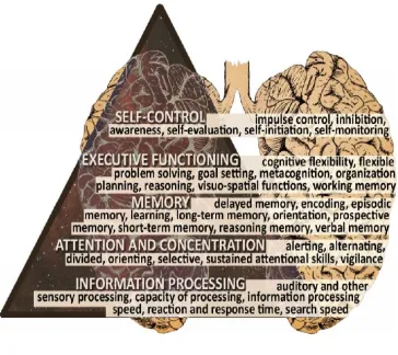
FIGURE - 4
COGNITION DOMAINS AND SPECIFIC FUNCTIONS
Executive function is the function of dorsolateral prefrontal cortex.
Executive functions play a role in planning and initiation of independent
activities, self monitoring, and performance, switching between tasks, inhibition
of inappropriate responses and planning complex motor and problem solving
for executive functions, more recent and advanced research has led to the
discovery of increased role played by extreme interconnections between
subcortical and cortical regions of brain. Executive functions are key to our
capacity, formulating our goals, planning and organising such goal directed
behaviours, carrying out such behaviours fully and effectively and monitoring
and self correcting ones behaviour as needed37
Attention or Vigilance is the ability to maintain attention over time.
Verbal learning and memory is the ability to learn more information, to retain
newly learned information over time, and recognising previously presented
material. Verbal fluency is the ability to produce as many words as possible.
Immediate or Working memory is the ability to hold a limited amount of
information for a brief period of time. Working memory is the core component
of neurocognition, which is mediated by prefrontal cortical regions.
Development of thought in a child was studied by Jean Piaget in 20th C, an
eminent Swiss epistemologist.
Knudsen38 describes a model which shows four processes of attention with working memory in the centre.
1. Working memory temporarily stores information for analysis.
2. Competitive selection that determines which information goes to working
3. Top-down sensitivity control by the content of working memory which
influences the selection of new information. This results in voluntary
control of attention in a recurrent loop – endogenous attention
4. Bottom-up filters which automatically enhance the response to infrequent
stimuli – exogenous attention.
The term "working memory" was coined by Miller, Galanter
and Pribram.39 Most working memory tasks recruit a network of prefrontal cortex and parietal areas. During a working memory task, the connectivity
between these areas increases.40
Working memory is a special short term memory store. It has three
component systems (Baddley and Hitch model):
a) Attentional control system: It focuses on perception of specific
happenings in the environment. It is located in the prefrontal cortex and
has very limited capacity. It regulates the flow of information to two
rehearsal systems which are thought to sustain memory for temporary
use.
b) Rehearsal systems- Articulatory loop: It refers to a storage system with
a quickly fading memory trace. Here, memory for numbers and words
can be sustained by subvocal speech e.g remembering a new mobile
c) Visuo-spatial sketch pad: It represents both the spatial location and the
visual properties of object to be remembered. For example, this system
allows a person to store the image of an individual’s face whom he met
at a dinner party.
The two rehearsal systems are situated in the posterior association areas.
The information processed in either of these systems has the possibility of
reaching long term memory. The cognitive state examination includes
orientation, attention, concentration, memory, general information, ad
intelligence.
NEUROPSYCHOLOGICAL TESTS
Mini Mental State Examination (MMSE):
MMSE (FOLSTEIN et al., 1975)41 takes 5-10 mins to administer and
test retest reliability is high. It provides a rough and ready index of cognitive
functioning. First part covers orientation, attention, concentration and memory.
Second part tests the ability to name common objects, follow verbal or written
commands, write a sentence spontaneously and copy a simple figure (eg:
overlapping pentagons) - visuospatial testing.
The total score is 3042, scores less than 24 indicates cognitive
impairment.
The basic aspect provided by the Stroop is that it has been associated with
cognitive flexibility, resistance to interference from outside stimuli, creativity,
psychopathology, and cognitive complexity. It clearly plays a role in many
interrelated cognitive processes which determine an individual's ability to
successfully cope with cognitive stress and to process complex input (Golden,
1978).
Inhibitory processing is the efficiency of the inhibitory process that
underlies selective attention. Inhibition allows a reduction of irrelevant
information to enter working memory (Hasher & Zacks, 1988). The area of the
brain which is usually affected by inhibition is the frontal lobe. The test
involves multiple areas including knowledge, attention, visual scanning and
acuity. As the impairment in executive function severely affects the quality of
life, the rehabilitation measures should be directly targeted on management of
executive functions after quantifying the problem (Crawford 2000)43. John
Ridley Stroop (1935) worked on the interference that can arise between word
reading and colour naming. It takes longer time to read printed colour names
when they are printed in coloured ink different from that of the coloured word.
This may be due to
1) response conflict
2) failure of response inhibition
The increase in time taken to perform the second task compared with the
first task is referred to as “the Stroop interference effect” (e.g., Davidson,
Zacks, & Williams, 2003; Moering, Schinka, Mortimer, & Graves, 2003). It
gives selective attention, cognitive flexibility and control, self correction and
speed of processing (Uttl & Graf, 1997) or executive functioning (Moering et
al., 2003).
Slowing with age has been consistently documented (Hasher & Zacks,
1988; Obler and Albert, 1985; Spreen and Strauss, 1991). The results also
reinforce the slowing due to decreased oxygen supply to the brain as identified
in those with COPD (Clark).
COGNITIVE EVOKED POTENTIAL STUDY
Evoked potentials refer to the action potentials generated from central
nervous system in response to a specific and adequate stimulus. They are small
and are buried in the background of spontaneous electrical activity (EEG). Their
details can only be studied and evaluated by repeated stimulation and averaging
the responses obtained after each stimulation. Evoked potentials have evolved
from a challenging scientific technique to a commonly applied tool in the
neurophysiology in the last few decades44. They establish objective evidence of
abnormality when signs and symptoms are equivocal.
Evoked potentials are classified into
2. Event related potentials (ERP) or Endogenous potentials 23
Stimulus related potentials:
They are generated in response to an exogenous stimulus – visual,
auditory, and somatosensory. They depend on the physical characteristics of the
stimulus. Commonly used stimulus related potentials are brainstem evoked
response audiometry, visual evoked potential and somatosensory evoked
potential.
Event related potentials:
In 1935-1936, Pauline and Hallowell Davis recorded the first known
ERPs on awake humans. In 1964, Grey Walter and colleagues reported
cognitive ERP component, called the contingent negative variation (CNV).
Sutton, Braren, and Zubin (1965) made advancement with the discovery of
the P3 component. In 1980s, the introduction of inexpensive computers opened
up a new door for cognitive neuroscience research. Currently, ERP is one of the
most widely used methods in cognitive neuroscience research to study the
physiological correlates of sensory, perceptual and cognitive activity associated
with processing information.
Over the past few decades, electrophysiology has contributed
substantially to the understanding of normal as well as abnormal brain function
deviations in psychopathological conditions. Electrophysiological techniques
resolution, providing the best methods to describe the time course of
brain-electrical activation during complex cognitive processes (such as conscious
sensory discrimination or semantic processing). They are elicited when the
subject is required to distinguish one stimulus (the target) from the other (non-
target). Since ERPs are related to cognitive processing associated with the
distinction of target from non-target stimuli, they are also called cognitive
evoked potential. ERPs are also referred to as P300, because they occur after a
latency period of approximately 300 milli seconds,45,46. ERPs are independent of
the physical characteristics of the stimulus. When a stimulus is presented to the
brain, it travels through the receiving sensory organ and the brainstem (except
for visual and olfactory stimuli) on its way to the primary cortical regions
mediating this particular sensory modality. ERPs recorded over primary sensory
cortex indicate how the stimulus was received and recorded by the brain,
thereby providing an index of the integrity of the underlying neural architecture.
For instance, the early waves arising immediately after (within about 15 ms) an
auditory stimulus are referred to as the brainstem auditory evoked potential.
These components reflect the intactness of the pathways that they traverse, are
useful for the diagnosis of sensory defects (such as hearing loss), and thus are of
great interest to neurologists.
Once received by the brain, a stimulus undergoes psychological
processing and evaluation, and these operations are reflected in later ERP waves
that are distributed to various scalp locations. These components are influenced