CLINICAL PROFILE ,ETIOLOGY,MANAGEMENT AND
OUTCOME OF SERUM ELECTROLYTE DISTURBANCES IN
CHILDREN ADMITTED IN PEDIATRIC INTENSIVE CARE
UNIT IN A TERTIARY CARE CENTRE
Dissertation submitted to
THE TAMILNADU DR.M.G.R.MEDICAL UNIVERSITY
In partial fulfilment of the regulations for the award of degree of
M.D DEGREE (PEDIATRICS) BRANCH VII
INSTITUTE OF SOCIAL PEDIATRICS
STANLEY MEDICAL COLLEGE
CHENNAI – 600 001
DECLARATION
I, Dr.J.BALAJI solemnly declare that the dissertation titled “CLINICAL PROFILE,ETIOLOGY,MANAGEMENT AND OUTCOME OF SERUM ELECTROLYTE DISTURBANCES IN CHILDREN ADMITTED IN PEDIATRIC INTENSIVE CARE UNIT IN A TERTIARY CARE CENTRE” was done by me at
Government Stanley Medical College during 2013- 2016 under the guidance and supervision of my chief Prof. S.SHANTHI M.D, D.C.H.
The dissertation is submitted to
The Tamilnadu Dr.M.G.R Medical
University
towards the partial fulfilment of the rules and regulations for the
M.D.
Degree Examination - BRANCH VII - in Pediatrics
.
Place : Chennai
signature of the candidate
CERTIFICATE BY THE GUIDE
This is to certify that the dissertation titled
“CLINICAL PROFILE,
ETIOLOGY,
MANAGEMENT AND OUTCOME OF SERUM
ELECTROLYTE DISTURBANCES IN CHILDREN ADMITTED IN
PEDIATRIC INTENSIVE CARE UNIT IN A TERTIARY CARE CENTRE ”
is a bonafide research work done under my guidance by
Dr.J.BALAJI
Postgraduate student,Department of Pediatrics, Government Stanley medical
college, The Tamilnadu Dr.M.G.R Medical University, Chennai, in partial
fulfilment of the requirement of the award for the degree of
M.D PEDIATRICS
-
BRANCH VII.
Place : Chennai Date :
Signature of the Guide
CERTIFICATE BY THE INSTITUTION
This is to certify that the dissertation titled
“CLINICAL PROFILE,
ETIOLOGY,
MANAGEMENT AND OUTCOME OF SERUM
ELECTROLYTE DISTURBANCES IN CHILDREN ADMITTED IN
PEDIATRIC INTENSIVE CARE UNIT IN A TERTIARY CARE CENTRE”
is submitted by
Dr.J.BALAJI
to
The Tamilnadu Dr.M.G.R Medical
University, Chennai
in partial fulfilment of the requirement of the award for the
degree of
M.D BRANCH VII (PEDIATRICS)
and is a bonafide work done by
him under our direct supervision and guidance, during the academic year
2013 -2016
DR.SHANTHI MD, DCH Professor and HOD
Institute of Social Pediatrics Stanley Medical College Chennai –600001
Prof .Dr.ISAAC CHRISTIAN MOSES MD Dean
Stanley Medical College Chennai –600001
ACKNOWLEDGEMENT
It is with immense pleasure and gratitude that I thank
Dr.ISAAC CHRISTIAN
MOSES
M.D.,DEAN, STANLEY MEDICAL COLLEGE
for bestowing me
the permission and privilege of presenting this study and for enabling me to avail
the institutional facilities.
I am gratefully indebted to
Prof.Dr.S,SHANTHI
M.D, DCH,Director, Department
of Pediatrics, Stanley Medical College for her valuable guidance and motivation .
I sincerely thank
Prof.Dr.SARAVANAN,
M.D,Department of Biochemistry,
Stanley Medical College for offering guidance and encouragement throughout the
study, and for permitting me to avail the lab facilities that has made this study
possible.
I am very grateful to both my chiefs,
Prof.Dr.Lakshmi
M.D, DCHand
Prof.Dr.Devimeenakshi
M.D, DCHand Prof.Dr.Vivekanandhan
M.D,DCHfor
guiding through my dissertation process and providing departmental resources for
the conductance of this study.
I am extremely thankful to
Dr.Elango
M.D, DCHMedical Registrar, for his
I express my gratitude to the Assistant Professor of PICU
Dr.Ekambaranath
M.Dfor his valuable help and guidance for this study.
I sincerely thank my Assistant Professor
Dr.P.Venkatesh
M.Dfor his timely help
and support throughout the course of this study.
I thank my Assistant Professors
Dr.Radhika
M.D, Dr.Raja
M.D,
Dr.Vinoth
M.D, Dr.Kumar
DCHand
Dr.Sankara Narayanan
M.Dand
Dr.Parveen kumar M.D
for their valuable support.
I express my heartfelt thanks to Late
Dr.V.Ezhil Srinivasan
M.Dwhose words of
knowledge and encouragement has stayed with me throughout this course of study.
I sincerely thank all the patients and their parents who participated in this study.
I thank all the PICU staff nurses for their immense help in conducting this study.
Finally I thank all the post graduates in the Department of Pediatrics in our Stanley
Medical College who have helped me through thick and thin. It was an immense
CLINICAL PROFILE,ETIOLOGY,MANAGEMENT AND OUTCOME OF SERUM ELECTROLYTE DISTURBANCES IN CHILDREN ADMITTED IN PEDIATRIC INTENSIVE CARE UNIT IN A TERTIARY CARE CENTRE
Dr J.Balaji postgraduate Pediatrics,Govt Stanley hospital
ABSTRACT :
Title : Clinical profile,etiology,management and outcome of serum electrolyte
disturbances in children in the Pediatric Intensive Care unit in a tertiary care centre.
Aims and objectives: This prospective study evaluated the frequency, causes
,management and outcome pattern of sodium, potassium, calcium, chloride disturbances.
Out of 227 patients studied from January 2015-august 2015,236 series (multiple
episodes) of electrolyte disturbances were noted, most common electrolyte abnormality
being hyponatremia (35.2% of cases) ,and is mostly of euvolemic type, the most common
age group being 1month-1year,more in male children, commonest cause being seizures
followed by poisoning. Not all children with electrolyte abnormality have symptoms or E
CG abnormalities
Conclusion :Administration of routine maintenance fluids which are hypotonic may
worsen hyponatremia.Electrolyte disturbances are common in PICU and high index of
suspicion is needed,so that we can prevent electrolyte abnormalities and its
complications.
CONTENTS
S.NO
TITLE
PAGENO
1
INTRODUCTION
1
2
AIM AND OBJECTIVES
4
3
REVIEW OF LITERATURE
5
4
MATERIALAND METHODS
32
5
OBSERVATION AND RESULTS
37
6
DISCUSSION
72
7
CONCLUSION
79
8
BIBLIOGRAPHY
9
PROFORMA
10
CONSENT FORM
11
ABBREVIATIONS
CLINICAL PROFILE,ETIOLOGY,MANAGEMENT AND OUTCOME
OF SERUM ELECTROLYTE DISTURBANCES IN CHILDREN
ADMITTED IN PEDIATRIC INTENSIVE CARE UNIT IN A
TERTIARY CARE CENTRE
INTRODUCTION:
Electrolytes are substances that ionizes when dissolved in suitable ionizing
solvents
eg :water
Electrolyte abnormalities are common in children who need intensive care ,
they occur in variety of conditions, may remain unrecognized and result in
morbidity and mortality irrespective of primary problem. Early recognition, a
high index of suspicion and a thorough understanding of common electrolyte
abnormalities is necessary to ensure their correction.
y Hyponatremia is particularly common in sick hospitalized children .
It is invariably associated with hypo-osmolality and normal hydration
and is attributed to SIADH. Acute hyponatremia poses an immediate
danger to central nervous system.
y Hypernatremia occurs less frequently than hyponatremia on other hand,
patients debilitated enough to develop hypernatremia carry a high
y A number of conditions predispose patients in the PICU to
hyperkalemia such as renal insufficiency, adrenal insufficiency,
insulin deficiency, resistance, tissue damage such as rhabdomyolysis,
burns or trauma.
y The development of many electrolyte disturbances in PICU can be
prevented by attention to use of intravenous fluids and nutrition.
y The development of acute renal failure continues to be a problem that
markedly affects outcome in critically ill children.despite advances in
treatment development of acute renal failure continues to be associated
with high mortality rates .
y Delayed resuscitation will lead to ongoing kidney injury which in turn
KNOWLEDGE GAP :
This study and the thesis presented here at Government Stanley Medical
College Hospital PICU gains significance as there hasn’t been any of it’s kind
prior to this.Hence survey regarding such electrolyte profile will lead to better
understanding towards electrolyte disturbances in our Government Stanley
Medical College PICU. Understanding such risks will enable us to provide
appropriate fluid management and reduce the incidence of electrolyte
disturbances in the PICU.
The osmolality and volume in the intravascular space are regulated by
independent systems of water balance that in turn determines osmolality
AIMS AND OBJECTIVES
y To know most commonly occurring electrolyte
abnormalities,etiology,management and outcome of serum electrolyte
disturbances in sick children admitted to pediatric intensive care unit to
Institute of Social Pediatrics Stanley medical college.
y To study the outcome pattern amongst the critically ill children seeking
emergency care with electrolyte abnormalities with respect to
REVIEW OF LITERATURE:
y S.D. Subba rao. et al and Biju Thomas et al analysed 305 patients in
St.john’s hospital aged between 1 month and 14 years , who were
admitted in PICU during the period. Ninety nine (32.45%) had
electrolyte abnormalities. Of these 24 (7.9%) had mixed electrolyte
imbalance.
y Hyperkalemia was the commonest found in 44 (14.4%)cases,
hyponatremia was seen in 11 (3.6%) cases which is second commonest
abnormality noted. Of the 99 patients with electrolyte imbalance, 24
(24.2%) expired. In these 24 patients 10 (41.6%) had hyperkalemia ,
6(25%) had hyponatremia , 5(20.8%) had hypernatremia , 3(12.5%) had
hypokalemia
y SV.S.S. Prasad , Sunit Singhi , K.S.Chugh et al analysed a total of 727
Children admitted in PICU the frequency distribution of serum sodium
concentration in 727 children , hyponatremia was present in
217(29.8%) children while severe hyponatremia ( serum sodium <125
mEq/l was found in 47(6.4%). Amongst those with severe hyponatremia
in 21 the serum sodium ranged between 121-125 mEq/l. in 17 between
116-120 mEq/l and in 9 patients it was 115mEq/l or less. hypokalemia
was found in 101 (13.9%) and 39 (5.4%) had hyperkalemia while In
their study pneumonia and diarrheal disease, each accounted for about
33% in meningoencephalitic illness, being higher in summer as
compared winter seasonal difference was observed
y 587 were normokalemic.
y Lamia. M Al Naama, Jawad Kadhum Abdul –Hassan et al, performed
case control study on 150 children (87 boys and 63 girls), of age group
between 2 months and 9yrs. 75 of them presented with acute CNS
manifestations while the rest were considered as control.
y Eight of 75 pediatric patients (10.7%) with acute CNS diseases had
hyponatremia syndrome, three were diagnosed with inappropriate
antidiuretic hormone secretion. The highest percentage of hyponatremia
(3 out of 6 patients) was found in patients with intracranial diseases.
Four out of 38 patients(15.5%) presented with CNS infections
y Singhi S et al Indian pedia 1996 Jan 33(1.8%)9-14. 43(14) patients had
54 episodes of hypokalemia .predisposing factors included the nature of
primary disease (renal disease 19%, acute diarrohea 14%,CCF &
meningoencephalities 12% each). The over all mortality among patients
with hypokalemia (25.6%) was significantly higher than that among the
remaining PICU patients(10.9%), all the patients receiving rapid
correction survived
y Cummings BM et al Intensive care medicine. 2013 june 6. A total of
512 patients had a potassium measurement. Of a total of 4484 potassium
40% of the admissions. Mild hypokalemia (3-3.4 mmol/l ) affected 24%
of the admissions. Moderate or severe hypokalemia (k<3.0mmol/l)
affected 16% of admissions.
y Hyperkalemia affected 29% 0f admissions. Mild hyperkalemia (5.1 –
6.0 mmol/l) affected 17% of admissions. Moderate or severe
hyperkalemia (>6.0 mmol/l) affected 12%. On univariate analysis,
severity of hypokalemia was associated with mortality
y Hoorn et al found a 30% overall incidence of pNa less than 136mmol/l
and a 38% incidence in the ICU. Severe hyponatriemia PNa less than
125mmol/l, was present in 3% of hospitalized patients and occurred
during hospitalization in fully half of cases of the cases
About two thirds of the human body is made up of water. In an infant and child
total body water is 65%.As the child grows older, TBW decreases to 60% in
male and 55% in female. The percentage of intracellular fluid compartment
remains the same in all age groups as 40%.Fall in TBW is reflected in ECF.
Sodium disturbances :
Normal sodium levels 135-145 meq/L
Serum sodium <135meq/L – hyponatremia
• Sodium Bulk cation of extracellular fluid Æ change in SNa reflects
change in total body Na+
• Principle active solute for the maintenance of intravascular & interstitial
volume
• Absorption: throughout the GI system via active Na,K-ATPase system
Excretion: urine, sweat & feces
• Kidneys are the principal regulator
– 2/3 of filtered Na+ is reabsorbed by the proximal convoluted
tubule, increase with contraction of extracellular fluid
– Countercurrent system at the Loop of Henle is responsible for Na+
(descending) & water (ascending) balance – active transport with
Cl
-– Aldosterone stimulates further Na+ re-absorption at the distal
convoluted tubules & the collecting ducts
– <1% of filtered Na+ is normally excreted but can vary up to 10%
if necessary
• Major component of serum osmolality
– Sosm= (2 x Na +
) + (BUN / 2.8) + (Glu / 18)
– Normal: 285-295
Hyponatremia :
The age and sex of the child does not affect the frequency of hyponatremia but
a significant seasonal difference was apparent in the present study. Incidence of
hyponatremia was higher in summer as compared to winter existed through
most of the diagnostic categories,one exception being diarrhea.Higher amount
of water and salt loss through sweating might have contributed to this
differences. The observation implies suspicion of hyponatremia in summer
could be enormously increased and this should be anticipated .The commonest
cause of low sodiumdue to bronchopneumonia,diarrhea,meningitis with
encephalitis.
In contrast to this, the frequency that was reported as hyponatremic
dehydration was only 10% in children with acute diarrhea in the western
countries.this lead to prediction that hyponatremia in diarrhea may be
hypovolemic type that may be due to excessive sodium loss in gastro-intestinal
secretions; intake of salt free drinks and increased loss of salt through sweating
in our climate might have contributed.
Apparently, hyponatremia occurs frequently without having significant changes
in extracellular fluid volume in children with infectious diseases needing
hospitalisation and needs to be looked into and should be managed
Symptoms of hyponatremia include nausea, abdominal cramps, vomiting,
headache Edema ,muscle weakness, tremor, paralysis, disorientation, slowed
breathing, seizures coma.
Studies on the mechanism of hypotonic hyponatremia with euvolemia have
showed a role of syndrome of inappropriate ADH secretion (SIADH) in those
children. Almost every patients having hypotonic hyponatremia with
euvolemia could be classified as having SIADH as these patients had normal
hydration status, decreased plasma osmolality , increased urine osmolality and
normal renal function.There is inappropriately higher concentration of plasma
vasopressin than that is expected for the degree of hyposmolality has been
demonstrated in asso-ciation with euvolemic hyponatremia in children with
meningitis, asthmaand hospitalized adults.
Thus, ADH mediated renal salt loss and water retention could be the cause of
hyponatremia in such patients. However, severehyponatremia may occur in
association with SIADH without unusual loss of sodium from the body or
dilution of the plasma sodium. Evidence supporting redistribution and
accumulation of sodium within the cells has also been presented. Recently,
Hannon and Bos-ton have shown significant intracellular shifts of sodium
chloride and water in septic animals. They suggested that in these animals
hyponatremia and hypoosmolality was caused by a combination of intracellular
shift of sodium and water, and dilution of extracellular space probably as a
Data from our ongoing research suggest a significant increase in RBC sodium
in the presence of hyponatremia in septicemic children. Further studies are
needed to clarify the mechanism responsible for hyponatremia in acute
infections. In conclusion, hyponatremia occurs frequently and should be looked
for in all sick children. It is of hypotonic-euvolemic type in almost all the acute
infections except diarrhea and should be managed accordingly.
HYPERNATREMIA
Hypernatremia defined as serum sodium >145 meq/l have a deficit of water in
relation to the body sodium stores, net water loss or a hypertonic sodium gain.
most cases are due to net water loss, where as hypertonic sodium gain results
from sodium loading accidentally or some interventions.Increased sodium in
extracellular compartment leads to increased plasma tonicity, this in turn leads
to water movement across cell membrane result in cellular dehydration.
Mechanism leading to hypernatremia or along with the same includes the
following
x Water depletion eg:diabetes insipidus
x Depletion of water exceeds depletion of sodium eg:diarrhea
x Excess of sodium eg:salt poisoning
the risk is highest among infants and ventilated patients.
Infants with large surface area with respect to height or weight in
diarrhea and infant formula that is improperly prepared or inadequate infant
mother interaction during breastfeeding have higher incidence of
hypernatremia. There will be increased efflux of water from cellular
compartment inorder to maintain osmolality that is equal inside and outside
the cell.cell contraction make the brain cells excessiblyvulnerable.Therefore
hypernatremic dehydration causes shrinkage of brain, tear cerebral
bloodvessels, cerebral hemorrhage, paralysis, seizures and encephalopathy.
Rapid rehydration of hypernatremic patients with hypotonic fluids may
cause cerebral edema that can lead in coma, seizures or death.
Causes of hypernatremia include ,
1) hypovolemic hypernatremia due to increased loss of hypotonic fluids
Increased loss of hypotonic fluid coupled with insufficient water intake is
the commonest cause of hypernatremia in children.
There is decreased total body sodium ,body content of water is decreased
further.
There is shift of water from ICF to ECF as a compensation to increased
serum osmolality. Here the circulating volume is maintained til there is
marked water deficits. Osmotic diuretics like glycerol or mannitol and
diuresis in diabetes mellitus can cause hypernatremia due to increased urine
losses.children with hypernatremic dehydration due to nonrenal losses have
2) Hypernatremia due to water deficit :
Central or nephrogenic diabetes insipidus, excessive sweating, fever,
sustained hyperventilation, urine is concentrated with low urine sodium.
3) Hypernatremia due to excess sodium euvolemic hypernatremia :
Seen in solutions for rehydration with increased sodium,concentrated
formula,iatrogenical,urine concentrated normally,with increased sodium
losses.
Potassium disturbances:
Potassium values normal range 3.5-4.5 meq/L
• Largely contained intra-cellularÆSKdoes not reflect total body K
• Important roles: contractility of muscle cells, electrical responsiveness
• Principal regulator: kidneys
• Complete absorption in the upper GI tract
• Kidneys regulate balance
• 10-15% filtered is excreted
• Aldosterone: increase K+& decrease Na+excretion
• Mineralocorticoid & glucocorticoid Æincrease K+& decrease Na+
excretion in stool
• Solvent drag
• 0.6 SK/ 10 of Sosmo
• Evidence of solvent drag in diabetic ketoacidosis
• Acidosis
• Low pH Æshifts K+out of cells (into serum)
• Hi pH Æshifts K+intocells
• 0.3-1.3 mEq/L K+change / 0.1 unit change in pH in the opposite
direction
Causes of hyperkalemia
Spurious
Difficult blood draw Æhemolysis Æ false reading
Increase intake
Iatrogenic: IV or oral
– Blood transfusionsDecrease excretion
» Renal failure
» Adrenal insufficiency or CAH
» Hypoaldosteronism
» Urinary tract obstruction
» ACE inhibitors
» Potassium sparing diureticsTrans-cellular shifts
» Acidemia
» Rhadomyolysis; Tumor lysis syndrome; Tissue necrosis
» Succinylcholine
» Malignant hyperthermia
HYPERKALEMIA :
– Intestinal ischemia shock was induced either by temporary occlusion of
the three splanchnic arteries for 40 min (SAO-shock) or by temporary
occlusion of the portal vein for 35-40 min (PVO-shock). In both types of
shock, life can be considerably prolonged (5-8-fold) by treatment with
rat plasma plus glucose. Eventually, death is caused by heart failure due
amount of K+ causing this hyperkalemia is estimated at roughly 10% of
the total body K+. Acidosis, low blood pressure, reduced kidney
function, and disintegration of erythrocytes in the gastrointestinal (GI)
tract probably are of no or the skeletal muscles, or the erythrocytes
release K+. Although the K+ concentration of the contents of the GI
tract as well as the K+ transport by the portal vein were increased, the
source of the excess K+ remains obscure. Removal of the contents of the
stomach and small intestine, followed by flushing of the gastrointestinal
tract, may have a favorable effect on the course of plasma K+ (and
plasma glucose) concentration, indicating that toxic products from the
damaged intestines may be important lethal factors
– minor importance in causing this extreme hyperkalemia. No indication
was found that the liver, the skeletal muscles, or the erythrocytes release
K+. Although the K+ concentration of the contents of the GI tract as
well as the K+ transport by the portal vein were increased, the source of
the excess K+ remains obscure. Removal gastrointestinal tract, may
have a favorable effect on the course of plasma K+ (and plasma glucose)
concentration, indicating that toxic products from the damaged intestines
may be important lethal factors.
– Clinical features of hyperkalemia includes weakness,nausea,abdominal
HYPOKALEMIA:
POTASSIUM disturbance especially hypokalemia, during admission was
known to occur in a number of patients . Low potassium level can have effects
that are profound on electrical activity in cardiac, skeletal and smooth muscle.
These disturbances if severe, result in life threatening conditions like
arrhythmias, cardiac-arrest, respiratory failure, paralytic ileus and paralysis of
muscles.Hypokalemia appears to be one of the most common electrolyte
disturbances in sick children. Studies addressing its incidence and outcome are
few and chiefly from developed countries.
Hypokalemia is common in critically ill children and have a significantly
higher mortality. Patients having preexisting kidney disease,
septicemia,bronchial asthma, cardiac disease with congestive cardiac failure,
diarrhea that is severe and meningoencephalitis were most likely to show
evidence of hypokalemia. The apparent cause for occurance of hypokalemia in
most patients being a loss of potassium from the body through gastrointestinal
or urinary tract , because of underlying disease process, or because of
associated use of drugs like diuretics,corticosteroids and asthma drugs. In a
patient with DKA hypokalemia would be due to correction of acidosis and
insulin use . In critically sick children hypokalemia may be caused by
Hypokalemia Causes
– Distribution from ECF
» Hypokalemic periodic paralysis
» Insulin,Ǻ-agonists, catecholamines, xanthine
– Decrease intake
– Extra-renal losses
» Diarrhea
» Laxative abuse
» Perspiration
– Excessive colas consumption
– Renal losses
– DKA
– Diuretics: thiazide, loop diuretics
– Drugs: amphotericin B, Cisplastin
– Hypomagnesemia
– Alkalosis
– Hyperaldosteronism
– Licorice ingestion
Hypokalemia
- Flattened or inverted T-wave
- U wave: prolonged repolarization of the Purkinje fibers
- Depressed ST segment and widen PR interval
- Ventricular fibrillation can happen
Symptoms of hypokalemia include weakness,paralysis,increased
urination,arrhythmias,orthostatic hypotension,muscle pain ,tetany
Calcium disturbances :
Normal range of total calcium is 8.8-10.8 mg/dl (2.2-2.7mmol/L)
HYPOCALCEMIA :
Total calcium <8mg/dl or ionized calcium <1mmol/L seen in seriously ill
children.ionized calcium if possible should be measured.
Causes for hypocalcemia :
Multiple transfusion –chelation of calcium by citrate.ionized calcium is low
But total calcium is normal.high alkalosis causes more calcium to be bound
to albumin leading to decreased ionized calcium.increased phosphate,septic
shock,
Rhabdomyolysis,tumourlysis,acutepancreatitis,hypoparathyroidism,
Pseudohypoparathyroidism,decreased magnesium levels,nutritional like
malabsorption,hepatic diseases,antiepileptics therapy,kidney failure.
Clinical features of hypocalcemia :neuromuscular activity like
seizures,tremors,laryngospasm,carpopedal spasm,muscle cramps.
Hypotension,cardiac failure.increased risk of digitalis toxicity,sapsi like
syndrome.
Calcium is the most abundant mineral in the body. 99% of the total body
calcium is stored in bone, and serum levels constitute less than 1%. Various
factors regulate the homeostasis of calcium and maintain serum calcium within
renal function (for conversion of vitamin D to active metabolites), and serum
phosphate and magnesium levels.
Serum calcium is present in two forms: the free (ionized) and the bound form.
Only about 50% of circulating calcium is present in the physiologically free
form. The rest is either bound to proteins (40%) or complexed (10%) with
bicarbonate, citrate, and phosphate. The ionized calcium level varies based on
the level of serum albumin, blood pH, serum phosphate, magnesium, and
bicarbonate levels, the administration of transfused blood containing citrate and
free fatty acid content in total parenteral nutrition. The normal range for
ionized calcium is 1-1.25 mmol/L (4-5 mg/dL).
The concentration of calcium in the serum is critical to many important
biologic functions, including the following:
x Calcium messenger system by which extracellular messengers regulate cell
function
x Activation of several cellular enzyme cascades
x Smooth muscle and myocardial contraction
x Nerve impulse conduction
x Secretory activity of exocrine glands
Symptoms of hypocalcemia :
Hypocalcemia manifests as central nervous system (CNS) irritability and poor
muscular contractility. Low calcium levels decrease the threshold of excitation
Because neuronal excitability occurs in sensory and motor nerves,
hypocalcemia produces a wide range of peripheral and CNS effects, including
paresthesias, tetany (i.e., contraction of hands, arms, feet, larynx, bronchioles),
seizures, and even psychiatric changes in children.
Muscle excitability is depressed because hypocalcemia impedes acetylcholine
release at neuromuscular junctions and, therefore, inhibits muscle contraction.
However, the increase in neuronal excitability overrides the inhibition of
muscle contraction. Cardiac function may also be impaired because of poor
muscle contractility.
Hypercalcemia :
Total calcium >10.5mg/dl or ionized calcium >1.2mmol/L
Hypercalcemia seen in oncology patients, primary hyperparathyroidism
prolonged immobilization.
Symptoms of hypercalcemia :
CNS- slowed nerve conduction :CNS depression,fatigue, seizures and coma
CVS- arrhythmias ,ventricular ectopy,myocardial depression
GIT – constipation,nausea,vomiting,peptic ulcer disease
• Hypercalcemia: Clinical presentation
– Groans: constipation
– Moans: psychic moans (fatigue, lethargy, depression)
– Bones: bone pain
– Stones: kidney stones
– Psychiatric overtones: depression & confusion
– Fatigue, anorexia, nausea, vomiting, pancreatitis
– ECG: short QT interval, widened T wave
Chloride :
• with Na+ and H2O to help maintain cellular integrity, fluid balance and
osmotic pressure
• Affects acid/base balance (enzyme activator, serves as buffer in
exchange of O2 and CO2 in RBC’s)
• Major ECF anion
• Serum level: 95-108 mEq/L
Function; circulates In conjunction with Ca+, Mg+, helps maintain nerve
transmission/muscle function
• Obtained primarily from foods (processed) and table salt, daily need:
~750mg.90% excreted by kidney
MAINTENANCE FLUID AND ELECTROLYTES
-Based on the caloric expenditure model, each calorie expended requires
provision of water in the ratio of 1 ml/cal metabolized/day at rest.
-Also, according to the caloric expenditure model maintenance sodium and
potassium ranges/100 ml of maintenance fluid/day have been determined.
Upper limit has been chosen for sodium and lower limit has been chosen for
potassium to be placed in the “Segar box”.
-Common names for commercially available saline solutions and their
sodium concentrations are:
Normal saline (0.9% NaCl/L) 154 mEq Na+/L
One-half normal saline (0.45% NaCl/L) 77 mEq Na+/L
One-third normal saline (0.33% NaCl/L) 57 mEq Na+/L
One-quarter normal saline (0.2% NaCl/L) 34 mEq Na+/L
Ringer’s lactate 130 mEq Na+/L
(Contains 4 mEq K+, 109 mEq Cl-, 28 mEq bicarb equivalent all/Liter, and
3mg/dl of Ca++)
-Addition of glucose to each of the above at a minimum of 5 gm/100 ml
are somewhat “spared” from providing substrate for gluconeogenesis.
Ketosis from fat metabolism is also prevented.
An intravenous fluid is the commonest intervention administered to a
Hospitalized children. The indications for administering intravenous fluids
are to either expand a contracted extracellular fluid or as maintenance
fluids to
replace urine output and insensible water loss in a fasting patients.the
administration of IVF is considered as an invasive procedure and should be
dealt with the same respect and vigilance as any drug prescription.
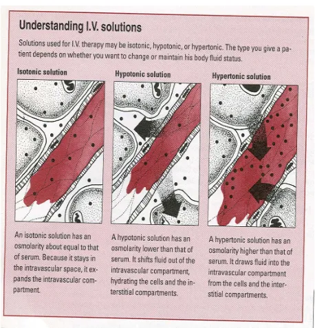
Objective of intravenous fluid therapy is to fill the intravascular
compartment. Fluids that are isotonic include normal saline NS or Ringers
lactate RL will be retained in the vascular compartment more than other
fluids.
Fluids that are hypotonic include ½ GNS, ¼ or 1/5GNS whwn
administered will stay in vascular compartment in small quantities.
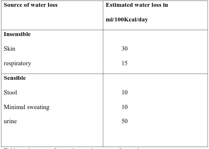
Source of water loss Estimated water loss in
ml/100Kcal/day
Insensible
Skin
respiratory
30
15
Sensible
Stool
Minimal sweating
urine
10
10
[image:34.595.100.531.71.377.2]50
Table no.1,source of water loss and amount of water loss
Osmolality :
Osmolality is the number of osmotically active particles present in a solution
per kilogram of solvent.
Osmolarity :
Osmolarity is the number of osmotically active particles present per litre of
solution.
These two terms are often used interchangeably.
Tonicity :
Tonicity is the effective osmolality of a solution and is equal to the sum of the
semipermeable membrane that is impermeable solutes eg sodium.Tonicity is
the property of the solution with reference to its membrane.osmolality and
osmolarity both is the property of a solution independent of any membrane
because it includes both impermeable and permeable solutes like urea.
One example 5%dextrose is isoosmolar initially with plasma but in normal
conditions,dextrose is permeable across the membrane and an ineffective
solute that readily enters the cell. Therefore 5% dextrose is isoosmolar with
plasma but hypotonic with reference to cell membrane.
The most potent stimuli for ADH secretion are an increase in serum
osmolality, hypovolemia and hypotension. Non osmotic stimuli like pain,
drugs, anesthetic agents, stress, nausea, and vomiting can also cause increase in
ADH secretion .ADH checks the renal water excretion even if there is low
plasma osmolality. This impaired ability to excrete hypotonic urine coupled
with positive balance of electrolyte free water leading to hyponatremia.
Therefore administration of hypotonic fluids will exacerbate the fall in sodium
concentration.
Sodium is the main cation in the extracellular fluid,thereby determining the
ECF volume.An isotonic solution will have approximately 154 meq/L
monovalent cations .Normal tonicity is 270-290 mosm/kg.Drastic variation in
the tonicity of the extracellular compartment causes water shifts that leads to
Sodium determines the extracellular fluid volume ,regulates the movement of
water across the cell membrane thereby determining intracellular edema that
occurs in the presence of hyponatremia.Children have a larger intracellular
fluid volume per total skull volume,therefore greater risk of neurological
sequelae secondary to hyponatremia.No single fluid rate or composition is
ideal for all children ,however isotonic solutions may be the most physiological
and therefore the safest empirical choice for maintenance IV fluids in the
critical care setting.
The choice of a solution in the acute setting should not be for the purpose of
satisfying the calculation of the daily sodium or caloric requirements for a
healthy child ,but should aim to maintain tonicity balance in the acute phase of
illness and in the postoperative period when the patients are at highest risk of
fluid and electrolyte abnormalities, in particular hyponatremia.Excretion of
electrolyte free water is limited in these patients and thus further administration
of exogenous electrolyte free water in the form of hypotonic solutions
increases the risk of acute hyponatremia and its associated morbidity.
Isotonic intravenous fluids should be considered or hypotonic solutions should
be avoided /contradicted in patients for whom a higher effective osmolality
needs to be maintained or a fall in effective osmolality should be avoided
during their critical period of illness , eg .CNS injury ,Diabetic keto acidosis.
Isotonic solutions are also indicated in the postoperative period and in patients
with gastroenteritis,especially when accompanied by evidence of elevated
a positive balance for electrolyte free water when there is an occasion of
electrolyte free water deficit that may occur with large water or osmotic
diuresis,or if there is non renal loss via the gastrointestinal tract or skin.
Fluids used for hydration contain higher concentrations of chloride that induce
or increase hyperchloremia and metabolic acidosis and decrease glomerular
filtration rate and kidney vasoconstriction .
Stewarts theory states that 3 independent variables determine the pH , in
manner of changing the degree of dissociation of water into hydrogen ions . 3
variables include strong ion differences,pCO2,the charge from weak acids.
A decrease in strong ion differences or an increase in the Pco2,or the charge
from weak acids has an acidifying effect on plasma.one of the effect that
plasma chloride has on Ph, can be assessed after looking the strong ion
differences that is calculated as the charge differences between the sum of
measured cations and measured anions .A strong ion is defined as the one that
is almost completely dissociated at physiological Ph,.As both Na+ and cl- are
the major strong ions in plasma their relative ratio to one another largely
Figure no.1,type of fluid and its relation to vascular compartment
An increase in the plasma chloride to sodium will reduce the plasma strong ion
differences ,hence increasing the dissociation of water into hydrogen ions.That
is the smaller the strong ion differences ,lower the pH .Therefore 0.9% Normal
saline is in equimolar distribution of sodium and chloride and therefore has an
saline will reduce the strong ion differences thereby producing a
hyperchloremic metabolic acidosis.
Both normal saline and colloid preparations such as 5% albumin have an
acidifying effect on plasma and therefore are not physiological.Ringer
lactate,Hartmann’s and plasmalyte solutions with multicarbon anions that
contain choride and sodium in the concentration similar to plasma are more
physiological and may be less likely to acidify the plasma.
Ideal maintenance fluid should be Balance solutions that are isotonic to plasma
and strong ion diffences must not be altered. To tackle the problem of
atmospheric CO2 loss,most manufacturers of balanced salt solutions substitute
organic anions for HCO3.Example include L-Lactate,acetate,gluconate malate
and citrate.There are particular considerations unique to critically sick child
that need a reduction as much as 40-50% of the currently recommended
volumes,once the patient is intravascular replete.
METHODOLOGY
y Duration of study: January2015 to August2015
y Sample size and design: Two hundred and twenty seven Children
admitted in PICU over a period of 8 months fulfilling inclusion and
exclusion criteria.
y Study design : cross sectional study
y Study place:
The Pediatric Intensive Care Unit
Institute of Social Pediatrics
Government Stanley Medical College Chennai – 600 001
x Study population:
Children of the age group between 1 month and 12 years admitted in the PICU.
Inclusion criteria:
y >1 month to 12 years age , both sexes
y All children admitted in PICU
Exclusion criteria :
y Those who have not given consent.
Sample size:
Cross sectional study for prevalence
Sample size is calculated using CDC Atlanta widely used by statistician
Sample size calculation :
Sample Size for Frequency in a Population
Population size(for finite population correction factor or fpc)(N): 500 Hypothesized % frequency of outcome factor in the population (p):50%+/-5 Confidence limits as % of 100(absolute +/- %)(d): 5% Design effect (for cluster surveys-DEFF): 1
Sample Size(n) for Various Confidence Levels
ConfidenceLevel(%) Sample Size
95% 218
80% 124
90% 176
97% 243
99% 286
99.9% 343
99.99% 377
Equation
Sample sizen = [DEFF*Np(1-p)]/ [(d2/Z21-Į*(N-1)+p*(1-p)]
Results from OpenEpi, Version 3, open source calculator--SSPropor
Sample size taking 10% as nonresponse=0.9%
Previous study shows prevalence of hyponatremia to be 200
200/0.9 =222.2 sample size =222 (225)
Based on the previous study ,the sample size of the present study is
METHOD OF STUDY
This study of electrolyte disturbances was conducted in the PICU of a
tertiary care referral hospital attached to a medical college in NorthChennai.
This was done for a period of 8months. This hospital caters to the medical
needs of a population, the majority of whom belong to a lower socio-economic
status. Being a referral institute with numerous feeding hospitals in the
surrounding locality, the patient turnover in the PICU is very rapid. Hence it is
only the very sick children who get admitted to thePICU.
Ethical clearance for the study was obtained from the Institutional ethics
Committee.
Informed written consent was obtained from the parent of the child
included in the study. Detailed history and clinical examination of all the
patients taken up for the study were done at the time of admission to the PICU
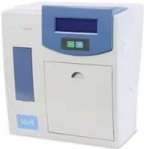
Electrolyte analyser machine
Figure no.2,electrolyte analyser machine
Time of collection:
At the time of admission the patients clinical picture is recorded in
prefixed proforma. Venous blood sampling is obtained from each patient
enrolled in the study and is sent for estimation of Electrolytes, Blood urea,
Glucose levels. Serum osmolality (calculated), Urine osmolality, Urine spot
sodium,potassium,were done in selected patients. Imaging studies as
Measurement of electrolytes
Electrolytes are measured by a process known as potentiometry. This
method measures the voltage that develops between the inner and outer
surfaces of an ion selective electrode. The electrode (membrane) is made
of a material that is selectively permeable to the ion being measured. For
example, sodium electrodes are made from a special glass formula that
selectively binds sodium ions.
y The inside of the electrode is filled with a fluid containing sodium ions,
and the outside of the glass membrane is immersed in the sample.
y A potential difference develops across the glass membrane that is
dependent upon the difference in sodium concentration (activity) on the
inside and outside of the glass membrane.
y This potential is measured by comparing it to the potential of a reference
electrode. Since the potential of the reference electrode is held constant,
the difference in voltage between the two electrodes is attributed to the
concentration of sodium in the sample. Ion selective membranes can be
OBSERVATION AND RESULTS
Analysis of the data collected has been done and statistical significance
of the risk factors are established as discussed in the following section.
During the study period from January 2015 to august 2015, the total
number of PICU admission was 227 patients. Among 227 patients ,236
episodes of serum electrolyte disturbances were noted.
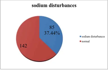
SODIUM DISTURBANCES:
Among this 227 children studied, 85 children had sodium disturbances
that included both hyponatremia(80 children) and hypernatremia(5 children)
depicted in the pie chart. This number accounts for 37.44% of the patients
[image:45.595.144.525.384.634.2]admitted to PICU.
Figure no.3,number of sodium disturbances and normal electrolyte
values
85
37.44%
142
sodium disturbances
sodium disturbances
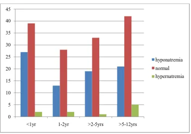
Age distribution :
Of the children with sodium disturbances, distribution based on age
were analysed and the corresponding chart is shown as below.Maximum
number (27 children) of patients who developed sodium disturbances were in
the age group of 1month to 1year and the mean age in years of the study
population was calculated to be 1month -2yrs,most common electrolyte
[image:46.595.145.526.336.604.2]imbalance being hyponatremia 80 cases.
Figure no.4,Incidence of sodium disturbances age wise distribution
0 5 10 15 20 25 30 35 40 45
<1yr 1-2yr >2-5yrs >5-12yrs
hyponatremia
normal
Among 80 cases of hyponatremia, Incidence of hyponatremia is more
common among infants( about 27 cases).Among 5 cases of hypernatremia , it is
more common among children aged >5 yrs.
Incidence of hyponatremia is least among 1-2yrs.on the contrary incidence of
[image:47.595.112.542.243.493.2]hypernatremia is least among age group of 2-5yrs
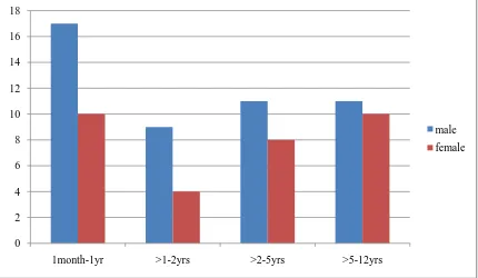
Figure no.5,Sex wise distribution of hyponatremia
Total number of hyponatremia was 80 children
Incidence of hyponatremia is more common among male child about 48
cases,most common age group being 1month-1year (17cases). Out of the 80
patients who developed hyponatremia 48 patients were male that is 60 % and
32 patients were females that is 40 % were female with male :female ratio
1.5:1.
0 2 4 6 8 10 12 14 16 18
1month-1yr >1-2yrs >2-5yrs >5-12yrs
male
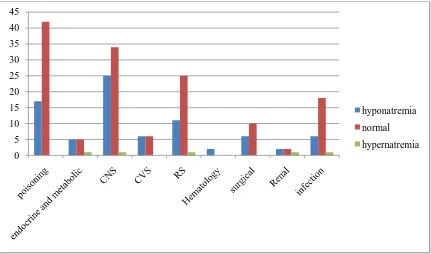
ETIOLOGY AND SODIUM DISTURBANCES
Figure no.6: Etiology and Sodium abnormalities
The most common cause of hyponatremia is CNS disorder 25 patients followed by poisoning 17 patients forming 31.25% and 21.25 % respectively.
Poisoning and CNS disorders accounted for 21.25% and 31.25% of
hyponatremia respectively; others were accounted for by bronchopneumonia
(13.75%) septicemia (7.5%), and renal(2.5%),cardiac(7.5%).
Poisoning includes kerosene poisoning,liquid detergent poison,camphor insecticide poisoning,
Endocrine /metabolic causes include diabetic ketoacidosis,metabolic liver disease,hepatic encephalopathy,neuronal ceroidal lipofuschinosis
CNS causes include seizure disorder,acute CNS infection
CVS causes include cyanotic/acyanotic heart diseases,cardiomyopathy,arrhythmias
RS causes include bronchopneumonia/bronchiolitis,aspiration pneumonia ,acute severe asthma
0 5 10 15 20 25 30 35 40 45
hyponatremia
normal
Hematological causes include sickle cell anemia .malignancies,hemolytic anemia
Surgical causes include post VP shunt ,head injury,hepatoma,extrahepatic biliary atresia,thoracotomy/ICD pneumothorax
[image:49.595.118.524.244.637.2]Endocrine ,CNS disorders,Respiratory system,renal, infection each contributing 1 case for hypernatremia.the
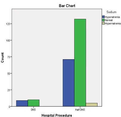
Figure no.7,maintenance fluid and sodium abnormalities
9 patients with hyponatremia received DNS that is about 11.25% of
Most common age group of hyponatremia was 1month-1yr (33.75%)
Clinical evaluation and concurrent plasma and urinary osmolality and urine
sodium suggested that hyponatremia associated with pneumonia,
meningitis/encephalitis, septicemia, seizures and miscellaneous diseases was of
euvolemic (dilutional) type in more than 80% patients while in all children with
acute diarrhea it was of hypovolemic type. The study has shown that
hyponatremia occurs frequently in sick children requiring emergency care, and
should receive appropriate attention in the management plan.
The patients were grouped on the basis of serum sodium concentration into
normonatremic (serum sodium > 135mEq/L) and hyponatremic (serum sodium
<135mEq/L or less). Serum sodium concentration of 125 mEq/L or less was
classified as severe hyponatremia. Hyponatremia was further categorized into
five types: euvolemic (normal hydration, plasma osmRODOLW\ P2VPNJ
hypovolemic (with dehydration), edematous (with edema), due to renal failure
and hyperglycemic (fictitious) hyponatremia.
The frequency distribution of serum sodium concentration in 227 study
children was analysed.Hyponatremia serum sodium <135mEq/L was present in
FKLOGUHQZKLOHVHYHUHK\SRQDWUHPLDVHUXPVRGLXPP(T/
was found in 5 chidren (6.25%). Amongst those with severe hyponatremia all
the 5 children were in the serum sodium ranged between 121-125 mEq/,none
Among patients with hyponatremia, symptomatic hyponatremia was found in
4 children. In children with seizures,around 18 patients inaddition to those 4
patients who were symptomatic were given 3% NACL correction.The
commonest cause of hyponatremia is cerebral salt wasting and Syndrome of
Inappropite ADH secretion.
Among patients with hyponatremia, 9 patients (11.25%) received DNS and 71
patients (88.75%) received ½ DNS. This difference had no statistical
significance despite giving this.
Among 25 patients with hyponatremia who died,16 patients (64%) had with
seizures died 9 patients (36%) with seizures survived.
Studies on the mechanism of euvolemic hyponatremia have emphasized a role
of syndrome of inappropriate ADH secretion (SIADH) in these patients. By
conventional criteria, almost all of our patients with euvolemic hyponatremia
could be plasma osmolality, high urine osmolality and normal renal function.
Inappropriately higher concentration of plasma vasopressin than that expected
for the degree of hypo-osmolality has been demonstrated in association with
euvolemic hyponatremia in children with meningitis, asthma and hospitalized
adults.
Thus, ADH mediated renal salt loss and water retention could be the cause of
hyponatremia in such patients. However, severe hyponatremia may occur in
dilution of the plasma sodium. Evidence supporting redistribution and
accumulation of sodium within the cells has also been presented.
Recently, Hannon and Boston have shown significant intracellular shifts of
sodium chloride and water in septic animals. They suggested that in these
animals hyponatremia and hypo-osmolality was caused by a combination of
intracellular shift of sodium and water, and dilution of extracellular space
probably as a result ofclassified as having SIADH as they had normal
hydration, low plasma osmolality
Data from our ongoing research suggest a significant increase in RBC sodium
in the presence of hyponatremia in septicemic children. Further studies are
needed to clarify the mechanism responsible for hyponatremia in acute
Figure no.8, Sex wise distribution of hypernatremia:
Among 5 cases of Hypernatremia,1 case occurred in both male and
female between 1month -1yr and >1-2yrs.There were no case of hypernatremia
over the age group of 5-12yrs.out of 5 patients with hypernatremia,3 patients
were males, 2 patients were females that is 60 % were males,40 % were
females with male to female ratio1.5:1
0 0.2 0.4 0.6 0.8 1 1.2
1month-1yr >1-2yrs >2-5yrs >5-12yrs
male
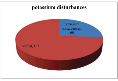
Potassium disturbances
Figure no.10,potassium disturbances and normal electrolyte values
Out of the 227 children,60 children had abnormalities of serum potassium that
included both hypokalemia (49 cases)and hyperkalemia(11 cases) accounted
for 26.43% of the total.22 patients with mixed electrolyte disturbances that is
both sodium and potassium disturbances.
potassium disturbances,
60
normal, 167
Figure no.11,Age wise incidence of Potassium disturbances:
Out of 11 cases of hyperkalemia,Incidence of hyperkalemia is more
common among infants between 1month and 1yr of age.the occurrence of
hyperkalemia is almost nil in the age group of 1-5yrs.Hereagain higher
incidence of hyperkalemia is noted in the age group of 1month to
1year.Incidence of hypokalemia is least among 1-2yr age group.
0 5 10 15 20 25 30 35 40 45 50
1month-<1yr 1-2yrs >2-5yrs >5-12yrs
hypokalemia
normal
Figure no.12,sexwise distribution of Potassium disturbances
Hypokalemia is more common among male children of age group>5-12yrs that
constitutes 18.36 % of hypokalemia caes reported.Among females 5cases each
in the age group of 1month-1yr, >2-5yrs and > 5-12yrs. 60 patients developed
potassium disturbances that accounted for 26.43% of incidence of total
electrolyte disturbances.Data on all those patients, who had hypokalemia
documented on at least one occasion during their PICU stay, was analyzed
further.
Details of patients regarding their age, sex, weight, diagnoses, clinical course,
and outcome were obtained from the records. Details of bio-chemical
0 1 2 3 4 5 6 7 8 9 10
parameters, i.e., electrolytes, acid-base status and renal function parameters,
along with ECG and details of treatment were noted.
Hypokalemia was graded as mild if serum potassium ranged between 3.0-3.4
mEq/L, moderate if between 2.0-2.9 mEq/L and severe if <2.0 mEq/L.
Among patients with hypokalemia,28 patients were male children,16 patients
were female children,moderate hypokalemia is seen in 3 children in the age
group 1-5yrs,none of them were in the severe hypokalemia range.
All the patients received slow intravenous correction in form of increased
potassium content of intravenous fluids to 40 to 60 mEq/L.One patient had
ECG changes,that received rapid potassium correction.Rapid infusion was
given at a rate of 0.3 mEq/kg/h .
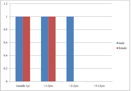
Sexwise distribution of hyperkalemia
0 1 2 3 4 5 6
1month-1yr >1-2yrs >2-5yrs >5-12yrs
male
Incidence of hyperkalemia is higher in male children in the age group of
1 month-1 year about 5 cases reported about 4 cases occurred in females of the
same age group 1month -1yr.no case was reported in both males and females in
the age group of 1-2yrs.There was 2 cases of hyperkalemia occurred in the age
group of >5-12yrs among female children and none in the male children.
Among 49 patients with hypokalemia ,31 patients were males,18 were
females that is 63.26% were males,36.73% were females with male to female
ratio 1.7:1
Among 11 patients with hyperkalemia ,5 patient were male ,6 patients
were female
etiology and potassium disturbances
Among 49 patients with hypokalemia, most common etiology was
CNS(seizures and acute CNS infection)19 cases contributing 38.78% of
hypokalemia cases, followed by poisoning 14 cases 28.57% of
hypokalemia.bronchopneumonia 6 cases (12.24%),cardiac renal septicemia
each contributing 1 case (2.04%) of hypokalemia.
Among 16 patients with hypokalemia 6 patients died due to CNS disorders like
seizures and acute CNS infection,4 patient died due to poisoning.
0 5 10 15 20 25 30 35 40 45 50
hypokalemia
normal
CALCIUM DISTURBANCES
Among the 227 children admitted ,77 children had calcium
disturbances,that accounted for 33.92% of total children admitted.
77
150
calcium abnormalities
calcium disturbances
Age wise incidence of Calcium disturbances
It is shown in the above chart that incidence of hypocalcemia is more
common in the age group of 1month-1yr.Hypercalemia occurs less
commonly and maximum in the age group of 1-2yrs.hypocalcemia
incidence is least among 1-2yrs,hypercalcemia is least among 2-5yrs age
group.
0 5 10 15 20 25 30 35 40 45 50
1month-1yr >1yr-2yr >2yrs-5yrs >5yrs
hypocalcemia
normal
Sex wise disrtribution of hypocalcemia
Among 71 patients with hypocalcemia,38 patients were male,33 patients were
females that is 53.52% were males,46.47% were females male to female ratio
1.15:1
Maximum number of hypocalcemia 13 cases occurred in the male children of
1month-1yr age group.About 10 cases each occurred in the female children
1month-1yr and again > 2-5yrs age group.Incidence of hypocalcemia is least
among female children of age group 5-12yrs.
0 2 4 6 8 10 12 14
1month-1yr >1-2yrs >2-5yrs >5-12yrs
male
Among 6 patients with hypercalcemia ,5 patients were males,1 patient were
female,83.33% patients were males,16.67% were females,with male to female
ratio 5:1
Among 2patients with hypochloremia 1 patient male and 1 female with male to
female ratio 1:1
Etiology and calcium disturbances
Among patients with hypocalcemia the most frequent cause is established to
be poison about 16 20.78% f/b Respiratory like bronchopneumonia 15cases
19.48% f/b CNS 14 cases 18.18%..Among 6 patients with hypercalcemia
poison contributed the maximum number 3 (50%) f/b CNS disorders
1(16.67%)
0 5 10 15 20 25 30 35 40 45 50
hypocalcemia
normal
Sexwise distribution of hypercalcemia
Maximum number of hypercalcemia about 3 cases occurred in male children of 1-2yrs age group with nil cases reported in the 2-5yrs age group.Among female 1 case reported in 1month -1yr age group
Chloride disturbance
227 children admitted 14 children had chloride abnormalities that accounted for 6.1% of the total.
0 0.5 1 1.5 2 2.5 3 3.5
1month-1yr >1-2yrs >2-5yrs >5-12yrs
male
female
14
213
chloride abnormalities
chloride abnormalities
Age wise incidence of chloride disturbances:
2 case of hypochloremia occurred 1 each in age group of 1month-1yr and
>5-12yr.there were 12 hyperchloremia cases maximum in the age group of
1month -1yr about 4 cases.
0 10 20 30 40 50 60 70
1month-1yr >1-2yrs >2-5yrs >5yrs-12yrs
hypochloremia
normal
About one case of hypochloremia each of age group 1month-1yr in male and
5-12yrs in female children respectively
Sexwise distribution of hyperchloremia
0 0.2 0.4 0.6 0.8 1 1.2
1month-1yr >1-2yrs >2-5yrs >5-12yrs
male
female
0 0.5 1 1.5 2 2.5 3 3.5 4 4.5
1month-1yr >1-2yrs >2-5yrs >5-12yrs
male
Among 12 patients with hyperchloremia ,9 patients were male,3 patients were
female,75 % were male,25% were females, with male to female ratio of 3 :1
Incidence of hyperchloemia is highest among male children of 1 month-1yr
about 4 cases and least in the age group of 2-12yrs about 1 case each.Among
female children maximum number of cases occurred in the age group of
>5-12yrs.there were no cases reported in the age group of 1month -5yrs agegroup
in females.
Maximum number of cases of hyperchloremia was caused by poisoning and
CNS disorders like seizure disorder,acute CNS infection
0 10 20 30 40 50 60
hypochloremia
normal
STATISTICAL ANALYSIS
The collected data were analysed with SPSS 16.0 version.To describe
about the data, descriptive statistics, frequency analysis, percentage analysis
were used for categorical variables and for continuous variables the mean and
S.D were used. To find the significant difference between the bivariate samples
in independent groups, the independent t test was used. To find the significance
in categorical data, Chi-Square test was used. In all the above statistical tools,
the probability value of<0.05 was considered as significant level.
P-Value +LJKO\6LJQLILFDQWDW3
P-Value 6LJQLILFDQWDW3
P-value No Significant at P >.05
Etiology * Sodium Crosstabulation Sodium
Total Hyponatremia Normal Hypernatremia
Etiology Poisoning Count 17 42 0 59
% within clinical history
28.80% 71.20% 0.00% 100.00%
Endocrine and
Metabolic
Count 5 5 1 11
% within clinical history
45.50% 45.50% 9.10% 100.00%
% within clinical history
41.70% 56.70% 1.70% 100.00%
CVS Count 6 6 0 12
% within clinical history
50.00% 50.00% 0.00% 100.00%
RS Count 11 25 1 37
% within clinical history
29.70% 67.60% 2.70% 100.00%
Hematology Count 2 0 0 2
% within clinical history
100.00% 0.00% 0.00% 100.00%
Surgical Count 6 10 0 16
% within clinical history
37.50% 62.50% 0.00% 100.00%
Renal Count 2 2 1 5
% within clinical history
40.00% 40.00% 20.00% 100.00%
Infection Count 6 18 1 25
% within clinical history
[image:69.595.87.552.67.587.2]24.00% 72.00% 4.00% 100.00%
Table no.2,etiology and sodium disturbances
Etiology * Potassium Crosstabulation Potassium Total Hypokalem ia Normal Hyperkalem ia Etiolog y
Poisoning Count 14 45 0 59
% within clinic al histor y
23.70% 76.30% 0.00% 100.00 %
Endocrine and
Metabolic
Count 0 11 0 11
% within clinic al histor y 0.00% 100.00 % 0.00% 100.00 %
CNS Count 19 35 6 60
% within clinic al histor y
31.70% 58.30% 10.00% 100.00 %
CVS Count 1 11 0 12
% within clinic al histor y
8.30% 91.70% 0.00% 100.00 %
RS Count 6 27 4 37
% within clinic al histor y
16.20% 73.00% 10.80% 100.00 %
Hematolog y
Count 0 2 0 2
histor y
Surgical Count 4 12 0 16
% within clinic al histor y
25.00% 75.00% 0.00% 100.00 %
Renal Count 1 4 0 5
% within clinic al histor y
20.00% 80.00% 0.00% 100.00 %
Infection Count 4 20 1 25
% within clinic al histor y
16.00% 80.00% 4.00% 100.00 %
Table no.3,etiology and potassium disturbances
38.75 % of hypokalemia is due to CNS disorders and 54.54% of hyperkalemia is due to CNS disorders this again no statistical significance p value >.05 (0.120) is noted
Etiology * Calcium Crosstabulation Calcium
Total Hypocalcemia Normal Hypercalcemia
Etiology Poisoning Count 16 40 3 59
% within clinical history
27.10% 67.80% 5.10% 100.00 %
Endocrine and
Metabolic
Count 3 8 0 11
% within clinical history
27.30% 72.70% 0.00% 100.00 %
% within clinical history
23.30% 75.00% 1.70% 100.00 %
CVS Count 6 6 0 12
% within clinical history
50.00% 50.00% 0.00% 100.00 %
RS Count 15 21 1 37
% within clinical history
40.50% 56.80% 2.70% 100.00 %
Hematology Count 1 1 0 2
% within clinical history
50.00% 50.00% 0.00% 100.00 %
Surgical Count 5 10 1 16
% within clinical history
31.30% 62.50% 6.30% 100.00 %
Renal Count 1 4 0 5
% within clinical history
20.00% 80.00% 0.00% 100.00 %
Infection Count 10 15 0 25
% within clinical history
40.00% 60.00% 0.00% 100.00 %
Total Count 71 150 6 227
% within clinical history
[image:72.595.107.564.65.667.2]31.30% 66.10% 2.60% 100.00 %
Table no.4,etiology and calcium disturbances
Etiology * Chloride Crosstabulation Chloride Total Hypochlore mia Normal Hyperchlore mia clinic al histor y
Poisoning Count 0 57 2 59
% withi n clinic al histor y
0.00% 96.60% 3.40% 100.00 %
Endocrine and
Metabolic
Count 0 11 0 11
% withi n clinic al histor y 0.00% 100.00 % 0.00% 100.00 %
CNS Count 0 55 5 60
% withi n clinic al histor y
0.00% 91.70% 8.30% 100.00 %
CVS Count 1 11 0 12
% withi n clinic al histor y
8.30% 91.70% 0.00% 100.00 %
RS Count 1 33 3 37
% withi n clinic al histor y
2.70% 89.20% 8.10% 100.00 %
gy % withi n clinic al histor y 0.00% 100.00 % 0.00% 100.00 %
Surgical Count 0 15 1 16
% withi n clinic al histor y
0.00% 93.80% 6.30% 100.00 %
Renal Count 0 5 0 5
% withi n clinic al histor y 0.00% 100.00 % 0.00% 100.00 %
Infection Count 0 24 1 25
% withi n clinic al histor y
0.00% 96.00% 4.00% 100.00 %
Table no.5,etiology and chloride disturbances
Etiology Outcome Crosstabulation Outcome Total Death Improved clinical history
Poisoning Count 5 54 59
% within clinical history
8.50% 91.50% 100.00%
Endocrine and
Metabolic
Count 6 5 11
% within clinical history
54.50% 45.50% 100.00%
CNS Count 16 44 60
% within clinical history
26.70% 73.30% 100.00%
CVS Count 6 6 12
% within clinical history
50.00% 50.00% 100.00%
RS Count 12 25 37
% within clinical history
32.40% 67.60% 100.00%
Hematology Count 0 2 2
% within clinical history
0.00% 100.00% 100.00%
Surgical Count 4 12 16
% within clinical history
25.00% 75.00% 100.00%
Renal Count 2 3 5
% within clinical history
40.00% 60.00% 100.00%
% within clinical history
16.00% 84.00% 100.00%
Total Count 55 172 227
% within clinical history
24.20% 75.80% 100.00%
Table no.6,etiology and outcome
29 % of patients with CNS disorders died f/b 21.81 % of Respiratory problem died, results were analysed using Pearson chisquare test found to be not statistically significant.
Hospital Procedure * Sodium Crosstabulation Sodium Total Hyponatremi a Norma l Hypernatremi a Hospital Procedur e DN S
Count 9 10 0 19
% within Hospital Procedur e 47.40% 52.60 % 0.00% 100.00 % Half DN S
Count 71 132 5 208
% within Hospital Procedur e 34.10% 63.50 % 2.40% 100.00 %
Total Count 80 142 5 227
[image:76.595.105.451.67.214.2]% within Hospital Procedur e 35.20% 62.60 % 2.20% 100.00 %
Table no.7 ,maintenance fluids and sodium disturbances
Hospital Procedure * Outcome Crosstabulation Outcome Total Death Improved Hospital Procedure
DNS Count 5 14 19
% within Hospital Procedure
26.30% 73.70% 100.00%
Half DNS
Count 50 158 208
% within Hospital Procedure
24.00% 76.00% 100.00%
Total Count 55 172 227
% within Hospital Procedure
[image:77.595.106.498.73.320.2]24.20% 75.80% 100.00%
Table no.8,maintenance fluid and outcome patter