JOURNAL OFVIROLOGY, Aug. 2003, p. 8957–8961 Vol. 77, No. 16 0022-538X/03/$08.00⫹0 DOI: 10.1128/JVI.77.16.8957–8961.2003
Copyright © 2003, American Society for Microbiology. All Rights Reserved.
Conditional Suppression of Cellular Genes: Lentivirus
Vector-Mediated Drug-Inducible RNA Interference
Maciej Wiznerowicz and Didier Trono*
Department of Genetics and Microbiology, CMU, Faculty of Medicine, University of Geneva, 1211 Geneva 4, Switzerland
Received 27 March 2003/Accepted 16 May 2003
RNA interference has emerged as a powerful technique to downregulate the expression of specific genes in cells and in animals, thus opening new perspectives in fields ranging from developmental genetics to molec-ular therapeutics. Here, we describe a method that significantly expands the potential of RNA interference by permitting the conditional suppression of genes in mammalian cells. Within a lentivirus vector background, we subjected the polymerase III promoter-dependent production of small interfering RNAs to doxycycline-con-trollable transcriptional repression. The resulting system can achieve the highly efficient and completely drug-inducible knockdown of cellular genes. As lentivirus vectors can stably transduce a wide variety of targets both in vitro and in vivo and can be used to generate transgenic animals, the present system should have broad applications.
The externally controllable expression of exogenous cDNAs can be readily obtained in cells or in animals owing to tech-niques pioneered more than a decade ago (2, 8). Recently, it was demonstrated that the knockdown of endogenous genes could be achieved by RNA interference, and plasmid- or viral vector-based delivery systems for the stable expression of small interfering RNAs (siRNAs) were rapidly created (1, 3, 5, 7, 9, 17). In many situations, however, it is desirable to suppress genes in a regulated fashion, for instance, to study cellular factors that play essential roles during differentiation or devel-opment. On the basis of this premise, we created a lentivirus vector-based system for drug-inducible production of siRNAs in stably transduced mammalian cells.
MATERIALS AND METHODS
Vector construction.Vectors were constructed by using standard cloning
pro-cedures. The pSUPER and pSUPER-p53 constructs were described previously (5). pSUPER-siGFP was provided by F. Iseni (Geneva, Switzerland), and pSUPER-siLamin was a gift from R. Oggi (Lausanne, Switzerland). pLV-H was constructed by inserting the H1 promoter from pSUPER into the 3⬘long ter-minal repeat (LTR) of pWPXL (http://www.tronolab.unige.ch/). To construct pLV-TH, thetetO cassette was excised from pUHD13-3 (obtained from H. Bujard, Heidelberg, Germany) and cloned into pLV-H, upstream of the H1 promoter. Finally, the H1 promoter cassette in pLV-H and pLV-TH was re-placed by the H1-siRNA cassette excised from pSUPER-siRNA, generating pLV-H/siRNA and pLV-TH/siRNA, respectively. The sequence encoding tTR-KRAB (kindly provided by P. Lorenz and H.-J. Thiesen, Rostock, Germany) was cloned into pWPXL, replacing the green fluorescent protein (GFP) marker (pLV-tTR-KRAB), or as part of a bicistronic unit also encodingDiscosomasp. Red, using the encephalomyocarditis virus 5⬘internal ribosome entry site.
The lentivirus vectors described here are available upon request (www .tronolab.unige.ch/).
Cell culture and transduction with lentivirus vectors.The 293T, HeLa, and
MCF-7 cell lines were cultured in Dulbecco’s modified Eagle’s medium supple-mented with 10% fetal calf serum. All recombinant lentiviruses were produced
by transient transfection of 293T cells according to standard protocols (21). Briefly, subconfluent 293T cells were cotransfected with 20g of a plasmid vector, 15g of pCMV-⌬R8.91, and 5g of pMD2G-VSVG by calcium phos-phate precipitation. After 16 h medium was changed, and recombinant lentivirus vectors were harvested 24 h later.
To analyze the regulation of GFP, a HeLa cell clone carrying a single copy of the WPXL-GFP provirus (HeLa-GFP) was used. For transduction, HeLa-GFP, MCF-7, or HeLa cells were plated on 24-well plate (20⫻104cells/well), and
after 16 h medium containing recombinant lentivirus vectors was added. Follow-ing 16 h of incubation, the cells were washed and split, and doxycycline (DOX) was added to half of the transduced cells at a final concentration of 5g/ml. Five days later the cells were harvested and analyzed by fluorescence-activated cell sorting (FACS).
Western blotting.Cell extracts were prepared in radioimmunoprecipitation
assay lysis buffer (25 mM Tris [pH 7.5], 1% Triton X-100, 0.5% sodium deoxy-cholate, 5 mM EDTA, 150 mM NaCl) containing a cocktail of protease inhibitors (Sigma). The protein samples (10g) were separated on sodium dodecyl sulfate– 4 to 20% gradient polyacrylamide gels, electroblotted to polyvinylidene difluo-ride membranes (Perkin-Elmer), and exposed to antibodies against p53 (Santa Cruz Biotechnology), lamin A/C (Santa Cruz Biotechnology), GFP (Clontech), and actin (Calbiochem). Antibodies conjugated with horseradish peroxidase (Amersham) and enhanced chemiluminescence (Amersham) were used for de-tection.
FACS analysis.Harvested HeLa-GFP cells transduced with lentivirus vectors
carrying⌬NGFR cDNA were incubated with monoclonal antibody specific for human nerve growth factor receptor NGFR (Becton Dickinson PharMingen) labeled with phycoerythrin, washed twice, and analyzed with a FACSscan (Bec-ton Dickinson) for green (GFP) and red (NGFR-phycoerythrin) fluorescence. MCF-7 and HeLa cells cotransduced with LV-THsi/p53 or LV-THsi/lamin and pLV-tTR-KRAB-Red and cultured in presence or absence of DOX were har-vested and analyzed with a FACSscan for green and red (dsRed) fluorescence.
Immunofluorescence.MCF-7 and HeLa cells cotransduced with LV-THsi/p53
or LV-THsi/lamin and pLV-tTR-KRAB-Red and cultured for 5 days in the presence or absence of DOX were fixed with methanol (10 min,⫺20°C), blocked with phosphate-buffered saline–1% bovine serum albumin, and stained with antibodies against p53 (Santa Cruz Biotechnology) or lamin A/C (Santa Cruz Biotechnology), using secondary antibodies conjugated with Alexa 633 (Molec-ular Probes) for detection. Images were acquired by using three-color confocal microscopy (LSM 510; Carl Zeiss) and analyzed with Zeiss software.
RESULTS AND DISCUSSION
We took advantage of a tetracycline-controlled hybrid pro-tein, tTR-KRAB, in which the tetracycline repressor (tTR)
fromEscherichia coli Tn10is fused to the KRAB domain of
* Corresponding author. Mailing address: Department of Genetics and Microbiology, CMU, Faculty of Medicine, University of Geneva, 1 rue Michel-Servet, 1211, Geneva 4, Switzerland. Phone: 41 22 702 5720. Fax: 41 22 702 5721 or 41 22 702 5702. E-mail: didier.trono @medecine.unige.ch.
8957
on November 8, 2019 by guest
http://jvi.asm.org/
human Kox1 (6, 8). KRAB is an approximately 75-amino-acid transcriptional repression module found in many zinc finger-containing proteins, which can suppress, in an orientation-independent manner, both polymerase II- and polymerase III-mediated transcription within a distance of up to 3 kb from its binding site, presumably by triggering the formation of hetero-chromatin (4, 6, 12, 14, 19). When linked to the DNA-binding domain of tTR, KRAB can modulate transcription from an integrated promoter juxtaposed with tet operator (tetO)
se-quences (6). In the absence of DOX, tTR-KRAB binds spe-cifically totetOand suppresses the activity of the nearby pro-moter. Conversely, in the presence of DOX, tTR-KRAB is sequestered away fromtetO, thus permitting gene expression (6).
We used human immunodeficiency virus type 1-derived len-tivirus vectors (designated LV) as delivery vehicles because we aimed for a system that would be easy to apply to a broad variety of cellular targets, both ex vivo (cell lines, primary cells
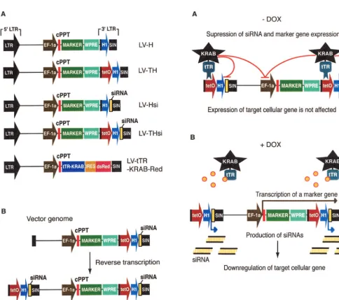
FIG. 1. (Left panels) A lentivirus vector-based system for conditional gene suppression with DOX-inducible siRNAs. (A) Schematic drawing of lentivirus vector plasmids used in this work. Cassettes consisting of the H1 promoter without (LV-H) or with (pLV-TH) the upstreamtetO sequence, H1-siRNA (LV-Hsi), andtetO-H1-siRNA (LV-THsi) were cloned in the 3⬘U3 region of pWPXL (http://www.tronolab.unige.ch/). All of the vectors contain an internal marker cDNA under transcriptional control of the EF-1␣promoter. (B) Double-copy design of siRNA lentivirus vectors. During reverse transcription, the U3 region of the 5⬘LTR is synthesized by using its 3⬘homologue as a template, which results in a duplication of the siRNA cassette in the provirus integrated in the genome of transduced cells.
FIG. 2. (Right panels) Mode of action of the DOX-controllable transrepressor. (A) In the absence of DOX, tTR-KRAB binds totetOand suppresses H1-mediated siRNA transcription, thus allowing normal expression of the cellular target gene (on). (B) In the presence of DOX, tTR-KRAB cannot bind totetOand hence siRNAs are produced, leading to downregulation of their target (off). The internal marker contained in the siRNA vectors provides an inverse monitoring device, as it is on in the presence of DOX and off in its absence.
on November 8, 2019 by guest
http://jvi.asm.org/
[image:2.603.47.532.60.490.2]including stem cells, fertilized oocytes, and blastocysts) and in vivo (e.g., brain and liver) (9, 13, 15, 16–18, 20), and because
tetO-linked transcriptional units are repressed by tTR-KRAB only when integrated in the genome. The tTR-KRAB cDNA was expressed from the ubiquitously active EF1-␣promoter as part of a bicistronic transcript also producing the dsRed marker (Fig. 1A, LV-tTR-KRAB). The regulated siRNA vec-tors were constructed by inserting atetO-H1 promoter-siRNA cassette into the U3 region of the 3⬘LTR of a self-inactivating lentivirus vector (Fig. 1A, LV-THsi). During reverse transcrip-tion, the vector RNA 3⬘U3 region serves as the template for the synthesis of its 5⬘DNA homologue, so that thetetO -H1-siRNA cassette is duplicated in the integrated provirus (Fig. 1B). We chose this double-copy configuration to obtain higher rates of siRNA synthesis. Sequences encoding siRNA hairpin precursors were designed as described previously (5). Control vectors carried either a constitutively active H1-siRNA cassette (LV-Hsi) or the H1- ortetO-H1 transcriptional elements with-out downstream siRNA-coding sequence (LV-H and LV-TH, respectively). All siRNA and control vectors also carried a marker gene downstream of an internal EF1-␣promoter. We predicted (Fig. 2A) that cells cotransduced with LV-THsi and LV-tTR-KRAB would normally express the gene targeted by the siRNA when maintained in the absence of DOX, owing to tTR-KRAB-mediated suppression of siRNA synthesis. In con-trast, addition of the drug would relieve this inhibition and allow for target gene downregulation (Fig. 2B). Expression of the internal marker gene would also be subjected to condi-tional tTR-KRAB repression, thus providing an internal mon-itoring device.
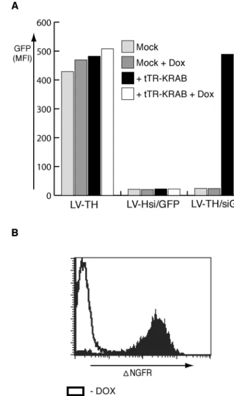
In a first series of experiments, we probed the ability of this system to regulate the production of GFP in HeLa cells stably expressing this fluorophore (Fig. 3A). Vectors were used at a multiplicity of infection of 10 to ensure good rates of (co)trans-duction. HeLa-GFP cells transduced with the empty LV-TH vector remained strongly GFP positive irrespective of their culture conditions. In contrast, cells transduced with the con-stitutively active LV-Hsi/GFP vector exhibited a strong down-regulation of the marker. In cells transduced with the control-lable LV-THsi/GFP vector, GFP expression was observed only in the presence of tTR-KRAB and in the absence of DOX (Fig. 3A). Correspondingly, in the absence of drug, tTR-KRAB suppressed the expression of the vector’s⌬NGFR in-ternal reporter gene (Fig. 3B). As expected, the tTR-KRAB-mediated suppression of siRNA production was equally efficient whether tetOwas inserted in the sense or antisense orientation and upstream or downstream of the H1 promoter (data not shown).
Next, we tested our system for the regulation of truly en-dogenous genes. We the chose p53 and lamin genes as targets because highly effective siRNAs directed against these genes were previously identified and well characterized (5, 7). MCF-7 breast cancer cells were used as substrates for p53 downregu-lation studies (Fig. 4, left panels). Cells cotransduced with LV-tTR-KRAB and LV-THsi/p53 produced wild-type levels of p53 when cultured in the absence of DOX, indicating full repression of siRNA synthesis (lower blot, lane 7). This repres-sion was mediated by tTR-KRAB, since p53 was undetectable in cells transduced only with LV-THsi/p53, whether or not DOX was present in the culture medium (upper blot, lanes 7
[image:3.603.300.536.72.471.2]and 8). In contrast, addition of the drug to the dually trans-duced cells resulted in rates of p53 downmodulation as robust as those observed in cells containing the constitutively active LV-Hsi/p53 vector (compare lane 8 in the lower blot with lanes 5 and 6 in both blots). Similar results were obtained for lamin in HeLa cells transduced with the corresponding siRNA len-tivirus vectors (Fig. 4, right panels). It is noteworthy that in both settings the drug-induced production of the siRNAs, and hence the suppression of the p53 or lamin target gene, corre-lated with the expression of the lentivirus vector internal GFP
FIG. 3. Regulation of GFP expression by using DOX-inducible siRNA. (A) HeLa cells carrying a single copy of a lentivirus vector expressing GFP from the EF-1␣promoter (HeLa-GFP) were trans-duced with a control lentivirus vector (LV-TH) or with vectors pro-ducing a GFP-specific siRNA in a constitutive (LV-Hsi) or regulated (LV-THsi) manner, with or without LV-tTR-KRAB (lacking the in-ternal ribosome entry site-dsRed cassette) and/or DOX as indicated. A truncated form of NGFR (⌬NGFR) served as an internal reporter in the siRNA vectors. (B) Conditional expression of the internal marker gene. HeLa-GFP cells dually transduced with THsi/GFP and LV-tTR-KRAB were maintained in the presence or absence of DOX before FACS analysis with a monoclonal antibody specific for the extracellular domain of NGFR.
VOL. 77, 2003 DRUG-INDUCIBLE PRODUCTION OF siRNAs 8959
on November 8, 2019 by guest
http://jvi.asm.org/
marker, whether examined by Western blotting (Fig. 4) or by FACS or confocal microscopy (data not shown).
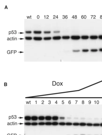
[image:4.603.62.522.70.363.2]Taken together, these results indicate that the tTR-KRAB-regulated, lentivirus vector-mediated delivery of siRNAs al-lows for the controllable suppression of cellular genes both with a high degree of efficacy and without significant leakiness. To complete the characterization of this system, we defined its kinetics and DOX dose responsiveness (Fig. 5). We chose p53 as target for these analyses because the half-life of this protein is relatively short, around 12 h. In MCF-7 cells dually trans-duced with the LV-THsi/p53 and LV-tTR-KRAB vectors, p53 steady-state levels started to decrease as early as 12 h after addition of 5g of DOX per ml to the culture medium and became undetectable by Western blotting within 36 h (Fig.
FIG. 4. Regulation of endogenous genes by using DOX-inducible siRNAs. Left panels, downmodulation of p53. MCF-7 cells were infected with the indicated lentivirus vectors as described in Materials and Methods. Western blotting was performed with monoclonal antibodies against p53, GFP, or actin (as a control). Right panels, downmodulation of lamin A/C. The same experiment as for the left panels was performed with HeLa cells, using lamin-specific siRNA vectors and antibodies.
FIG. 5. Kinetics and dose responsiveness of DOX-inducible RNA interference. (A) MCF-7 cells were cotransduced with LV-THsi/p53 and LV-tTR-KRAB as described in Materials and Methods. Five days later, DOX was added at a concentration of 5 g/ml. Cells were harvested just before DOX treatment (lane 0) and then at indicated time points. wt, nontransduced cells. Whole-cell extracts were analyzed by Western blotting with p53-specific antibodies. (B) At 5 days post-transduction as described for panel A, cells were placed in medium containing the following concentrations of DOX (in micrograms per milliliter): 0 (lane 1), 0.0005 (lane 2), 0.001 (lane 3), 0.002 (lane 4), 0.004 (lane 5), 0.008 (lane 6), 0.016 (lane 7), 0.063 (lane 8), 0.25 (lane 9), 1 (lane 10), and 5 (lane 11). wt, nontransduced cells. Western blot analyses of whole-cell extracts were performed after another 5 days.
on November 8, 2019 by guest
http://jvi.asm.org/
[image:4.603.309.527.434.721.2]5A). This suggests that RNA interference was fully effective in less than 24 h, implying that the DOX-mediated sequestration of tTR-KRAB rapidly unleashes high rates of siRNA produc-tion from the integrated H1 promoters. A dose-response anal-ysis further revealed an extreme sensitivity to DOX control, while pointing to the possibility of some tuning of the gene suppression. Indeed, whereas p53 downregulation was already apparent at the low DOX concentration of 0.004g/ml, full-blown suppression was achieved only at a dose of 0.25g/ml (Fig. 5B). The anti-p53 siRNA used in this experiment being very efficient, a greater range of DOX concentrations may allow for a modulation of the degree of gene knockdown with siRNAs of lower specific activity.
In summary, we provide a system for the conditional sup-pression of genes in mammalian cells. The versatility of its mode of delivery suggests very broad uses, as lentivirus vectors can transduce a wide range of targets, including stem cells, and can be used for generating transgenic animals from several species. In the latter setting, the system described here should offer significant advantages over currently available conditional knockout techniques, among which are its reversibility and simplicity of use. While the lentivirus vector-mediated delivery of drug-inducible RNA interference may thus be of particular interest for the study genes involved in development and dif-ferentiation, it is likely to be useful in many other areas of biology as well.
ACKNOWLEDGMENTS
We thank P. Lorenz and H.-J. Thiesen for tTR-KRAB cDNA, F. Iseni for pSUPER-siGFP, R. Oggi for pSUPER-siLamin, S. Vianin for technical assistance, and M.-O. Sauvain, other members of our labo-ratory, and J. Szulc for helpful discussions.
This work was supported by the Swiss National Science Foundation under the auspices of the National Center for Competence in Re-search Frontiers in Genetics program and by the Institut Clayton de la Recherche.
REFERENCES
1. Abbas-Terki, T., W. Blanco-Bose, N. Deglon, W. Pralong, and P. Aebischer.
2002. Lentiviral-mediated RNA interference. Hum. Gene Ther.13:2197– 2201.
2. Agha-Mohammadi, S., and M. T. Lotze.2000. Regulatable systems:
appli-cations in gene therapy and replicating viruses J. Clin. Investig.105:1177– 1183.
3. Barton, G. M., and R. Medzhitov.2002. Retroviral delivery of small
inter-fering RNA into primary cells. Proc. Natl. Acad. Sci. USA99:14943–14945.
4. Bellefroid, E. J., D. A. Poncelet, P. J. Lecocq, O. Revelant, and J. A. Martial.
1991. The evolutionarily conserved Kruppel-associated box domain defines a subfamily of eukaryotic multifingered proteins. Proc. Natl. Acad. Sci. USA 88:3608–3612.
5. Brummelkamp, T. R., R. Bernards, and R. A. Agami.2002. A system for
stable expression of short interfering RNAs in mammalian cells. Science 296:550–553.
6. Deuschle, U., W. K. Meyer, and H.-J. Thiesen.1995. Tetracycline-reversible
silencing of eukaryotic promoters. Mol. Cell. Biol.15:1907–1914.
7. Elbashir, S. M., J. Harborth, W. Lendeckel, A. Yalcin, K. Weber, and T.
Tuschl.2001. Duplexes of 21-nucleotide RNAs mediate RNA interference in
cultured mammalian cells. Nature411:494–498.
8. Gossen, M., and H. Bujard.1992. Tight control of gene expression in
mam-malian cells by tetracycline-responsive promoters. Proc. Natl. Acad. Sci. USA89:5547–5551.
9. Jacque, J. M., K. Triques, and M. Stevenson.Modulation of HIV-1
repli-cation by RNA interference. Nature418:435–438.
10. Kafri, T., U. Blomer, D. A. Peterson, F. H. Gage, and I. M. Verma.1997.
Sustained expression of genes delivered directly into liver and muscle by lentiviral vectors. Nat. Genet.17:314–317.
11. Lois, C., E. J. Hong, S. Pease, E. J. Brown, and D. Baltimore.2002. Germline
transmission and tissue-specific expression of transgenes delivered by lenti-viral vectors. Science295:868–872.
12. Margolin, J. F., J. R. Friedman, W. K. Meyer, H. Vissing, H.-J. Thiesen, and
F. J. Rauscher III.1994. Kruppel-associated boxes are potent transcriptional
repression domains. Proc. Natl. Acad. Sci. USA91:4509–4513.
13. Miyoshi, H., K. A. Smith, D. E. Mosier, I. M. Verma, and B. E. Torbett.1999.
Transduction of human CD34⫹cells that mediate long-term engraftment of NOD/SCID mice by HIV vectors. Science283:682–686.
14. Moosmann, P., O. Georgiev, H.-J. Thiesen, M. Hagmann, and W. Schaffner.
1997. Silencing of RNA polymerases II and III-dependent transcription by the KRAB protein domain of KOX1, a Kruppel-type zinc finger factor. Biol. Chem.378:669–677.
15. Naldini, L., U. Blomer, P. Gallay, D. Ory, R. Mulligan, F. H. Gage, I. M.
Verma, and D. Trono.1996. In vivo gene delivery and stable transduction of
nondividing cells by a lentiviral vector. Science272:263–267.
16. Pfeifer, A., M. Ikawa, Y. Dayn, and I. M. Verma.2002. Transgenesis by
lentiviral vectors: lack of gene silencing in mammalian embryonic stem cells and preimplantation embryos. Proc. Natl. Acad. Sci. USA99:2140–2145.
17. Qin, X. F., D. S. An, I. S. Chen, and D. Baltimore.2003. Inhibiting HIV-1
infection in human T cells by lentiviral-mediated delivery of small interfering RNA against CCR5. Proc. Natl. Acad. Sci. USA100:183–188.
18. Rubinson, D. A., C. P. Dillon, A. V. Kwiatkowski, C. Sievers, L. Yang, J.
Kopinja, M. Zhang, M. T. McManus, F. B. Gertler, M. L. Scott, and L. Van
Parijs.2003. A lentivirus-based system to functionally silence genes in
pri-mary mammalian cells, stem cells and transgenic mice by RNA interference. Nat. Genet.33:401–406.
19. Senatore, B., A. Cafier, I. Di Marino, M. Rosati, P. P. Di Nocera, and G.
Grimaldi.1999. A variety of RNA polymerases II and III-dependent
pro-moter classes is repressed by factors containing the Kruppel-associated/ finger preceding box of zinc finger proteins. Gene234:381–394.
20. Tiscornia, G., O. Singer, M. Ikawa, and I. M. Verma.2003. A general
method for gene knockdown in mice by using lentiviral vectors expressing small interfering RNA. Proc. Natl. Acad. Sci. USA100:1844–1848.
21. Zufferey, R., D. Nagy, R. J. Mandel, L. Naldini, and D. Trono.1997. Multiply
attenuated lentiviral vector achieves efficient gene delivery in vivo. Nat. Biotechnol.15:871–875.
VOL. 77, 2003 DRUG-INDUCIBLE PRODUCTION OF siRNAs 8961