A Thesis in General Surgery
RIGHT ILIAC FOSSA MASS A
CLINICAL STUDY
Submitted in partial fulfilment of the
Requirements for the Degree of
M.S General Surgery
(Branch I)
Kilpauk Medical College
The Tamil Nadu Dr. M.G.R Medical University
Chennai
DECLARATION BY THE CANDIDATE
I hereby declare that this dissertation titled
“RIGHT ILIAC FOSSA
MASS A CLINICAL STUDY”
is a bonafide and genuine research workcarried out by me under the guidance of Dr.V.Ramalakshmi M.S., Professor,
Department of General Surgery, Kilpauk Medical College, Chennai.
This dissertation is submitted to THE TAMIL NADU DR. M.G.R.
MEDICAL UNIVERSITY, CHENNAI in partial fulfillment of the requirements
for the degree of M.S. General Surgery examination to be held in April 2016.
Date :
CERTIFICATE BY THE GUIDE
This is to certify that the dissertation titled
“RIGHT ILIAC
FOSSA MASS A CLINICAL STUDY”
is a bonafide research workdone by Dr. G. VIMALA., Post Graduate in M.S. General Surgery,
Kilpauk Medical College, Chennai under my direct guidance and
supervision in my satisfaction, in partial fulfillment of the requirements for
the degree of M.S. General Surgery
.
Date : Dr. V.Ramalakshmi M.S.,
Place : Professor,
ENDORSEMENT BY THE HOD AND
HEAD OF THE INSTITUTION
This is to certify that the dissertation titled “RIGHT ILIAC FOSSA
MASS A CLINICAL STUDY” is a bonafide research work done by Dr. Vimala
G. Post Graduate in M.S. General Surgery, Kilpauk Medical College, Chennai
under the guidance of Dr.V.Ramalakshmi M.S., Professor, Department of
General Surgery, Kilpauk Medical College, Chennai.
Dr.P.N.Shanmugasundaram M.S., Dr.R.Narayana Babu M.D., DCH .,
Professor and Head, Dean,
Department of General Surgery, Kilpauk Medical College, Kilpauk Medical College, Chennai-10 Chennai-10
Date: Date:
ACKNOWLEDGEMENT
My sincere thanks to Prof. Dr. R. Narayana Babu, M.D., DCH., Dean,
Kilpauk Medical College and Hospital for allowing me to conduct this study in
the Department of General Surgery, Government Royapettah Hospital, Chennai.
I am extremely grateful to Dr.P.N.Shanmugasundaram, M.S, Professor and
Head of the Department of General Surgery, Government Kilpauk Medical
college for his encouragement and permission in granting unrestricted access to
utilising the resources of the Department.
I thank my mentor and guide Dr.V.Ramalakshmi, M.S, Professor of
General Surgery, Government Royapettah Hospital for her valuable guidance
during the tenure of my course.
I thank my Professors Dr. R.Kannan and Dr. Balakrishnan for their support
and guidance.
I also acknowledge my Assistant professors Dr.Thirunavukkarasu and
Dr.Suresh Babu for their valuable support and timely help rendered to complete
this study.
I thank my colleagues Dr. Varun, Dr.Kenny, Dr.Deepan, Dr. Lingesh ,
Dr.Kadhirvel who helped me throughout my study.
My utmost thanks to all my patients who cooperated to complete my
dissertation. Without their help it would have been impossible for me to complete
this study.
I thank my family for their great help and support. Last but not the least, I
ABSTRACT
BACKGROUND AND OBJECTIVE
Mass in the right iliac fossa is a common clinical entity encountered in surgical
practice. It is one diagnosis that has a varied range of pathologies and fits in aptly to
the description that the abdomen is a pandora’s box.
The main intention of this study is to know the varying modes of presentation,
different modalities of diagnosis, treatment and management of right iliac fossa mass
and to identify factors which can help in better management of these cases.
METHODS
Fifty patients presenting to the general surgery department of Govt. Royapettah
Hospital and Kilpauk Medical hospital, Chennai with a clinical diagnosis of Right
Iliac Fossa Mass were included in the study. Period of Study was from November
2014 to August 2015.
Inclusion criteria:
Masses in right iliac fossa arising from the appendix, caecum, terminal ileum,
retroperitoneal connective tissue and psoas abscesses are included
Exclusion criteria:
1. Female patients with pathology related to uterus and its appendages.
2. Right iliac fossa masses secondary to extra-abdominal pathology.
3. Masses from structures which abnormally present in the right iliac fossa.
5. Patients with right iliac fossa mass who are terminally ill.
6. Children less than 10 years of age.
A detailed clinical history was elicited and a careful general physical and
systemic examination was carried out along with the necessary investigations.
Appropriate management was done
Follow up period – 1 month
RESULTS
The Data obtained in the study was analysed, and it was found that the male to
female ratio was 2.8:1. Most patients were of appendicular pathology. Appendicular
mass was seen in 22 patients and appendicular abscess in 6 patients. Appendicular
pathology was seen in younger age groups and Carcinoma caecum was common in
older group. Patients were also analysed based on clinical parameters, few laboratory
TABLE OF CONTENTS
S.No CONTENTS Page No.
1 INTRODUCTION 1
2 AIMS & OBJECTIVES 2
3 REVIEW OF LITERATURE 3
4 METHODOLOGY 66
5 OBSERVATION AND RESULTS 68
6 DISCUSSION 88
7 CONCLUSION 102
8 SUMMARY 103
9 BIBLIOGRAPHY 105
10 ANNEXURE
PHOTOGRAPHS
PROFORMA
CONSENT FORM
MASTER CHART
KEY TO MASTER CHART
111
114
124
125
LIST OF TABLES
S.No CONTENTS Page No.
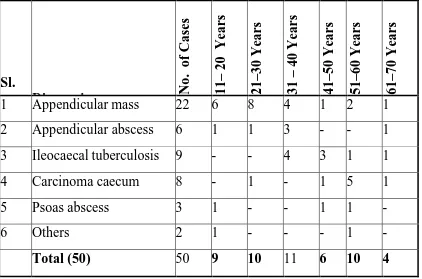
1 INCIDENCE OF VARIOUS CONDITIONS 68
2 AGE INCIDENCE OF RIF MASS 69
3 SEX INCIDENCE OF RIF MASS 71
4 OCCUPATION BASED DISTRIBUTION 72
5 DURATION OF SYMPTOMS 75
6 MASS ABDOMEN (SYMPTOM) 75
7 SYMPTOMS OF RIF MASS 77
8 CLINICAL FINDINGS 79
9 HEMOGLOBIN % 81
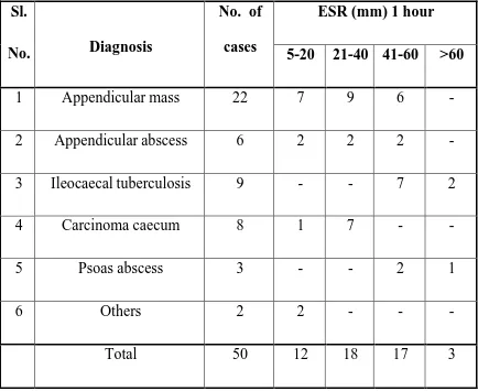
10 ESR 82
11 USG & BARIUM STUDIES 83
12 MODE OF TREATMENT 84
13 TYPES OF SURGERY 86
LIST OF FIGURES
S.No CONTENTS Page No.
1 INCIDENCE OF VARIOUS CONDITIONS 68
2 AGE INCIDENCE OF RIF MASS 69
3 SEX INCIDENCE OF RIF MASS 71
4 OCCUPATION BASED DISTRIBUTION 72
5 DURATION OF SYMPTOMS 75
6 MASS ABDOMEN (SYMPTOM) 75
7 SYMPTOMS OF RIF MASS 77
8 CLINICAL FINDINGS 79
1
INTRODUCTION
Mass in the abdomen, by reason of their wide spread implications, has
since long inspired the minds of many workers. Mass in the right iliac fossa is
an common entity. Pandora's box-hackneyed phraseology is apt in case of mass in
the right iliac fossa.
Patient with mass in the right iliac fossa may confront the surgeon,
pediatrician obstetrician and gynaecologist. A thorough understanding of the
anatomy and pathological processes that may occur within the abdomen are
essential for an accurate diagnosis and management. Some patients will need
immediate surgical intervention, whereas others will improve with conservative
treatment.
This challenging task of finding certain well defined clinicopathological
aspects of mass in the right iliac fossa has inspired me in undertaking this study.
The purpose of the present study is to finding certain well defined
clinicopathological entities, in mass in the right iliac fossa, the relative
occurence of various pathologies, as seen in Govt. Royapetah Hospital, Kilapuk
Medical College, Chennai in the overall endeavour to reduce morbidity a n d
2
OBJECTIVES
• To study various diseases which can presents as mass in the right iliac
fossa.
• To study age and sex distribution of various conditions.
• To study percentage of various diseases presenting as mass in the right iliac
fossa.
• To study various modes of management.
• To analyse the efficiency of current treatment and its prognosis in our
setup.
• To follow up the studied cases for further management and to detect
3
REVIEW OF LITERATURE
The history of disease is at least as old as the history of mankind. One
can assume that surgical disease or the surgical response to disease, is of similar
age. The progress of human ability to improve the well being of humanity by
means of surgery is fascinating indeed. It was not until the introduction of
anaesthesia and antisepsis that abdominal surgery became a practical therapeutic
approach for patients. Important contribution to the success of abdominal surgery
included the introduction and development of antibiotics after World War II
and developments in the metabolic care of the post operative patient started in the
late 1940’s and continues still.
Lorenz Heister, German surgeon published in 1718 a case of appendicitis.
He discovered it when he was dissecting the body at Altodotf.
Claudius Amyand, British surgeon, recorded first successful
appendicectomy in 1736.
The term “appendicitis” was coined by Reginald Heber Fitz, a Boston
Surgeon in 1886.
Charles McBurney, in November 1889, described the point of maximum
tenderness in acute appendicitis 5 years later in 1894, McBurney described his
4
It should be noted that the understanding of this common entity and
the operative procedures for it were worked out by the co-operative
efforts of physicians of several specialities. The first book on appendicitis
appeared in 1895. It was written not by a surgeon, but by a physician Dr. Herbert
Hawkins.
Sir Frederick Treves, was great advocatein the plan of conservative
management with interval operation.
Lord Moynihan of Leeds in England and others at the end of the century
leads the vigorous campaign for withholding purgatives.
A new approach for the management of peritonitis appeared in 1902
when A.J. Oschner of Chicago recommended conservative therapy for diffuse
peritonitis resulting from perforated appendix.
R.T Shackelford in 1955 said the most common tumour of the appendix is
mucocele which may occur spontaneously or after an attack of appendicitis.
In 1908 CA. Williams reported 19 cases of primary carcinoma of appendix.
Obern Dorfer in 1907 used the term carcinoid in 1907 and in 1911 Aschoff
recognised a distinctive histological pattern of carcinoid tumours.
Intestinal tuberculosis is indeed the commonest form of tubercular lesion
5
W.J. Mayo emphasized that in some instances the hyperplastic lesions
could only be differentiated from carcinoma with the help of a microscope.
Gershon Gohen used double contrast enema to diagnose early intestinal
tuberculosis.
In 1932 Carbinand and his associates published their theory on regional
ileitis and about the revolutionary change in the concept of primary
ileocaecal tuberculosis.
In 1941 Trevedi and Gupta came to the conclusion, after study of autopsy
findings and reports of 4,000 hospital cases, that 41 percent of pulmonary
tuberculosis cases had secondary intestinal involvement and 5.1 percent of cases
were primary in origin.
In 1950 the classical paper of Hoon et al. showed mainly that ileocaecal
tuberculosis was a definite entity and mentioned some mentionable differences
between tuberculosis, sarcoidosis and Crohn’s disease.
In India, intestinal tuberculosis is still a relatively more common condition
(Prakash et al., 1975).
That in India tuberculosis of the intestine is the commonest granulomatous
lesion and Crohn’s disease is rare was emphasised by Wig and Bawa in 1953. The
6
Hyperplastic type of tuberculosis was first described by Duget in 1889.
Granulomatous inflammatory bowel disease (IBD) was established as a distinct
entity in 1932 with the presentation of the classic paper by Crohn, Ginzburg and
Oppenheimer from the Mount Senai, Hospital.The constant incidence of mass in
the right iliac fossa they observed usually requires surgical intervention
Moor said “The best operative results are obtained by careful dissection of
involved bowel followed by its complete removal with suitable anastamosis
usually an ileo transverse colostomy.
Crohn and associates said that the best line of procedure was division
of ileum, three feet from ileocaecal valve closing both ends and implanting
proximal terminus of ileum by side to side anastamosis to transverse colon.
In 1963 Moor said free perforation rarely occurs whereas fistula
between loops of bowel is common.
At the Mayo clinic from 1945-1955 there were 257 patients with the
condition and 86% had resection and primary anastamosis.
Ramesh C. Bharti et al. (1996) concluded that though basic treatment of
abdominal tuberculosis remains medical yet role of surgery is for complications
and its management.
Kelly J. et al. (1999) said that ileocaecal tuberculosis should be sought first
in patients with appropriate clinical features even if classical risk factors for
7
Adalla S.A. et al. (1996) advocated conservative treatment for appendicular
mass and said it is not an indication for interval appendicectomy.
Milland F.C. et al. (1991) correctly identified organ of origin of mass in
97% cases by ultrasonography.
Hurme T. (1995) noted that if appendicular abscess is operated on in acute
8
ANATOMY
Abdomen is divided into nine regions by two vertical lines passing through
midclavicular lines superiorly and these lines extending inferiorly through
midinguinal points and two horizontal lines namely transpyloric and
transtubercular lines.
Thus right iliac fossa is the region in the right lateral side and lower most
quadrant.
Boundaries of this region are from superficial to deep by skin,
subcutaneous tissue, external oblique aponeurosis, transverse abdominis muscle
and internal oblique muscles anteriorly. Posterior boundary is formed by psoas
and quadratus lumborum muscles and thoracolumbar fascia.
Inferiorly bounded by posterior part of ilium and iliacus muscle. Laterally
it is bounded by external oblique, internal oblique, transverse abdominis and
fascia transversalis.
Structures normally present in the right iliac fossa are appendix, caecum,
terminal ilium, part of ascending colon, iliac lymphnodes, iliac vessels,
retroperitoneal connective tissue, iliopsoas muscle and sheath.
Structures which can abnormally present in the region are unascended or
dropped kidney, undescended testes, masses from uterus and its appendages,
bladder, gall bladder, etc. Appendix, caecum and terminal part of ilium form an
9
APPENDIX
It is the starting of large gut. At an early embryonic stage, it has same
diameter as caecum. It is formed by excessive growth of the right wall of
caecum which thus pushes appendix to the medial side.
Vermiform process of appendix is attached to caecum about 2.5 cm below
ileocaecal junction on posteromedial border and can be located through tracing
the anterior longitudinal band distally.
It is uniformly cylindrical and usually varies in length from 2.5 cm to 10
cm and is about 3 to 8 mm in diameter. Its layers are similar to those of large
gut, but muscular coat may be deficient in parts so that peritoneum and mucous
membrane are separated only by connective tissue through which infection may
spread from mucous membrane to peritoneum. Its wall contains much lymphoid
tissue. Its orifice into the caecum is guarded by a cresentric mucosal fold,
absence of which may predispose for presence of faecal matter in its lumen.
Mesoappendix is a triangular fold of peritoneum. It attaches appendix to
terminal part of left inferior layer of mesentry of ileum and its free cresentric
edge contains appendicular branch of posterior caecal. artery. If mesentry is
incomplete, artery lies on the wall of the appendix in its distal part and wall of
the vessel may get eroded in suppurative appendicitis or early thrombosis can
10
Base of appendix has a constant relation to caecum and is represented on
surface by McBurney’s point which is located at the junction of lateral 1/3rd and
medial 2/3rd of spinoumbilical line. Various positions of appendix have been
described. Tip of appendix in relation to caecum is variable and has been said
similar to hands of a clock. Appendix can sometimes cross psoas muscle and its
apex can hang over the pelvic brim into true pelvis.
Inflammed appendix gets fixed to psoas muscle and stretching of muscle
by extension of thigh causes pain. If inflammed appendix hangs over the pelvic
brim, it rests on pelvic fascia overlying obturator internus and on flexing and
rotating the thigh medially it causes pain.
It can also cause irritation of bladder causing strangury and rectum causing
passage of mucous per anum and tenesmus. In women symptoms might be less
because of presence of uterus.
Appendix is in pelvic position in 21 % of cases. It is in retrocaecal position
in 74% cases. It is the safest position as organ may be partly or completely behind
peritoneum.
Ileal position is the most dangerous and is in 1.5%. It is completely
intraperitoneal and can be preileal in 1% and postileal in 0.5% cases. If
inflammed in this region it may affect distal part of the ileum and can cause
vomiting, or even obstruction of small bowel which in turn can cause general
11
Blood supply
The artery to appendix, branch of lower division of ileo-colic artery runs
behind the terminal part of ileum and enters the mesoappendix a short distance
from base of the appendix. Here it gives off a recurrent branch, which
anastomoses at the base of appendix with a branch of the posterior caecal artery.
An aberrant artery, branch of posterior caecal artery is also seen in 50%
cases.
Lymphatic system
The appendix drains from its lymphatic follicles through the muscle wall
into nodes in the mesoappendix. These drain into paracolic nodes lying along
ileo-colic artery and then to the superior mesentric group.
TERMINAL ILEUM
Terminal part of ileum usually lies in the pelvis except the last 5 cms
which is fixed in the right iliac fossa. It ascends over psoas muscle and right iliac
vessels to end by opening into medial side of junction of caecum and ascending
colon. It is suspended by its mesentry allowing very free movement.
Blood supply
It is by inferior branch of ileo-colic artery which later ends by
anastomosing with the termination of superior mesentric artery. There are 2
12
Lymphatic system
Lymph drains from aggregated lymphatic follicles (Peyer’s patches) of
mucous membrane through the muscle wall into the mesentry (ileo-colic group of
nodes) from where they drain into the superior mesentric group of nodes.
CAECUM
It is the blind pouch of large intestine projecting downwards from the
commencement of ascending colon, below ileocaecal junction. It is about 6 cm
long and 7.5 cm wide and lies over floor of right iliac fossa i.e., over iliacus
and psoas fascia. Its lower end lies at the pelvic brim and when distended, its
anterior surface touches the parietal peritoneum of anterior abdominal wall and
when collapsed, coils of ileum lie, between it and the parietal peritoneum. In
obstruction anywhere in the colon, caecum eventually leads to distension because
of presence of ileocaecal valve and it may rupture.
Caecum is partly covered by peritoneum and may have a short mesentry or
even two mesentries, with a retrocaecal fossa extending upwards between them.
As in rest of the colon, positions of Taenia coli are anterior, posteromedial and
posterolateral. All these 3 converge over base of appendix and form outer
13 Blood supply
Ileocolic artery after giving off ileal and colic branch divides to supply
the caecum. The anterior caecal artery is smaller of the two terminal branches and
ramify over anterior surface of caecum, posterior caecal artery supplies posterior
wall of caecum as well as medial and lateral walls of that part of the gut.
Lymphatic system
Caecum drains from its lymphatic follicles into nodes lying along left side
of gut. These epicolic nodes which drain into paracolic nodes situated along
ileocolic and right colic arteries. From here they drain to superior mesentric group
of pre-aortic lymphnodes.
ILEO-CAECAL VALVE
Ileocaecal valve is situated at· the entrance of the ileum into the large
intestine opposite the junction of caecum and ascending colon.
Orifice is circular and the circular muscle of the small gut, covered with
the mucous membrane points into the large gut.
Valve consists of an upper and lower segment formed by duplication of
wall of small and large bowel. The thickened circular muscle of terminal ileum
14 ASCENDING COLON
It varies about 15 cm in length and extends upwards from ileocaecal
junction to the right colic flexure. It is invested with peritoneum on its anterior,
lateral and medial surfaces; but posteriorly it is devoid of peritoneum.
Posteriorly it lies on iliac fascia and anterior lamella of lumbar fascia
being connected and fixed to them by fibrous tissue of extra peritoneal fascial
envelope.
Anteriorly it is in relation with coils of ileum, possibly the right edge of
greater omentum and the anterior abdominal wall.
Blood supply
Ascending colon and right flexure are supplied by ileo-colic and right
colic arteries, branches of superior mesentric artery. Ileocolic artery divides into
ileal and colic branches where colic branch anastomoses with the descending
branch of right colic artery. Ascending branch of right colic artery anastomoses
with middle colic artery.
Lymphatic system
Lymphatic drainage is into epicolic, paracolic, intermediate colic and
15
RETROPERITONEUM
It consists of that portion of the body bound anteriorly by peritoneum,
posteriorly by spine and psoas with quadratus lumborum muscles. Superiorly it is
bounded by the 12th rib and attachments of the diaphragm. Inferiorly it is bounded
by brim of pelvis. Lateral margins of this space correspond to lateral borders of
quadratus lumborum muscles.
These limits marks potential space containing some very important organs
and structures like kidneys,ureters,adrenals, pancreas, abdominal aorta, inferior
vena cava, portions of autonomic and peripheral nervous systems, spermatic or
ovarian vessels, lymphatics, lymphnodes and certain portions of gastrointestinal
tract like duodenum.
Retroperitoneal space in relation to right iliac fossa contains common iliac
artery and vein and also external iliac artery and vein. Common iliac and external
iliac lymphnodes are situated along these vessels.
a) Relation of duodenum to colon
Second part of duodenum curves downwards over hilum of right kidney
covered infront by the peritoneum and crossed by attachment of transverse
mesocolon, so that its upper and lower half lie in the right supracolic and
16 b) Relation of Gonadal vessels
They have similar origin and course in both sexes. Both testicular and
ovarian arteries arise from anterior part of aorta, below renal arteries but well
above the origin of inferior mesentric artery. They run steeply downwards over
psoas muscle crossing ureter and supplying its middle portion and being
themselves crossed by colic vessels and peritoneum of infracolic compartments.
They reach pelvic brim about half way between sacroiliac joint and inguinal
ligament after which course is different in both sexes.
In the male testicular artery runs along pelvic brim above external iliac
artery and enters deep inguinal ring. In the female ovarian artery crosses
pelvic brim and runs down the lateral wall of pelvis to enter the
infundibulopelvic fold of peritoneum and passes to the ovary and uterine tube.
Gonadal veins accompany the arteries and are usually paired. As they run up on
psoas muscle two venae comitantes usually unite and the right vein enters
inferior venacava an inch or so below renal vein, Left vein enters left renal vein.
c) Relation of ureter in retroperitoneum
Ureter or duct of kidney is 25 cm long. Its upper half is in abdomen and
its lower half is in the pelvis. Its abdominal part extends almost vertically from
17
Ureter lies in sub-peritoneal areolar tissue and attaches to peritoneum.
When peritoneum is mobilised ureter is in danger as it moves with it.
Ureter descends on psoas fascia and crosses genitofemoral nerve. Inferior
vena cava is close to the medial side of right ureter. Its anterior relations are
vessels of testis/ovary second part of duodenum, right colic and ileocolic arteries,
root of mesentry and terminal part of ileum.
VI. KIDNEY
• Unascended kidney
• Dropped kidney
VII. UNDESCENDED TESTES
VIII. PELVIC ABSCESS
IX. URINARY BLADDER – DIVERTICULUM
X. DISTENDED GALL BLADDER
EXCLUSION CRITERIA
• Ilium bone tumours
• Mass from uterus and its appendages
18
ETIOPATHOLOGY
APPENDICULAR MASS AND APPENDICULAR ABSCESS
Appendicitis is yet a common surgical emergency which is more common
in upper and middle class people, probably because of intake they take which is
rich in meat and scanty in cellulose.
Appendicitis is of two types: Non-obstructive and Obstructive.
Appendicitis in majority of cases results from obstruction following
infection, approximately 60% of cases are related to hyperplasia of submucosal
lymphatic follicles (Non-obstructive) 35% are related to presence of faecal stasis
or faecoliths, 4% are due to presence of foreign body or round worm or
threadworm (obstructive). Abuse of purgatives and violent peristaltic action
which results therein favours and often determines perforation of inflammed
appendix causing appendicular mass or generalized peritonitis. So purgation
means perforation is a vice adage.
Usually on the 3rd day (rarely sooner) after the start of attack of acute
appendicitis, a tender mass can be frequently felt in right iliac fossa beneath some
rigidity of overlying musculature. Mass which at this time is not yet an
appendicular abscess, and may never become one, it is composed mainly of the
greater omentum, oedematous caecal wall and oedematous portions of small
19
appendix. By 4th or 5 th
day mass becomes more circumscribed. During the
following days (i.e. 5th to 10th day) swelling either becomes larger and an
appendix abscess results or it becomes smaller and subsides slowly as
inflammation resolves.
Bacteriology
Cultures from inflammed appendices usually reports that the infection is
mixed and there is hardly a pyogenic organism which has not been isolated from
such cultures. Most common organisms are mixture of Escherichia coli (85%),
Enterococci (30%), Non-haemolytic streptococci, Anaerobic streptococci,
together with Clostridium welchi (30%) and bacteroides.
ILEOCAECAL TUBERCULOSIS:
Tuberculosis is caused by acid-fast bacilli called mycobacterium
tuberculosis.Organism was first discovered by Robert Koch in 1882. It may be
human, bovine or avian type. Human type of infection is air borne (i.e.
tuberculosis of lung). Bovine type spreads by infected cow's milk. Intestinal
tuberculosis in India is caused by human type of bacilli which is secondary to
pulmonary tuberculosis.
Poverty is the main contributory factor of aetiology, which profoundly
20
The terminal part of the ileum and adjacent part of caecum become infected
by swallowed bacilli derived either from lungs or contaminated food.
Haematogenous spread is also likely.
Focus of infection is most commonly in ileocaecal region because of the
following factors:
• Increased physiologic stasis
• Most abundant lymphoid tissue
• Increased rate of absorption
There are two types of lesions commonly seen in gastrointestinal tuberculosis.
1. Hyperplastic type
2. Ulcerative type
Ulcerative :
Usually secondary to pulmonary tuberculosis associated with high virulent
organisms and low resistant host. In terminal ileum there are multiple ulcers,
long axis of which lie transversely along the lymphatic vessels. Ulcers are
shallow with undermining edges. Overlying serous coat is infected, thickened with
multiple tiny Tubercles. Fibrosis is characteristic and strictures of ileum are
21
Hyperplastic type
It is common in ileo-caecal region and occurs in patient who has already
developed resistance against mycobacterium tuberculosis. Caecum and terminal
two inches of ileum are usually affected. Infection first starts in lymphoid follicles
and spreads to the submucous and subserous planes. Chronic inflammation sets
in and the intestinal wall becomes thickened. This thickening is partly due to
tubercular granular Tissue and oedema, but is mostly due to excess fibrous
tissue, causing narrowing of the affected part. Regional lymphnodes are involved
early in the disease and may caseate. Ascending colon is shortened and ileum rises
to enter it vertically so that ileum and colon lie in straight line instead of at right
angles to each other. Sooner or later sub-acute or even acute obstruction may be
established above the constriction and is often precipitated by deposition of
faecolith or food particles.
Histology
Diffuse fibroblast reaction with giant cell of tuberculoid type. Frank
caseation which is pathognomonic of tuberculosis is rare and is reported in only
35-50% cases of operated specimens.
Caseation is more often seen in lymphnodes and is reported in 40- 60%
22
There are distinct entity of granulomas at ileo-caecal region which do not
give direct evidence of tuberculosis. These cases are labelled as indeterminate
group of granulomas.
Non-specific regional ileocolitis / non specific enteritis probably
represent the end result of a successfully controlled tubercular process.
In tuberculosis glands at first are firm, discrete, but when periadenitis adds
they become matted. Cut surface is grey and translucent but later becomes yellow,
opaque and caseous.
Further caseous material may breakdown to produce cold abscess.
Microscopically endothelial cells and lymphocytes can be seen. Giant cells are
seen within many nuclei arranged like a horse shoe. As the healing occurs,
fibroblasts proliferate and dense collagen fibres are laid down.
CARCINOMA CAECUM AND ASCENDING COLON
It occurs in 12% of all colo-rectal cancers (Mayoclinic Statistics). Surgery
has always been and is most likely to continue as first line of treatment. It
develops as a sequelae to ulcerative colitis (10% incidence after 10 years rising
to 45% after 25 years) or to polyposis of the colon. There is an indeterminate
relation to other types of adenomas but in great majority of cases no aetiological
factor can be demonstrated. Rarely there appears to be familial incidence (Love et
al., 1976). Of late there have been hypothesis stressing the implication of
23 Environmental factors
There have been 3 main theories advanced over the past 2 decades to
explain the cause of the tumour. The first of these proposed by Denis Burkitt and
his colleagues 1976 states that populations with diet low in fibre content have a
higher incidence of colo-rectal cancer.
The next one is due to dietary factors. An epidemiological association has
been demonstrated between Beef consumption and its contaminated animal fat and
frequency of bowel cancer. The third theory and most recent one was developed
by Bjel K. and it showed that high levels of selenium - metal found in soil and
foliage is related to a low incidence of colon cancer.
Genetic factors
In a minority of cases hereditary factor t r u l y contributes. Familial
multiple polyposis, Gardener’s syndrome and cancer – a family syndrome are
three hereditary disorders having an autosomal dominant mode of transmission.
Lynch H.T. et al., 1976; Rider et al., 1964 have shown that colonic carcinoma
occurs with 5 times greater frequency in polyp patients than in normal
individuals. There is frank evidence that colonic carcinoma is more common in
24 Age incidence
Carcinoma of the colon may occur at any age although it is usually seen
between the ages of 50 and 80, the peak age incidence being in the 6th and 7th
decades. It is not uncommon to find the disease in patients between ages 20
and 30 or even in children and adolescents.
Pathology
Macroscopic type of colonic cancers include:
1. Proliferative
2. Annular
3. Ulcerative
4. Mucoid
5. Primary linitis plastica
6. Multiple primary carcinoma of the colon
The proliferative type is most commonly seen in caecum and ascending
colon. It forms fleshy bulky polypoid mass that bulges into the lumen of bowel.
It is a malignancy having slow growth and shows no tendency to metastasise to
25
Obstruction is a late phenomenon because
Proximal colon is more spacious
1. Its contents are liquid in nature
2. The papilliferous tumour obstructs only by nature of its bulk.
LYMPHADENITIS
Most of the times lymphadenitis is non-specific and designated as acute or
chronic nonspecific lymphadenitis. Chronic inflammation may be specific. The
specific nodes involved are iliac lymphnodes. Iliac lymphadenitis is very
common In India where most of the population walk without protection.
Acute lymphadenitis and chronic lymphadenitis are caused by virulent
bacteria like streptococci and staphylococci. Chronic specific lymphadenitis is
either due to tuberculosis or filariasis.
If the causative organism is of relatively low virulence, lymphadenitis may
remain c o n f i n e d to nodes immediately proximal to the site of infection. The
nodes are enlarged, painful and tender. Rarely when infection is more severe,
the proximal barriers are overwhelmed thus in the cases of infected foot,
painful enlarged lymphnodes will be external iliac group instead of inguinal
group and may provocate acute abdominal symptoms closely resembling acute
appendicitis, a differential diagnosis that fears the surgeon. Macroscopically, the
26
changes in the perinodal tissue. Histologically there is prominence of lymphoid
follicles and large germinal centres.
LYMPHOMAS
May arises as a primary neoplasm at any level of gastro intestinal tract. As
primary lesions they most often affect stomach and ileum, less commonly colon
and rectum. Involvement of bowel may also appear as a part of dissemination of
systemic lymphomatosis. In such instances gastro-intestinal lesions are typically
multifocal whereas primary lymphomas are usually solitary masses in a
particular segment of gut. 11% of lesions are confined to ileo-caecal region.
Spread is by direct extension or by lymphatics. Distant metastasis ensues lately.
Lymphomas are common in ileum coinciding with the abundance of Peyer’s
patches.
Pathology
The tumour presents as ulcerating or infiltrating type and polypoid tumours
being least common. They sometimes become bulky and involve long segment
of the bowel which becomes rigid. Regional lymphnodes are usually enlarged but
not always by tumour deposits and sometimes show changes of reactive
hyperplasia alone. Although usually lot difficult to diagnose, these tumours may
27
The lymphomas as a group vary from well differentiated less malignant
forms one extreme to the anaplastic on the other extreme. Giant follicular
lymphoma is the most benign histological pattern followed by lymphosarcoma,
Hodgkin’s para granuloma, Hodgkin’s disease reticulum cell sarcoma in the order
of increasing malignancy.
APPENDICULAR MASS
Appendicitis is particularly common in highly civilised people like
European and Americans, while it is rare in Africans. This is attributed to the diet
which is rich in meat and scant in cellulose.
Appendicitis in majority of the cases result from obstruction following
injection, approximately 60% of the cases are related to hyperplasia of
submucosal lymphatic follicles, 35% to the presence of faecal stasis or
faecolith, 4% due-to the presence of foreign body or roundworm or thread worm.
Abuse of purgatives and violent peristaltic action which results there in,
predisposes and often leads to perforation of inflamed appendix leading to
appendicular mass or generalised peritonitis. So “purgation means perforation ‟ is
a wise adage.
Usually on 3rd day (rarely sooner) after the commencement of attack of
acute appendicitis a tender mass appears in right iliac fossa with overlying
rigidity, remaining part of abdomen being free from rigidity. The mass at this
28
mainly of greater omentum, edematous caecal wall and edematous portion of
small intestine. In its middle is a perforated or inflammed appendix.
During the ensuring days (5th to 10th) the swelling either become larger,
resulting in appendicular abscess or it becomes smaller and subsides slowly as the
inflammation resolves.
Bacteriology
Cultures from the inflammed appendix usually reveal that infection is
mixed. The most common organisms present are mixture of E. coli. 85%,
Enterococci 30%, Non-hemolytic streptococci, Anaerobic streptococci, together
with Clostridium welchii 30% and bacteroides.
AMOEBOMA
About 20% of population of world suffers from infestation with E.
histolytica. Most interesting to surgeon is Amoeboma. Though uncommon
it may be mistaken for carcinoma or if the symptoms are acute for an
appendicular abscess. The caecum and ascending colon are affected most often
followed by sigmoid and rectum. Although Amoebiasis is very common in
India very few cases of Amoeboma have been accounted. The common lesions are
discrete, shallow ulcers with a yellow base, a bright red edge and normal mucosa
29
The early and less severe forms of disease heal without scarring and in
more chronic forms granulomatous masses are formed due to the formation of
concentric strictures and para-colonic inflammatory masses termed Amoeboma,
most commonly found in caecum. Caecal Amoeboma present as hard slightly
tender, nodular and relatively fixed masses.
To summarise amoeboma is a thick edematous swelling of the wall of the
limited segment of the colon often without mucosal ulceration.
ACTINOMYCOSIS
Ileo-caecal actinomycosis was first reported by Ransom in 1892 and it
is the commonest form of abdominal actinomycosis. The organisms enter the
sub-mucosa through small breach of surface either in the appendix or through an
ulcer in the wall of the caecum. A foreign body seems sometimes responsible
for the breach which acts as a route of entry.
Secondary infection rapidly follows the invading organisms and the
typical granuloma develops in the bowel wall. An inflammatory honey comb of
dense and almost cartilaginous fibrous tissue results with multiple abscesses and
cavities.
In this inflammatory mass the caecum, ileum and appendix lie enveloped.
30
The fungus is Actinomycosis israeli an anaerobic gram positive
branching filamentous organism. It does not spread by lymphatic channels, and
hence regional lymphnodes are not involved. The inflammatory process spreads
by direct extension to the serous surface, a peritoneal reaction follows and soon
the abscess extends to the muscle layers of abdominal wall to burst open as
sinuses. In the late cases liver may be involved through portal vein.
CROHN’S DISEASE
Although no causative organism has been found in the lesion or in the
stools, abnormal forms of E. coli have been discovered in most of the patients. It is
considered to be hereditary possibly Crohn’s disease develops in patients with
relatively complete genotype. Ulcerative colitis is common in relatives of patients
with Crohn’s disease but the converse is not common. Like ulcerative colitis it is
believed that Crohn's disease is a precancerous condition. Pathologically there is
cicatrising inflammation with ulceration of the mucosa.
It usually commences at or near the ileo-caecal wall and extends upwards
along the ileum for about 30 cm. In acute cases the affected intestine is seen to
be swollen, bright pink in colour with fibrinous exudate on its peritoneal surface.
On palpation the intestinal wall feels like hose-pipe. The mesentery of the
involved intestine is much thickened, edematous and contains enlarged fleshy
31
Histologically, a characteristic finding is granulomatous infiltration of
lymphatics of the submucosa with the presence of non-caseating giant cells. In
late stages of the disease fibrosis extends into and obliterates the sub-mucosa.
PSOAS ABSCESS PRESENTING AS RIGHT ILIAC FOSSA MASS
Pathology
Tuberculous osteomyelitis of the spine in most cases results from
hematogenous spread into the marrow cavity. One or more vertebral bodies in the
dorso lumbar region are affected most often while the intervening fibrocartilage is
eroded and absorbed at an early stage. The primary cause is tubercular end
arteritis, the marrow is converted into myxomatous tissue which provides an ideal
nidus for the growth of the tuberculous bacilli. In the devitalised tissue a tubercle
follicle develops until it is visible to the naked eye as a small yellowish nodule.
As this nodule grows the lamellae over a wide area are progressively rarified and
eventually disappear.
The centre of the body of the vertebra being caseous the superimposed
weight of the vertebral column is borne by the fragile shell of compact bone
which sooner or later When the body of the vertebra collapses following things
32
1. Tubercular debris containing granulation tissue
2. Caseous material
3. Disintegrated lamellae and bone marrow
4. There is a cold abscess and is the commonest cause of Pott’s disease
occurring in 20% of cases.
This cold abscess tends to spread downwards under the influence of
gravity influenced by surrounding anatomy. Usually extending within the psoas
sheath coming to the surface above the inguinal ligament.
RETRO PERITONEAL TUMOURS PRESENTING AS RIGHT ILIAC
FOSSA MASS
These lesions may arise from a multitude of tissue types including muscle,
fat, connective tissue, vascular tissue, sympathetic nervous tissue. But in general
the finding of a retroperitoneal mass is an omnious one i.e.
malignant lesions out number benign lesions in the ratio of 4:1. The
commonest of the connective tissue tumours encountered are fibrosarcoma arising
from ileo psoas sheath and chondrosarcoma arising from iliac bone itself.
TUMOURS OF THE MESENTERY
Primary tumours of the mesentery may be cystic or solid. Of these cystic
33
tissues vascular, nervous and connective tissue are the sources of these tumours.
In addition cystic tumours may arise from embryonic rests (Dermoid) from the
developmental defect (Chylous or serous retention cyst) or following trauma
(Haemorrhagic cysts). Benign tumours have a great tendency to develop at the
periphery near the intestine and 2/3rd of them develop near the ileum. Malignant
solid tumours arise near the root of mesentery and spread by local extension or
peritoneal implants. The mobility of mesentery permits both benign and
malignant tumours to grow to a very large size before causing symptoms.
Origin Benign Malignant
1. Cystic Tumours
a) Developmental defects Chylous cyst serous cysts
b) Lymphatic tissue Lymphangioma Lymphangiosarcoma
c) Trauma Traumatic Cyst
d) Embryonic rests Enteric cyst dermoid Malignant teratoma
2) Solid Tumours
a) Adipose tissue Lipoma Liposarcoma
b) Fibrous tissue Fibroma Fibrosarcoma
c) Nerve elements
Neurilemmoma
Neurofibroma
Malignant
Schwannomma
Neurofibrosarcoma
d) Smooth muscle Leiomyoma Fibromyoma Leimyosarcoma
34
DISEASES OF ORGANS ABNORMALLY PRESENT IN RIGHT
ILIAC FOSSA
Ectopic kidney: This abnormality occurs in some degree in approximately 1%
of individuals. It is more common on the right side than in the left. The kidney
is normal ascent near pelvic brim. The ectopic kidney may be mistaken for an
abdominal tumour.
Diverticulum of bladder: A huge diverticulum of bladder may invade the
right iliac fossa as well. A diverticulum can be congenital or acquired and can
occur anywhere in the bladder.
Mal descended testis: The testis develops below the kidney in the mesoderm of
the Wolffian ridge.
The primitive testis is attached to the posterior abdominal wall by a narrow
mesentery (mesorchium) in which its vessels run. In the normal course the testis
descends through the retroperitoneum, comes to occupy the scrotal position after
traversing through the inguinal canal.
It can present as a mass in the right iliac fossa, when its descent is blocked
retroperitoneally just above the internal inguinal ring.
The malpositioned testis is more prone for trauma, torsion, inflammation
and malignancy. All types of malignant tumours have been seen in undescended
testis. Seminoma is the commonest. Benett- Jones and Harrison estimated
35
Gall Bladder: Practically every article on biliary tract surgery features the
frequent variations found in biliary tract anatomy (Dowdy, the biliary tract). A
hugely distended gall bladder-Hydrops and a gall bladder with a mesentery may
descend as low as right iliac fossa.
Common conditions are:
1) Floating gall bladder and
2) Mucocoele of gall bladder
CLINICAL FEATURES, DIAGNOSIS AND MANAGEMENT
ILEOCAECAL TUBERCULOSIS
Clinical Presentation
Attacks of abdominal pain with intermittent diarrhoea are the premonitory
symptoms.
Age – 20-40 years
Females are affected more often than males. Frequently presentation is that
of blind loop syndrome. The ileum above the partial obstruction is distended,
leading to stasis and consequent infection leading to steatorrhoea, anaemia and
loss of weight.
Sometimes the presenting picture is that of a mass in right iliac fossa
in a patient with vague ill health and evening rise of temperature. Sub-acute or
chronic intestinal obstruction is the commonest presentation in 35 to 50% of
36
Patients have a long history of dull aching pain, constipation, vomiting and
borborgymi patients may show distension of abdomen and typical stepladder
pattern of visible peristalsis. The patient then presents with acute abdominal pain,
vomiting, distension and constipation.
Investigations
In general the results of haematological and biochemical investigations will
indicate a chronic inflammatory process.
The blood picture will show normocytic, normochromic type of anaemia,
High E.S.R. is present.
Iron deficiency anaemia may also be encountered. Plasma proteins are
disturbed with low albumin due to increased loss through inflammed intestinal
mucosa, Gamma globulins are raised (Mehrothra and Agarwal, 1968) IgG and
IgM are raised.
Tuberculin test
The test is negative in active miliary tuberculosis. This occurs because
immunological systems get suppressed due to toxaemia. In adults positive
tuberculin test is of some value when it is more than 20 mm diameter.
Ascitic fluid examination
This is of confirmatory value when proteins are more than 3 gm% and cells
37 Radiology
It is a helpful diagnostic procedure. Plain X-ray may show dilated coils of
intestine, calcified enterolith, calcification in mesenteric lymph nodes.
Contrast X-ray
Barium meal follow through is relatively contraindicated in presence of
subacute obstruction for the fear of causing total obstruction.
Otherwise it is carried out to see accelerated transit time, thickening or
irregularity of intestinal mucosa, areas of small bowel obstruction as manifested
by dilated loops with delay in emptying, filling defects in small bowel and large
bowel. Sterling’s sign i.e., failure to retain Barium in diseased segment may be
seen.
String’s sign i.e., narrowing of the terminal ileum which is visualised as a
straight line.
Barium enema examination reveals a long, narrow smooth filling defect
tapering at its end ileum and ascending colon lie almost vertically in straight line.
Caecum is drawn-up under the liver.
Gallium citrate scanning, Thoracic duct cannulation, Laparoscopy may
help in early diagnosis.
Antitubercular chemotherapy is alone needed and surgery is not
recommended unless complications adds. A doubtful diagnosis is another
38
With new techniques available for diagnosis of gastro intestinal
tuberculosis more patients are getting chemotherapy, only few surgery.
Medical therapy
General therapy includes improvement in diet, iron and vitamin
supplementation.
Antituberculous chemotherapy is the most important measure in the
treatment of all forms of tuberculosis.
Chemotherapy
The following five drugs – Rifampicin, isoniazid, streptomycin, ethambutol
and pyrazinamide are considered in the initial treatment of tuberculosis. The
main regimens followed are:
1. Long Term Therapy
Duration 9 months.
a) Initial phase (2 months)
(i) Rifampicin 450 mg (less than 50 kg) given in a single dose on empty
stomach. 600 mg (more than 50 kg)
(ii) Isoniazid: 300 mg daily in a single dose
(iii) Streptomycin 0.75 gm given parenterally in a single dose or ethambutol 25
39
b) Continuation phase (7 months)
(i) Isoniazid (plus pyridoxine 10 mg to prevent
peripheral neuropathy)
(ii) Rifampicin
2. Short term chemotherapy
It is now possible to shorten the chemotherapy regimen by using two or
more “bactericidal” drugs. With this it is possible to produce rapid sputum
conversion to negativity. The possibility of the emergence of drug resistance and
bacteriological relapse is negligible. The duration of the treatment is 6 months.
Initial phase (2 months)
Rifampicin + INH + streptomycin or ethambutol Continuation phase (4
months) INH + Rifampicin
3. Inexpensive treatment regimen
This treatment is adopted in developing countries for economic reasons.
The following form is effective if administered for 12 months.
1) Streptomycin 1 gm by intramuscular injection plus INH 300 mg by
mouth on 2 days per week. This is 90-95% effective. If daily
treatment with standard dose of streptomycin and INH can be afforded
40
2) INH 300 mg plus thiacetazone 150 mg given in a single daily dose by
mouth is extremely cheap and is 80-95% effective.
Treatment of resistant tuberculosis:
Such cases are treated with additional drugs like:
1) Sodium aminosalicylate (PAS 5 gm bd)
2) Proethionamide (0.75-1 g) once daily
3) Capreomycin (0.75 to 1 g once daily 1M)
4) Cycloserine (0.75-1g once daily by mouth)
Prevention of tuberculosis
1) Following control measures are important
2) Improvement in socioeconomic conditions in respect of adequate
housing,ventilation and nutrition.
3) Case finding by mass radiography, sputum smear examination, contact
examination.
4) Proper use of modern highly effective chemotherapy.
5) BCG vaccination by administration of freeze dried vaccine (0.1 ml)injected
at the junction of the upper and middle third of upper arm. It should not be
given in presence of immunodeficiency. The duration of protection is upto
41
6) Chemoprophylaxis: Using INH 5 mg/kg by mouth daily for 1 year in (1)
non BCG vaccinated tuberculin positive children under 3 years of age.
(2) unvaccinated individual who have recently become tuberculin positive
(3) patients on immunosuppressive drugs.
Surgical Treatment
Principle indication for surgery are:
1) Management of complications
2) Diagnostic procedures
There are instances when gastro intestinal tuberculosis presents as an
emergency.
Indications for surgical treatment:
1) Perforation of tuberculous ulcer
2) Perforation with localised abscess
3) Obstruction by cicatricial stenosis or shortening of mesentery resulting in
kinking of bowel.
4) Localised hyperplastic tuberculosis with diminishing calibre of lumen.
Medical treatment should always precede and follow surgery.
The classical surgical treatment of ileocaecal tuberculosis when patient
42
anastomosis of ileum and ascending colon or transverse colon as the case may
be. If the general condition of patient does not allow such a procedure a simple
bypass i.e., ileo transverse colostomy followed by formal resection at a later date
is indicated.
Ileo-transverse colostomy leaves behind a long segment of blind loop and is
to be avoided as far as possible in the treatment of tuberculosis. Right
hemicolectomy was felt as a standard procedure previously. It involves removal
of the small intestine 8 inches proximal to the ileo caecal junction upto the point
where proximal 3rd of transverse colon meets the middle third. If the disease is
extensive and suspicion of malignancy cannot be ruled out right hemicolectomy is
indicated. Nowadays conservative resection limited to 2 inches on either side of the
growth is felt needed.
Kataria R.B., Sood S., Rao P.G. et al. have described plastic correction of
tuberculous strictures. Stricturoplasly has the advantage of:
1) Relieving obstruction
2) Preventing blindloop formation
3) Avoiding another surgery in future
4) It is quick and less shock producing to a already weak patient.
Ileo caecoplasty is indicated in ileocaecal strictures. The technique involves
43
Closure is done by 2 layered technique horizontally, inner layer by catgut, outer
layer by silk.
CARCINOMA CAECUM
Clinical presentation
The clinical features of a carcinoma colon vary according to the type and
grade of growth. Proliferative, ulcerative or annular and its situation in the
proximal or distal part of colon. The chief complaints of the patients are:
1) Abdominal pain
2) Alteration of bowel habits
3) Bleeding per rectum
4) Anorexia and weight loss
5) Palpable mass
6) Vomiting
7) Anaemia
8) Partial or complete obstruction
9) Melaena
10) Perforation with abscess formation or spreading
44
1) The most presentable feature however is often a change in bowel
habit. Later there is slight but persistent dyspepsia with some pain and
tenderness felt over the caecum.
2) Anaemic group: The patients are markedly anaemic, asthenic, lethargic
and toxic. In about 10% of cases of carcinoma caecum, macroscopic
blood is observed in stools on examination.
3) Mass Group: During routine physical examination a palpable mass is felt in
the right iliac fossa. A mass in the right iliac fossa may be the first sign of
disease. In approximately 70% of the patient with carcinoma caecum and
ascending colon a mass can be found on examination.
Diagnostic aids
Occult blood in the stool is a diagnostic tool in special investigation.
Radiological examination - Barium enema examination shows a constant
short irregular filling defect. Negative radiography comparatively in early cases is
not by any means conclusive of absence of growth. In 75 cases of carcinoma
colon examined radiologically no pathology was found in 8% of cases. Tumours
of the caecum are more commonly to be discovered by barium meal rather than
45 a. Barium enema
The malmo technique – the malmo double contrast enema demonstrates
lesions as positive visual images rather than negative filling defects.
The technique is based upon following principles:
Thorough bowel cleaning, the use of barium and air contrast and
standardised radiography.
b. Colonoscopy
The fibre optic colonoscopy as an extremely valuable diagnostic and
therapeutic method is intermediate between barium enema and laparotomy. Most
colonic diseases start from the mucosal aspect of the bowel and can be better
assessed by looking and taking biopsies through endoscope than by surgeons hand
at operation. Recent myocardial infarction and early pregnancy are strong
contraindications. Colonoscopy is particularly helpfulful in patients with
persistent bleeding and negative findings on radiology.
c. Exfoliative cytology
When the diagnosis of the carcinoma is suspected on Barium enema, on
clinical grounds and proctoscopy is negative, irrigation through a colonoscope
will provide cells for cytological study. Returning fluid is collected and
centrifuged. Films are prepared and stained from sediment.
46
Hepatic scanning and chest X-ray are helpful in assessing distant
metastasis
Prognostic factors
The prognosis of the patient with cancer of the colon is dependent on:
1) The extent of bowel involvement with modified Dukes classification.
Dukes A.carries – a 5-year survival rate of 100% and B1 - 66%. B2 and
C1 - 53% and 42 % respectively whereas C2 carries very poor prognosis.
Dukes D has very bad prognosis.
2) Presence or absence of spread to lymph nodes and number of positive
lymphnodes.
3) Tumour size – invasive, infiltrating variety carry poor prognosis
because of their tendency to metastasise. Large bulky tumours i.e.
proliferative type carry good prognosis.
4) The histological differentiation of lesion: Undifferentiated infiltrating,
perineural invasions carry poor prognosis. Well differentiated lesions carry
good prognosis.
Carcinoembryonic antigen (CEA)
The levels are of more value in detecting tumour recurrence or to know
47 Treatment
After the bowel is prepared, abdomen is opened through right para median
incision. Liver is palpated for secondary deposits, the presence of which is not a
contra indication to resection as the best palliative treatment for carcinoma of
colon is removal of tumour. Peritoneum is palpated for neoplastic implantations.
Various groups of lymphnodes that drain the involved segment are
palpated.Their enlargement does not mean metastasis, for it may be inflammatory.
Then the neoplasm is examined to ascertain if it is fixed or free and if it is
operable. Lesser resections are indicated, should hepatic metastasis makes the
condition incurable. With no evidence of secondaries and if the tumour is free,
radical right hemicolectomy is the preferred choice.
Following structures are removed, 5-8 cms of terminal ileum, caecum,
ascending colon, appendix, junction of the right 1/3rd with left 2/3rd of transverse
colon and leaf of peritoneum containing vessels and lymphnodes. Care must be
taken to avoid injury to the duodenum, right ureter, right spermatic or right
ovarian vessels.
Cancer chemotherapy
In 1990, the National Institute of Health (NIH) consensus conference on
adjuvant therapy for patients with colon cancer made the following
48
• The most favourable additional therapy for patients with stage-III and high
risk stage-II carcinoma of colon is unknown. Such patients should be
entered into clinical trials.
• If this is not possible, patients with stage-III can be treated with 5
flouro-uracil and levamisole if there are no medical or psychosocial contra
indications.
• Patients with stage-I and low risk stage-II cancers do not need additional
therapy.
• Radiation therapy should not be used as adjuvant therapy for patients with
colon cancer.
Dosage
Each cycle containing
5 flouro-uracil 600 mg/m2 IV bolus over 1 hour
Leucovorin 500 mg/m2 in 2 hours IV infusion in saline Each cycle is
repeated every week for 6 weeks.
LYMPHADENITIS
Clinical Presentation
In severe infection picture is of acute abdomen. Patient complains of acute
49
many cases tender, nodular masses are palpable. If suppuration supervenes it
resembles appendicular abscess with evidence of psoas spasm.
The blood picture shows polymorphonuclear leucocytosis, and raised
E.S.R. In chronic lymphadenitis, lymphocytosis is a feature. In filariasis,
eosinophilia dominates the picture. An E.S.R. of more than 30 mm/1st hour is
suggestive of tubercular Lymphadenitis.
Treatment
In nonspecific cases a course of suitable antibiotics for a period of 3 weeks will
be sufficient. In specific infections like tubercular lymphadenitis following
measures are adopted.
1. Attention to nutrition and general health.
2. Tubercular material is aspirated for culture and drug sensitivity tests. A
specimen must be obtained before anti tubercular drugs are started.
3. Antitubercular drugs are given immediately after aspiration and
confirmation.
When the patient’s condition begins to improve, breaking down tubercular
lymphnodes must be removed because ,the drugs will not reach the organisms in
the avascular caseous material. In filarial lymphadenitis diethyl carbamazine in
the dose of 12 mg per kg body weight in divided doses for a period of 21
50 LYMPHOMA
Clinical presentation
The most common presentation is painless, progressive lymphnode
enlargement in the cervical or supra clavicular regions which may or may not be
associated with malaise, fever, weight loss and pruritis.
Bony pain indicates metastasis into the bone. Abdominal symptoms like
pain, vomiting and mass in the right iliac fossa with palpable lymph nodes in
abdomen with splenomegaly may be present.
Important investigations
Node excision biopsy for accurate histological grading is necessary. Chest
X-ray to demonstrate enlarged mediastinal growth. Intravenous pyelography to
demonstrate compression displacement of renal calyces by retroperitoneal lymph
nodes growth are helpful.
Bipedal lymphangiography
Ultrasonography
CT Scan
Barium meal follow through
51 Treatment
If the tumour is within 20 cm of ileocaecal wall a right hemicolectomy
should be performed with atleast 90 cm of ileum being removed in all.
Continuity of bowel being maintained by end to end ileocolic anastamosis.
Chemotherapy can often produce long remissions in non resectable cases.
Treatment depends on stage of the disease and is best carried out by combined
radiotherapy and chemotherapy if the disease is of stages I, II and IIIa.
Radiotherapy
Tumoricidal dose of 3500 rads in 3½ weeks to 4400 rads in 4 weeks with a
boost upto 5000 rads in 5 to 6 weeks to exceptionally large or slowly regressing
lymphnode masses.
Chemotherapy along with radiotherapy may be used for rapid relief of
pressure on vital structures. This is given before definitive treatment with
radiotherapy.Various combination chemotherapy regimes are
1. MOPP regimen
a) Nitrogen mustard 6 mg/m2 IV on days 1 and 8
b) Vincristine (Oncovin) 1.4 mg/m2 IV on days 1 and 8.
c) Procarbazine 100 mg/m2 orally on days 1 to 14 (inclusive)
d) Prednisolone 40 mg/m2 orally on days 1 to 14 (inclusive)