The subcellular localization of the hepatitis C virus
non-structural protein NS2 is regulated by an ion
channel-independent function of the p7 protein
Philip Tedbury,
13
Sarah Welbourn,
24
Arnim Pause,
2Barnabas King,
1Stephen Griffin
11
and Mark Harris
1Correspondence Mark Harris [email protected]
Received 24 September 2010 Accepted 16 December 2010
1Institute of Molecular and Cellular Biology, Faculty of Biological Sciences and Astbury Centre for
Structural Molecular Biology, University of Leeds, Leeds LS2 9JT, UK
2McGill Cancer Centre and Department of Biochemistry, McGill University, Montreal, Quebec,
Canada
The hepatitis C virus (HCV) p7 ion channel and non-structural protein 2 (NS2) are both required for efficient assembly and release of nascent virions, yet precisely how these proteins are able to influence this process is unclear. Here, we provide both biochemical and cell biological evidence for a functional interaction between p7 and NS2. We demonstrate that in the context of a genotype 1b subgenomic replicon the localization of NS2 is affected by the presence of an upstream p7 with its cognate signal peptide derived from the C terminus of E2 (SPp7). Immunofluorescence analysis revealed that the presence of SPp7 resulted in the targeting of NS2 to sites closely associated with viral replication complexes. In addition, biochemical analysis demonstrated that, in the presence of SPp7, a significant proportion of NS2 was found in a detergent (Triton X-100)-insoluble fraction, which also contained a marker of detergent resistant rafts. In contrast, in replicons lacking p7, NS2 was entirely detergent soluble and the altered localization was lost. Furthermore, we found that serine 168 within NS2 was required for its localization adjacent to replication complexes, but not for its accumulation in the detergent-insoluble fraction. NS2 physically interacted with NS5A and this interaction was dependent on both p7 and serine 168 within NS2. Mutational and pharmacological analyses demonstrated that these effects were not a consequence of p7 ion channel function, suggesting that p7 possesses an alternative function that may influence the coordination of virus genome replication and particle assembly.
INTRODUCTION
Hepatitis C virus (HCV) infects over 170 million individuals and is a major cause of chronic liver disease, resulting in cirrhosis and hepatocellular carcinoma (Lavanchy, 1999). Current therapies based on a combina-tion of pegylated alpha interferon and ribavirin are ineffective in around 50 % of the cases due to the high prevalence of genotype 1 resistant strains. This has driven intensive research for the development of virus-specific therapies. Subgenomic replicon (SGR)-based approaches
have expedited the development of compounds targeting virus genome replication, but it is only recently, following the development of an infectious culture system for HCV based on the genotype 2a isolate, JFH-1 (Wakita et al., 2005), that it has become possible to study the processes involved in virus assembly.
HCV is a member of the genusHepacivirus of the family Flaviviridae. It possesses a positive-sense RNA genome of ~9.6 kb and forms an enveloped particle ~60 nm in diameter. The genome contains a single ORF and the translation product is cleaved into 10 mature products by both host and viral proteases. The core (C) and envelope (E1/E2) proteins together with the genomic RNA comprise the infectious virion, whereas the non-structural (NS) proteins NS3–NS5B are both necessary and sufficient for viral genome replication and form membrane associated replication complexes in infected cells (Moradpour et al., 2007). The two remaining proteins, p7 and NS2, are dis-pensable for HCV RNA replication, but have been shown 3Present address:Virus-Cell Interaction Section, HIV Drug Resistance
Program, National Cancer Institute at Frederick, MD 21702-1201 USA.
4Present address: Laboratory of Molecular Microbiology, Viral Biochemistry Section, National Institute of Allergy and Infectious Diseases, NIH, Bethesda, MD 20892-0460 USA.
to play a critical role in the production of infectious virus particles (Joneset al., 2007; Steinmannet al., 2007a).
p7 (63 aa) comprises twotrans-membrane domains and a conserved basic cytosolic loop (Carre`re-Kremeret al., 2002; Linet al., 1994). It possesses ion channel activity in vitro (Griffinet al., 2003; Pavlovic´et al., 2003; Premkumaret al., 2004) and forms hexa- or heptameric channels (Clarke et al., 2006; Luiket al., 2009), which are blocked by several classes of small molecules (Griffin, 2010). These inhibitors block virion release in cell culture, strongly implicating a functional requirement for ion channel activity during this process (Griffinet al., 2008; Steinmannet al., 2007b). Deletion of p7 or specific point mutations abrogate HCV particle release in culture (Jones et al., 2007; Steinmann et al., 2007a), yet the specificity of these mutations regard-ing ion channel activity or an as-yet undefined alternative role for p7 has not been established. We recently demon-strated that p7 proton channel function is directly responsible for the enhancement of infectious virion pro-duction (Wozniak et al., 2010). Interestingly, p7 point mutants can be rescued by the vATPase inhibitor, bafilomycin A, or by exogenous expression of the influenza M2 proton channel, whereas this is not the case for p7 deletants, suggesting that non-ion channel functions of p7 are disrupted (Wozniaket al., 2010; Brohmet al., 2009).
NS2 possesses a highly hydrophobic N-terminal region (residues 1–93), proposed to contain three trans -mem-brane helices, as well as a C-terminal cytosolic auto-protease domain (residues 94–217) that functions to cleave the NS2–3 precursor (Grakouiet al., 1993). Although NS2 is not required for genome replication, if present (in either NS2 containing SGR or full-length virus) replication is dependent on NS2–3 cleavage (Joneset al., 2007; Welbourn et al., 2005). This is probably due to a requirement for mature NS3, as artificial separation of NS2–3 results in replication-competent genomes and renders HCV inde-pendent of mutations that disrupt the NS2 protease active site (Joneset al., 2007). Nevertheless, deletion of NS2, and specifically its C terminus, causes a profound defect in HCV particle production providing clear evidence of a role for mature NS2 in assembly separate to its auto-protease function.
There is increasing genetic and biochemical evidence for cooperation between p7 and NS2 during assembly. Chimeric HCV genomes, where structural proteins from other genotypes are joined to the JFH-1 non-structural region, only efficiently produce particles when the N terminus of NS2 originates from the same sequence as the structural proteins (Pietschmannet al., 2006). Similarly, p7 chimeras were only seen to replicate in chimpanzees when the p7 termini remained parental in origin (Sakai et al., 2003). Several adaptive mutations in both p7 and NS2 have also been described that independently enhance particle production and/or rescue mutations in other regions of the genome (Russellet al., 2008; Yiet al., 2007). Recently, using a recombinant virus expressing a p7–GFP fusion separated
from NS2 by an IRES, a physical interaction between p7 and NS2 was demonstrated (Ma et al., 2011). NS2 has also been proposed to interact with other non-structural proteins, providing a potential link between replication and assembly (Dimitrovaet al., 2003; Flajoletet al., 2000). Furthermore, p7 and NS2 are generated by an inefficient signallase-mediated cleavage of the E2–p7–NS2 precursor, suggesting an advantage in regulating the amount of mature protein in the cell (Carre`re-Kremer et al., 2004). The C-terminal region of p7 has also been proposed to act as a signal peptide for NS2, although both p7 and NS2 are able to associate with endoplasmic reticulum (ER) mem-branes independently of an upstream signal sequence (Carre`re-Kremeret al., 2002; Griffinet al., 2005; Santolini et al., 1995).
Here, we provide evidence for p7-mediated targeting of NS2 to a compartment closely associated with HCV replica-tion complexes, concomitantly causing it to accumulate in raft-like, detergent-insoluble membranes. This effect is independent of p7 ion channel function and could provide a means by which these proteins link HCV replication with the assembly pathway.
RESULTS
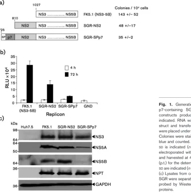
Characterization of SGRs containing NS2 and p7 sequences
may also facilitate correct membrane insertion of p7 and NS2.
The presence of p7 results in an altered distribution of NS2
To gain insight into the potential effects of p7 on NS2 function we established stable Huh7.5 cell lines harbouring SGR-NS2 and SGR-SPp7. The SGR-NS2 harbouring cells expressed the HCV non-structural proteins to similar levels as the FK5.1 (SGR-NS3) cells (Fig. 1c). However, consistent with the differences in replication efficiency (Fig. 1b), in the SGR-SPp7 harbouring cells, non-structural protein expression was lower. Of note, levels of neomycin phos-photransferase were equivalent, suggesting that SPp7 was not having a global inhibitory effect on protein translation from the bicistronic replicon RNA. These observations suggested that p7 was indeed influencing the function of the non-structural proteins, so we therefore examined the subcellular localization of NS2, NS3 and NS5A by immunofluorescence. In cells harbouring SGR-NS2, both
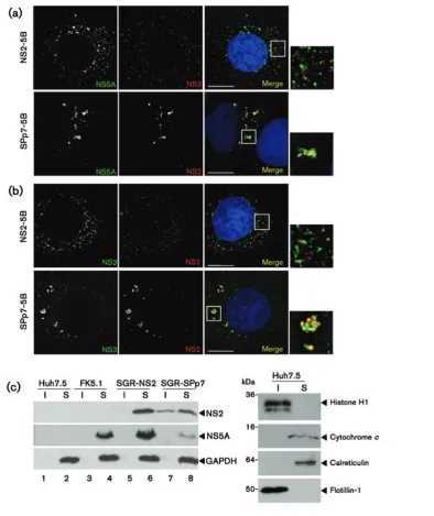
NS2 and NS5A exhibited a punctate cytoplasmic local-ization, but did not colocalize (Fig. 2a). Contrastingly, in cells harbouring SGR-SPp7, NS2 and NS5A were less widely dispersed within the cytoplasm and exhibited a high degree of colocalization, marked by a close apposition and overlap of the NS2 and NS5A signals. This difference was also observed when NS2 and NS3 distribution were analysed – in the SGR-NS2-harbouring cells there was again a lack of colocalization between NS2 and NS3, whilst in the SGR-SPp7-harbouring cells they were less widely dispersed and significantly colocalized (Fig. 2b). These data are consistent with the hypothesis that the presence of p7 directs NS2 to a location proximal to sites of viral genome replication.
[image:3.595.57.452.68.472.2]the detergent-insoluble fraction contained raft-like mem-branes as indicated by the presence of Flotillin-1, as well as nuclear material (histone H1) (Fig. 2c). As NS2 could be clearly shown by immunofluorescence to be absent from the nucleus (Fig. 2a), we conclude that p7 targets at least a proportion of NS2 to a detergent-insoluble fraction. The detergent-soluble fraction contained ER and mitochon-dria-derived membranes (indicated by calreticulin and
[image:4.595.118.513.63.533.2]cytochrome c, respectively) as well as cytosolic proteins (GAPDH). Contrary to previous reports (Gaoet al., 2004; Shi et al., 2003), the detergent-insoluble fraction did not contain NS5A (Fig. 2c). This further supports the con-clusion that in the presence of p7, NS2 is not being incorporated into replication complexes per se, but rather is being targeted to a location adjacent to these sites of viral genome replication.
Serine 168 of NS2 is required for the p7-mediated alteration in NS2 distribution, but not for the association with the detergent-insoluble fraction
Previous reports have implicated a role for serine 168 within NS2 in virus assembly as a mutation at this residue impairs virion production (Jirasko et al., 2008; Yi et al., 2009), and this residue can be phosphorylated by casein kinase 2 (CK2), leading to proteosomal degradation (Franck et al., 2005). As our data suggested that p7 affected the trafficking of NS2, we therefore asked whether serine 168 also played a role in this aspect of NS2 function. SGR-NS2(S168A) and SGR-SPp7(S168A) replicons were replica-tion competent (data not shown), and could establish stable, replicon harbouring cell lines. In cells harbouring SGR-NS2(S168A), the distribution of NS2 as observed by fractionation (Fig. 3a) or immunofluorescence (Fig. 3b, compare to Fig. 2a, top panel) was unchanged. However, fractionation analysis revealed that in the context of SGR-SPp7, S168A had no effect on the presence of NS2 in the detergent-insoluble fraction (Fig. 3a, compare lanes 7 and 8, and 9 and 10). In contrast, in the context of SGR-SPp7, the S168A mutation resulted in a drastic alteration in the distribution of NS2 such that it closely resembled SGR-NS2 with very little colocalization between NS2 and NS5A (Fig. 3c). These data suggest that serine 168 is not required for the targeting of NS2 to the detergent-insoluble fraction, but it does play a role in the p7-mediated localization of NS2 to sites proximal to NS5A and replication complexes seen in SGR-SPp7 (Fig. 3c).
These data also implied that serine 168 might be involved in a physical interaction between NS2 and components of the replication complexes. To test this we performed a coimmunoprecipitation analysis. As NS5A is a key com-ponent of the replication complex and also plays a major role in assembly (Hughes et al., 2009; Masakiet al., 2008; Tellinghuisen et al., 2008), we immunoprecipitated cell lysates with an NS5A antibody and analysed the pre-cipitates by Western blot. NS2 coprecipitated with NS5A from lysates of SGR-SPp7 harbouring cells (Fig. 3d, lane 3), but not from either SGR-NS2 or SGR-SPp7(S168A) lysates (lanes 2 and 4). Furthermore, when lysates were immuno-precipitated with an NS2 antibody, NS5A was only found to precipitate with NS2 in the context of SGR-SPp7 and was absent in the S168A mutant (Fig. 3d, right hand panel). These data are consistent with a direct interaction between NS2 and NS5A, which requires both p7 and serine 168 within NS2. However, we cannot rule out the possibility that this interaction is mediated via additional interactions between NS2, NS5A and other viral or cellular proteins.
The presence of NS2 in the detergent-insoluble fraction is independent of p7 ion channel activity
The ion channel activity of p7 has been shown to be required during virus assembly (Joneset al., 2007; Steinmannet al., 2007a), and it has recently been shown to be critical in protecting intracellular virions from acid pH (Wozniak,
et al., 2010). We therefore sought to investigate whether the altered distribution of NS2 required the ion channel function of p7. To this end we disrupted p7 function by introducing specific point mutations into SGR-SPp7, which have previously been demonstrated to disrupt p7 ion channel activityin vitro(StGelaiset al., 2009) as well as in mammalian cells (Griffinet al., 2004). Mutation of the basic charges on the p7 cytosolic loop (K33A/R35A) has been shown to disrupt both p7 function and secretion of infec-tious virus (Steinmann et al., 2007a), yet our recent data indicate that it also disrupts polyprotein processing and dramatically reduces the abundance of p7 and NS2 (S. Griffin, data not shown), which may be linked to inefficient membrane insertion observed for this mutant in vitro (StGelais et al., 2009). We therefore also introduced two alternative mutations; H17A and G39A, which do not cause such defects in mammalian cells yet abrogate p7 activityin vitrowithout affecting membrane insertion (StGelaiset al., 2009). We established stable, replicon harbouring cell lines for each of these mutants and assessed the distribution of NS2 both by fractionation and fluorescence as described above. The effects of mutating the basic loop [ SGR-SPp7(KR)]were immediately apparent, with NS2 abundance being reduced below detectable levels by Western blot (Fig. 4a: lanes 5 and 6, Fig. 4c). Consistent with the low abundance of NS2 in Western blots, immunofluorescence revealed an SGR-NS2 type staining pattern for NS5A in cells harbouring SGR-SPp7(KR) and NS2 fluorescence was barely detectable above background (Fig. 4c). The other mutations, however, behaved as the parental SGR-SPp7; NS2 being present in the detergent-insoluble fraction and localizing adjacent to NS5A in bright foci (Fig. 4a: lanes 7–10, Fig. 4c). Interestingly, G39A resulted in a reduction in the amount of NS2 in the insoluble fraction – this is consistent with its more profound effect on ion channel activity (StGelaiset al., 2009), perhaps reflective of a more dramatic effect on p7 structure and ion channel-independent functions. Previous in vitro studies showed that the H17A defect could be partially overcome by increasing the concentration of purified p7 protein, whereas for G39A this was not the case (StGelaiset al., 2009). As an additional control, SGR-SPp7 harbouring cells were treated with 50mM rimantadine, which inhibits p7 ion channel activity bothin vitroand in cell culture (Griffin et al., 2008; StGelais et al., 2007; Wozniak et al., 2010); again this did not affect the localization of NS2 (Fig. 4b: lanes 9–12, Fig. 4c). These data confirm that the altered distribution of NS2 by p7 does not require the ion channel activity of the latter protein.
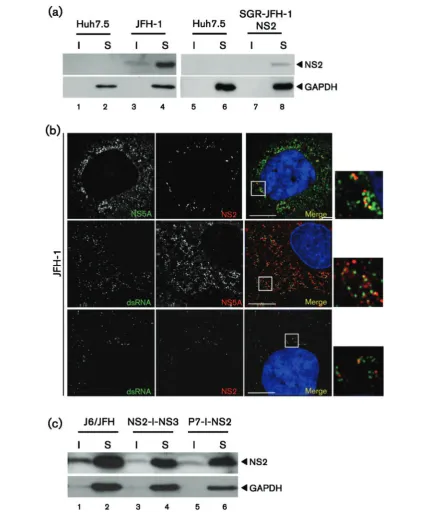
The p7-mediated distribution of NS2 is observed in the context of the full-length virus
transiently transfected with the full-length infectious JFH-1, were fractionated as previously described. Whereas in the context of full-length infectious JFH-1, NS2 was seen in
[image:6.595.116.492.66.577.2]7 and 8), even upon overexposure of the Western blot (data not shown). We were unable to detect NS2 in the SGR-JFH-1-SPp7 harbouring cells as the anti-NS2 serum exhibited reduced sensitivity for genotype 2a NS2 in Western blot, compared with the 1b protein. Additionally, it was not possible to detect NS2 in any of the JFH-1 SGR harbouring cells by immunofluorescence. However, the higher viral protein expression levels in cells transfected with full-length JFH-1 RNA (Fig. 5a) permitted direct detection of NS2 by immunofluorescence (Fig. 5b), and we again confirmed that it localized adjacent to NS5A. The high level of virus replication also permitted the use of the J2 antibody raised to dsRNA. As shown in Fig. 5(b), similar to that observed previously (Targett-Adamset al., 2008), NS5A showed a significant degree of colocalization with dsRNA. This was less apparent for NS2, although NS2 foci were predominantly observed adjacent to sites containing dsRNA (Fig. 5b). This is again consistent with the targeting of NS2 to sites adjacent to replication complexes.
Targeting of NS2 does not require the p7-NS2 precursor
Although the targeting of NS2 appeared p7-dependent, it was not clear whether this occurred prior to, or follow-ing, cleavage of the p7-NS2 precursor. It was therefore necessary to demonstrate whether this precursor mediated NS2 targeting, or whether it occurred via a protein–protein interaction between the mature proteins. We addressed this question by transfecting Huh7.5 cells with RNAs derived from modified chimeric J6/JFH-1-derived genomes in which p7 and NS2 were separated by an IRES [ J6/JFH-1(p7-I-NS2)] (Jones et al., 2007). Consistent with the recent observation that NS2 will functionin trans during HCV particle assembly (Yiet al., 2009), NS2 was targeted to the detergent-insoluble fraction in cells transfected with p7-I-NS2 RNA, as well those transfected with an RNA in which NS2 and NS3 were separated by an IRES (NS2-I-NS3) (Fig. 5c). The data from these experiments confirm that p7 can mediate the altered distribution of NS2in trans, and the uncleaved p7-NS2 precursor is not required for this observed effect.
DISCUSSION
A key unresolved question in HCV biology is how the processes of genome replication and assembly are coordi-nated and linked, such that infectious virus particles can be
generated. In this context, HCV mutants lacking p7 or NS2 are able to undergo genome replication but cannot pro-duce new virions (Jones et al., 2007; Steinmann et al., 2007a); it is likely therefore that both proteins function at the interface between genome replication and packaging. Our data are consistent with the hypothesis that p7 targets NS2 to sites adjacent to replication complexes where it interacts with NS5A and potentially other replication com-plex components, thereby facilitating the coordination of genome replication and virion assembly.
et al., 2009). It will be of great interest to establish the effect of p7 on the phosphorylation state of NS2.
The observation that p7 directs NS2 to a site adjacent to replication complexes also provides a potential solution to a paradox of HCV biology, namely that membrane-bound RNA replication complexes isolated from NS3-5B replicon cells are resistant to both nuclease and protease digestionin vitro(Aizakiet al., 2004; Yang et al., 2004). How then do nascent genomes get exported from this compartment in order to undergo assembly into virus particles? It may be that NS2 provides such a mechanism by positioning at an exit site of the replication compartment – explaining both the juxtaposition of NS2 and the replication complexes in SGR-SPp7 harbouring cells, and the previously observed requirement for NS2 in the production of infectious virus particles (Jones et al., 2007). Implicit in this hypothesis would be interactions between NS2 and both the structural and non-structural proteins. Consistent with this, it has recently been shown that NS2 interacts with E1, E2, p7, NS3 and NS5A (Maet al., 2011) and it is likely therefore that NS2 functions to mediate the interaction between the replication complex and the structural proteins, perhaps acting in concert with the recently defined interaction between NS5A and core (Masakiet al., 2008). Alternatively, NS2 may function in the release of RNA from the repli-cation complex and allow its encapsidation into nascent HCV particles. As well as the direct physical interaction, there is genetic evidence for a p7–NS2 interaction from studies of chimeric HCV genomes based on JFH-1. These viruses only produce infectious particles when the N-terminaltrans-membrane helix of NS2 is derived from the same isolate as the other structural proteins (including p7) (Pietschmannet al., 2006). In addition, our finding that complementing proton channel activity is insufficient to rescue p7 deletants, yet efficiently restores particle pro-duction to point mutants confirms that a p7-specific protein–protein interaction is likely to be required for an early stage of virion morphogenesis (Wozniaket al., 2010). The data herein provides additional support for an interac-tion between p7 and NS2 and furthermore imply that the resultant redistribution of NS2 into close apposition with replication complexes is a critical event during the assembly of infectious virus particles.
METHODS
HCV replicon constructs.AnRsrII–BsrGI fragment containing the
EMCV IRES and the 59end of the NS3 coding region was subcloned from
the culture-adapted Con1 NS3–NS5B SGR, FK5.1 (Kriegeret al., 2001)
into Litmus38. Within this subclone a uniqueNcoI site incorporating the
AUG of the HCV ORF was used in conjunction withBsrGI to introduce
additional sequences to the N terminus of NS3. HCV-derived sequences
were amplified from the genotype 1b Con1 infectious clone usingPfu
polymerase (Stratagene).PmeI–BsrGI fragments of the subclone were
transferred back into the FK5.1 backbone. Constructs carrying firefly luciferase were generated by excising the neomycin phosphotransferase
codingAscI–PmeI fragment and replacing it with the corresponding
luciferase coding fragment from pFK5.1luc (Kriegeret al., 2001).
To generate JFH-1-derived SGRs, a subclone of the JFH-1 SGR (Kato et al., 2003) was created by cloning thePmeI–SpeI fragment into a
modified Litmus28 vector in which theNcoI site was replaced with
PmeI (Litmus28P). This was linearized withNcoI, blunted with mung
bean nuclease (New England Biolabs) then digested withKasI. The
NS2 coding region of JFH-1 was amplified by PCR using a forward primer containing a start codon and a reverse primer extending to the KasI site in NS3, then digested with KasI and ligated into the
subclone. ThePmeI–ClaI fragment of the subclone was replaced in
the JFH-1 SGR creating SGR-JFH-1-NS2.
Mammalian cell culture.Huh7.5 cells were cultured in Dulbecco’s modified Eagle’s medium, supplemented with 10 % (v/v) FBS, 100 U
penicillin ml21, 100mg streptomycin ml21, 2 mM L-glutamine and
non-essential amino acids (Gibco) at 37uC, 5 % CO2, in a humidified
incubator. Stable cell lines were maintained with 500mg G418 (Melford)
ml21
. Where indicated rimantadine (provided by GlaxoSmithKline) was prepared as a stock solution (40 mM) in DMSO.
Transfection of Huh7.5 cells. Templates for transcription were
prepared by linearization with eitherScaI (FK5.1) orXbaI (JFH-1),
the latter were also mung bean nuclease treated. RNA was transcribed using Ribomax Express (Promega). RNA was transfected into cells as
described previously (Lohmann et al., 1999). Briefly, Huh7.5 cells
were trypsinized, washed twice in ice-cold PBS then resuspended
in ice-cold PBS at 1 or 26107 cells ml21 for replicon or virus,
respectively. RNA (1mg for replicons, 10mg for virus) was mixed with
400ml cell suspension and electroporated at 270 V and 950mF. Cells
were recovered in pre-warmed medium and seeded as required. Colony formation assays were performed as described previously
(McCormicket al., 2004).
Luciferase assay.Cells were harvested by the addition of Passive Lysis Buffer (Promega). Luciferase Assay Reagent (Promega) was
added (30ml per 50ml of cell lysate) and luminescence was measured
by using a BMG plate reader.
Antibodies.The polyclonal rabbit anti-NS2 serum, 4106, was raised against purified, NS2 cytosolic domain (detailed protocols available upon request). Sheep antisera against HCV non-structural proteins
NS3 and NS5A have been described previously (Aoubalaet al., 2001;
Macdonaldet al., 2003), rabbit anti-NS5A serum was provided by
Ralf Bartenschlager. Antibodies for cellular proteins were obtained commercially and used as described by the manufacturers; mAbs to calreticulin (Calbiochem), flotillin-1 (BD Biosciences), glyceralde-hyde-3 phosphate dehydrogenase (GAPDH; Abcam), and polyclonal
sera to cytochromec and histone H1 (Abcam). HRP- (Sigma) or
Alexa-Fluor (Invitrogen)-conjugated secondary antibodies were used for Western blotting or immunofluorescence, respectively.
Cell fractionation.Cells were harvested, washed twice with PBS, then
lysed in GLB[1 % TX-100, 120 mM KCl, 30 mM NaCl, 5 mM MgCl2,
10 % glycerol (v/v) and 10 mM PIPES-NaOH, pH 7.2], supplemented
with Complete protease inhibitor cocktail (Roche), on ice for 15 min.
Insoluble material was pelleted by centrifugation at 500gfor 5 min at
4uC. Lysates were clarified by a second centrifugation at 500g, and the pellets were washed twice in GLB. Normalized lysate and pellet fractions were analysed by Western blotting.
Immunoprecipitation.Cells were harvested and lysed in IP buffer
[20 mM Tris/HCl pH 7.4, 135 mM NaCl, 1 % TX-100, 0.5 % sodium
deoxycholate and 10 % glycerol (v/v)], supplemented with Complete
protease inhibitor cocktail (Roche). Lysates were diluted to 2 mg total
protein ml21then 500mg protein was mixed with 2ml antiserum at
at 4uC. Beads were washed three times in IP buffer then eluted by
boiling in 26Laemmli buffer for 5 min.
Indirect immunofluorescence microscopy. Cells, seeded onto coverslips 24 h prior to fixation, were washed twice with PBS then fixed with 4 % paraformaldehyde for 10 min at room temperature. Following washing, cells were permeabilized with 0.2 % TX-100 in PBS for a further 10 min, washed twice with PBS, then incubated with primary antibody diluted in PBS/10 % FBS in for 1 h at room temperature. The coverslips were washed with PBS/10 % FBS then incubated with Alexa-Fluor-conjugated secondary antibody. The coverslips were then stained with a second primary antibody, if required, by the same method. Before mounting onto slides, the coverslips were incubated with Hoechst 33342 (Molecular Probes) diluted 1 : 10 000 in PBS for 5 min to stain nuclei, then washed twice in PBS. Coverslips were mounted with Citifluor AF1 (Agar Scientific). Images were captured using an
Olympus IX71 microscope with a6100 oil immersion objective with a
numerical aperture of 1.35 (DeltaVision – Applied Precision).Z-stacks
were collected comprising optical slices of 0.2mm and deconvolved by
using Softworx software (Applied Precision).
ACKNOWLEDGEMENTS
This work was supported by a grant to M. H. and S. G. from the Wellcome Trust (082812). S. G. is the recipient of a Medical Research Council New Investigator Award (G0700124). We thank Ralf Bartenschlager (University of Heidelberg) for the FK5.1 replicon constructs and rabbit anti-NS5A serum, Takaji Wakita (National Institute for Infectious Diseases, Tokyo) for pJFH-1 and pSGR-JFH-1 and Charles Rice (The Rockefeller University, New York) for the Huh7.5 cells and J6/JFH-1 virus constructs.
REFERENCES
Aizaki, H., Lee, K. J., Sung, V. M., Ishiko, H. & Lai, M. M. (2004). Characterization of the hepatitis C virus RNA replication complex
associated with lipid rafts.Virology324, 450–461.
Aoubala, M., Holt, J., Clegg, R. A., Rowlands, D. J. & Harris, M. (2001). The inhibition of cAMP-dependent protein kinase by full-length
hepatitis C virus NS3/4A complex is due to ATP hydrolysis. J Gen
Virol82, 1637–1646.
Brohm, C., Steinmann, E., Friesland, M., Lorenz, I. C., Patel, A., Penin, F., Bartenschlager, R. & Pietschmann, T. (2009). Characterization of determinants important for hepatitis C virus p7 function in
morpho-genesis by using trans-complementation.J Virol83, 11682–11693.
Carre`re-Kremer, S., Montpellier-Pala, C., Cocquerel, L., Wychowski, C., Penin, F. & Dubuisson, J. (2002).Subcellular localization and topology
of the p7 polypeptide of hepatitis C virus.J Virol76, 3720–3730.
Carre`re-Kremer, S., Montpellier, C., Lorenzo, L., Brulin, B., Cocquerel, L., Belouzard, S., Penin, F. & Dubuisson, J. (2004). Regulation of hepatitis C virus polyprotein processing by signal peptidase involves structural determinants at the p7 sequence
junc-tions.J Biol Chem279, 41384–41392.
Clarke, D., Griffin, S., Beales, L., Gelais, C. S., Burgess, S., Harris, M. & Rowlands, D. (2006).Evidence for the formation of a heptameric
ion channel complex by the hepatitis C virus p7 proteinin vitro. J Biol
Chem281, 37057–37068.
Dimitrova, M., Imbert, I., Kieny, M. P. & Schuster, C. (2003). Protein-protein interactions between hepatitis C virus nonstructural Protein-proteins. J Virol77, 5401–5414.
Flajolet, M., Rotondo, G., Daviet, L., Bergametti, F., Inchauspe´, G., Tiollais, P., Transy, C. & Legrain, P. (2000).A genomic approach of
the hepatitis C virus generates a protein interaction map.Gene242,
369–379.
Franck, N., Le Seyec, J., Guguen-Guillouzo, C. & Erdtmann, L. (2005). Hepatitis C virus NS2 protein is phosphorylated by the protein kinase CK2 and targeted for degradation to the proteasome. J Virol79, 2700–2708.
Gao, L., Aizaki, H., He, J. W. & Lai, M. M. (2004).Interactions between viral nonstructural proteins and host protein hVAP-33 mediate the formation of hepatitis C virus RNA replication complex on lipid raft. J Virol78, 3480–3488.
Grakoui, A., McCourt, D. W., Wychowski, C., Feinstone, S. M. & Rice, C. M. (1993). A second hepatitis C virus-encoded proteinase. Proc Natl Acad Sci U S A90, 10583–10587.
Griffin, S. (2010).Inhibition of HCV p7 as a therapeutic target.Curr Opin Investig Drugs11, 175–181.
Griffin, S. D. C., Beales, L. P., Clarke, D. S., Worsfold, O., Evans, S. D., Jaeger, J., Harris, M. P. G. & Rowlands, D. J. (2003).The p7 protein of hepatitis C virus forms an ion channel that is blocked by the antiviral
drug, Amantadine.FEBS Lett535, 34–38.
Griffin, S. D., Harvey, R., Clarke, D. S., Barclay, W. S., Harris, M. & Rowlands, D. J. (2004).A conserved basic loop in hepatitis C virus p7 protein is required for amantadine-sensitive ion channel activity in mammalian cells but is dispensable for localization to mitochondria. J Gen Virol85, 451–461.
Griffin, S., Clarke, D., McCormick, C., Rowlands, D. & Harris, M. (2005).Signal peptide cleavage and internal targeting signals direct the hepatitis C virus p7 protein to distinct intracellular membranes. J Virol79, 15525–15536.
Griffin, S., StGelais, C., Owsianka, A. M., Patel, A. H., Rowlands, D. & Harris, M. (2008).Genotype-dependent sensitivity of hepatitis C virus
to inhibitors of the p7 ion channel.Hepatology48, 1779–1790.
Hughes, M., Gretton, S., Shelton, H., Brown, D. D., McCormick, C. J., Angus, A. G., Patel, A. H., Griffin, S. & Harris, M. (2009).A conserved proline between domains II and III of hepatitis C virus NS5A
influences both RNA replication and virus assembly. J Virol 83,
10788–10796.
Jirasko, V., Montserret, R., Appel, N., Janvier, A., Eustachi, L., Brohm, C., Steinmann, E., Pietschmann, T., Penin, F. & Bartenschlager, R. (2008). Structural and functional characterization of nonstructural protein 2 for
its role in hepatitis C virus assembly.J Biol Chem283, 28546–28562.
Jones, C. T., Murray, C. L., Eastman, D. K., Tassello, J. & Rice, C. M. (2007). Hepatitis C virus p7 and NS2 proteins are essential for
production of infectious virus.J Virol81, 8374–8383.
Kato, T., Date, T., Miyamoto, M., Furusaka, A., Tokushige, K., Mizokami, M. & Wakita, T. (2003). Efficient replication of the
genotype 2a hepatitis C virus subgenomic replicon.Gastroenterology
125, 1808–1817.
Krieger, N., Lohmann, V. & Bartenschlager, R. (2001).Enhancement of hepatitis C virus RNA replication by cell culture-adaptive mutations. J Virol75, 4614–4624.
Lavanchy, D. (1999).Global surveillance and control of hepatitis C. Report of a WHO Consultation organized in collaboration with the
Viral Hepatitis Prevention Board, Antwerp, Belgium.J Viral Hepat6,
35–47.
Lin, C., Lindenbach, B. D., Pra´gai, B. M., McCourt, D. W. & Rice, C. M. (1994). Processing in the hepatitis C virus E2-NS2 region: identification of p7 and two distinct E2-specific products with
different C termini.J Virol68, 5063–5073.
Lohmann, V., Ko¨rner, F., Koch, J. O., Herian, U., Theilmann, L. & Bartenschlager, R. (1999). Replication of subgenomic hepatitis C
Luik, P., Chew, C., Aittoniemi, J., Chang, J., Wentworth, P., Jr, Dwek, R. A., Biggin, P. C., Ve´nien-Bryan, C. & Zitzmann, N. (2009).The 3-dimensional structure of a hepatitis C virus p7 ion channel by
electron microscopy.Proc Natl Acad Sci U S A106, 12712–12716.
Ma, Y., Anantpadma, M., Timpe, J. M., Shanmugam, S., Singh, S. M., Lemon, S. M. & Yi, M. (2011).Hepatitis C virus NS2 protein serves as a scaffold for virus assembly by interacting with both structural and
nonstructural proteins.J Virol85, 86–97.
Macdonald, A., Crowder, K., Street, A., McCormick, C., Saksela, K. & Harris, M. (2003). The hepatitis C virus NS5A protein inhibits activating protein-1 function by perturbing Ras-ERK pathway signall-ing.J Biol Chem278, 17775–17784.
Masaki, T., Suzuki, R., Murakami, K., Aizaki, H., Ishii, K., Murayama, A., Date, T., Matsuura, Y., Miyamura, T. & other authors (2008). Interaction of hepatitis C virus nonstructural protein 5A with core
protein is critical for the production of infectious virus particles.J Virol
82, 7964–7976.
McCormick, C. J., Challinor, L., Macdonald, A., Rowlands, D. J. & Harris, M. (2004).Introduction of replication-competent hepatitis C virus transcripts using a tetracycline-regulable baculovirus delivery
system.J Gen Virol85, 429–439.
Moradpour, D., Penin, F. & Rice, C. M. (2007).Replication of hepatitis
C virus.Nat Rev Microbiol5, 453–463.
Pavlovic´, D., Neville, D. C., Argaud, O., Blumberg, B., Dwek, R. A., Fischer, W. B. & Zitzmann, N. (2003).The hepatitis C virus p7 protein forms an ion channel that is inhibited by long-alkyl-chain iminosugar
derivatives.Proc Natl Acad Sci U S A100, 6104–6108.
Pietschmann, T., Kaul, A., Koutsoudakis, G., Shavinskaya, A., Kallis, S., Steinmann, E., Abid, K., Negro, F., Dreux, M. & other authors (2006). Construction and characterization of infectious
intragenotypic and intergenotypic hepatitis C virus chimeras. Proc
Natl Acad Sci U S A103, 7408–7413.
Premkumar, A., Wilson, L., Ewart, G. D. & Gage, P. W. (2004). Cation-selective ion channels formed by p7 of hepatitis C virus are blocked
by hexamethylene amiloride.FEBS Lett557, 99–103.
Russell, R. S., Meunier, J. C., Takikawa, S., Faulk, K., Engle, R. E., Bukh, J., Purcell, R. H. & Emerson, S. U. (2008).Advantages of a single-cycle production assay to study cell culture-adaptive mutations
of hepatitis C virus.Proc Natl Acad Sci U S A105, 4370–4375.
Sakai, A., Claire, M. S., Faulk, K., Govindarajan, S., Emerson, S. U., Purcell, R. H. & Bukh, J. (2003).The p7 polypeptide of hepatitis C virus is critical for infectivity and contains functionally important
genotype-specific sequences.Proc Natl Acad Sci U S A100, 11646–
11651.
Santolini, E., Pacini, L., Fipaldini, C., Migliaccio, G. & Monica, N. (1995).The NS2 protein of hepatitis C virus is a transmembrane
polypeptide.J Virol69, 7461–7471.
Shi, S. T., Lee, K. J., Aizaki, H., Hwang, S. B. & Lai, M. M. (2003). Hepatitis C virus RNA replication occurs on a detergent-resistant
membrane that cofractionates with caveolin-2.J Virol77, 4160–4168.
Steinmann, E., Penin, F., Kallis, S., Patel, A. H., Bartenschlager, R. & Pietschmann, T. (2007a).Hepatitis C virus p7 protein is crucial for
assembly and release of infectious virions.PLoS Pathog3, e103.
Steinmann, E., Whitfield, T., Kallis, S., Dwek, R. A., Zitzmann, N., Pietschmann, T. & Bartenschlager, R. (2007b).Antiviral effects of amantadine and iminosugar derivatives against hepatitis C virus. Hepatology46, 330–338.
StGelais, C., Tuthill, T. J., Clarke, D. S., Rowlands, D. J., Harris, M. & Griffin, S. (2007). Inhibition of hepatitis C virus p7 membrane
channels in a liposome-based assay system.Antiviral Res76, 48–58.
StGelais, C., Foster, T. L., Verow, M., Atkins, E., Fishwick, C. W., Rowlands, D., Harris, M. & Griffin, S. (2009). Determinants of hepatitis C virus p7 ion channel function and drug sensitivity
identifiedin vitro.J Virol83, 7970–7981.
Targett-Adams, P., Boulant, S. & McLauchlan, J. (2008). Visualization of double-stranded RNA in cells supporting hepatitis
C virus RNA replication.J Virol82, 2182–2195.
Tellinghuisen, T. L., Foss, K. L. & Treadaway, J. (2008).Regulation of hepatitis C virion production via phosphorylation of the NS5A
protein.PLoS Pathog4, e1000032.
Wakita, T., Pietschmann, T., Kato, T., Date, T., Miyamoto, M., Zhao, Z., Murthy, K., Habermann, A., Kra¨usslich, H. G. & other authors (2005). Production of infectious hepatitis C virus in tissue culture from a
cloned viral genome.Nat Med11, 791–796.
Welbourn, S., Green, R., Gamache, I., Dandache, S., Lohmann, V., Bartenschlager, R., Meerovitch, K. & Pause, A. (2005).Hepatitis C virus NS2/3 processing is required for NS3 stability and viral
RNA replication.J Biol Chem280, 29604–29611.
Wozniak, A. L., Griffin, S., Rowlands, D. J., Harris, M., Yi, M., Lemon, S. M. & Weinman, S. A. (2010).Intracellular proton conductance of the hepatitis C virus p7 protein and its contribution to infectious
virus production.PLoS Pathog6, e1001087.
Yang, G., Pevear, D. C., Collett, M. S., Chunduru, S., Young, D. C., Benetatos, C. & Jordan, R. (2004).Newly synthesized hepatitis C virus replicon RNA is protected from nuclease activity by a
protease-sensitive factor(s).J Virol78, 10202–10205.
Yi, M., Ma, Y., Yates, J. & Lemon, S. M. (2007). Compensatory mutations in E1, p7, NS2, and NS3 enhance yields of cell
culture-infectious intergenotypic chimeric hepatitis C virus.J Virol81, 629–
638.
Yi, M., Ma, Y., Yates, J. & Lemon, S. M. (2009). Trans-complementa-tion of an NS2 defect in a late step in hepatitis C virus (HCV) particle